© 2011 Museu de Ciències Naturals ISSN: 1578–665X
Azevedo, E., Caeiro, M. F., Rebelo, R. & Barata, M., 2011. Biodiversity and characterization of marine mycota from Portuguese waters. Animal Biodiversity and Conservation, 34.1: 205–215.
Abstract
Biodiversity and characterization of marine mycota from Portuguese waters.— The occurrence, diversity and similarity of marine fungi detected by the sum of direct and indirect observations in Fagus sylvatica and Pinus pinaster baits submerged at two Portuguese marinas are analyzed and discussed. In comparison with the data already published in 2010, the higher number of specimens considered in this study led to the higher number of very frequent taxa for these environments and substrata; the significant difference in substrata and also in fungal diversity detected at the two environments is also highlighted, in addition to the decrease in fungal similarity. Because the identification of Lulworthia spp., Fusarium sp., Graphium sp., Phoma sp. and Stachybotrys sp. down to species level was not possible, based only on the morphological characterization, a molecular approach based on the amplification of the LSU rDNA region was performed with isolates of these fungi. This was achieved for three isolates, identified as Fusarium solani, Graphium eumorphum and Stachybotrys chartarum. To achieve this with the other isolates which are more complex taxa, the sequencing of more regions will be considered.
Key words: Marine fungi, Wood baits, Fungal diversity, Ascomycota, Anamorphic fungi, Sequence alignment. Resumen
Biodiversidad y caracterización de los hongos marinos de las aguas portuguesas.— Se analiza y discute la presencia, la diversidad y la similitud de los hongos marinos detectados mediante la suma de observaciones directas e indirectas utilizando cebos de Fagus sylvatica y Pinus pinaster sumergidos en dos puertos deportivos portugueses. En comparación con los datos ya publicados en 2010, el mayor número de especímenes aquí considerados condujo a un mayor número de taxones muy frecuentes en estos sustratos y medios ambien-tales; también debe destacarse la diferencia significativa en los sustratos y también en la diversidad fúngica en los dos medios ambientales, además de la disminución de la similitud fúngica. Dado que no fue posible la identificación de Lulworthia spp., Fusarium sp., Graphium sp., Phoma sp., y Stachybotrys sp. hasta el nivel de especie, basándose únicamente en la caracterización morfológica, se llevó a cabo un estudio molecular basado en la amplificación de la región LSU ADNr con extractos de dichos hongos. Ello se consiguió en tres extractos puros, identificados como de Fusarium solani, Graphium eumorphum y Stachybotrys chartarum. Para llevar a cabo este proceso con otros extractos puros pertenecientes a taxones más complejos, se considerará la secuenciación de más regiones.
Palabras clave: Hongos marinos, Cebos de madera, Diversidad fúngica, Ascomycota, Hongos anamórficos, Alineación de secuencias.
E. Azevedo, R. Rebelo, M. F. Caeiro & M. Barata, Fac. de Ciências, Univ. de Lisboa.– E. Azevedo, R. Rebelo & M. Barata, Centro de Biologia Ambiental (CBA), Fac. de Ciências, Univ. de Lisboa.– E. Azevedo & M. F. Caeiro, Centro de Estudos do Ambiente e do Mar (CESAM), Univ. de Aveiro.
Corresponding author: Egidia Azevedo, Depto. de Biologia Vegetal, Fac. de Ciências, Univ. de Lisboa, Campo Grande, Edifício C2, 4º Piso, 1749-016 Lisboa, Portugal. E–mail: egazd@hotmail.com
Biodiversity and characterization
of marine mycota
from Portuguese waters
Introduction
Fungi have been known to exist in marine environ-ments since early times. Hyde et al. (2000) highlighted the first reports of marine fungi up until 1846; however, interest in marine mycology only increased worldwide with Barghoorn & Linder (1944).
In natural marine environments many substrata are good sources for marine fungi detection. The most studied have been wood substrata (Barghoorn & Linder, 1944; Koch, 1974; Koch & Petersen, 1996; Gonzálvez et al., 2001; Lintott & Lintott, 2002; Jones et al., 2006; Ravikumar et al., 2009), halophytes as Spartina spp. (Gessner & Kohlmeyer, 1977; Barata, 1997, 2002; Torzilli et al., 2006), Phragmites australis (Poon & Hyde, 1998; Wong & Hyde, 2002) and Juncus roemarianus (Kohlmeyer & Volkmann Kohlmeyer, 2001, 2002), as well as algae (Kohlmeyer & Volkmann–Kohlmeyer, 2003; Zucaro et al., 2008) and sea foam (Kohlmeyer & Kohlmeyer, 1979; Steinke & Lubke, 2005). Marine mycota as-sociated to sand dunes plants (e.g. Arundo donax, Agropyron junceiforme and Ammophila arenaria) are poorly explored (Kohlmeyer & Kohlmeyer, 1979; Jones et al., 2009). Other substrata like corals, tropical sea grasses, crustacean and mollusk shells and soft rocks, are yet to be intensively investigated (Hyde et al., 2000; Jones et al., 2009).
Considerable progress has been made in inventory-ing endophytes from marine hosts includinventory-ing seagrass (Alva et al., 2002; Sakayaroj et al., 2010). The diversity found comprises mostly anamorphic fungi and sterile mycelia and some isolates revealed to be producers of cellulases and xylanases (Alva et al., 2002).
The baiting method, often used by mycologists for ecological studies, also yields pure cultures of marine fungi, representative of particular and/or selected environments (Vrijmoed et al., 1982, 1986; Alias & Jones, 2000; Azevedo et al., 2010). Pinus spp. and Fagus sylvatica are woods often used to inventory marine fungi in submerged conditions (Byrne & Jones, 1974; Grasso et al., 1990; Vrijmoed et al., 1982, 1986; Azevedo et al., 2010), because lignocellulosic substrata are colonized throughout submersion by a great variety of lignicolous species.
Among the marine fungi isolated by Azevedo et al. (2010), five taxa could not be identified to spe-cies level based only on morphology: Lulworthia spp., Fusarium sp., Graphium sp., Phoma sp. and Stachybtrys sp.
In temperate waters Lulworthia species are among the most frequently detected fungi in sub-merged woods (Byrne & Jones, 1974; Mouzouras et al., 1985; Grasso et al., 1985, 1990; Azevedo et al., 2010) and halophytes (e.g. S. maritima) (Barata, 1997). Several investigations reported Fusarium species in sediments, sand dunes and recovered submerged twigs of Tamarix aphylla (Jones et al., 2009), on coral reefs (Morrison–Gardiner, 2002) and in wood baits submerged in marine environment (Azevedo et al., 2010). Graphium species are also found in wood in marine environments (Vrijmoed et al., 1982, 1986; Gonzálvez et al., 1998; Maria
& Sridhar, 2003; Azevedo et al., 2010). Phoma species are widespread, occurring in a variety of environments and ecological niches; they are less explored in marine environment in which Phoma species completely new to science are regularly found (Aveskamp et al., 2010). Finally, Stachybotrys species are also detected in marine environment (Landy & Jones, 2006; Jones et al., 2009; Azevedo et al., 2010), having been considered important by Jones et al. (2009) to document the occurrence of these taxa in the sea and to discover their ecologi-cal role.
One goal of this work was to present and analyze, in a comprehensive manner, the data from the sur-vey of Azevedo et al. (2010) concerning the occur-rence, diversity and similarity of the marine mycota detected in wood baits before and after incubation in moist chambers (direct and indirect observations respectively). We further proposed to compare the results from this and other surveys carried out in temperate waters.
A second goal was to present and discuss the results of a molecular approach performed to cha-racterize the isolates that had not been possible to identify down to species level based only on morpho-logical characters.
Material and methods Sampling strategies
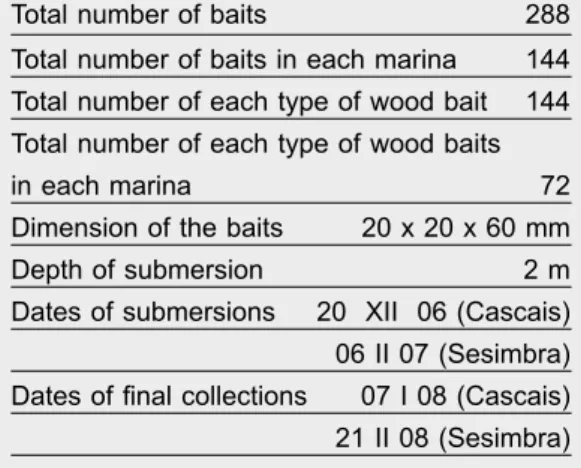
Two marinas located on the western coast of Portugal, Cascais (38º 40' N 09º 25' E) and Sesimbra (38º 26' N 09º 06' W), were selected for the submersion of wood baits from Pinus pinaster Aiton and Fagus sylvatica L., as described by Azevedo et al. (2010) and shown in figure 1. The experimental design of baits is presented in table 1 and figure 1.
The wood baiting technique involved a previous overnight soaking of the baits in distilled sterilized water followed by 20–minute autoclave sterilization at 121ºC.
After submersion, collections were performed periodically each eight to 10 weeks, on a total of six collections, at each marina, The baits were examined as soon as possible after collection under the dis-secting microscope to detect spores and fruit bodies. Microscopic characterizations were performed under the light microscope (Leitz Laborlux S with Normar-ski) in slides prepared with seawater as mounting media and microphotographs were taken (fig. 2). Thereafter, identifications were made following the dichotomous keys of Kohmeyer & Kolhmeyer (1979), Kohlmeyer & Volkmann–Kolhmeyer (1991) and Hyde & Sarma (2000).
The baits were analyzed by direct observation and then incubated in moist chambers for 12 months. They were re–examined on a monthly basis, following the procedures described by Vrijmoed (2000). The isolates of marine fungi subjected to molecular analysis were obtained by the single spore method (Azevedo et al., 2010).
Analysis of fungal occurrence, diversity and similarity Frequencies of occurrence, expressed as percenta-ges, were calculated taking the results from direct and indirect observations together. Marine fungi were classified as 'very frequent', 'frequent' or 'infrequent' based on Tan et al. (1989).
The average numbers of fungi per bait, species richness (S), Shannon (H’) and evenness (E) diver-sity indices, as well as the Sorenson similarity index (Cs), were calculated as described by Figueira & Barata (2007). The values of Shannon Index were compared applying a t–test as proposed by Hutch-eson (Zar, 1999).
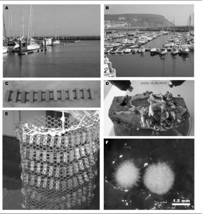
Fig. 1. A. Cascais marina; B. Sesimbra marina; C. Set of wood baits before submersion; D. Pinus pinaster bait colonized with marine organisms after six months of submersion; E. Box of wood baits at the moment of submersion; F. Fagus sylvatica bait colonized with basidiocarps of Nia vibrissa.
Fig. 1. A. Puerto deportivo de Cascais; B. Puerto deportivo de Sesimbra; C. Serie de cebos de madera, antes de sumergirlos; D. Cebo de Pinus pinaster colonizado por organismos marinos después de seis meses de inmersión; E. Caja de cebos de madera en el momento de sumergirlos; F. Cebo de Fagus sylvatica colonizado con basidiocarpos de Nia vibrissa.
A B
C D
E
F
DNA extraction, PCR amplification and sequencing The cultures selected for molecular analysis were grown in Malt extract broth prepared with sea water on a rotary shaker at 200 rpm for 6–15 days at 20ºC. Fungal biomass was harvested, washed three times with sterile distilled sea water and frozen in liquid nitrogen to be ground into a fine powder with a mortar and pestle. DNA was extracted following the instructions of Nucleospin Plant DNA extraction Kit (Machery–Nagel, Germany).
A partial LSU rDNA sequence was amplified with LROR and LR5 primers (Viglays & Sun, 1994) and
PCR reactions were carried out in a total volume of 25 µl with Phire Hot Start DNA polymerase (Finnzymes Oy., now Thermo Scientific) and 1 µl DNA sample, following the manufacturer’ instructions.
The amplification program consisted of an initial 3–minute denaturation step at 98ºC followed by 35 cycles of (i) denaturation (98ºC for 10''), (ii) an-nealing (58.5ºC for 10'') and (iii) elongation (72ºC for 30 '') and a final extension of 1' at 72ºC. After a sample being resolved on 0.7% agarose gel, PCR products were purified by Jet quick DNA Clean Up Kit (Genomed GmbH), according to the manufac-turer’s instructions, and sent to be sequenced by a commercial lab.
Direct sequencing was performed by STAB VIDA (Portugal), using the same set of primers and the the big dye terminator kit on ABI automated DNA sequencer.
BioEdit Sequence Alignment Editor v7.0.9.0 (Hall, 1999) and ClustalW (Thompson et al., 1997) with default parameter settings were used for align-ment and to obtain the consensus sequences. The obtained consensus sequences were compared to data in GenBank (National Center for Biotechnology Information, Bethesda, USA) online (www.ncbi.nih. gov), with GenBank BLASTn search engine.
Results
Marine fungi occurrence, diversity and similarity Table 2 presents the marine fungi detected by direct and indirect observations. The taxa are listed by decreasing values of frequency of occurrence in the ensemble of the two marinas; only infrequent fungi for both marinas were not listed. Diversity and simi-larity indices per environment and per substratum are presented respectively in tables 3 and 4.
Table 1. Experimental design of the baits. Tabla 1. Diseño experimental de los cebos.
Total number of baits 288
Total number of baits in each marina 144 Total number of each type of wood bait 144 Total number of each type of wood baits
in each marina 72
Dimension of the baits 20 x 20 x 60 mm
Depth of submersion 2 m
Dates of submersions 20 XII 06 (Cascais) 06 II 07 (Sesimbra) Dates of final collections 07 I 08 (Cascais) 21 II 08 (Sesimbra)
Fig. 2. Lulworthia sp.: A. 15 day–old colony on corn meal agar made with 50% seawater; B. Ascocarp; C. Asci; D. Ascospores with conic apical chambers (arrow). Fusarium solani (JF746155): E. Eight day– old colony on potato dextrose agar (PDA) made with distilled water; F. Macroconidia with five septa; G. Monophialide with a slimy head of microconidia; H. Macro and microconidia. Graphium eumorphum (JF746156): I. 15 day–old colony on PDA; J. Synemmata; K. Annellidic cells with conidia (arrow); L. Co-nidia. Phoma sp. (JF746158): M. 15 day–old colony on PDA; N. Pycnidium; O, P. CoCo-nidia. Stachybotrys chartarum (JF746157): Q. Eight day–old colony on PDA; R. Rough dark conidiophore (arrow); S. Conidia in wet mass (arrow); T. Rough dark spores.
Fig. 2. Lulworthia sp.: A. Colonia de 15 días de edad sobre harina de maíz agar hecho con 50% de agua de mar; B. Ascocarpo; C. Ascos; D. Ascosporas con cámaras apicales cónicas (flecha). Fusarium solani (JF746155): E. Colonia de ocho días de edad sobre agar papa dextrosa (PDA) hecho con agua destilada; F. Macroconidios con cinco septos; G. Monophialide con conidios agregados en una masa mucilaginosa; H. Macro y microconidios. Graphium eumorphum (JF746156): I. Colonia de 15 días de edad sobre PDA; J. Synemmata; K. Células anelídicas con conidios (flecha); L. Conidios. Phoma sp. (JF746158): M. Colonia de 15 días de edad sobre PDA; N. Picnidio; O, P. Conidios. Stachybotrys charta-rum (JF746157): Q. Colonia de ocho días de edad sobre PDA; R. Conidióforo oscuro y rugoso (flecha); S. Conidios agregados en una masa mucilaginosa (flecha); T. Esporas oscuras y rugosas.
A B C D E F G H I J K L M N O P Q R S T 252.86 µm 51.67 µm 17.5 µm 39.06 µm 36.67 µm 26.67 µm 98.3 µm 43.75 µm 31.25 µm 262.2 µm 43.75 µm 43.75 µm 25.0 µm 25.0 µm 29.02 µm
Table 2. Frequency of occurrence of marine fungi (in %): C. Cascais marina (144 baits); S. Sesimbra marina (144 baits); C + S. Cascais + Sesimbra marinas (288 baits); Fs. Fagus sylvatica (144 baits); Pp. Pinus pinaster (144 baits).
Tabla 2. Frecuencia de presencia de hongos marinos (en %): C. Puerto deportivo de Cascais (144 cebos); S. Puerto deportivo de Sesimbra (144 cebos); C + S. Puertos deportivos de Cascais + Sesimbra (288 cebos); Fs. Fagus sylvatica (144 cebos); Pp. Pinus pinaster (144 cebos).
Environment Substrata C + S C S Fs Pp
Lulworthia sp. 71.88 74.31 69.44 97.92 45.14
Cirrenalia macrocephala (Kohlmer.) Meyers & Moore 46.18 43.06 49.31 13.19 79.17 Corollospora maritima Werdermann 36.81 41.67 31.94 27.78 45.83
Zalerion maritima Anastasiou 36.81 31.94 41.67 14.58 59.03
Cerisosporopsis halima Linder 33.33 44.44 22.22 28.47 38.19
Halosphaeria appendiculata Linder 29.51 37.50 21.53 46.53 12.50 Trichocladium achrasporum (Meyers & Moore) Dixon 17.01 15.28 18.75 3.47 28.86
Periconia prolifica Anastasiou 11.81 18.75 4.86 20.83 1.39
Remispora quadriremis (Hohnk) Kohlm. 10.07 9.72 10.42 – 19.44
Richness (S) 26 15 23 19 22
Total number of specimens 949 477 472 415 530
Average number of fungi per bait 3.30 3.31 3.28 2.88 3.68
Marine fungal diversity was higher at Sesimbra than at Cascais (table 3), the difference being highly signifi-cant for both types of baits: F. sylvatica (t322.2 = –3.73; P < 0.001) and P. pinaster (t521.2 = –4.49; P < 0.001).
Considering the total number of baits, the fungal diversity was higher for P. pinaster than for F. sylvatica (t813.1 = – 2.46; P < 0.01) (table 3). This is a significant difference that was also observed separately in each marina: for Cascais (t453.8 = –3.66; P < 0.001) and for Sesimbra (t335 = –1.92; P < 0.05).
Comparing the two marinas for mycota similarity, the Sorenson index presented a mean value for all analyzed situations (tables 3, 4), except for the com-parison between the two types of baits submerged at Sesimbra marina (table 4).
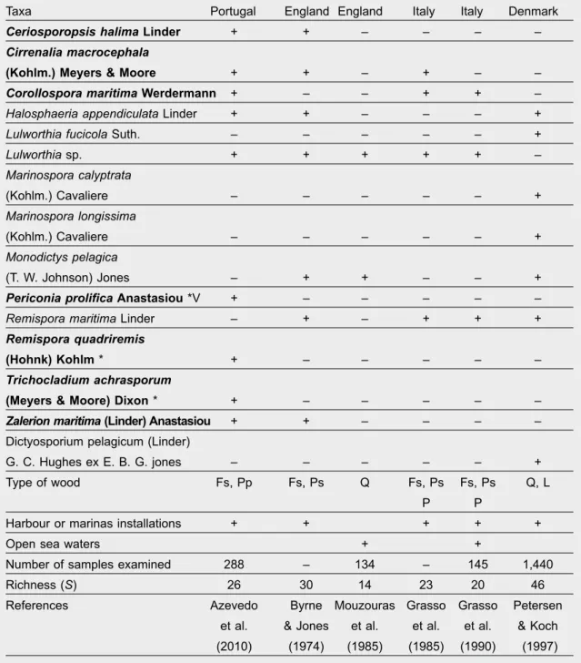
Lignicolous marine mycota occurrence in temperate locations
Table 5 lists the very frequent and frequent marine fungi recorded in this and in other surveys carried out with submerged woods in temperate waters. Sequence analysis of the selected fungi
Comparisons were made between partial sequences of the LSU rDNA region from our isolates and sequen-ces from Genbank. Our sequensequen-ces ranged between 886 and 915 base pairs.
Concerning Lulworthia spp., the results from align-ments and comparisons of sequences from selected
isolates are until now inconclusive to achieve species level (data not shown).
Our isolate of Fusarium sp. (JF746155) shared 99% maximum identity with Fusarium solani (Mart.) Sacc. (EU719659, AY097317, AY097316 and FJ34532), as well as with Fusarium lichenicola C. Massal (AY097325) with query coverage of 99%.
Our isolate of Graphium sp. (JF746156) evidenced maximum identities of 98% with Scedosporium apiospermum Sacc. ex Castell. & Chalm. (FJ345358) and 99% with Pseudallescheria boydii (Shear) Mc-Ginnis, A. A. Padhye & Ajello (AY882372) with query coverage of 100% and 95%, respectively.
The consensus sequence of our isolate of Phoma sp. (JF746158) showed 98% of maximum identity with Loratospora aestuarii Kohlm. & Volkm.– Kohlm. (GU301838) and Coniothyrium obiones Jaap (DQ678054) with query coverage of 99%. When BLASTn was directed to 'Phoma', the results pointed out values of 98% maximum identity and 96% query coverage, with Phoma septicidalis Boerema (GQ387600, GQ387599, GQ387601), Phoma glaucispora (Delacr.) Noordel. & Boerema (GU238078), Phoma violicola P. Syd. (GU238156), Phoma fallens Sacc. (GU238074), Phoma vasinfecta Boerema, Gruyter & Kesteren (GU238151), Phoma dimorphospora (Speg.) Aa & Kesteren (GU238069), Phoma carteri Gruyter & Boerema (GQ387594, GQ387593), Phoma flavigena Constant. & Aa (GU238076), Phoma betae A. B. Frank (EU754179, EU754178), Phoma heteromorphospora Aa & Kesteren (EU754188, EU754187), and Phoma
Table 3. Comparison of diversity indices per environment. Tabla 3. Comparación de los índices de diversidad por ambiente.
Cascais Sesimbra Cascais Sesimbra Cascais Sesimbra Total (144 baits) F. sylvatica (72 baits) P. pinaster (72 baits)
S 15 23 10 18 12 20
H’ 2.21 2.48 1.87 2.27 2.10 2.41
E 0.82 0.79 0.81 0.77 0.84 0.81
CS 0.58 0.64 0.63 (J = 11, a = 15, b = 23) (J = 9, a = 10, b = 18) (J = 10, a = 12, b = 20)
Table 4. Comparison of the diversity indices per substratum: Fs. Fagus sylvatica; Pp. Pinus pinaster. Tabla 4. Comparación de los índices de diversidad por sustrato: Fs. Fagus sylvatica; Pp. Pinus pinaster.
Cascais + Sesimbra Cascais Sesimbra (144 baits) (144 baits) (72 baits) (72 baits) (72 baits) (72 baits) Fs Pp Fs Pp Fs Pp S 19 22 10 12 18 20 H’ 2.16 2.32 1.87 2.10 2.27 2.41 E 0.73 0.76 0.81 0.84 0.77 0.81 CS 0.73 0.64 0.79 (J = 15, a = 19, b = 22) (J = 7, a = 10, b = 12) (J = 15, a = 18, b = 20) apiicola Kleb. (GQ387601) all of them with a query
coverage of 96%.
The isolate of Stachybotrys sp. (JF746157) shared 100% maximum identity with Stachybotrys chartarum (Ehrenh, ex Link) Hughes (AY489712) with query coverage of 99%.
Discussion
Marine mycota collected from wood baits submerged in temperate regions
This analysis includes the total mycota detected on the survey of Azevedo et al. (2010).
The data of frequency of occurrence highlight the increase of the very frequent fungi (four taxa) in rela-tion to the results reported by Azevedo et al. (2010) because C. maritima and Z. maritima (very frequent fungi) and R. quadriremis (frequent fungus) were not detected by direct observation.
The average number of fungi per Fagus sylvatica and Pinus pinaster baits increased respectively from
1.70 to 2.88 and from 1.92 to 3.68. This shows how the incubation on moist chambers significantly contrib-uted for the differentiation of reproductive structures from the marine fungi mycelia already present when direct observations were carried out (Azevedo et al., 2010; table 2).
The diversity was significantly higher at Sesimbra than at Cascais and in P. pinaster than in F. sylvatica baits; a highly significant value was obtained when comparisons were done only with baits from Cascais. It is to be stressed that no significant differences were found between the two types of baits from Sesimbra marina when comparisons were made only with results of direct observations (Azevedo et al., 2010).
The values of fungal similarity (Cs) decreased for all analyzed situations when compared with the results presented by Azevedo et al. (2010). This evidences the advantages of using different types of substrata and subjecting them to long incubation periods in order to achieve better inventories of marine fungal communities. Evenness values indicate that individuals recorded for each species were more evenly abundant in Cas-cais marina and for P. pinaster baits.
Table 5. Very frequent and frequent marine fungi recorded in submerged wood at temperate locations: Fs. Fagus sylvatica; Pp. Pinus pinaster; Ps. Pinus sylvestris; Q. Quercus sp.; P. Populus sp.; L. Larix sp.; + Present; – Absent; In bold, exclusive taxa to baiting method; * This paper.
Tabla 5. Hongos marinos frecuentes y muy frecuentes registrados en maderas sumergidas en áreas templadas: Fs. Fagus sylvatica; Pp. Pinus pinaster; Q. Quercus sp.; P. Populus sp.; L. Larix sp.; + Presente; – Ausente; en negritas, taxones exclusivos del método de los cebos; * Este estudio.
Taxa Portugal England England Italy Italy Denmark
Ceriosporopsis halima Linder + + – – – –
Cirrenalia macrocephala
(Kohlm.) Meyers & Moore + + – + – –
Corollospora maritima Werdermann + – – + + –
Halosphaeria appendiculata Linder + + – – – +
Lulworthia fucicola Suth. – – – – – +
Lulworthia sp. + + + + + – Marinospora calyptrata (Kohlm.) Cavaliere – – – – – + Marinospora longissima (Kohlm.) Cavaliere – – – – – + Monodictys pelagica (T. W. Johnson) Jones – + + – – +
Periconia prolifica Anastasiou *V + – – – – –
Remispora maritima Linder – + – + + +
Remispora quadriremis
(Hohnk) Kohlm * + – – – – –
Trichocladium achrasporum
(Meyers & Moore) Dixon * + – – – – –
Zalerion maritima (Linder) Anastasiou + + – – – –
Dictyosporium pelagicum (Linder)
G. C. Hughes ex E. B. G. jones – – – – – +
Type of wood Fs, Pp Fs, Ps Q Fs, Ps Fs, Ps Q, L
P P
Harbour or marinas installations + + + + +
Open sea waters + +
Number of samples examined 288 – 134 – 145 1,440
Richness (S) 26 30 14 23 20 46
References Azevedo Byrne Mouzouras Grasso Grasso Petersen et al. & Jones et al. et al. et al. & Koch (2010) (1974) (1985) (1985) (1990) (1997)
Studies in temperate open coastal waters relative to wood inhabiting fungi are based both in submerged and in drift or intertidal wood. When comparing the results of the survey of Azevedo et al. (2010) with
other surveys carried out in temperate waters, differ-ences found in fungal richness (table 5) could be due to the different nature of the woods used, to duration and depth of submersions in sea water and also to
different abiotic conditions (oxygen, temperature, salinity) to which the woods were subjected as well as to the number of analyzed samples.
Lulworthia species were the most common fungi (present in five surveys), followed by Remispora maritima (observed in four surveys) C. maritima, H. appendiculata and M. pelagica (observed in three surveys) (table 5). The most common species can be considered species that play an important role in wood degradation (Alias & Jones, 2000). Additionally, considering the results expressed in table 2, it is to be emphasized that, for some of these taxa, there are references to production of enzymes and bio-compounds. Bucher et al. (2004) reported production of cellulase, xylanase and peroxidase for one isolate of Lulworthia sp., and laccase for T. achrasporum. In relation to C. maritima, Jensen & Fenical (2002) found that an isolate of this fungus was able to pro-duce a new secondary metabolite (Corollosporine) and Bucher et al. (2004) referred the production of cellulase and xylanase.
Sequence analysis of the selected fungi
The sequence data obtained suggest that our isolate of Fusarium sp. is closely related to Fusarium solani and F. lichenicola. However, the morphological cha-racters are only compatible with the descriptions of Domsch & Gams (1980) and Samson et al. (2002) for F. solani as well as with the dichotomous key pre-sented by Samson et al. (2002) for Fusarium species. Taking together morphological and molecular data, our isolate was considered to be Fusarium solani.
Concerning Graphium sp., the result indicating identity with Scedosporium apiospermum was evalu-ated, although the morphological features of our iso-late (figs. 2I, 2J, 2K, 2L) did not correspond with the description of this fungus (www.mycobank.org). The molecular results also revealed a close relation to the teleomorph Pseudollescheria boydii. It is worth noth-ing that Graphium eumorphum (Sacc.) is described as anamorph of this fungus (www.mycobank.org). The morphological features of our isolate are in accordance with the original description of Saccardo (www.index-fungorum.org), with slight differences on the length of conidia. For this reason, our isolate was considered Graphium eumorphum.
For the isolate of Phoma sp., our molecular re-sults pointed out members of two other genera (L. aestuarii and C. obiones) as well as 11 species of Phoma that have never been described for marine habitats (Jones et al., 2009) Eight of these species of Phoma are included in clade 7 (Leptosphaeriaceae and Pleosporaceae) in the study performed with 159 species of Phoma and its associated teleomorphs by Aveskamp et al. (2010). These authors recognize the complexity of this group, which is considered to be one of the largest fungal genera. This explains why a better identification of our isolate was not achieved, also because only one DNA region was accessed by sequence data.
Concerning our isolate of Stachybotrys sp., com-parisons of sequence data support the coincidence
found between our morphological characterization and the one made by Samson et al. (2002) for Stachybotrys chartarum (= S. atra corda). S. atra was referred by Jones et al. (2009) for marine environ-ments, however indicating conidia dimensions slightly smaller. For this reason our molecular data were de-terminant in considering our isolate as Stachybotrys chartarum (= S. atra).
Finally, regarding Lulworthia spp., our results pointed out the necessity of further analysis to accom-plish the objective of characterizing the Portuguese isolates. The phylogenetic trees recently proposed for Lulworthiales comprise many isolates to be identified down to species level (Campbell et al., 2005; Jones et al., 2009) as well. We intend to contribute for the establishment of phylogenetic relationships within this taxon with the molecular characterization (still currently underway) of our isolates.
In conclusion, this molecular approach pursuing a contribution for the identification of these Portu-guese isolates down to species level showed to be valuable as this goal could be achieved for three of them (Fusarium solani, Graphium eumorphum and Stachybotrys chartarum). However, the sequenc-ing of more regions always allows more accurate results. This procedure will be mandatory for more complex taxa such as Lulworthia spp., and Phoma sp., those that, in this study, remain to be better characterized.
Acknowledgements
We thank Francisco Caeiro for reviewing this ma-nuscript. This work was partially funded by CBA and CESAM.
References
Alias, S. A. & Jones, E. B. G., 2000. Colonization of mangrove wood by marine fungi at Kuala Selangor stand, Malaysia. Fungal Diversity, 5: 9–21. Alva, P., Mckenzie, E. H. C., Poiting, S. B., Pena–
Murrala, R. & Hyde, K. D., 2002. Do sea grasses harbour endophytes? In: Fungi marine environ-ments: 167–178 (K. D. Hyde, Ed.). Fungal Diversity Press, Hong Kong.
Aveskamp, M. M., De Gruyter, J., Woudenberg, J. H. C., Verkley, G. J. M. & Crous, P. W., 2010. High-lights of the Didymellaceae: A polyphasic approach to characterise Phoma and related pleosporalean genera. Studies in Mycology, 65: 1–60.
Azevedo, E., Rebelo, R., Caeiro, M. F. & Barata, M., 2010. Diversity and richness of marine fungi on two Portuguese marinas. Nova Hedwigia, 90(3–4): 521–531.
Barata, M., 1997. Fungos marinhos superiores as-sociados com Spartina maritima em estuários da Costa Portuguesa. Ph. D. Thesis, Lisbon Univ. – 2002. Fungi in the Halophyte Spartina maritima
in salt marsh. In: Fungi marine environments: 179–193 (K. D. Hyde, Ed.). Fungal Diversity Press,
Hong Kong.
Barghoorn, E. S. & Linder, D. H., 1944. Marine fungi: their taxonomy and biology. Farlowia, 1: 395–467. Bryne, P. J. & Jones, E. B. G., 1974. Lignicolous marine fungi. Veroff. Inst. Meeresforsch. Bremerh. Suppl, 5: 301–320.
Bucher, V. V. C., Hyde, K. D., Pointing, S. B. & Reddy, C. A., 2004. Production of wood decay enzymes, mass loss and lignin solubilization in wood by marine ascomycetes and their anamorphs. Fungal Diversity, 15: 1–14.
Campbell, J. Volkmann–Kohlmeyer, B., Grafenhan, T. Spatafora, J. W. & Kohlmeyer, J., 2005. A re– evaluation of Lulworthiales: relationships based on 18S and 28S rDNA. Mycological Research, 109(5): 556–568.
Domsch, K. H. & Gams, W., 1980. Compedium of soil Fungi. Volume I. Academic Press, London. Figueira, D. & Barata, M., 2007. Marine fungi on two
sandy beaches. Mycologia, 99(1): 20–23. Gessner, R. V. & Kohlmeyer, J., 1977. Seasonal
Oc-currence and distribution of fungi associated with Spartina alterniflora from Rhode Islands estuary. Mycologia, 69: 477–491.
Gonzálvez, M. C., Herrera, T., Uloa, M. & Hanlin, R., 1998. Abundance and diversity of microfungi in three coastal beaches of Mexico. Mycoscience, 39: 115–121.
Gonzálvez, M. C., Hanlin, R. T. & Ulloa, M., 2001. A checklist of higher marine fungi of Mexico. Myco-taxon, LXXX: 241–253.
Grasso, S., Panebianco, C. & La Ferla, R., 1990. Lignicolous marine fungi in the straits of Messina, Italy. Hydrobiologia, 206: 149–154.
Grasso, S. & La Ferla, R. & Jones, E. B. G., 1985. Lignicolous marine fungi in a Harbour Environments (Millazo). Botanica Marina, XXVIII: 259–264. Hall, T. A., 1999. BioEdit: a user–friendly biological
sequence alignment editor and analysis program for Windows 95/98/NT, Nucleic Acids Symp. Ser, 41: 95–98.
Hyde, K. D. & Sarma, V. V., 2000. Pictorial Key to higher marine fungi. In: Marine Mycology–A practi-cal approach: 205–270 (K. D. Hyde & S. B. Point-ing, Eds.). Fungal Diversity Press, Hong Kong. Hyde, K. D., Sarma, V. V. & Jones, E. B. G., 2000.
Morphology and taxonomy of higher marine fungi. In: Marine Mycology–A practical approach: 172– 204. (K. D. Hyde & S. B. Pointing, Eds.). Fungal Diversity Press, Hong Kong.
Jensen, P. R. & Fenical, W., 2002. Secondary me-tabolites from marine fungi. In: Fungi in Marine environments: 293–315 (K. D. Hyde, Ed.). Fungal Diversity Press, Hong Kong.
Jones, E. B. G., Pilantanapak, A., Chatmala, I., Sakayaroj, J., Phongpaichit, S. & Choeyklin, R., 2006. Thai marine fungal diversity. Songklankarin J. Sci. Technol., 28(4): 687–708.
Jones, E. B. G., Sakayaroj, J., Suetrong, S., Somirithipol, S. & Pang, K. L., 2009. Classifica-tion of marine Ascomycota, Anamorphic fungi and Basidiomycota. Fungal Diversity, 35: 1–187. Koch, J., 1974. Marine fungi on driftwood from the
west coast of Jutland Denmark. Friesia, X(4–5): 209–250.
Koch, J. & Petersen, K. R. L., 1996. A check list of higher marine fungi on wood from Danish coast. Mycotaxon, 15: 397–414.
Kohlmeyer, J. & Kohlmeyer, E., 1979. Marine Mycol-ogy–the higher fungi. Academic Press, New York. Kohlmeyer, J. & Volkmann–Kohlmeyer, B., 1991.
Il-lustrated key of filamentous higher marine fungi. Botanica Marina, 34: 1–61.
– 2001. Fungi on Juncus roemarianus. 16. More New. Coelomycetes, including Tetranacriella. gen. nov. Botanica Marina, 44: 147–156.
– 2002. Fungi on Juncus and Spartina: New mari-ne species of Anthostomella, with a list of fungi Know for Spartina. Mycological Research, 106(3): 365–374.
– 2003. Marine Ascomycete from algae and animal hosts. Botanica Marina, 46: 285–306.
Landy, E. T. & Jones, M. G., 2006. What is the Fun-gal Diversity of Marine Ecosystems in Europe. Mycologist, 20: 15–21.
Lintott, W. H. & Lintott, E. A., 2002. Marine fungi from New Zeland. In: Fungi in Marine Environments 285–292 (K. D. Hyde, Ed.). Fungal Diversity Press, Hong Kong.
Maria, G. L. & Sridhar, K. R., 2003. Diversity of fila-mentous fungi on woody litter of five mangrove plant species from the southwest coast of India. Fungal Diversity, 14: 109–126.
Morrison–Gardiner, S., 2002. Dominant fungi from Australian coral reefs. Fungal Diversity, 9: 105–121. Mouzouras, R., Jones, E. B. G., Venkatasamy, R. &
Moss, S. T., 1989. Decay of wood by micro–organ-isms in marine environments. B. W. P. A. Annual Convention.
Petersen, K. R. L. & Koch, J., 1997. Substrate pref-erence and vertical zonation of lignicolous marine fungi on Mooring posts of Oak (Quercus sp.) and Larch Larix sp. in Svanemollen (Denmark). Bo-tanica Marina, 40(1–6): 451–464.
Poon, M. O. K. & Hyde, K. D., 1998. Biodiversity of Intertidal estuarine fungi on Phragmites at Mai Po Marshes, Hong Kong. Botanica Marina, 41: 141–155.
Ravikumar, M., Sridhar, K. R.., Sivakumar, T., Ka-ramchand, K. S., Sivarkumar, N. & Vellauyan, R., 2009. Diversity of filamentous fungi on coastal debris after the tsunami of southeast coast of India. Czech Mycol, 61(1): 107–115.
Sakayaroj, J., Preedanon, S., Supaphon, O., Jones E. B. G. & Phongpaichit, S., 2010. Phylogenetic diversity of endophyte assemblages associated with the tropical seagrass Enhalus acoroides in Thailand. Fungal Divers, 42: 27–45.
Samson, R. A., Hoekstra, E. S., Frisvad, J. C., Filten-borg, O., 2002. Introduction to food–and Airborne Fungi. Sixth edition, Centraalbureau Voor Schim-melcultures, Utrecht.
Steinke, T. D. & Lubke, R. A., 2003. Arenicolous marine fungi from Southern Africa. South African Journal of Botany, 69(4): 540–546.
Succession of fungi on wood Avicennia alba and A. lanata in Singapore. Canadian Journal Botany, 67: 2686–2691.
Thompson, J. D., Gibson, T. J., Plewniak, F., Jean-mougin, F. & Higgins, D. G., 1997. The CLUSTALX windows interface: flexible strategies for multiple sequence alignment aided by quality analysis tools, Nucleic Acids Research, 25: 4876–4882.
Torzilli, A. P., Sikaroodi, M., Chalkley, D. & Gillevet, P. M., 2006. A comparison of fungal communities four salt marsh plant using automated ribosomal intergenic spacer analysis (ARISA). Mycologia, 98(5): 690–698.
Vrijmoed, L. L. P., 2000. Isolation and culture of higher fungi In: Marine Mycology – a practical approach: 1–20 (K. D. Hyde & S. B. Poiting, Eds.). Fungal Diversity Press, Hong Kong.
Vrijmoed, L. L. P., Hodgkiss, I. J. & Thrower, L. B.,
1982. Seasonal patterns of primer colonization of lignicolous marine fungi in Hong Kong. Hydrobio-logia, 89: 253–262.
– 1986. Occurrence of fungi on submerged pine and teak blocks in Hong Kong coastal waters. Hydrobiologia, 135: 109–122.
Zar, J. H., 1999. Biostatistical Analysis. Fourth Edition. Prentice Hall, New Jersey.
Zuccaro, A., Schoch, C. L., Spatafora, J. W., Kohlmeyer, J., Draeger, S. M. & Mitchell, J. I., 2008. Detection and Identification on Fungi intimately associated with the brown Seaweed Fucus serratus. Applied and Environmental Microbiology, 74(4): 931–941. Wong. M. K. M. & Hyde, K. D., 2002. Fungal saprobes
on standing grasses end sedges in a subtropical aquatic habitat. In: Fungi in Marine Environments: 195: 212 (K. D. Hyde, Ed.). Fungal Diversity Press, Hong Kong.