IN ST IT U T O D E C IÊ N C IA S B IO M ÉD IC A S A B EL S A LA Z A R
Sa
ra P
in
to d
a S
ilv
a T
ele
s. D
isc
lo
sin
g (
ep
i)g
ene
tic m
ec
ha
nis
m
s
w
ith po
te
nti
al v
alu
e a
s p
re
di
cti
ve m
ar
ke
rs o
f t
he
ra
py r
es
po
nse
in
G
as
tric
C
an
ce
r
Di
sc
lo
sin
g (
ep
i)g
en
et
ic me
ch
an
ism
s w
ith p
ot
en
tial
val
ue a
s p
re
di
ctiv
e m
ar
ke
rs o
f t
he
ra
py r
es
pon
se i
n
G
as
tr
ic C
an
ce
r
Sa
ra P
in
to d
a S
ilv
a T
eles
Disclosing (epi)genetic mechanisms with
potential value as predictive markers of
therapy response in Gastric Cancer
Sara Pinto da Silva Teles
M
2018M
.ICB
AS
2018
MESTRADO ONCOLOGIA MOLECULARSara Pinto da Silva Teles
Disclosing (epi)genetic mechanisms with potential value as
predictive markers of therapy response in Gastric Cancer
Tese de candidatura ao grau de Mestre em
Oncologia Molecular submetida ao Instituto de
Ciências Biomédicas Abel Salazar da Universidade
do Porto
Orientador Doutora Carla Oliveira
Categoria Investigadora Principal e Professora
Afiliada
Afiliação Instituto de Investigação e Inovação
em Saúde/ Instituto de Patologia e
Imunologia Molecular da
Universidade do Porto,
Faculdade de Medicina da
Universidade do Porto
Co-orientador Doutora Patrícia Oliveira
Categoria Investigadora
Afiliação Instituto de Investigação e Inovação
em Saúde/ Instituto de Patologia e
Imunologia Molecular da
Universidade do Porto
Co-orientador Doutora Fátima Gärtner
Categoria Professora Catedrática e
Investigadora
Afiliação Instituto de Investigação e Inovação
em Saúde/ Instituto de Patologia e
Imunologia Molecular da
Universidade do Porto, Instituto de
Ciências Biomédicas Abel Salazar
da Universidade do Porto
Agradecimentos
O trabalho que apresento é o culminar de 3 anos de aprendizagem no grupo de investigação da Dra. Carla Oliveira, no qual tive a oportunidade de desenvolver capacidades importantes como autonomia, comunicação científica, pensamento crítico e trabalho de equipa.
Em primeiro lugar, queria agradecer às minhas orientadoras. À Dra. Carla Oliveira por me ter dado a oportunidade de realizar o estágio da licenciatura em Biologia no seu grupo, e mais tarde a dissertação de mestrado. Se não me tivesse dado essa primeira oportunidade, não teria tido depois esta experiência valiosa de 3 anos e evoluído tanto cientificamente. À Patrícia Oliveira, que estará para sempre na base de todo o meu conhecimento científico. Todas as capacidades que desenvolvi nos últimos 3 anos estão diretamente relacionadas com os ensinamentos que a Patrícia me passou e, sem ela, não teria metade das qualidades que considero ter hoje. Agradeço toda a preocupação e entrega que sempre me dedicou, esperando ser, depois de todo esse esforço, um motivo de orgulho.
Todo o grupo ERiC contribuiu para o meu desenvolvimento nestes anos e aprendi um pouco com todos os seus membros. Em especial, agradeço à Sara Rocha por ter estado sempre ao meu lado e pronta para me transmitir todo o seu conhecimento e experiência, tendo sempre os melhores conselhos para me dar. Agradeço também à Joana Carvalho, por disponibilizar sempre o seu tempo e conhecimento para me ajudar, tendo sido crucial para a minha evolução e a do meu trabalho durante o mestrado. Obrigada à Carla Pereira por todo o apoio que também me deu ao longo dos 3 anos, sempre com a paciência que lhe é caraterística. Obrigada à Marta Ferreira por todo o contributo bioinformático que deu, tão importante neste trabalho, e obrigada à Anabela Ferro por torcer sempre por mim! Finalmente, quero agradecer aos meus pais e ao João, por acompanharem todas as minhas conquistas e dificuldades, estando intrinsecamente ligados à minha confiança e capacidade de continuar em frente. Sem eles, não seria possível concretizar esta nova etapa.
Table of Contents
Agradecimentos 2 Table of Contents 3 Abstract/Resumo 5 I. Introduction 9 1. Gastric cancer 91.1. Clinical and histological characterization 9
1.2. Molecular characterization 10
1.3. Current management of the disease 11
2. Targeted therapy in gastric cancer 12
2.1. Current targeted therapies approved for Gastric Cancer
treatment: anti-HER2 and anti-VEGFR2 12 2.2. Targeted therapies tested in Gastric Cancer without approval 14
2.2.1. Targeting VEGFA in GC 14
2.2.2. Targeting EGFR in GC 16
2.2.3. Targeting MET/HGF in GC 17
2.2.4. Targeting FGFR2 in GC 18
3. FGFR2 and VEGFR2 signalling in Gastric Cancer 19
3.1. FGFR2 20
3.1.1. FGFR2 signalling in the normal and cancer contexts 20 3.1.2. ESRP1 and the alternative splicing of FGFR2 22 3.2. VEGFR2 signalling in the normal and cancer contexts 23 4. Epigenetic mechanisms controlling gene expression in gastric cancer 25
4.1. DNA methylation 26
4.2. How to explore gene-specific and genome-wide DNA
methylation 27
II. Rationale and Aims 30
III. Materials and Methods 33
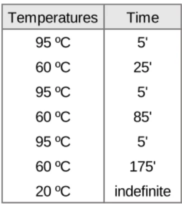
1. Gastric cancer samples 33
2. Cell culture 33
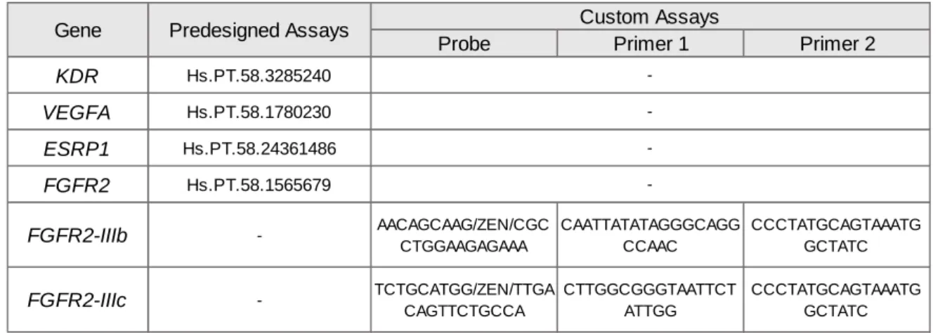
3. DNA extraction and methylation analysis 33 4. Copy number variation quantification by qRT-PCR 37
5. RNA extraction and expression quantification 37
6. 5-Aza-2′-deoxycytidine treatment 38
7. Bioinformatics Approach using TCGA Cohorts 2 and 3 39
IV. Results and Discussion 40
PART 1: KDR/VEGFR2 and VEGFA genetic, epigenetic and transcriptomic events in Gastric Cancer
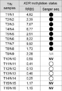
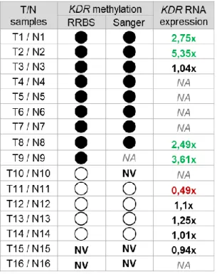
40 1.1. Correlating KDR promoter methylation status with KDR RNA expression in gastric tumour vs. normal samples
40
1.2. Correlating VEGFA copy number with VEGFA RNA expression in gastric tumour vs. normal samples
45
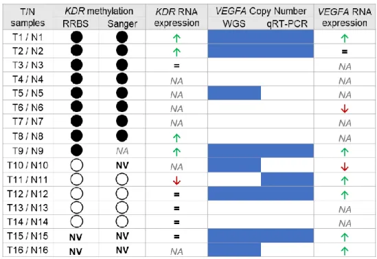
1.3. Correlating KDR promoter methylation status and RNA expression with VEGFA copy number and RNA expression in gastric tumour vs. normal samples
49
1.4. Correlating KDR promoter methylation with VEGFA copy number and VEGFA and KDR RNA expression in other GC cohorts
51
PART 2: FGFR2 and ESRP1 epigenetic and transcriptomic events in Gastric Cancer
58 2.1. Correlating FGFR2 promoter methylation status with total
FGFR2, FGFR2-IIIb and FGFR2-IIIc RNA expression in tumour vs.
normal samples
58
2.2. Correlating ESRP1 promoter methylation status with ESRP1 RNA expression in tumour vs. normal samples
63
2.3. Correlating ESPR1 promoter methylation status and RNA expression with FGFR2 promoter methylation and FGFR2-IIIb RNA expression in tumour vs. normal samples
64
2.4. Correlating FGFR2 and ESRP1 promoter methylation with their RNA expression in other GC cohorts
66
PART 3: Exploring the role of ESRP1 in the expression regulation of FGFR2-IIIb expression in GC using in vitro models
74
3.1. Characterizing ESRP1 and FGFR2 promoter methylation status and RNA expression in MCF10A and GC cell lines
74
3.2. Determining the effect of global DNA demethylation of MCF10A E-cells in ESRP1 and FGFR2 RNA expression
76
V. Conclusion 80
VI. Bibliographic References 81
Abstract
Gastric Cancer (GC) is one of the most incident and deadliest cancer types. Most cases are diagnosed in advanced stages, with few treatment options available. Several clinical trials have taken place, using antibodies targeting proteins frequently altered in GC, such as the receptor tyrosine kinases HER2, VEGFR2 and FGFR2, or growth factors like VEGFA.
Trastuzumab (anti-HER2) and Ramucirumab (anti-VEGFR2) are clinically approved
targeted therapies in GC, however their impact in patient overall survival is poor. Whereas anti-HER2 therapy is chosen based on the overexpression and/or genetic amplification of
ERBB2/HER2 (15-30% of GC cases), no biomarker has been found that could predict
response to anti-VEGFR2, anti-VEGFA or anti-FGFR2 therapies for GC treatment. This is important to select cases for clinical trials and may explain the failure of some of them or the low therapy response to the targeted therapies already tested in these trials.
Although the overexpression of VEGFR2, VEGFA and FGFR2 has been reported in GC at various frequencies, the underlying genetic or epigenetic mechanisms are still unclear. Therefore, our general aim was to explore the potential of the promoter methylation status of KDR (codifying VEGFR2) and FGFR2 as novel predictive biomarkers for anti-VEGFR2 and anti-FGFR2 therapies in GC.
A genome wide preliminary analysis assessing promoter methylation and copy number variants was performed for a cohort of GC and matched normal gastric samples (n=47, Cohort 1), by RRBS and WGS, respectively. This study revealed that the majority of tumour samples presented KDR (encoding for VEGFR2) promoter hypermethylation, normal
VEGFA copy number (17% displayed VEGFA amplification) and FGFR2 promoter
hypomethylation in comparison with their corresponding normal samples. Moreover, the promoter of ESRP1, a known splicing regulator of FGFR2 responsible for the differential expression of FGFR2-IIIb and FGFR2-IIIc isoforms, was also hypomethylated in tumour samples. Considering this preliminary data, and to address our general aim, we established two specific aims: in specific aim 1, we investigated the co-occurrence of variations in KDR promoter methylation status, VEGFA copy number and KDR and VEGFA RNA expression and; in specific aim 2, we investigated the co-occurrence of variations in FGFR2 and ESRP1 promoter methylation status and RNA expression of both genes as well as FGFR2 isoforms. Both specific aims were explored using selected cases from Cohort 1 for which the promoter methylation status of each gene was validated by bisulfite Sanger sequencing, copy number status validated using copy number qRT-PCR assays and RNA expression was assessed using qRT-PCR. Results were further investigated using tumour samples from two larger TCGA-derived cohorts, referred as Cohorts 2 and 3.
Results from specific aim 1 showed that KDR and VEGFA were found to be frequently upregulated in tumour samples. However, there was no correlation between KDR promoter methylation, VEGFA copy number status and KDR/VEGFA RNA expression in GC tumour samples. These data support the relevance of exploring KDR and VEGFA RNA expression as putative predictive biomarkers for anti-VEGFR2 and/or anti-VEGFA therapies. Results from specific aim 2 revealed a co-occurrence between FGFR2 and ESRP1 promoter hypomethylation, together with the increased RNA expression of ESRP1 and the epithelial isoform IIIb and decreased RNA expression of the mesenchymal isoform FGFR2-IIIc. This was true in gastric tumour samples and in vitro models. Therefore, the promoter methylation status of FGFR2 and ESRP1, as well as the RNA expression of FGFR2-IIIb,
ESRP1 and FGFR2-IIIc could be promising biomarkers for the selection of GC patients for
anti-FGFR2/FGFR2-IIIb therapies.
Overall, our results demonstrate the importance of considering different alterations within GC tumour cells, rather than only genetic amplification or protein overexpression, to improve the selection of patients that would benefit the most from a given therapy. In the future, further studies using larger GC cohorts with paired normal samples and clinical data available should be performed to understand the potential of RNA expression and/or promoter methylation status as novel predictive biomarkers for anti-VEGFR2, anti-VEGFA and anti-FGFR2/FGFR2-IIIb therapies.
Resumo
O Cancro Gástrico (CG) é um dos tipos de cancro com maior incidência e dos que mais mortes provoca. A maioria dos casos são diagnosticados em estádios avançados da doença, com poucas opções terapêuticas disponíveis. Vários ensaios clínicos foram realizados, usando anticorpos direcionados a proteínas frequentemente alteradas em CG, como os recetores tirosina cinase HER2, VEGFR2 e FGFR2, ou fatores de crescimento como o VEGFA. Trastuzumab (anti-HER2) e Ramucirumab (anti-VEGFR2) são terapias dirigidas aprovadas em CG que, mesmo assim, têm pouco impacto na sobrevivência global dos pacientes. Enquanto que a terapia anti-HER2 é escolhida mediante a sobreexpressão proteica/amplificação genética de ERBB2/HER2 (15-30% dos casos de CG), nenhum biomarcador existe ainda para predizer a resposta a terapias anti-VEGFR2, anti-VEGFA ou anti-FGFR2 para o tratamento de CG.
Embora a sobreexpressão de VEGFR2, VEGFA e FGFR2 já tenha sido reportada em CG em várias frequências, o mecanismo genético ou epigenético subjacente permanece por esclarecer. Portanto, o nosso objetivo geral foi explorar o potencial do estado de metilação dos promotores do KDR (que codifica o VEGFR2) e FGFR2 como novos biomarcadores preditivos para terapias anti-VEGFR2 ou anti-FGFR2 em CG.
Uma análise genómica preliminar a avaliar a metilação de promotores e variantes no número de cópias genéticas foi executada usando uma coorte de amostras tumorais de CG pareadas com amostras normais (n=47, Coorte 1), por RRBS e WGS, respetivamente. Este estudo revelou que a maioria das amostras tumorais apresentava hipermetilação do promotor do KDR, número normal de cópias do VEGFA (17% exibia amplificação do
VEGFA) e hipometilação do promotor do FGFR2, comparativamente com as amostras
normais correspondentes. Para além disso, o promotor do ESRP1, um importante regulador do splicing do FGFR2 responsável pela expressão diferencial das isoformas
FGFR2-IIIb e FGFR2-IIIc, estava também hipometilado nas amostras tumorais.
Considerando estes dados preliminares, e para responder ao nosso objetivo geral, dois objetivos específicos foram estabelecidos: no objetivo específico 1, investigámos a co-ocurrência de variações no estado de metilação do promotor do KDR, no número de cópias do VEGFA e na expressão de ARN do KDR e VEGFA e; no objetivo específico 2, investigámos a co-ocurrência de variações no estado de metilação do promotor do FGFR2 e ESRP1 e na expressão de ARN de ambos os genes, assim como das isoformas do
FGFR2. Ambos os objetivos específicos foram explorados em casos selecionados da
Coorte 1, para os quais o estado de metilação de cada gene foi validado por sequenciação de Sanger de ADN tratado com bissulfito, o número de cópias foi validado por qRT-PCR e
a expressão de ARN foi avaliada por qRT-PCR. Estes resultados foram ainda investigados usando amostras tumorais de duas coortes maiores derivadas do TCGA, referidas como Coortes 2 e 3.
Os resultados do objetivo específico 1 mostraram que os níveis de expressão de ARN do
KDR e VEGFA estavam frequentemente aumentados nas amostras tumorais. No entanto,
não havia nenhuma correlação entre a metilação do promotor do KDR, o número de cópias do VEGFA e a expressão de ARN do KDR e VEGFA em amostras de CG. Estes resultados suportam a relevância de explorar a expressão de ARN do KDR e VEGFA como putativos biomarcadores preditivos para terapias anti-VEGFR2 e/ou anti-VEGFA. Os resultados do objetivo específico 2 mostram uma co-ocurrência da hipometilação do promotor do FGFR2 e ESRP1, em conjunto com o aumento da expressão de ARN do ESRP1 e da isoforma epitelial FGFR2-IIIb e a diminuição de expressão de ARN da isoforma mesenquimal
FGFR2-IIIc. Estas co-ocurrências foram observadas em amostras tumorais gástricas e
modelos in vitro. Assim, o estado de metilação do promotor do FGFR2 e ESRP1, assim como a expressão de RNA do FGFR2-IIIb, ESRP1 e FGFR2-IIIc poderão ser biomarcadores promissores para a seleção de pacientes para terapias anti-FGFR2/FGFR2-IIIb.
Em geral, os nossos resultados demostraram a importância de considerar diferentes alterações nas células tumorais de CG, para além da amplificação genética ou sobreexpressão proteica, para melhorar a seleção dos pacientes que mais irão beneficiar de uma determinada terapia. No futuro, mais estudos usando coortes de CG maiores, com amostras normais pareadas e dados clínicos disponíveis, deverão ser realizados para entender o potencial da expressão de ARN e/ou do estado de metilação dos promotores de genes como novos biomarcadores preditivos para terapias anti-VEGFR2, anti-VEGFA e anti-FGFR2/FGFR2-IIIb.
I. Introduction
1. Gastric cancer
Every year, Cancer threatens millions of people throughout the world, being one of the leading causes of death and one of the biggest concerns of the 21st century. As our lifestyle
habits continue to boost cancer risk and our population grows and ages, the number of people affected by this disease is expected to rise [1, 2]. Each type of cancer presents its own therapeutic challenges, as each represents a disease with different molecular and phenotypical characteristics. As a result, new therapies are being developed based on specific molecular alterations observed in each tumour type, providing tailored solutions for each patient and/or type of cancer. In cases such as breast or lung cancer, finding a specific molecular alteration to target led to major improvements in the survival of selected cancer patients [3, 4]. However, many other types of cancer lack alternatives to standard chemotherapy regimens and therefore patients have little chance of survival. Such is the case of Gastric Cancer (GC).
Gastric cancer is the fifth most incident and third deadliest type of cancer in the world [5]. These numbers reflect mostly the late diagnosis of this disease, often due either to the lack or late presentation of specific symptoms over the course of the disease, and high intra- and inter-tumour heterogeneity [6]. As a result, most patients present advanced stages of the disease at diagnosis and available treatment is largely ineffective. Some of the main risk factors for GC are Helicobacter pylori infection, smoking, male gender, high intake of salted and smoked foods, obesity and Gastroesophageal Reflux Disease (GERD) [7]. The majority of GC cases are adenocarcinomas of the stomach while a small percentage of patients present adenocarcinoma of the gastroesophageal junction. Although most GC cases are of sporadic nature, approximately 10% of patients present familial aggregation, associated with the syndromes Hereditary Diffuse Gastric Cancer (HDGC), Gastric Adenocarcinoma and Proximal Polyposis of the Stomach (GAPPS) and Familial Intestinal Gastric Cancer (FIGC) [8, 9].
1.1. Clinical and histological characterization
The histological classification of GC is mostly governed by the Laurén and World Health Organization (WHO) systems [10, 11]. The Laurén classification divides tumours in two main types: intestinal and diffuse GC. GC cases that do not entirely fit in these two categories are classified as mixed or undetermined types. Tumours of the intestinal type arise generally from intestinal metaplasia, a pre-cancerous lesion characterized by the
replacement of normal gastric cells for differentiated intestinal cells in the stomach epithelium. Tumours of the diffuse type are characterized by undifferentiated and poorly cohesive cells, with no or low gland formation and are considered more aggressive than intestinal GC tumours [12, 13]. The WHO system is composed by four main histological types: tubular and papillary (corresponding to the intestinal type from the Laurén classification), mucinous, poorly cohesive (equivalent to the diffuse type from Laurén classification) and mixed carcinomas [11]. However, this classification does not significantly correlate with clinicopathological or prognostic characteristics [6]. In addition, although the Laurén classification shows clinically relevant GC subgroups, it is still insufficient to improve the current therapeutic landscape. For this reason, unveiling the molecular complexity of
GC tumours could reveal new molecular markers and consequently lead to novel therapies, improving the current outcome of this disease.
1.2. Molecular characterization
A plethora of molecular alterations contribute to the tumorigenesis of GC and are part of the complex biology of this disease. Like all tumours, GC has several characteristic gene mutations, copy number alterations, epigenetic modifications and transcriptional or translational changes. For example, TP53 mutations, amplification/overexpression of
ERBB2/HER2 and E-cadherin expression deregulation are often observed in GC [14-16].
Several studies have aimed at combining different molecular alterations in the hope of finding GC molecular signatures with clinical significance. A seminal paper was published in 2014, by The Cancer Genome Atlas research network (TCGA) combining data derived from whole exome sequencing, array-based DNA methylation and copy number analysis [17], proposing 4 GC subtypes:
• Epstein-Barr virus (EBV) positive tumours, which revealed high levels of DNA promoter hypermethylation (~9%);
• Tumours with microsatellite instability (MSI), with elevated mutational rates and hypermethylation (~22%);
• Genomically stable tumours (~20%);
• Tumours with chromosomal instability (CIN, ~50%).
Although relevant, the clinical significance of this proposed GC molecular stratification has yet to be proven and a consensus on clinically relevant subtypes still needs to be established. Another study aimed at understanding the molecular signatures of GC, was published by the Asian Cancer Research Group (ACRG) which defined 4 GC groups with
different molecular alterations correlated with patient overall survival and recurrence patterns [18]:
• Mesenchymal-like tumours, classified as MSS/EMT, that include those of the diffuse type with worst prognosis and the highest recurrence rates (15%);
• Tumours with microsatellite instability (MSI), presenting hyper-mutated intestinal tumours that also display the best prognosis and lowest recurrence rates (22%); • Tumours with microsatellite stability and active tumour protein 53, referred as
MSS/TP53+, and intermediate prognosis and frequency of recurrence (26%);
• Tumours with microsatellite stability and inactive tumour protein 53, referred as MSS/TP53-, and intermediate prognosis and frequency of recurrence(36%).
These four GC groups derived from distinct expression signatures known to be relevant in the context of GC and were further characterized by somatic alterations. Additionally, the authors verified that the 4 ACRG groups were associated with distinct overall survival benefits, with patients in the MSS/EMT subgroup having the worst prognosis and highest recurrence rates. This study focused on a private GC cohort and later on expanded to two other cohorts to understand the reproducibility of the ACRG proposed GC groups: the TCGA and the Gastric Cancer Project ’08 Singapore cohorts. Despite differences between the cohorts, the ACRG proposed groups were considered applicable to all three cohorts.
Overall, the TCGA and ACRG studies enlighten the heterogeneity of GC reflected in the 4 GC subtypes or groups proposed. However, these classifications are not currently used in the clinical context, although proposed groups may encompass novel therapeutic targets relevant for GC patient management.
1.3. Current management of the disease
Currently, the diagnosis of GC heavily relies on the results obtained from biopsies collected during an endoscopy procedure. After determining the tumour stage, a careful treatment plan is organized by multidisciplinary teams including oncologists, surgeons and radiologists. According to the European Society for Medical Oncology (ESMO) guidelines, patients with early-stage GC are usually fit for surgery with curative potential. In some cases, tumours can be endoscopically removed. However, other patients might need to remove the entire stomach (total gastrectomy) together with pre- and/or postoperative chemotherapy [19]. Nevertheless, the majority of GC cases are diagnosed in advanced stages of the disease. Therefore, surgery is no longer a possibility. In a first-line setting, these patients are treated with a doublet or triplet platinum and fluoropyrimidine regimen.
This type of treatment varies geographically, with the double combination being preferred in Asian countries and the triple regimen being more often used in western countries. This difference likely contributes for the differences observed in the disease outcome between the two populations [20]. In combination with the standard chemotherapy regimen, selected GC patients may be submitted to two targeted therapies: Trastuzumab in a first-line setting and Ramucirumab in a second-line setting. Trastuzumab, the first targeted therapy to be introduced in the treatment plan of GC patients [21], is given to patients with GC tumours expressing the Human Epidermal Growth Factor Receptor 2 (HER2). Ramucirumab, approved for the treatment of metastatic GC, is an antibody targeting the Vascular Endothelial Growth Factor Receptor 2 (VEGFR2) given to patients as a second-line therapy without prior selection [22, 23]. Nevertheless, the survival improvement of GC patients
submitted to these targeted therapies is poor, hence GC patients remain without effective long-term solutions.
2. Targeted therapy in gastric cancer
2.1. Current targeted therapies approved for Gastric Cancer treatment: anti-HER2 and anti-VEGFR2
Over the last decades, several attempts have been made to establish new targeted therapies for GC treatment and improve the current outcome of this disease. However, most clinical trials have failed to present any significant advantage in the addition of these new therapies to the standard chemotherapy regiments. Currently, only two targeted therapies have been approved for GC treatment: the monoclonal antibodies Trastuzumab and
Ramucirumab.
Trastuzumab targets HER2, a known tyrosine kinase receptor involved in many cellular
functions important for tumour development [15]. Unlike other members of the HER protein family, HER2 activation is independent of ligand binding. This receptor is either constitutively activated or dimerizes with itself or other HERs, resulting in the initiation of several signalling pathways that contribute to increased cell proliferation, differentiation and invasion [15]. Trastuzumab targets the extracellular domain of HER2, impairing receptor
dimerization and consequent activation of the tyrosine domain. Moreover, this receptor usually undergoes cleavage, leaving behind a phosphorylated portion that can lengthen the stimulus induced by the HER2 activation. Trastuzumab also prevents this cleavage and further pathway stimulation and is believed to induce cell-mediated cytotoxicity and endocytosis [24].
Trastuzumab was firstly introduced to the clinical practice as a new targeted therapy for
advanced breast cancer patients with tumours overexpressing HER2 [25]. Years later, the focus turned to GC given the similar rates of HER2 overexpression (15-30%), which is mainly caused by gene amplification, resulting in the worse prognosis of patients carrying these alterations [15, 26]. Consequently, Trastuzumab became the first approved targeted therapy for GC treatment after completion of the Trastuzumab for Gastric Cancer (ToGA) clinical trial [21]. In this trial, patients with inoperable locally advanced tumours were randomly assigned to two treatment groups: chemotherapy plus Trastuzumab and; chemotherapy alone. The authors observed that the first group presented a significant increase of 2.7 months in median overall survival. Next, a stratification of tumours based on HER2 expression was performed using an adaptation of the score described by Hofmann
et al [27]. The post-hoc analysis showed that by dividing patients with tumours with HER2
high versus patients with HER2 low expression, the difference in survival was even stronger: 4.2 months [21]. Therefore, the current consensus is to screen GC patients diagnosed with inoperable advanced disease for HER2 protein expression by immunohistochemistry (IHC) and ERBB2/HER2 gene amplification by fluorescence in-situ hybridisation (FISH). For IHC, tumours are scored from 0 to 3+, with tumours classified as 2+ or 3+ presenting either a weak to moderate (2+) or strong (3+) staining for HER2 in 10% or more tumour cells. Tumours that are classified as HER2 IHC 3+ or tumours presenting both HER2 IHC 2+ and FISH-assay positive results are considered eligible for a combination of chemotherapy and Trastuzumab [19].
Ramucirumab is a monoclonal antibody that targets VEGFR2 and has been vastly
investigated as a new anti-angiogenic therapy for a variety of cancer types [28]. VEGFR2 is one of the main regulators of angiogenesis, i.e. the formation of new blood vessels from pre-existing ones and one of the most important factors driving tumour growth [29]. This receptor, like the other VEGFR protein family members, can be activated in two distinct manners, both causing tyrosine phosphorylation and downstream signalling initiation: 1) through the canonical pathway, by VEGF ligand binding and; 2) through the non-canonical pathway, by non-VEGF ligands or mechanical forces, such as shear stress [30].
Ramucirumab is highly specific for the extracellular domain of VEGFR2, having a great
binding affinity for this receptor, impairing VEGFR2 interaction with ligands [31].
To understand the relevance of targeting VEGFR2 in the setting of GC, a pre-clinical trial was performed using a mouse-specific anti-VEGFR2 antibody in mouse GC xenograft models [32]. This study revealed that by blocking VEGFR2, tumour growth in vivo was inhibited, strengthening the therapeutic importance of targeting VEGFR2 [32]. Subsequent safety and dose-finding trials were performed in cancer patients and Ramucirumab was
eventually approved for GC treatment after completion of two phase III studies: the REGARD and RAINBOW trials [32]. In the REGARD trial, 355 patients with metastatic or inoperable gastric or gastroesophageal cancer with disease progression after first-line treatment were randomly assigned to receive Ramucirumab or placebo. Patients treated with the antibody presented an increase of 1.4 months in overall survival [22]. Simultaneously, the RAINBOW trial initiated the safety and efficacy assessment of
Ramucirumab combined with Paclitaxel in patients with gastric or gastroesophageal
adenocarcinoma who had progressed after first-line chemotherapy. Patients randomly assigned to the Ramucirumab plus Paclitaxel group presented a significantly improved median overall survival in comparison with the Paclitaxel alone group: 9.6 months vs. 7.4 months, respectively [23]. The results from these two trials culminated in the approval of
Ramucirumab as a second-line therapy, combined with chemotherapy, for patients with
gastric or gastroesophageal cancer after disease progression. Nevertheless, contrarily to
Trastuzumab, there are currently no biomarkers to predict response to Ramucirumab
therapy. The authors from both trials performed exploratory analyses to find potential predictive biomarkers [33, 34]. However, the level of assessed VEGFRs, VEGFs or other cytokines in either tumour or serum samples failed to present any significant association with Ramucirumab efficacy. Therefore, there is still the need for appropriate predictive biomarkers to select patients that will benefit the most from Ramucirumab therapy.
2.2. Targeted therapies tested in Gastric Cancer without approval
Although Trastuzumab and Ramucirumab are currently approved for GC treatment, the poor survival benefit underlies the continuous effort for development of novel targeted therapies. However, most have yet to present any significant survival benefit for GC patients. The majority of these unsuccessful therapies target known cancer-associated receptors/ligands such as the Vascular Endothelial Growth Factor A (VEGFA), Epidermal Growth Factor Receptor (EGFR), Hepatocyte Growth Factor Receptor (MET) and Fibroblast Growth Factor Receptor 2 (FGFR2).
2.2.1 Targeting VEGFA in GC
VEGFA was the first cytokine and member of the VEGF family to be discovered as a tumour-associated angiogenic factor, being the principal regulator of this complex process [35]. This family also encompasses VEGFB, VEGFC, VEGFD and placental growth factor (PlGF), which have different affinities to the VEGF-receptors VEGFR1, VEGFR2 and VEGFR3,
which in turn have distinct kinase activities. Together, VEGFs and VEGFRs are key regulators of important processes such as vasculogenesis, angiogenesis, lymphangiogenesis and vascular permeability, both in the physiological and pathological context [36]. In GC, VEGFA overexpression and amplification was seen in up to 58% and 7% of cases, respectively [37]. Many studies have tried to correlate VEGFA protein expression with patient prognosis, however presented contradictory results [38]. More recently, a meta-analysis showed that VEGFA expression was associated with poor overall survival and disease-free survival of GC patients [38]. Hence, successfully targeting VEGFA in GC continues to be an objective of researchers that try to implement new effective treatments for these patients.
Bevacizumab was the first monoclonal antibody designed to target VEGFA and is currently
approved for treatment of several cancer types, such as advanced colon, lung, breast, ovarian, endometrial and clear cell renal carcinoma [39-43]. In advanced GC, Bevacizumab was the first anti-angiogenic therapy tested: phase II trials supported the anti-angiogenic effect of Bevacizumab in GC patients, reporting a 42-67% overall response rate (ORR) in the test group, as well as a median progression free survival (PFS) of 6.6-12 months and an overall survival (OS) of 8.9-16.2 months [44]. For this reason, the Avastin (or
Bevacizumab) for Advanced Gastric Cancer (AVAGAST) trial was initiated [45]. In this
study, patients with unresectable advanced adenocarcinoma of the stomach or gastroesophageal junction were randomly assigned to receive either chemotherapy (fluoropyrimidine-cisplatin) plus Bevacizumab or chemotherapy alone, as a first-line therapy. Despite the first group presenting a significant ORR and PFS, there was no overall survival benefit in the combination of Bevacizumab with standard chemotherapy. Further analyses showed there were geographical differences in the efficacy of this therapy, with Asian patients presenting no advantage from this treatment. Moreover, the same authors published a posteriori exploratory study to find possible biomarkers for Bevacizumab efficacy [46]. Circulating VEGFA in blood samples was evaluated, as well as the expression of VEGFA, VEGFR1, VEGFR2 and the co-receptor neuropilin-1 in tumour samples. In addition, HER1 and HER2 were also assessed as potentially prognostic markers, given the fact that these patients did not receive Trastuzumab. The authors concluded that high baseline VEGFA plasma levels and low baseline neuropilin-1 tumour expression were candidate biomarkers for Bevacizumab efficacy after observing an increased OS in advanced GC patients who had been given this therapy. However, there was no correlation between VEGFA plasma and tumour expression levels and the observed trends for OS and PFS were again mostly seen in non-asian patients.
Further exploring the efficacy of Bevacizumab, the AVATAR clinical trial investigated the addition of this antibody to first-line chemotherapy in 202 Chinese patients with advanced GC [47]. The authors obtained a similar result to the AVAGAST study: the addition of this antibody to the standard chemotherapy presented no advantage for GC patients. Interestingly, the AVATAR Chinese population therapy response was closer to European and Pan-American GC patients from the AVAGAST trial than to its Asian subgroup, constituted by 90% of Japanese GC patients. This highlighted the differences between the Chinese and Japanese populations, which have very distinct GC clinical management strategies. Overall, these studies demonstrate the importance of elucidating the heterogeneity associated with Asian and non-Asian populations in GC that may confound the results obtained in these trials, as well as the need for proper biomarkers to better predict Bevacizumab efficacy [46]. In summary, no predictive biomarkers for anti-VEGFA
therapies currently exist for GC patient stratification and treatment.
2.2.2 Targeting EGFR in GC
EGFR is part of the HER receptor family, encoded by HER1 gene, being overexpressed in 2.3-40% and amplified in up to 10% of GC cases [17, 37, 48]. Two major monoclonal antibodies have been tested in GC patients as potential new targeted therapies: Cetuximab and Panitumumab [49]. Cetuximab binds to the extracellular domain of EGFR with higher affinity than the natural ligands, preventing the initiation of signal transduction. In the EXPAND trial, 904 advanced gastric or gastroesophageal cancer patients were randomly assigned to receive Cetuximab plus chemotherapy or chemotherapy alone [49]. The addition of this antibody to the chemotherapy regimen brought no benefit in terms of OS, PFS or ORR. Concurrently, the REAL3 trial tested the efficacy of Panitumumab (added to chemotherapy) in a group of advanced or inoperable esophagogastric cancer patients [50].
Panitumumab targets the extracellular portion of EGFR and had previously shown a survival
benefit in advanced colorectal cancer patients. However, in the REAL3 study Panitumumab was shown to be associated with lower OS and PFS. The authors associated this unexpected effect with the need to lower the chemotherapy dosage and not with the presence of the antibody being tested. In addition, a retrospective analysis of tumour EGFR expression was also performed by immunohistochemistry. EGFR presented consistently low expression across samples and there was no difference in OS or PFS in patients between the two treatment groups. Other molecular events could be associated with this lack of efficacy: for example, in metastatic colorectal cancer, KRAS mutations have been shown to confer resistance to anti-EGFR antibodies [51]. However, in GC, KRAS mutations
occur in low frequency (3-10%) [50]. Nevertheless, mutations in other genes may have similar significance in GC and remain unexplored. In summary, there is a clear lack of
patient stratification that is likely contributing to the inefficiency of anti-EGFR therapies.
2.2.3 Targeting MET/HGF in GC
MET is a receptor mainly expressed in epithelial tissues that, after binding of its ligand Hepatocyte Growth Factor (HGF) or autodimerization, is activated and commands a signalling pathway responsible for many functions important for morphogenesis, such as cell proliferation, survival and migration [52]. In cancer, MET and/or HGF can be overactivated, resulting in increased tumour growth and invasion. In GC, MET can be amplified and overexpressed in 4-10% and approximately 50% of cases, respectively [44]. Three promising monoclonal antibodies addressing MET/HGF-related activity in GC were tested: Rilotumumab, targeting HGF; Onartuzumab and ABT-700 both targeting MET [44]. After a phase II study demonstrating a correlation between higher MET expression and
Rilotumumab efficacy, two phase III studies were initiated (the 1 and
RILOMET-2) testing the addition of this antibody to chemotherapy in advanced GC patients [53, 54]. However, RILOMET-1 reported an increased number of deaths in the Rilotumumab plus chemotherapy group and both trials were stopped.
Onartuzumab targets the extracellular portion of MET, impairing HGF binding. One phase
II trial reported high toxicity levels related to the presence of this antibody [55]. Consequently, the METGastric phase III trial that had already begun was stopped [56]. Nonetheless, subgroup analyses from both trials suggested that Ornatuzumab did not add any benefit to the intent-to-treat or MET-overexpressing patient population.
ABT-700 has been shown to have anti-tumour activity in four patients with gastric or
gastroesophageal cancer in a phase I study [57]. More recently, another study demonstrated that this antibody prevents MET activation by both preventing HGF binding and MET autodimerization. Furthermore, ABT-700 induced apoptosis and suppressed tumour growth in tumour cell lines and cancers harbouring MET amplification [58]. Although the sample size was small, this antibody was considered a good candidate for pre-screened MET-amplified GC patients, however no phase II/III trials are currently underway [59].
Overall, anti-MET and anti-HGF therapies have lacked success. Nevertheless, these targets remain biologically relevant for GC therapy as novel clinical trials are expected to be developed.
2.2.4. Targeting FGFR2 in GC
Another popular target in GC is FGFR2, a member of the FGFR family that includes the Receptor Tyrosine Kinases (RTKs) FGFR1, FGFR3 and FGFR4. Together with their large
group of FGF ligands, FGFRs are in the centre of command of many important developmental programmes such as gastrulation and organogenesis, regulating functions such as cell proliferation, survival and migration [60]. With different affinities, FGFs bind to FGFRs which dimerize and induce the phosphorylation of their tyrosine kinase domains, activating downstream signalling pathways. In cancer, this FGF/FGFR signalling is dysregulated, owning to molecular alterations such as activating mutations, amplifications, fusion genes and FGFs or FGFRs overexpression, creating autocrine or paracrine loops that influence tumour growth by promoting cell proliferation, survival and angiogenesis [60, 61]. Therefore, FGFRs are desirable targets for the treatment of many types of cancer.
The role of FGFR2, initially referred as K-sam, has been studied in GC since its amplification was first detected in the gastric cancer cell line KATO-III [62]. Subsequent studies demonstrated that FGFR2/K-sam codified two different isoforms that differed in the third immunoglobulin domain and had distinct binding affinities to FGFs and presented tissue-specific expression [63-65]. The FGFR2-IIIb isoform (also called KGFR) was mainly expressed in carcinoma cell lines, while the FGFR2-IIIc isoform (also called Bek) was only expressed in nonepithelial cancer cells [65]. Later, high FGFR2/K-sam protein expression was confirmed in gastric cancer tumours [66]. However, reports addressing FGFR2 protein expression in GC have been controversial, with studies detecting FGFR2 overexpression from 0% up to 51% of GC cases [67-69]. This disparity could be due to high intra-tumour heterogeneity, differences in protein detection techniques, regional differences between the populations being tested or other underlying genetic events: in fact, FGFR2 is amplified in less than 10% of GC cases [17, 70, 71]. However, the correlation between FGFR2 copy number status and clinicopathological data of patients is still debated [70, 72, 73]. Some studies have tried to correlate FGFR2 gene amplification with FGFR2 or FGFR2-IIIb protein overexpression [74, 75]. In one report, FGFR2 was amplified in 2.7% of GC cases and all cases presented FGFR2-IIIb protein overexpression [74]. In another study, 4% of GC cases displayed FGFR2-IIIb overexpression and 92% of these also presented FGFR2 amplification [75]. Although FGFR2 genetic amplification could explain some GC cases with FGFR2 overexpression, these constitute less than 10% of all GC cases. Therefore, the molecular mechanisms underlying FGFR2 overexpression in GC remain largely unclear. Anti-FGFR2 therapies in GC have yet to reveal clinical benefits. Several inhibitors have been tested, from compounds targeting all FGFRs (pan-FGFR), to specific antibodies
designed to recognize FGFR2 or the FGFR2-IIIb isoform [76]. The last promising clinical trial to report results was the SHINE study that tested the efficacy of AZD4547 in combination with chemotherapy, as a second-line therapy, in patients with locally advanced or metastatic GC [77]. Although AZD4547 recognized FGFR1, FGFR2 and FGFR3, tumours were required to have FGFR2 amplification for patients to enter the study. However, neither the overall nor the stratified population presented any significant response to this therapy. Moreover, a biomarker analysis performed by the same authors showed that both amplified and non-amplified tumour samples presented similar FGFR2 expression levels, with only 6 out of 24 amplified GC tumours showing FGFR2 overexpression. This could be due to the intra-tumour heterogeneity of FGFR2 expression in these cases, as reported by the authors. Moreover, setting a correct cut-off for FGFR2 amplification is still needed for patients that present this alteration and respond to therapy [78]. In summary, anti-FGFR2 therapies have
yet to prove their clinical relevance, a problem likely associated with the lack of relevant biomarkers to predict efficient therapy response.
Overall, the findings on currently unapproved targeted therapies in GC highlight the unmet need to find proper biomarkers to select available and developing therapies. This could be achieved by uncovering new molecular mechanisms regulating the expression of currently GC actionable genes, such as the previously described VEGFA, EGFR, MET and FGFR2.
3. FGFR2 and VEGFR2 signalling in Gastric Cancer
As occurred for clinical trials testing anti-VEGFA, anti-EGFR, anti-MET and anti-FGFR2 targeted therapies, most other targeted therapies are tested in GC patients without their stratification based on any predictive markers of therapeutic response. Moreover, studies that search for a correlation between a given molecular feature and therapy response, recurrently consider only genetic amplification and/or protein overexpression of the molecule being targeted, ignoring other mechanisms. Until now, only Trastuzumab has a predictive biomarker associated with its higher efficacy in the treatment of GC:
ERBB2/HER2 amplification and/or protein overexpression. Therefore, other molecular
alterations or partners regulating the expression of popular targets in GC, such as VEGFR2, VEGFA, EGFR, MET and FGFR2, should be studied as potential predictive biomarkers of therapy response. In the next subchapter, we explored FGFR2 and VEGFR2 signalling pathways as well as mechanisms of expression control in GC, as examples.
3.1. FGFR2
3.1.1. FGFR2 signalling in the normal and cancer contexts
FGFR2 and the other FGFR family members have been deeply studied in several types of cancer, including GC. There are five FGFRs belonging to the same family, with FGFR1, FGFR2, FGFR3 and FGFR4 being RTKs constituted by an intracellular tyrosine kinase domain, a transmembrane domain and an extracellular ligand-binding domain composed by three immunoglobulin-like structures (IgI-III) [79]. FGFs are a family of 23 proteins, with 18 constituting active FGFR ligands (3 hormone-like and 15 canonical FGFs) [80]. The majority of FGFs (canonical FGFs) are seized at the extracellular matrix by Heparan Sulphate Proteoglycans (HPSG). After being released, FGFs bind to FGFRs present at the membrane in a ternary complex with HPSGs. Hormone-like FGFs bind poorly to HPSGs, thus depending on the help of Klotho proteins to bind to FGFRs. After binding, these receptors usually undergo dimerization and the tyrosine residues of the FGFRs are transphosphorylated. This allows adaptor proteins such as FGFR substrate 2 (FRS2) to bind. FRS2 acts as a docking site to several other proteins, including SOS (Son of Sevenless) and GRB2 (growth factor receptor-bound 2) that activate the RAS-MAPK-ERK pathway. Furthermore, another GRB2-related protein, GRB2-associated binding protein 1 (GAB1) can bind to FRS2 and activate the PI3K-AKT signalling pathway. In a different portion of the intracellular domain of FGFRs, phospholipase Cy (PLCy) also binds to the phosphorylated carboxy-terminal tail of the receptors and eventually reinforces the RAS-MAPK-ERK pathway [60].
In cancer, the enhanced activation of FGFRs results from multiple molecular alterations, from gene amplification to mutations. FGFR1 is the most frequently amplified gene in the FGFR family: amplifications have been reported in 6-17% and 5-15% of lung and breast cancer cases, respectively [81-83]. FGFR2 is amplified in 5-10% and 2-4% of gastric and breast cancer cases, respectively [73, 81, 84]. Somatic activating mutations are more frequently seen in FGFR2 and FGFR3. FGFR2 presents mutations in non-small-cell carcinomas and endometrial, gastric and urothelial cancer, while FGFR3 is mutated in 75% of non-muscle invasive urothelial cell carcinomas and in 15% and 5% of high-grade invasive urothelial cancers and cervical cancers, respectively. FGFR oncogenic fusions have also been reported and most of the fusion partners encompass dimerization domains which lead to ligand-independent dimerization of the receptors and activation of downstream pathways. These mechanisms can lead to FGF or FGFR overexpression, causing an overactivation of the signalling pathways important for tumour growth.
The intricacy of FGF/FGFR signalling arises from the different homo- and heterodimers that can be formed, which associate with different affinities to FGFs, as well as several alternative splicing events that add variability to this process [85, 86]. FGFRs are able to dimerize in the presence or absence of ligands and, regardless of ligand binding FGFRs can become phosphorylated [86-88]. For example, FGFR2 has been shown to form homodimers, as well as heterodimers with FGFR1 and FGFR3 in the presence or absence of ligands, leading up to its activation via phosphorylation [86, 87]. This ability to be in a phosphorylated state without the need for a ligand, supports the pathogenicity associated with FGFR overexpression. In addition, the heterodimerization capacity of FGFRs supports the need for assessment of all family members in a disease context for an accurate inference of FGF/FGFR pathway status. Furthermore, the relevance of FGFRs is likely different between cancer types and hence further investigation is required.
Another important feature of FGF/FGFR signalling is FGFR alternative splicing events. In particular, the alternative splicing of exons 7, 8 and 9, which encode the extracellular Ig-III region of FGFR1, FGFR2 and FGFR3, has been shown to be relevant in different cancer types [89]. Exons 7, 8 and 9 correspond to the IIIa, IIIb and IIIc protein domains, respectively. The IIIb and IIIc regions encode for the C-terminal portion of the receptors and the alternative inclusion of either IIIb or IIIc originates different isoforms of each receptor with distinct biological consequences [90]. For example, the deletion of the IIIc region in
FGFR1 and the deletion of IIIb in FGFR2 are both embryonically lethal. These isoforms are
of particular importance during embryogenesis and organogenesis given that each participates in tissue-specific programs. The IIIb isoform is mainly expressed in epithelial tissues, while the IIIc isoform is mostly expressed in mesenchymal tissues. This is particularly true for FGFR2, whose isoforms strictly follow this expression pattern [91]. FGFs are key partners in this expression diversity, given the different binding affinities to each isoform, with FGFR-IIIb variants having the strictest binding-pattern [90]. In GC, FGF7/FGFR2-IIIb signalling has been shown to be associated with disease progression. FGF7 exerts its effect in a paracrine manner being produced by fibroblasts and activating FGFR2-IIIb-expressing gastric cancer cells [92]. However, the relevance of the correlation between expression of FGFs and that of FGFR2 or its isoforms for the prediction of therapy response has yet to be proven. Moreover, and as previously mentioned, it has not yet been revealed whether expression or molecular alterations affecting FGFRs are predictive biomarkers for targeted therapy efficacy in GC [70, 74]. In fact, FGFR2 genetic amplification, alternative splicing and/or protein overexpression have proven not to be adequate biomarkers in GC. Nevertheless, FGFR2 and FGFR2-IIIb protein expression has been found deregulated in GC using different cohorts [74, 75]. This observation supports the
study of other genes/proteins known to be involved in the alternative splicing of FGFR2. A key molecule in this process is the RNA binding protein ESRP1 (Epithelial Splicing Regulatory Protein 1), a protein seldomly explored in the context of cancer.
3.1.2. ESRP1 and the alternative splicing of FGFR2
A decade ago two new important epithelial cell-type specific hnRNPs were found to regulate the splicing of a variety of transcripts with tissue-specific expression: The Epithelial Splicing Regulatory Protein 1 and 2 (ESRP1 and ESRP2) [93, 94]. The authors first showed that expression of ESRP1 and ESRP2 was necessary for the expression of the epithelial FGFR2-IIIb isoform. Knocking down ESRPs led to an isoform switch favouring FGFR2-IIIc expression, with ESRP1 showing great relevance in this process [93]. Subsequent studies showed that ESRPs regulated the alternative splicing of CD44, CTNND1 and ENAH, genes with differential expression during the Epithelial-to-Mesenchymal Transition (EMT). EMT is a known process important for embryogenesis, organogenesis and wound healing in the normal context, as well as tumour invasion and migration in the cancer context [95]. ESRPs are considered key regulators of EMT, as they regulate the genetic switch between epithelial or mesenchymal isoforms of proteins with functions such as regulation of actin cytoskeleton, cell polarity, adhesion and migration [96]. This discovery contributed for the revelation of a major alternative splicing program that occurs during EMT, involving not only RBPs promoting an epithelial splicing pattern, such as ESRPs and RBM47, but also RBPs promoting a mesenchymal splicing program, such as QKI and RBFOX2 [97, 98].
With such a responsibility in EMT and given the association between EMT and cancer, it was hypothesized that ESRPs could also have an important role in cancer progression. Following up on this, some studies have reported increased ESRPs expression in carcinomas [99, 100]. However, the correlations found between ESRPs overexpression and overall survival of patients were contradictory, with ESRPs being associated both with cancer progression and anti-tumorigenic effects [100, 101]. Moreover, the authors of one study showed that ESRPs presented a plastic expression during breast cancer progression, being expressed in carcinomas in situ and in metastases, while losing their expression at the invasive front, thus regulating negatively cell motility [99]. Further studies were conducted exploring this role in cell motility but presented conflicting results as well [96, 100, 102]. Overall, ESPRs may have opposite roles in cancer progression depending on cancer type, presenting a favourable prognostic in some cases, while aiding cell invasion in others [103].
The expression regulation of ESRPs and ESRP-promoted alternative splicing of FGFR2 is therefore worth further investigation, particularly in the setting of FGFR2-deregulated cancers, such as GC. Recently, one study showed that ESRP1-overexpressing colorectal cancer cells had also increased FGFR1 and FGFR2 expression and promoted the formation of macrometastases in mice [104]. In GC, there is only one hint of ESRP1-related malignancy: a region in chromosome 8 that included the ESRP1 locus presented an increased copy number, an observation that was correlated with poor overall survival of patients [105]. Given the known FGFR2 deregulation in GC, it would be interesting to understand whether ESRP1 expression deregulation is contributing to FGFR2/FGFR2-IIIb-related malignancy in gastric tumours.
3.2. VEGFR2 signalling in the normal and cancer contexts
As previously mentioned, VEGFR2 is one of only two molecules with a targeted therapy approved for GC treatment. Throughout the years, VEGF/VEGFR signalling has been a popular target for the development of therapies across different cancer types and often successful due to its central role in neoangiogenesis. VEGFs encompass five growth factors, VEGFA, VEGFB, VEGFC, VEGFD and PlGF, which bind with different affinities to three receptor tyrosine kinases VEGFR1, VEGFR2 and VEGFR3 that contain an intracellular tyrosine kinase domain, a transmembrane domain and seven extracellular immunoglobulin homology domain repeats [106]. In general, VEGFRs are activated after binding of a VEGF ligand. Similarly to other RTKs, this induces receptor dimerization that leads to phosphorylation of the tyrosine kinase domain which then acts as binding site for other molecules involved in several signalling pathways. For VEGF/VEGFR mediated signalling several factors are at play: besides the fact that these receptors and ligands can be regulated by alternative splicing, forming different protein isoforms, VEGFRs are able to form homo- and heterodimers, changing their binding affinities and the transduced signal. Moreover, VEGFR activation and signalling can also be modulated by: binding of co-receptors such as Neuropilin proteins 1 and 2 (NRP1, NRP2); binding of non-VEGF ligands and; mechanical forces sensed by the cell [30, 107-110]. Consequently, VEGF/VEGFR signalling is greatly diverse, being able to induce different biological outcomes. Exploring this mechanistic diversity has brought new insight into anti-angiogenic therapy.
VEGFR2 is known to be the principal regulator of VEGF/VEGFR signalling in blood cells. As a result, the mechanisms behind VEGFR2 expression regulation in the normal and pathological context have been thoroughly explored. Through the canonical pathway, VEGFR2 is mainly activated by binding of VEGFA, VEGFC or VEGFD. However, the
binding affinity of each growth factor to VEGFR2 is influenced by the receptor dimerization, being able to form both homodimers as well as heterodimers with VEGFR1 and VEGFR3 [30]. Moreover, VEGFR co-receptors NRP1 and NRP2 form distinct complexes that are able to modulate VEGFR activation and signalling. In the non-canonical pathway, VEGFR2 can be activated after binding of different non-VEGF ligands. In addition, by forming a mechanosensory protein complex, mechanical forces can induce VEGFR2 phosphorylation and activate the receptor. Regardless of how VEGFR2 is activated, different key signalling pathways for endothelial biology are initiated: the phospholipase Cy (PLCy)-ERK1/2, PI3K-AKT-mTOR and small GTPases pathway [30]. These pathways regulate important functions such as cell survival, migration, polarization and vascular development [30, 110]. These processes, overlapping known hallmarks of cancer [29], underly the importance of VEGFR2 deregulation in cancer progression. Nevertheless, finding a biomarker within VEGFR2 deregulation in GC has proven to be difficult. In GC, some studies have reported VEGFR2 expression, although its clinical significance has been contradictory. In one study, which evaluated the expression of several growth factors and VEGFRs in GC by immunocytochemistry, VEGFR2 was expressed in just 4.4% of gastric adenocarcinomas and was associated with poor overall survival [111]. In another study, VEGFR2 expression was not detected in cancer cells, however it was present in 53% of stromal cells [112]. Moreover, no association between VEGFR2 protein expression and therapy efficacy was observed in a biomarker analysis performed following up on the REGARD trial, one of the studies responsible for the approval of Ramucirumab for GC treatment [33]. In fact, VEGFR2 expression in tumour cells was minimal, in line with the previously mentioned studies. Even with almost 87% of GC samples presenting VEGFR2 expression in tumour vessels, patients with high or low expression had no significant difference in survival. Therefore, a proper biomarker for anti-VEGFR2 therapies is still lacking.
Several studies have highlighted the relevance of FGFR2 and VEGFR2 signalling in GC. Although only anti-VEGFR2 therapy is currently approved for GC treatment, strong evidences support the continued relevance of FGFR2 and its isoforms as important actionable molecules. Nevertheless, strong predictive biomarkers, beyond protein expression or gene amplification, are still lacking to effectively stratify GC patients and improve therapy response. For example, the methylation status of the promoter of these genes and the presence of known alternative isoforms could represent novel FGFR2 and/or VEGFR2 molecular biomarkers.
4. Epigenetic mechanisms controlling gene expression in gastric cancer
Cancer results mostly from structural alterations in the DNA sequence of several genes that induce abnormal expression patterns and disturb normal cell phenotype and behaviour [29]. However, there are other modifications that can occur without altering the DNA sequence and still regulate expression patterns, both in the normal and cancer context. Epigenetics studies these expression-based alterations that result from a complex collaboration between non-coding RNAs, DNA methylation, histone and nucleosome modifications and transcription factors [113]. In cancer, these mechanisms have been thoroughly explored as key molecular events underlying cancer progression and pinpointed novel therapies. For example, recognizing increased DNA methylation and loss of histone acetylation in tumours led to the approval of different therapeutic agents, such as azacitidine and decitabine in leukemia, with encouraging results for cancer patients [113-115]. In GC, genetic and histological profiles have yet to show enough potential to predict therapy response and impact the outcome of this disease. However, like other types of cancer, many epigenetic modifications are involved in the carcinogenesis and malignancy of GC at the 1) RNA; 2) histone and; 3) DNA levels [116]:
1) At the RNA level, the existence of numerous micro-RNAs (miRNAs) with oncogenic or tumour suppressor functions, as well as a histological type-specific miRNA signature have been discovered in GC [116-118]. Moreover, miRNAs have been extensively investigated as potential diagnostic and prognostic biomarkers in liquid biopsies, with increasing sensitivity when compared with other standard cancer biomarkers [117].
2) Several histones have shown to correlate with either active or repressive chromatin and transcriptional states. Currently, studying the global influence of histone modifications is possible using either microarray or next-generation sequencing technologies. For example, one study found over 100 loci affected by one suppressive histone modification in GC samples [119]. Therefore, exploring histone alterations in GC could open new avenues to stratify GC patients and uncover new therapeutic targets.
3) Studying DNA methylation in GC has provided a deeper understanding on the expression regulation of several genes important for carcinogenesis such as tumour suppressors or cell cycle regulators [120, 121]. One example of the importance of this type of analysis was the discovery of the CpG Island Methylator Phenotype (CIMP) in GC, originally described in colorectal cancer [122, 123]. The CIMP phenotype was shown to be associated with the TCGA-defined EBV and MSI
groups, called EBV-CIMP and Gastric-CIMP tumours, respectively, and presented different molecular characteristics [17]. Therefore, DNA methylation analysis could have an impact in both patient stratification, as well as in the discovery of new predictive biomarkers for therapy response.
4.1. DNA methylation
DNA methylation generally consists in the addition of a methyl group to a cytosine linked to a guanine by one phosphate bond called CpG site/dinucleotide [124]. It is one of the most studied epigenetic deregulations in cancer and is largely focused on CpG site methylation analysis. CpGs are often located near Transcription Start Sites (TSSs), however they can also be found across intergenic regions [125]. DNA regions rich in CpG sites are called CpG islands and are usually located near TSS/gene promoters [124]. In the normal context, DNA methylation is responsible for several important functions such as silencing genetic imprinted genes, X chromosome inactivation and maintenance of genetic stability [126]. These patterns, that can be inherited across cell divisions or established de novo, are regulated by the enzymes DNA methyltransferases (DNMTs) [126, 127]. In cancer, loss of DNA methylation in tumours (hypomethylation) across the genome was the first described epigenetic change in cancer [128]. During tumour formation, DNA hypomethylation occurs in many genomic regions such as repetitive sequences and retrotransposons, leading to increased genomic instability [129]. Increased methylation (hypermethylation) was also observed in tumours, in specific regions of the genome such as CpG islands in the promoter of genes. In particular, this promoter hypermethylation was often observed in tumour suppressor genes, important for the regulation of cell proliferation and survival, that led to their transcriptional inactivation, contributing for tumour formation [129]. However, this direct and inverse correlation between DNA methylation and gene expression has been further explored and sometimes contradicted [130, 131]. For example, Guillaumet-Adkins et al. revealed that acute myeloid leukemia cell lines presented hypermethylation of the promoter of Wilms’ tumour 1 gene (AWT1), however it was associated with increased RNA expression [131]. Moreover, researchers have also investigated the role of hypomethylation in the promoter of genes, in correlation with increased expression, as an activation mechanism of cancer-related genes [132, 133]. For example, Yuille MR et al. suggested that promoter hypomethylation of the TCL1 gene may be an alternative activating mechanism, other than chromosomal rearrangement, of its expression as it correlated with TCL1 increased expression in mature B-cell malignancies [134].