Universidade de Lisboa
Faculdade de Ciências
Departamento de Química e Bioquímica
Phosphorylation of CFTR and Other
Membrane Proteins: a Role in
Biogenesis and Traffic?
Francisco Maria de Sousa de Macedo
Malta Romeiras
Mestrado em Bioquímica
(ramo Bioquímica Médica)
Dissertação orientada pelo
Professor Doutor Carlos Farinha
Preface
Cystic Fibrosis (CF), also known as mucoviscidosis, is the most common lethal genetic disease in Caucasians, affecting children and young adults, with a frequency of 1 in every 2500 newborns. The characteristic dysfunction in CF is the production of abnormally thick mucus by several epithelial cells, including the cells lining the respiratory tract, leading to obstruction of the pulmonary airways, and the development of recurrent bacterial infections. Progressive loss of lung function is responsible for 95% of the mortality. CF is also characterized by exocrine pancreatic insufficiency, male infertility and abnormal function of sweat glands, leading to an elevated electrolyte concentration in sweat, namely NaCl, which allows CF diagnostic at an early stage. Life expectancy and quality of life of CF patients have been improved in the past years, and the current management of the disease includes physical therapy, antibiotic administration and pancreatic enzyme replacement.
The molecular basis of CF, which was elucidated in 1989 with the cloning of cystic fibrosis gene (CFTR), is defective Cl- transport in epithelia. The gene encodes the protein cystic fibrosis transmembrane conductance regulator (CFTR), a protein which functions as a cAMP and protein kinase A (PKA) regulated Cl- channel in the apical membrane of epithelial cells, and belongs to the ABC transporters superfamily.
Despite the fact that there are more than a thousand mutations described, the most common mutation found in CF patients is a single deletion of a phenylalanine residue at position 508 (F508del), found in 70% of all CF chromosomes, which leads to the retention of CFTR in the endoplasmic reticulum (ER).
20 years of intensive scientific research revealed the complexity of the molecular mechanisms underlying CFTR trafficking and function and its role in CF pathophysiology, motivating scientists to study this
monogenic disease continuously, in order to improve CF patients life expectancy and quality of life.
In the past years, novel proteins that interact with CFTR have been intensively investigated for their roles in regulating CFTR traffic and function. Particular interest has been devoted to protein kinases, as they can generally become easily druggable. Among these kinases are adenosine monophosphate stimulated kinase (AMPK), casein-kinase 2 (CK2) and spleen tyrosine kinase (SYK) as well as LMTK2. The MSc work described in this thesis focuses on the effects of the phosphorylation of CFTR by those kinases.
The state-of-the-art concerning CF pathophysiology, CFTR structure, biogenesis, trafficking and function and the objectives of this work are presented in Chapter I, while Chapter II presents an overall description of the experimental procedures used throughout this work.
Chapter IIII includes functional assays of CFTR variants with point mutations in putative phosphorylation consensus sites by CK2 or SYK, and the functional characterization of a non-disease causing CFTR variant, found in diabetic patients with pancreatitis.
An integrative discussion of all results presented in this thesis is the issue of Chapter IV – including future directions and perspectives of the present work.
Acknowledgements / Agradecimentos
Gostaria de agradecer ao Professor Carlos Farinha, em primeiro lugar, pela sua orientação científica e disponibilidade, pela contribuição para o meu gosto pela investigação desde os tempos da licenciatura, pela confiança que teve em mim, e pela sua coerência.
Agradeço à Professora Margarida Amaral por me ter recebido no seu laboratório e pela dedicação e preocupação que sempre mostrou comigo e com o meu trabalho.
Ao Departamento de Química e Bioquímica da Faculdade de Ciências da Universidade de Lisboa, agradeço o acolhimento, na pessoa da sua actual presidente, a Professora Doutora Maria de Lurdes Mira.
To Professor Karl Kunzelmann (Institute of Physiology, University of Regensburg, Regensburg, Germany): I am grateful for receiving me in his Laboratory, for all the guidance and support, for teaching me and continuously motivating me for electrophysiology.
I also would like to thank to Doctor Rainer Schreiber for all his practical assistance during the time that I was in Regensburg. To Patthara (Aim), for helping me with the DEVC experiments and for performing some of the experiments presented here. This work would not be complete without Tiny’s pratical help with solutions and other technical work.
To Ji, Fadi, Tina, René, Diana, Joana Martins, Joana Almaça and Inna for helping my adaptation to Regensburg and to electrophysiology. I am very grateful for all the support and friendship that you always showed. I would also like to thank Christine, a friend I will never forget.
Às portuguesas em Regensburg e ao René, obrigado por todas as conversas Regensburg, pelo dia-a-dia no laboratório, e pelos grandes momentos (em termos de tempo e qualidade) que passámos juntos: essencialmente pelas grandes amizades que se criaram.
Obrigado Marta, Margarida, Luka e Simão por toda a ajuda, amizade e disponibilidade: principalmente pelos momentos únicos de riso que tivemos, durante os nossos longos dias de laboratório.
Gostaria também de agradecer a todos os meus colegas no INSA pela ajuda e amizade que demonstraram desde o início. À Marisa pelos efluxos de iodeto e gargalhadas, à Anabela pelos conselhos, à Ana pela boa-disposição, à Carla pela exemplo de boa-vontade, ao Toby pelas conversas cheias de humor.
Aos meus amigos e família, pela sua presença constante e por tudo o que representam.
Aos meus pais e irmãos, pela constante dose de loucura e amizade, essenciais para manter uma vida sã.
Aos meus avós, um agradecimento especial, pelo seu apoio incondicional e exemplo de vida.
Table of Contents
Preface ... i
Acknowledgements / Agradecimentos ... iii
Table of Contents ... v Summary ... viii Resumo ... x Abbreviations ... xv Chapter I –Introduction ...1 1. Cystic Fibrosis ...1 1.1. – Historical overview ...1 1.2. Pathophysiology ...2
1.3. Diagnosis and treatment ...4
2. CFTR ...5
2.1. CFTR structure...6
2.2. CFTR Biogenesis, Processing and Trafficking ...6
2.3. Classes of mutations and mutation-specific therapies for treating CF ...9
2.4. CFTR function ... 12
2.4.1. CFTR as a chloride channel ... 12
2.4.2. CFTR and bicarbonate secretion in the pancreas . 15 2.5. CFTR regulation by phosphorylation ... 16 2.5.1. AMPK ... 17 2.5.2. CK2 ... 18 2.5.3. SYK ... 18 2.5.4. LMTK2 ... 19 3. Objectives ... 20
Chapter II – Methods and Materials ... 25
1. Production of CFTR variants ... 25
1.1 Characterization of the Biological material ... 25
1.1.2 Plasmid vectors... 25
1.2 Competent Bacteria – Production and Transformation 26 1.2.1 Production of competent bacteria ... 26
1.2.2 Transformation of competent bacteria ... 26
1.3 DNA Extraction ... 27
1.4 Mutagenesis ... 27
1.5 DNA Sequencing ... 29
2 Biochemical and functional assays of CFTR variants ... 30
2.1 Characterization, Culture and Maintenance of cell lines 30 2.2 Transfection using cationic lipossomes ... 31
2.3 RT-PCR ... 31
2.4 Iodide efflux assays ... 32
2.5 Double electrode voltage clamp experiments ... 33
2.5.1 Isolation of the oocytes ... 33
2.5.2 cRNA injection and voltage clamp ... 34
Chapter III – Results ... 39
1. Metformin increases the risk for pancreatitis in diabetes patients bearing the CFTR variant S573C ... 39
1.1 Effect of metformin upon IBMX/Forsk induced CFTR activation 41 1.2 Effect of metformin on low CFTR activation ... 42
1.3 Extracellular acidic pH upon CFTR activation ... 44
1.4 Intracellular pH upon CFTR activation ... 48
2. Analysis of the role of Casein Kinase II in CFTR function ... 52
2.1 Functional analysis of S422A/D-CFTR ... 52
2.2 Functional analysis of T471A/D-CFTR ... 55
2.3 Functional analysis of S511A/D-CFTR ... 58
3 Analysis of the role of the Spleen Tyrosine Kinase in CFTR function ... 58
4 LMTK2 expression in human epithelial cells ... 61
References ... 71 Appendix I ... 79
Summary
Cystic Fibrosis (CF), the most common lethal autosomal recessive disorder among Caucasians, is caused by mutations in the gene that encodes for the Cystic Fibrosis Transmembrane Conductance Regulator (CFTR). With more than 1600 mutations reported, a single mutation– the deletion of phenylalanine residue at position 508 – accounts for more than 70% of chromosomes worldwide. The presence of this mutation causes CFTR retention at the endoplasmic reticulum (ER) and its early degradation at the proteasome.
We have studied the effect of AMPK phosphorylation on a CFTR variant (S573C) found in non-CF patients with pancreatitis. This variant was found to be less functional than wt-CFTR and more sensitive to AMPK phosphorylation. Our results suggest that S573 plays an important role in channel gating.
We have also studied the effect of CK2 phosphorylation upon CFTR function and found that TBB inhibits wt-CFTR currents without affecting F508del-CFTR. Further insight was achieved by mutating three putative CK2-phosphorylation sites. (S422, S511 and T1471). Mutating S511 (A or D) did not have any detectable effect upon wt-CFTR function, while mutating S422 (A or D) significantly , decreases or increases CFTR function, respectively. Both T1471A- and T1471D-CFTR are less functional than wt-T1471D-CFTR, and significantly inhibitable by TBB, similarly to S422 mutants. These data suggest that CK2 may be a positive effector for CFTR biogenesis, trafficking or function.
Mutating Y512, the SYK consensus phosphorylation site, into an A results in a significant increase in CFTR function in both wt- and F508del backgrounds. This mutation may cause a conformational change that favours channel gating.
Altogether, these data provide novel insight into CFTR regulation by phosphorylation and thus to our understanding of the molecular mechanisms that govern its trafficking and/or function.
Key words: CFTR, Cystic Fibrosis, Phosphorylation, CK2, SYK,
Resumo
A fibrose quística é a doença autossómica recessiva mais comum na população caucasiana, com uma incidência de 1:2500. Esta doença monogénica letal é caracterizada pela produção de muco extremamente espesso pelas células epiteliais que revestem os pulmões, que leva consequentemente à obstrução das vias aéreas e ao aparecimento recorrente de infecções bacterianas. A perda de função pulmonar é responsável por 95% da mortalidade. Além disso, a fibrose quística é também caracterizada por insuficiência pancreática exócrina, infertilidade masculina e funcionamento incorrecto das glândulas sudoríparas - elevando a concentração de electrólitos no suor, observação que está na base do mais clássico método de diagnóstico da doença - o teste do súor.
Tanto a esperança média de vida como a qualidade de vida dos doentes de fibrose quística têm vindo a aumentar nos últimos anos, sobretudo devido aos avanços no controlo dos sintomas desta doença, sobretudo, incluindo fisioterapia, administração de antibióticos e suplementos enzimáticos.
A base molecular desta doença foi elucidada em 1989 com a clonagem do gene que codifica para a proteína CFTR (Cystic Fibrosis Transmembrane Conductance Regulator). Esta proteína pertence à família dos transportadores ABC (ATP-binding cassette) e funciona como canal de cloreto activado por cAMP e PKA (Protein Kinase A), na membrana apical de células epiteliais.
A ausência de secreção de cloreto pela proteína CFTR e a hiperabsorção de sódio através do canal epitelial de sódio (ENaC) levam a um aumento da absorção de água e consequente desidratação do muco que reveste as vias aéreas, aumentando a sua viscosidade e facilitando a colonização bacteriana. Desta forma, o organismo tende a desenvolver uma resposta inflamatória contínua,
levando a uma perda progressiva da função pulmonar e, em última análise, à morte.
Dados mais recentes indicam que a CFTR está também envolvida na secreção de bicarbonato pelas células do ducto pancreático, apesar do mecanismo exacto pelo qual actua não ser consensual. A secreção de bicarbonato pode ocorrer directamente através da CFTR ou através da troca com cloreto, que será continuamente secretado pela CFTR para o lúmen do ducto pancreático.
A CFTR contém 1480 resíduos de aminoácidos e é constituída por 5 domínios: 2 domínios transmembranares (TMD) que formam o canal iónico, 2 domínios de ligação a nucleótidos (NBD), cada um com a capacidade de ligar uma molécula de ATP, essenciais para a abertura do canal, e um domínio de regulação (R), fosforilado por diversos cinases e principal responsável na regulação da função da CFTR-
Apesar de existirem mais 1600 mutações descritas no gene CFTR, a mutação mais comum consiste na delecção de um resíduo de fenilalanina na posição 508 (F508del) da cadeia polipeptídica. Esta mutação, que está presente em 70% cerca dos cromossomas, causa a retenção da proteína CFTR no retículo endoplasmático e o seu rápido envio para degradação pela via da ubiquitina-proteassoma.
Nos últimos anos, têm-se identificado proteínas que interagem com a CFTR e que regulam a sua biogénese, trafégo intracelular e função. Entre estes alguns cinases como AMPK (monophosphate stimulated kinase), CK2 (casein-kinase 2) e SYK (spleen tyrosine kinase) têm merecido interesse crescente. Nesta dissertação, pretende-se estudar o efeito da fosforilação por estes cinases na função da CFTR.
O AMPK é um cinase ubíquo que fosforila resíduos de serina ou treonina. O AMPK é activado por uma diminuição dos níveis de ATP na célula, estimulando vias catabólicas que levam à produção de ATP e inibindo vias anabólicas que consomem ATP, quer através da fosforilação de enzimas metabólicos quer pela regulação da sua
expressão génica. O AMPK liga-se ao terminal carboxilo da proteína CFTR, fosforilando-a e inbindo a sua função como canal de cloreto. Os principais agonistas do AMPK são a metformina e fenformina, responsáveis por uma diminuição da produção de ATP na célula, através da inibição do complexo I da cadeia respiratória.
O CK2 é um cinase heterotetramérico, constituído por duas subunidades catalíticas α ou α’ e duas subunidades regulatórias β, que fosforila resíduos de serina e treonina com a sequência de consensus Ser/Thr – X – X – Glu/Asp. O CK2 fosforila proteínas envolvidas na síntese, folding e degradação proteica e é inibido especificamente pelo tetrabromobenzotriazolo (TBB). Por ser uma proteína antiapoptótica, é um alvo hipotético para a criação e actuação de novos fármacos anti-cancerígenos.
O SYK pertence à subfamília SYK/ZAP70 dos cinases de tirosina e participa em diversos processos de sinalização molecular através de receptores de células do sistema imunitário. O SYK é constituído por dois domínios SH2 seguidos de um domínio catalítico e fosforila resíduos de tirosina com a seguinte sequência de consensus Tyr – Asp/Glu – Asp/Glu.
Em primeiro lugar, estudou-se o efeito da fosforilação de uma variante da CFTR (S573C) pelo cinase ubíquo AMPK. Esta mutação tem uma incidência aumentada em pacientes de pancreatite que não tinham fibrose quística. Os nossos resultados mostram que a proteína mutante resultante (S573C-CFTR) é menos funcional do que a proteína nativa (wt-CFTR) e mais sensível à metformina, um agonista do cinase AMPK usado no tratamento da diabetes mellitus tipo II. Consequentemente, o mutante S573C-CFTR parece ser sensível à fosforilação pelo AMPK. Os nossos resultados mostram também que a S573C-CFTR é regulada por alterações no pH intracelular de uma forma diferente da wt-CFTR, o que sugere que este resíduo, localizado no NBD1 da CFTR, tem especial relevância na ligação ao ATP e abertura do canal iónico.
Posteriormente, estudou-se o efeito da fosforilação do CK2 na função da CFTR. A inibição específica deste cinase por TBB (4,5,6,7 – tetrabromobenziotriazolo) resultou numa diminuição da função da wt-CFTR. No entanto, este composto não teve nenhum efeito na função da F508del-CFTR.
Focámo-nos também o nosso estudo em três resíduos (S422, S511 e T1471), passíveis de fosforilação pelo CK2. Estes resíduos foram substituídos, por mutagénese dirigida, para resíduos de alanina (A) ou aspartato (D). A mutação do resíduo S511 quer para alanina quer para aspartato não teve nenhum efeito significativo na função da wt-CFTR. No entanto, a mutação do resíduo S422 para uma alanina diminuiu significativamente a função da wt-CFTR, enquanto que a mutação desta serina para aspartato a aumentou significativamente. Quanto ao resíduo T1471A, ambas as variantes produzidas são menos funcionais do que a wt-CFTR. Adicionalmente, os mutantes S422 e T1471 são inibidos pelo TBB, o que sugere que nenhum destes local é exclusivo para a fosforilação pelo CK2.
No seu conjunto, estes dados sugerem que o CK2 pode ser um efector positivo da biogénese, tráfego e/ou função da CFTR.
O nosso último objectivo foi estudar o efeito da fosforilação da CFTR pela SYK, mutando o único local de consensus encontrado na sequência da CFTR, o resíduo de tirosina na posição 512, para um resíduo de alanina (A), aspartato (D) ou fenilalanina (F).
A mutação do resíduo Y512 quer para aspartato quer para fenilalanina não produziu qualquer efeito na activação da CFTR pelo cAMP. No entanto, ao mutar este resíduo para alanina vê-se um aumento significativo da actividade quer da wt-CFTR quer da F508del-CFTR, o que sugere que esta mutação seja responsável por uma mudança conformacional na proteína que favorece a abertura do canal iónico.
No seu conjunto, estes resultado contribuem para a compreensão dos efeitos da fosforilação da CFTR por diferentes cinases, com o
objectivo de alargar a perspectiva sobre os mecanismos que regulam este canal iónico, e desta forma aumentar o conhecimento acerca dos mecanismos moleculares que regulam o processamento, tráfego e função da proteína CFTR.
Palavras-chave: CFTR, Fibrose Quística, Fosforilação, CK2, SYK,
Abbreviations
% v/v Percentage expressed in volume/volume % w/v Percentage expressed in weight/volume
A Ampere
A Adenine residue
aa. Aminoacid residue
ABC ATP-binding cassette
AFT Arginine-framed tripeptide
AMP Adenosine monophosphate
AMPK Adenosine monophosphate stimulated kinase ATP Adenosine triphosphate
Band B Core-glycosylated CFTR, ER-specific Band C Fully-glycosylated CFTR, post-Golgi BCR B-cell antigen receptor
BHK Baby hamster kidney (cells)
BSA Bovine serum albumin
C Cytosine residue
CA Carbonic anhydrase
Calu-3 Cancer lung 3 cells
cAMP cyclic adenosine monophosphate
CC Compound C
CCCP Carbonyl cyanide m-chloro phenyl hidrazone cDNA complementary DNA (to mRNA)
CF Cystic Fibrosis
CFBE Cystic fibrosis human bronchial epithelial cells CFTR Cystic Fibrosis transmembrane condutance regulator
CFTR Gene encoding CFTR
CK2 Casein Kinase 2
Cl- Chloride ion
COP Coat Protein Complex
cRNA Capped RNA
C-terminal Carboxyl-terminal
Da Dalton
del Deletion
DNA Deoxyribonucleic acid
dNTP Deoxynucleoside triphosphate
DOC Sodium deoxycholate
dsDNA Double-stranded DNA
DTT Dithiothreitol
dTTP Deoxythymidine triphosphate
E. Coli Escherichia coli
EDTA Ethylenediaminetetraacetic acid ENaC Epithelial sodium channel
ER Endoplasmic reticulum
ERAD ER associated degradation
ERQC ER quality control
EtBr Ethidium bromide
EtOH Ethanol
F508del Deletion of phenylalanine (F) residue at position 508
G Guanine residue
Glc Glucose
GlcNAc N-acetylglucosamine
Gm Conductance measured
HeLa Henrietta Lacks cervical cancer cells
hNBD1 Human NBD1
HRP Horseradish peroxidase
Hsc Heat shock cognate (protein)
Hsp Heat shock protein
I Electric current
I- Iodide ion
IBMX 3-Isobutyl-1-methylxantine
kb Kilobase (1000 base pairs)
kDa Kilodalton
LMTK2 Lemur Tyrosine Kinase 2
Man Mannose
Met Metformin
mRNA Messenger RNA
MSD Membrane spanning domain
MTX Metothrexate
Na+ Sodium ion
NBD Nucleotide binding domain
ng nanograms
NK Natural killer cell
N-terminal Amino-terminal
PAGE Polyacrilamide gel electrophoresis
PBS Phosphate buffer saline
PCR Polymerase chain reaction
Phen Phenformin
Pi Pirophosphate
PI3K Phosphoinositide 3-kinase
PKA Protein Kinase A
PKC Protein Kinase C
RD Regulatory domain
RNA Ribonucleic Acid
RT Room temperature
S Siemens
SDS Sodium dodecyl sulphate
SEM Standard error of mean
SH2 Src-homology 2 (domain)
SLC Solute-link carrier
SYK Spleen Tyrosine Kinase
T Timine residue
TBB tetrabromobenzotriazole, specific CK2 inhibitor
TCA Trichloroacetic acid
TEMED N,N,N,N’-tetrametilenodiamin Tris Tris(hidroximatil)aminometano
Tween 20 Polyoxyethylene (20) sorbitan monolaurate
UPP Ubiquitin proteasome system
UV Ultraviolet
V Volt
Chapter I –Introduction
1. Cystic Fibrosis
1.1. – Historical overview
Cystic Fibrosis (CF) was firstly recognized as a separate disease in 1938 when autopsy studies of malnourished infants revealed a disease of mucus plugging of the glandular ducts, which was named “cystic fibrosis of the pancreas”. Malabsorption of fat and proteins, poor growth and pulmonary infection were the major characteristics of this disease. The nutritional failure was considered to be caused by pancreatic damage and lack of pancreatic enzyme secretion, while the thick mucus clogging the gland ducts throughout the body raised the designation of “mucoviscidosis”. (Davis, 2006) (Quinton, 2007)
1944 was the year of the identification of CF as a genetic disorder, by Dorothy Andersen. (Andersen, 1949;Quinton, 2007)
In 1948, Paul di Sant’Agnese, a young assistant professor working with Andersen, was lead by a heat wave in New York to discover that the sweat of CF patients was abnormal and that there was an excess of sodium and chloride, up to a fivefold concentration. (di Sant'Agnese et al., 1953)
This finding was very important because it provided the first diagnostic test, which is considered very robust, due to the fact that nearly every patient with CF has a high electrolyte concentration in sweat. (Davis, 2006)
It was only in 1983 that the basic defect of CF was identified: Paul Quinton discovered that: “abnormally low Cl- permeability in cystic fibrosis leads to poor reabsorption of NaCl in the sweat duct, and hence to a high concentration of NaCl in the sweat” (Quinton, 1983).
Almost simultaneously, it was identified an increased absorption of sodium in the airways of CF patients as a common feature of CF. (Knowles et al., 1983)
In 1989, the CF gene was cloned and predicted to encode for a protein, Cystic Fibrosis Transmembrane Conductance Regulator (CFTR) with two similar motifs, each one with “(i) a domain having properties consistent with membrane association and (ii) a domain believed to be involved in ATP (adenosine triphosphate) binding”. (Riordan et al., 1989)
At the same time the most common mutation found in CF patients, F508del, was identified and found to have with an incidence of 70% – “A deletion of three base pairs that results in the omission of a phenylalanine residue at the center of the first predicted nucleotide-binding domain”. (Riordan et al., 1989)
1.2. Pathophysiology
Lung disease is the major cause of mortality in CF - airway obstruction by thick mucus and chronic infection by Pseudomonas aeruginosa, Burkholderia cepacia or Staphylococcus aureus, most of the time accompanied by severe inflammation. (Amaral and Kunzelmann, 2007;Accurso, 2006)
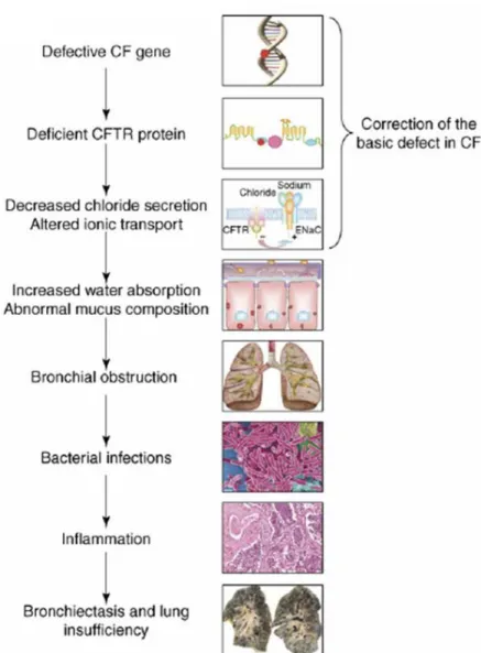
The failure of Cl- secretion and the hyperabsorption of Na+ through the epithelial Na+ channel (ENaC) lead to an osmotic absorption of H2O. This dehydration of the periciliary liquid layer enhances its viscosity, inhibiting mucociliary clearance. Bacterial colonization happens easily in such an environment. (Knowles and Durie, 2002). The organism tends to develop a continuous inflammatory response, which further augments respiratory failure, and leads ultimately to death. (Figure I.1). (Amaral and Kunzelmann, 2007)
Figure I.1. The CF pathogenesis cascade in the lung. A defective CFTR gene leads to an absent or deficient CFTR protein. The dysfunction in ionic transport leads to dehydration of periciliary layer, increasing its viscosity, impeding mucociliary clearance and allowing bacterial colonization. The continuous inflammatory response results in bronchiectasis and progressive lung insufficiency. From (Amaral and Kunzelmann, 2007)
Most airway injury in CF is believed to be mediated by neutrophil products, including proteases and oxidants, liberated by the abundance of neutrophils in the CF airway.(Accurso, 2006)
Although most patients die due to lung disease, non-respiratory manifestations are very important, such as exocrine pancreatic insufficiency, male infertility, and a form of intestinal obstruction named meconium ileus.(Collins, 1992)
CF-related diabetes, which is thought to further contribute to lung disease, undernutrition and CF-related bone disease are also important complications of CF. .(Accurso, 2006)
1.3. Diagnosis and treatment
The major diagnostic test is the sweat test. However, there are some CF patients that have normal sweat sodium chloride concentrations, and further testing is required. Azoospermia in men, evidence of intestinal obstruction, assessment of liver function, identification of pan-sinusitis, or measurement of nasal potential difference, may confirm the diagnosis. (Davis, 2006)
Nevertheless, definitive diagnosis depends on either sweat testing or identification of two mutant alleles in the CFTR.
When CF was described, patients’ lives “were very short and painful”. (Davis, 2006) Presently, both duration and quality of life of CF patients have increased, due to the available treatments.
Nutritional repletion is accomplished with pancreatic enzyme supplements, which were formulated in the late 1980s. However, the pancreatic supplements do not fully correct the malabsorption, due to acidification of intestinal contents, caused by impaired bicarbonate intestinal and pancreatic secretion in CF patients. Therefore, some blockers of gastric acid secretion may be prescribed to minimize stomach acidification. (Davis, 2006).
Relief of airway obstruction is a major concern. Human DNase is administered to CF patients and reduces mucus viscosity, because it
cleaves free DNA which is one of the molecules that contribute to the stickiness of the mucus. (Davis, 2006).
Administration of antibiotics is a major form of therapy in CF, and culture-specific antibiotics have been used for 60 years. The development of oral antibiotics such as the quinolone family for Pseudomonas and linezolid for Staphylococcus aureus has provided an effective alternative to intravenous administration. Nevertheless, formulation of antibiotics for aerosol use is preferentially chosen because higher antibiotic concentration is achieved and adverse effects are minimal. (Davis, 2006). (Accurso, 2006)
Infection control remains a key issue in CF, so most patients are administrated with steroids or high-dose ibuprofen. (Davis, 2006).
Most treatments are directed to the lower panels of Figure I.1. The current aim is to direct them at the upper panels.
2. CFTR
CFTR gene is located at the band 31 of the long arm of chromosome 7 (7q31).and spans a genomic region of 190 kb. (Tsui et al., 1985) (Tsui and Durie, 1997)
The gene consists of 27 exons and encodes a transcript of 6,5 kb. CFTR expression is detected in lung, colon, sweat glands, placenta, liver, parotid glands, pancreas an in nasal polyp tissue, by mRNA gel-blot hybridization. (Riordan et al., 1989)
The resultant protein CFTR is 1480 amino acids residues long and has a molecular weight of approximately 180kDa, when fully glycosylated. (Powell and Zeitlin, 2002)
Based on its structure, function and regulation, CFTR is a member of a large family of proteins – ATP-binding cassette (ABC) transporters, and is found in the apical side of epithelial cells, where it functions as a chloride channel. (Amaral and Kunzelmann, 2007)
2.1. CFTR structure
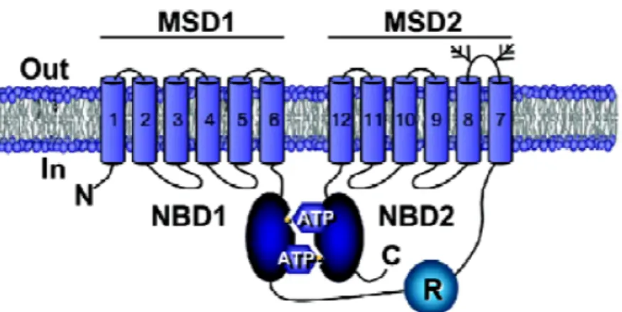
CFTR is composed of two membrane-spanning domains (MSDs), with six transmembrane α-helical segments each, and two nucleotide-binding domains (NBDs) (Figure I.2 and Figure I.3) (Sheppard and Welsh, 1999)
The presence of a regulatory domain (RD), which regulates CFTR activity upon phosphorylation, is a unique feature of CFTR, as a member of the ABC transporters family. (Riordan, 2005)
MSDs are linked by 6 extracellular and 4 intracellular loops. The loop between transmembrane segment 7 and transmembrane segment 8 contains two consensus N-glycosylation sites.
Figure I.2 - CFTR structure. Schematic representation of CFTR protein inserted into the cel membrane, showing the two membrane spanning domains, MSD1 and MSD2, the two nucleotide binding domains, NBD1 and NBD2 and the regulatory domain, RD. Glycosylation sites located in the extracellular loop between transmembrane segment 7 and transmembrane segment 8 are indicated by branched From (Li et al., 2007)
2.2. CFTR Biogenesis, Processing and
Trafficking
The most common CFTR mutation, a single deletion of a phenylalanine residue at position 508 (∆F508 or F508del), causes
protein misfolding, preventing CFTR from attaining its native global conformation. {Amaral, 2005). Therefore, F508del-CFTR is retained at the endoplasmic reticulum (ER) and sent to proteassomal degradation at an early stage. (Yang et al, 2003)
Worldwide, current strategies to treat CF focus on correcting misfolding and early degradation.
CFTR, along with other transmembrane glycoproteins, is co-translationally inserted into the ER and N-linked to glycosyl groups. (Farinha et al., 2002)
The chaperones Hsc70 (Heat shock cognate, 70 kDa) and Hsp70 (Heat shock protein, 70 kDa) bind to the polypeptidic chain, co-translationally, and assist the protein to acquire the proper folding. (Figure I.3). The presence of Hsp40 is required for CFTR stabilization and the prolonged retention of unfolded protein (F508del-CFTR, for instance) in the Hsc70 system targets it to degradation at an early folding checkpoint, involving CHIP and UbcH5a. (Farinha et al., 2002) (Farinha and Amaral, 2005)
CHIP promotes ubiquitylation and degradation of misfolded CFTR in the association with the cytosolic E2 ubiquitin conjugating enzyme UbcH5a. (Ameen et al., 2007)
CFTR seems to be particularly difficult in achieving a conformational state that fulfils all criteria that are necessary to proceed to the secretory pathway. While other ABC transporters, such as P-glycoprotein, mature and reach the membrane with great efficiency, CFTR matures inefficiently, as approximately 30% of the protein synthesised is fully glycosylated. (Riordan, 2008)
The core CFTR glycosylation consists in the addition of 14-unit oligosaccharidic branched structure to consensus sequences (Asn-X-Ser/Thr) in the nascent polypeptidic chain. These glycans are responsible for the interaction between the protein and different lectins, most of which participate in the ER quality control (ERQD). (Amaral, 2005)
Export from the ER may involve additional checkpoints, namely the AFT-mediated retrieval/retention. It was shown that simultaneous mutation of 4 AFTs is able to rescue F508del-CFTR. (Figure I.3) (Amaral, 2005) (Farinha and Amaral, 2005a) (Roxo-Rosa et al., 2006)
Figure I.3: Model of CFTR Biogenesis. CFTR is inserted in the ER membrane and binds Hsc70/Hsp40, and retention leads to proteasomal degradation, mediated by Hsc70-Chip-UbcH5a. Several rounds of glucose binding/release constitute the second checkpoint. When correctly folded, CFTR leaves the ER and proceeds to the secretory pathway, after being accessed for its folding at the last checkpoint. Adapted from (Farinha and Amaral, 2005)
If CFTR is correctly folded it proceeds to the secretory pathway, while misfolded CFTR which is identified by the ERQC and degraded by the ubiquitin-proteasome pathway (UPP). (Amaral, 2005)
The secretory pathway of eukaryotic cells is the sequential movement of a protein translocated into the ER through cis, medial¸ and trans Golgi apparatus.(Bannykh et al., 2000)
Conventionally, CFTR and other proteins of the secretory pathway interact with components of the COPII coat machinery forming
vesicular-tubular clusters (VTCs), which are then delivered at cis-Golgi. At this state VTCs-dependent recycling or COPI-independent retrieval may occur. However, the transport of cargo from VTCs along the Golgi is controversial, and Bannykh et al suggested that VTCs may bypass the cys- and medial-Golgi and reach the trans-Golgi or even endosomes. (Bannykh et al., 2000)
The population of wt-CFTR that reach Golgi and post-Golgi compartments is quite stable. Moreover, CFTR pool in the cell membrane is rapidly internalized, at a rate of 10% per minute, which implies that CFTR must be recycled to the cell surface. CFTR molecules that are internalized follow the clathrin-coated vesicle endocytic pathway and are recycled or sent to lysosomal degradation. (Riordan, 2008)
2.3. Classes of mutations and
mutation-specific therapies for treating CF
More than 1,600 mutations in CFTR have been described to date. (http://www.genet.sickkids.on.ca/cftr).
These mutations span throughout the entire gene but preferentially located in the regions that encode for the NBDs and the RD. F508del accounts for approximately 70% of chromosomes in CF patients, while the other CFTR mutations are very rare, with only four other mutations (G542X, N1303K, G551D and W1828X) having frequencies above 1%. (Rowntree and Harris, 2003)
Mutations in the CFTR gene have, generally, been classified into five different groups according to the molecular mechanisms that lead to disease.
Nowadays, several mutation specific strategies are under experimental/ clinical testing.
Class I mutations affect CFTR biosynthesis and are, therefore, the ones that include the most severe CF phenotype. These nonsense
(e.g., G542X), but also some frameshift and splicing mutations, prevent the biosynthesis of a stable protein or result in a truncated protein which is rapidly degradaded. (Rowntree and Harris, 2003) The strategy to suppress premature termination codons consists in the administration of aminoglycoside antibiotics, which enable the incorporation of an amino acid residue and permit the translation to continue. (Amaral and Kunzelmann, 2007)
Class II mutations, including F508del, result in CFTR misprocessing and intracellular retention with concomitant delivery to degradative pathways thus resulting in a lack of functional protein at the apical cell membrane. (Rowntree and Harris, 2003). To assist the folding and enable CFTR to be correctly located at the cell membrane, pharmacological, molecular or chemical chaperones are being studied. These compounds are generally referred as correctors. (Amaral and Kunzelmann, 2007)
CFTR bearing a class III mutation, G551D-CFTR for instance, reaches the cell membrane but does not respond to cAMP stimulation. Class III mutations are located in NBDs and are likely to prevent ATP binding or the coupling between ATP binding and channel gating. (Rowntree and Harris, 2003) To overcome the impaired gating, the strategy is to use compounds that stimulate pre-activated CFTR channel activity (potentiators) such as IBMX, an alkylxantine, and genistein, a naturally derived flavonoid (Amaral and Kunzelmann, 2007)
Class IV mutations, located in the MSDs, are the ones where CFTR is correctly located at the cell membrane and responds to cAMP stimulation but does not generate a proper Cl- conductance (R117H-CFTR for example). Compensation for reduced conductance can be achieved by simultaneous traffic promotion and potentiators administration. (Amaral and Kunzelmann, 2007)
Class V mutations result in a decrease of CFTR level and are often caused by mutations in the promoter that diminish transcription rate.
Splicing variants, such as G576A, which result in variable CFTR tissue-specific expression are also included in this class. (Rowntree and Harris, 2003) To overcome CFTR low levels, splicing factors that either promote normal exon inclusion or abnormal exon skipping should be used to augment protein synthesis. (Amaral and Kunzelmann, 2007)
Figure I.4 summarizes the functional classes found in CF patients.
Figure I.4 – Functional classes of CF mutations. Class I mutations prevent translation. Class II mutations result in misfolded proteins, retained in the ER and rapidly degraded. CFTR bearing a class III or class IV mutation reaches the membrane but does not respond to cAMP stimulation or does or results in decreased Cl- conductance. Class IV mutations result in CFTR decrease. Adapted from (Tsui and Durie, 1997)
2.4. CFTR function
When first identified, CFTR was considered to be the chloride channel defective in CF.
This protein is a unique ABC transporter since it functions as a Cl -channel and is essential for absorption and secretion of salt across epithelia. CFTR also plays an important role in bicarbonate secretion either by being permeant to bicarbonate or by stimulating Cl-/HCO3 -exchangers. (Riordan, 2008)
2.4.1.
CFTR as a chloride channel
As stated above, CFTR major function as a cAMP activated and Protein Kinase A (PKA) regulated Cl- channel, in the apical membrane of epithelial cells. (Amaral and Kunzelmann, 2007)
Single-channel analysis of wt-CFTR is characterized by bursts of channel openings, interrupted by brief closures and separated by longer closures between bursts. (Figure I.5) CFTR has a small single-channel conductance (6-10 pS) and that the anion permeability sequence is Br- > Cl- > I- channel openings.(Sheppard and Welsh, 1999).
When F508del-CFTR is correctly located in the cell membrane it forms a channel with a gating defect, which suggests that the region around the phenylalanine at position 508 is involve in the engine that is responsible for the conformational changes that lead to the opening and closure of the channel. (Li et al., 2007)
Figure I.5.: Representative single-channel traces of wt- and F508del-CFTR in excised inside-out membrane patches from BHK cells. The upper dot line represents the closed state, while downward deflections correspond to channel openings. From (Roxo-Rosa et al., 2006)
NBD1 and NBD2 use the energy of ATP hydrolysis to transport substrates across the cell membrane through diverse transmembrane pathways assembled from the MSDs, which form a Cl- selective channel gated by ATP binding. (Figure I.6)
The two NBDs of CFTR function as a head-to-tail dimer with the ATP-binding sites located at the interface of the two subunits (Figure I.5). The association of NBD1 and NBD2 is required for optimal ATPase activity and channel gating by CFTR. (Li et al., 2007)
CFTR is atypical both as an ion channel and as an ABC transporter since its activity is controlled by phosphorylation of the R domain. (Riordan, 2008)
It was also shown that that the energy of ATP binding in the two catalytic sites seems to drive NBD1-NBD2 dimerization. (Vergani et al., 2003) (Figure I.6)
Figure I.6.: Simplified model for CFTR-dependent Cl- transport across the cell membrane under quiescent and activated conditions. From (Li et al., 2007)
This structural arrangement leads to the opening of the Cl- permeation pathway in the transmembrane domains, and the NBD dimer would be stabilized by the two ATP complexes. Therefore, the hydrolosis of ATP at NBD2 would favor the dissociation of the complex and the closure of the pore and termination of the burst. (Figure I.7) (Vergani et al., 2003)
Figure I.7.: Simplified scheme illustrating the mechanism of CFTR channel gating, nucleotide binding and hydrolytic cycles. From (Vergani et al., 2003)
2.4.2.
CFTR and bicarbonate secretion in
the pancreas
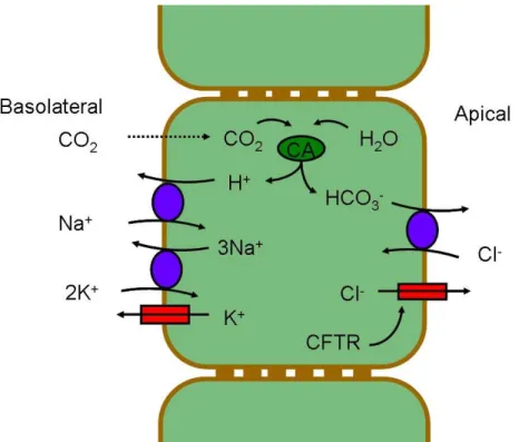
The pancreatic duct epithelium achieves the unique feature of secreting an almost isotonic sodium bicarbonate solution, in order to neutralize the gastric HCl. The low chime pH, as it reaches the small intestine, triggers secretion of the hormone secretin into the blood, which stimulates the bicarbonate secretion.
Bicarbonate secretion by the pancreatic duct cells is certainly dependent on CFTR, though its exact role is still controversial. Some believe that bicarbonate permeability of CFTR may provide a pathway for bicarbonate efflux, while others state that bicarbonate secretion happens in exchange for Cl-, which is continuously being exported through CFTR. (Steward et al., 2005)
The classic model for bicarbonate secretion by pancreatic duct cells, as it shown in Figure 2.3.1.1, considers that CO2 diffuses into the duct cell by the basolateral membrane, in first place. Then, carbonic anhydrase (CA) converts CO2 in H2CO3, which rapidly dissociates into H+ and HCO3- (bicarbonate). The resulting H+ leave the cell by an Na+/H+ exchanger, which is driven by the Na+ gradient, maintained by the Na+/K+-ATPase. The bicarbonate ions then leave the cell at the apical membrane in exchange for Cl- . The Cl- availability in the lumen is determined by CFTR. The membrane potential is maintained by basolateral K+ channels. (Steward et al., 2005)
Nevertheless, very recently, Ishiguro et al have shown, using an electrophysiological approach that “CFTR functions as a bicarbonate channel in pancreatic duct cells, and provides a significant pathway for bicarbonate transport across the apical membrane”. (Ishiguro et al., 2009)
Impaired pancreatic bicarbonate secretion in pancreas confirms that CFTR should play a very important in bicarbonate secretion. (Riordan, 2008)
Figure I.8. Classic model for HCO3− secretion by pancreatic duct
epithelium. In this model bicarbonate is secreted in exchange for Cl-, which is exported by CFTR. Adapted from (Steward et al., 2005)
CFTR regulation by phosphorylation
Phosphorylation and dephosphorylation are known to bo involved in the overall regulation of CFTR function. Phosphorylation of the R domain is the major mechanism that regulates CFTR activity.
Apart from protein kinase A (PKA) several other kinases phosphorylate and activate CFTR such as Ca2+-dependent protein kinase C (PKC) and cyclic guanosine monophosphate dependent kinase (PKG). Recently, Faria et al reported the activation of CFTR through stimulation of the metabotropic purinergic receptor P2Y2, which occurs through an unknown kinase that is different from PKA and other protein kinases downstream of P2Y2. (Faria et al., 2009)
Adenosine monophosphate stimulated kinase (AMPK) also regulates CFTR activity, inhibiting it through phosphorylation of two serine residues, S668 and S737. (Kongsuphol et al., 2009b)
Several sites for casein kinase 2 (CK2) phosphorylation are also present. Three of them have been described: two serine residues, at position 422 and 511 (S422, S511) and one threonine residue at position 1471 (T1471). (Luz 2008)
It has been shown by Pagano et al that S511 is not phosphorylated by CK2, which was found to phosphorylate S422 in NDB1, in vitro. (Pagano et al., 2008a) Moreover, T1471 was described as another CK2 putative phosphorylation site. (Ostedgaard et al., 2006).
Spleen tyrosine kinase (SYK) may also have a role in CFTR regulation, since there is a SYK phosphorylation consensus sequence in CFTR – a tyrosine residue at position 512 (Y512). (Luz, 2008).
2.5.1.
AMPK
AMP-activated protein kinase (AMPK) is an ubiquitous Ser/Thr kinase and downstream molecule of a protein kinase cascade that is sensitive towards cellular energy. Upon ATP depletion, AMPK is activated and stimulates catabolic pathways that ultimately lead to ATP production and downregulates anabolic pathways that consume ATP, by direct phosphorylation of metabolic enzymes or by regulating gene expression. (Hallows et al., 2000;Hallows et al., 2003)
AMPK binds to the cytoplasmic COOH terminal tail of CFTR and phosphorylates and inhibits CFTR channel activity predominantly through effects on CFTR gating. (Hallows et al., 2000;Hallows et al., 2003)
Several drugs have been used to activate AMPK in intact cells: metformin, phenformin and the nucleoside 5-aminoimidazole-4-carboxamide-1-β-D-ribofuranoside (AICAR). (Woolhead et al., 2007). Phenformin and metformin are biguanidines that promote
insulin-stimulated glucose uptake in muscle and lower hepatic glucose output. (Hawley et al., 2002;Mallick, 2004)
Phenformin was used in the clinical treatment of type II diabetes, but it has been withdrawn from clinical use due the high incidence of lactic acidosis on patients treated with this drug. It was replaced by metformin, which still has some risk of lactic acidosis though. (Woolhead et al., 2007) (Mallick, 2004)
Metformin, like phenformin, is believed to activate AMPK by inhibiting Complex I of the respiratory chain. (Hawley et al., 2002)
2.5.2.
CK2
CK2 is a pleiotropic kinase which phosphorylates serine and threonine residues with a consensus sequence Ser/Thr – X – X – Glu/Asp. This kinase phosphorylates molecular targets that are involved in protein synthesis, folding, degradation and proliferation processes. CK2 is well known for its anti-apoptotic role, suggesting that it might be a valuable target to develop anticancer drugs. (Pagano et al., 2008)
In general, CK2 is a heterotetramer consisting of two catalytic α or α’ subunits and two regulatory β subunits, and is specifically inhibited by tetrabromobenzotriazole (TBB). (Gotz et al., 2007)
2.5.3.
SYK
SYK belongs to the tyrosine protein kinase family, SYK/ZAP70 subfamily and plays a critical role in signalling through immune receptors, namely in B-cell antigen receptor (BCR) stimulated responses or in phosphoinositide 3-kinase (PI3K) dependent natural cytotoxicity by Natural killer cells (NK). (Jiang et al., 2002) (Turner et al., 2000)
SYK phosphorylates tyrosine residues followed by two amino acid residues with side chains negatively charged (Tyr – Asp/Glu – Asp/Glu).
Structurally, SYK comprises two N-terminal regulatory Src-homology 2 (SH2) domains followed by a catalytic region. (Arias-Palomo et al., 2007).
2.5.4.
LMTK2
LMTK2 (Lemur Tyrosine Kinase 2, also known as KPI-2 for Kinase/ Phosphatase/ Inhibitor-2) is a 1503-residue protein with two predicted transmembrane helices at the N terminus, a kinase domain, followed by a C-terminal domain. It has been described to associate with PP1C and Inh2 to form a regulatory complex that is localized at membranes. (Wang and Brautigan, 2002)
LMTK2 is strictly a Ser/Thr kinase that reacts with Ser either preceded by or followed by Pro residues but unlike other Pro-directed kinases does not strictly require an adjacent Pro residue. To this date, LMTK2 has been only shown to phosphorylate CFTR and and directly bind to myosin VI, an actin-based retrograde motor protein that is known to facilitate CFTR endocytosis. (Wang and Brautigan, 2006) (Swiatecka-Urban et al, 2004).
3. Objectives
CFTR regulation is highly dependent on phosphorylation and dephosphorylation by different kinases and phosphatases. However, novel kinases that interact with CFTR have been described and it is necessary to better understand those protein-protein interactions, in order to have a wider perspective of CFTR regulation and ultimately CF pathophysiology.
Therefore, in the main objective of the present work was to study the effect of AMPK, CK2 and SYK phosphorylation upon CFTR function.
For that we proposed:
a) To study the mechanism by which genotypic alterations in the CFTR gene enhance the predisposition to develop pancreatitis and its relation with AMPK phosphorylation;
b) To characterize the effect of CK2 and SYK phosphorylation upon CFTR biogenesis, trafficking and function, namely by studying the effect of mutating putative phosphorylation sites for these kinases.
Altogether, we expected to contribute to novel insights into CFTR regulation through phosphorylation by these kinases.
Chapter II –
Methods and Materials
1. Production of CFTR variants
1.1 Characterization of the Biological material
1.1.1 Bacterial strain
The bacterial strain used for DNA cloning and amplification was XL1-Blue (Stratagene, La Jolla, CA, USA). These Escherichia coli (E.coli) cells have tetracyclin and cloranfenicol resistance and exhibit the Hte phenotype, which increases the transformation efficiency of ligated and large DNA molecules. XL1-Blue cells are deficient in all known restriction systems [Δ(mcrA)183 Δ(mcrCB-hsdSMR-mrr)173]. The strain is endonuclease deficient (endA), greatly improving the quality of miniprep DNA, and recombination deficient (recA), helping to ensure insert stability. The lacIqZDM15 gene on the F´ episome allows blue-white screening for recombinant plasmids.
Genotype: Tetr∆(mcrA)183 ∆(mcrCB-hsdSMR-mrr)173 endA1 supE44
thi-1 recA1 gyrA96 relA1 lac Hte [F´ proAB lacIqZ∆M15 Tn10 (Tetr) Amy Camr].
1.1.2 Plasmid vectors
wt-CFTR and F508del -CFTR were introduced into pBluescript vector (Appendix I) by ligation into Sma I restriction site. All the other variants were produced by site-directed mutagenesis.
1.2 Competent Bacteria – Production and
Transformation
1.2.1 Production of competent bacteria
Bacteria were plated in LB-agar medium and a single colony was used to inoculate a small volume of LB medium overnight at 37°C with vigorous shaking (220 rpm). This pre-inoculum was then used to inoculate a larger volume of LB medium, typically 100 ml, which was also grown at 37°C (220 rpm) to final concentration of 5 x 10⁷ bacteria/mL (corresponding to an OD of 0.3 at 600 nm). Bacteria were transfered to ice and pelleted by centrifugation (1000 g for 15 min at 4°C). The bacterial pellet was then resuspended, incubated on ice for 15 min in RF1 buffer (100 mM RbCI, 50 mM Mn(OH)2, 30 mM KCH3COO, 10 mM Ca(OH)2, pH 7.5, 15 % (w/v) glycerol, pH 5.8; all from Sigma-Aldrich, St. Louis, MO, USA) - 1/3 of initial volume - and re-pelleted by centrifugation (1000 g for 15 min at 4°C). This second pellet was resuspended and incubated on ice for 15 min in RF2 buffer (10 mM RbCI, 75 mM Ca(OH)2,, 10 mM MOPS, 15 % (w/v) glycerol, pH 6.5; all from Sigma-Aldrich) – 1/12 of initial volume. 200 μl aliquots were then rapidly frozen with liquid nitrogen and stored at -80°C.
1.2.2 Transformation of competent bacteria
Bacteria were transformed by incubating a 200 μL aliquot of competent cells with DNA (~100 ng of ligation products or ~1 ng of purified plasmids) for 30 min on ice, performing a heat-shock (90 s at 42°C), further incubating the mixture for 2 min on ice and then allowing antibiotic resistance to be expressed by growth in antibiotic-free LB medium for 45 min at 37°C at 220 rpm. Bacteria were then pelleted (5000 g for 2 min), the supernatant was discarded and the pellet was resuspended in the remaining supernatant medium. This suspension
was then plated into LB-agar supplemented with the appropriate antibiotic (100 μg/mL ampicillin, Sigma-Aldrich, for pNUT and pBluescript) and left to grow overnight.
Transformed bacterial colonies were grown in LB medium supplemented with the appropriate antibiotic and used to extract plasmid DNA, which was screened by automatic DNA sequencing. After screening, positive clones were stored in liquid LB medium supplemented with 15 % (w/v) glycerol (Sigma-Aldrich) at - 80°C.
1.3 DNA Extraction
Small scale plasmid DNA was purified with commercial kit JETQUICK Plasmid Miniprep Spin Kit (Genomed, Lohne, Germany). This protocol is based on an alkaline lysis of the bacteria in the presence of SDS, to denature bacterial proteins, followed by a centrifugation step to remove cellular debris, genomic DNA and denatured proteins and adsorption of the plasmid DNA in the supernatant to an anionic exchange matrix in the presence of high saline concentrations. After adsorption, the DNA is washed and eluted in water or TE buffer (10mM Tris/HCl, pH8).
DNA concentration was determined by measurement of the absorbance at 260 nm (one absorbance unit corresponding to 50 μg/mL of dsDNA) and its purity was evaluated by assessment of the ratio A260/A280.
1.4 Mutagenesis
Point mutations were introduced into pBluescript wt-CFTR, using a combination of the QuickChange® Site-Directed Mutagenesis Kit (Stratagene) and the KOD Hot Start Kit (Novagene, Darmstadt, Germany) with complementary pairs of custom designed
HPLC-purified mutagenic primers (Thermo Electron Corporation, Waltham, MA, USA).
The amplification was confirmed by agarose gel electrophoresis and the resultant mutant plasmid was hydrolised with DpnI (Invitrogen, Carlsbad, CA, USA), a restriction enzyme that specifically hydrolyzes methylated and hemi-methylated DNA, thus removing all parental bacterial DNA.
After bacterial transformation (section II-1.2.2) and plasmid DNA extraction (section II-1.3), the presence of each mutation was verified by automatic DNA sequencing (section II-1.5).
Sense primers used in the mutagenesis reactions are presented in the Table 1.1. For each sense primer a complementary anti-sense primer was also used.
Table 1.4.1: Sense primers for mutagenesis reaction.
Name Sequence S422A s 5’- CAATAACAATAGAAAAACTGCTAATGGTGATGACAGCC -3’ S422D s 5’- CAATAGAAAAACTGATAATGGTGATGAC -3’ Y512A s 5’-CATCTTTGGTGTTTCCGCTGATGAATATAGATACAGAAGCGTC-3’ Y512D s 5’-CGCTTCTGTATCTATATTCATCATCGGAAACACCAAAGATG-3’ Y512F s 5'-CATCTTTGGTGTTTCCTTTGATGAATATAGATACAGAAGCGTC-3’
F508del Y512A s 5’-CGGTGTTTCCGCTGATGAATATAGATACAGAAGCGTCATC-3’
F508del Y512D s 5’-CATCGGTGTTTCCGATGATGAATATAGATACAGAGCG-3’
F508del Y512F s 5'-CATTGGTGTTTCCTTTGATGAATATAGATACAGAAGCGTC-3’
S573A s 5´-GCTGATTTGTATCTTCTAGACGCTCCTTTTGG-3’
S573C s 5´-GCTGATTTGTATCTTCTAGACTGTCCTTTTGGATAC-3’
T1471A s 5’-GCTGCTCTGAAAGAGGAGGCAGAAGAAGAGGTGCAAG-3’
1.5 DNA Sequencing
Plasmid DNAs were purified with the JETquick Plasmid Miniprep (Genomed), as described. (section II-1.3). The sequencing reactions were performed using the ABI Prism BigDye Terminator Cycle Sequencing Kit (Applied Biosystems, Foster City, CA, USA) according to the manufacturer’s instructions. The products were analyzed in the automatic sequencer 3130 XL Genetic Analyzer (Applied Biosystems). Normally, only forward primers were used in the sequencing reactions. The following table summarizes the primers used in the sequencing reactions (Table 1.2).
Table 1.5.1: Primers for DNA sequencing reaction
Name Sequence
Annealing position in CFTR
mRNA
CF-5'NC-f 5’- GCA TTA GGA GCT TGA GCC CA -3’ 72-96
CF Ex5.F 5’-CTC CTT TCC AAC AAC CTG AAC -3’ 679-699
B3R 5’- AAT GTA ACA GCC TTC TGG GAG -3’ 1318-1338
C2R 5’- AGC AGT ATA CAA AGA TGC TG -3’ 1812-1831
D1R 5’- GAC AAC AGC ATC CAC ACG AA -3’ 2490-2509
E1R 5’- AGA TTC TCC AAA GAT ATA GC -3’ 3055-3074
Ex18.F 5’- AAC TCC AGC ATA GAT GTG G -3’ 3574-3592
Ex 22.F 5’- AGC AGT TGA TGT GCT TGG C -3’ 4184-4202
For sequence analysis, the sequences obtained were analysed through comparison with the reference CFTR sequence (Genebank accession number: M26886). This comparative analysis was done using the softwares ChromasPro (http://www.technelysium.com.au) and BioEdit (http://www.mbio.ncsu.edu/BioEdit/bioedit.html).
2
Biochemical and functional assays of
CFTR variants
2.1 Characterization, Culture and Maintenance
of cell lines
The BHK 21 (Baby Hamster Kidney) cell line is a quasi-diploid established line of variant hamster cells, descendent from a clone of an unusually rapidly growing primary culture of new-born hamster kidney tissue (Stoker and Macpherson, 1964). BHK cells are usually described as fibroblasts and are widely used in cell physiology and biochemistry studies, being easy to grow and transfect.
BHK mutants (BHK wt-CFTR and F508del-CFTR) used in this study were obtained by stable transfection with pNUT wt-CFTR or pNUT F508del-CFTR respectively.
BHK cells were cultured in a 1:1 mixture of Dulbecco’s Modified Eagle Medium (DMEM) and Ham’s F-12 nutrient medium supplemented with 5 % (v/v) fetal calf serum, 100 U/mL penicillin and 100 mg/mL streptomycin (all from Invitrogen). The medium used with stable transfected cells also contained 500 µM methotrexate (MTX) (AAH Pharmaceuticals Ltd., Coventry, UK).
Calu-3 (cancer lung 3) and CFBE (cystic fibrosis human bronchial epithelial) cells stably transfected with wt-CFTR (CFBE-wt) and F508del-CFTR (CFBE-F508del) were cultured in Minimum Essential Media (MEM), containing 10 % (v/v) fetal calf serum, 100 U/mL penicillin and 100 mg/mL streptomycin (all from Invitrogen).
HeLa (Henrietta Lacks cervical cancer) cells were cultured in RPMI medium 1640 with 10 % (v/v) fetal calf serum, 100 U/mL penicillin and 100 mg/mL streptomycin (all from Invitrogen).
Continuous growth was permitted by pre-confluence enzymatic dissociation with trypsin (Invitrogen), an enzyme that hydrolyzes
proteins in the extracellular matrix. After dissociation, cells were resuspended and redistributed in new flasks or plates. Cell lines were stored in aliquots of cells frozen in 90 % (v/v) FCS (Invitrogen) and 10 % (v/v) DMSO (Sigma-Aldrich), a cryoprotectant that prevents the formation of ice crystals during the freezing process.
Cultures were maintained at 37°C in a humidified atmosphere of 5% (v/v) CO2.
2.2 Transfection using cationic lipossomes
BHK cells were transfected with 2 μg of plasmid DNA using the Lipofectin® reagent (Invitrogen), a cationic liposome formulation that forms DNA complexes able to fuse with the cell membrane. Selection for stable transfectants is achieved by adding the selective reagent MTX (500 µM) to the culture medium 48 h after transfection. Individual clones were isolated at 10-15 days in the selection medium. BHK stable cell lines expressing the CFTR variants were produced by. Simão Luz.
2.3 RT-PCR
´ Total RNA was isolated using the RNeasy extraction kit (Qiagen, Hilden, Germany) according to the manufacturer's instructions. Total RNA concentration was determined by measurement of A260 and was
treated with 1 U of RNAse-free DNAse I (Invitrogen) for 1h min at 37ºC to eliminate contamination with genomic DNA. RNA was annealed to 100 pmol of random hexamers (Invitrogen) and the mixture was incubated 10 min at 60°C and then chilled on ice. Following the addition of 5x first strand buffer (250 mM Tris-HCI pH 8.3, 375 mM KCI, 15 mM MgCI2) (Invitrogen), 0.01 M DTT (Invitrogen), 2.5 mM dNTP mix (Amersham Biosciences, Uppsala, Sweden) and 20 U RnaseOut
(Invitrogen), the mixture was incubated 2 min at 42°C. SuperScript II RNaseH- reverse transcriptase (200 U; Invitrogen) was added and the final mixture for incubated 60 min at 42°C. The reaction was stopped by heating at 70°C for 15 min. The PCR amplification of the cDNA products was carried out in a reaction that contained 5 μL of cDNA, PCR buffer (100 mM Tris-HCI pH 8.3, 500 mM KCL, 15 mM MgCl2, 0.01 % (w/v) gelatin) (Perkin Elmer, Norwalk, CT, USA), 25 mM dNTP mix (Amersham), 10 pmol of each primer and 1 U Taq polymerase (Perkin Elmer) or 1.5 U Pfu turbo polymerase (Stratagene).
To amplify cDNA, human LMTK2 primers amplifying a 239 bp fragment were used: forward primer TTGAGCACCTCATTGCAGTC-3 (2700-2719 of human LMTK2) and reverse primer, 5’-CTGTTATCCAGGCTGTGGGT-3’ (2919-2938 of human LMTK2)
2.4 Iodide efflux assays
CFTR-mediated iodide efflux was measured at room temperature, as described (Lansdell et al., 1998). BHK cells grown to confluence on 60 mm dishes were incubated for 1h in iodide loading buffer (136 mM NaI, 3mM KNO3, 2mM Ca(NO3)2, 11 mM glucose, 20 mm HEPES, pH 7.4-7.5; all from Sigma-Aldrich) and then thoroughly washed with efflux buffer (136 mM NaNO3, 3mM KNO3, 2mM Ca(NO3)2, 11 mM glucose, 20 mm HEPES, pH 7.4-7.5; all from Sigma-Aldrich). Efflux buffer aliquots were then added to the cells, left to equilibrate for 1 min, and collected for the iodide measurements. The first 4 aliquots were used to establish a baseline of iodide efflux, without CFTR simulation. CFTR was then stimulated with the cAMP agonist Forskolin 10 µM (Sigma-Aldrich) and with the CFTR potentiator genistein 50 µM (Sigma-Aldrich), for 4 minutes. Stimulation was absent in the remainder of the assay. The amount of iodide in each aliquot was measured with an iodide-selective electrode (MP225, Mettler Toledo, Thermo Electron
Corporation). Standard iodide solutions were also prepared freshly for electrode calibration and iodide concentration calculations. Date are expressed as mean ± SEM (standard error of the mean) of n observations. Statistical analyses were performed using Student’s unpaired t-test and a value of p < 0.05 was considered statistically significant.
2.5 Double electrode voltage clamp
experiments
2.5.1 Isolation of the oocytes
To isolate stage V-VI oocytes from Xenopus laevis mature female frogs (Xenopus Express, South Africa and Kaehler, Germany), the frog was anesthetized by immersion in 1 g/L 3-aminobenzoic acid ethyl ester for 30 min. The animal was placed on ice and a 1 cm incision was made on the abdominal wall (skin and muscle layer) approximately 1 cm over the hip. Ovarian lobes containing oocytes wrapped in connective tissue and blood vessels were removed with a pincet and the oocytes collected in OR-2 buffer (82.5 mM NaCl, 2 mM KCl, 1 mM MgCl2.6H2O, 5 mM HEPES, pH 7.4 – 7.5, all from Sigma-Aldrich). After removal of the ovarian lobe, the incisions were sutured closed with sterile needles. To remove the follicular cell layers and connective tissue, the oocytes were then incubated with 1.11 U/mL Collagenase A (Clostridium histolyticum, Clostridiopeptidase A, EC 3.3.24.3, Lypophilisate, Boehringer), for 1h at 18ºC. Defolliculated oocytes were washed 10 times with ND 96 buffer (96 mM NaCl, 2 mM KCl, 1.8 mM CaCl2.2H2O, 1 mM MgCl2.6H2O, 5 mM HEPES, 2.5 mM Na-Pyruvat, pH 7.4-7.5, all from Sigma-Aldrich) and placed in ND 97 buffer, previously autoclaved, (97 mM NaCl, 2 mM KCl, 1.8 mM
CaCl2.2H2O, 1 mM MgCl2.6H2O, 5 mM HEPES, 2.5 mM Na-Pyruvat, 0.5 mM Theophylin, 50 mg/ mL Gentamycin, pH 7.4-7.5, all from Sigma-Aldrich).
2.5.2 cRNA injection and voltage clamp
Double-electrode voltage clamp experiments were done was previously described. (Kongsuphol et al., 2009). Oocytes were injected with cRNA (10 ng, 47 nL of double-distilled water). Water-injected oocytes served as controls. 2–4 days after injection, oocytes were impaled with two electrodes (Clark Instruments Ltd., Salisbury, UK), which had a resistance of <1 MΩ when filled with 2.7 M KCl. Using two bath electrodes and a virtual-ground head stage, the voltage drop across the serial resistance was effectively zero. Membrane currents were measured by voltage clamping (oocyte clamp amplifier, Warner Instruments LLC, Hamden CT) in intervals from –60 to +40 mV, in steps of 10 mV, each 1 s. The bath was continuously perfused at a rate of 5 mL/min. All experiments were conducted at room temperature (22º) and conductances were calculated according to
V I G ∆ ∆ = , which
derives from Ohm’s law. Data are expressed as mean ± SEM (standard error of the mean) of n observations. Statistical analyses were performed using Student’s unpaired t-test and a value of p < 0.05 was considered statistically significant. These experiments were performed with Kongsuphol’s assistance.
Chapter III –
Results
1. Metformin increases the risk for
pancreatitis in diabetes patients bearing
the CFTR variant S573C
Phosphorylation is the major mechanism that regulates CFTR function as a Cl- channel. Several kinases phosphorylate and activate CFTR such as PKA, PKC, PKG and AMPK. (Faria et al., 2009) (Hallows et al., 2000;Hallows et al., 2003)
AMP-activated protein kinase (AMPK) is a Ser/Thr kinase, activated upon ATP depletion. AMPK stimulates catabolic pathways that ultimately lead to ATP production and down-regulates anabolic pathways that consume ATP. (Hallows et al., 2000;Hallows et al., 2003)
Metformin is a biguanidine compound used in the clinical treatment of type II diabetes, and it is described to activate AMPK, which co-localizes with CFTR and attenuates its function, through the inhibition of Complex I of the respiratory chain. In clinical use, it replaced phenformin which had higher incidence of lactic acidosis. (Hawley et al., 2002) (Kongsuphol et al., 2009)
Nonetheless, metformin still increases the risks of lactic acidosis and patients using this compound tend do develop pancreatitis secondary to metformin poisoning (or at therapeutic metformin doses, in the case of renal failure). (Mallick, 2004)
The pancreas contains exocrine tissue that produces several digestive enzymes that enter the small intestine through the pancreatic duct. These enzymes are delivered in an alkaline, bicarbonate-rich fluid that neutralizes the acidic chime. Bicarbonate secretion by the pancreatic duct cells is certainly dependent on CFTR, though its exact
role is still controversial, namely whether bicrbonate is directly transported by CFTR or by a transporter (SLC26A) which in turn is regulated by CFTR. (Steward et al., 2005)
Intriguingly, patients with pancreatitis have an increased incidence of genotypic alterations in the CFTR gene, including the non-disease causing variant S573C. (Keiles and Kammesheidt, 2006)
Therefore, we used double electrode voltage clamp (DEVC) to study the relation between pancreatitis and the increased genotypic alterations in the CFTR gene: whole cell chloride conductances were activated by 3-Isobutyl-1-methylxanthine (IBMX 1 mM) and forskolin (Forsk 2 µM) or Forsk 20 µM in Xenopus laevis oocytes expressing wt-, S573C- or S573A-CFTR.