UNIVERSIDADE DE LISBOA
Faculdade de Medicina Veterinária
REPRODUCTIVE MANAGEMENT IN CAPTIVE ELEPHANTS
SÓNIA ALEXANDRA DE JESUS FONTES
CONSTITUIÇÃO DO JÚRI
Doutor Luís Filipe Lopes da Costa Doutor George Thomas Stilwell
Doutor João Nestor das Chagas e Silva
ORIENTADOR
Doutor Thomas Bernd Hildebrandt CO-ORIENTADOR
Doutor George Thomas Stilwell
2017 LISBOA
UNIVERSIDADE DE LISBOA
Faculdade de Medicina Veterinária
REPRODUCTIVE MANAGEMENT IN CAPTIVE ELEPHANTS
SÓNIA ALEXANDRA DE JESUS FONTES
Dissertação de Mestrado Integrado em Medicina Veterinária
CONSTITUIÇÃO DO JÚRI
Doutor Luís Filipe Lopes da Costa Doutor George Thomas Stilwell
Doutor João Nestor das Chagas e Silva
ORIENTADOR
Doutor Thomas Bernd Hildebrandt CO-ORIENTADOR
Doutor George Thomas Stilwell
2017 LISBOA
i Acknowledgements
Aos meus pais, pelo constante apoio e incentivo. À minha mãe que me permitiu ser sempre sonhadora e confiante, ao meu pai que me ensinou a atingir todos os meus objectivos e a nunca parar de evoluir. À minha restante família por aceitarem as minhas constantes ausências e correrias, recebendo-me sempre com braço abertos e um sorriso caloroso.
Á Didi pela eterna paciência, apoio no design gráfico, mas acima de tudo por todas as gargalhadas e lágrimas partilhadas ao longo destes 13 anos! Ao Cajó, à Janocas e Jasko, por me aturarem mostrando sempre preocupação e carinho.
Á Renata Martins, por todas as noites em claro, a aproveitar os bons momentos em Berlim, ou a bater no teclado. Espero um dia poder retribuir todo o apoio que me deste.
To Professor Thomas Hildebrandt for all the amazing knowledge shared with me during this internship, for all the support given in my doubting phases, and for introducing me to the world of research.
Ao Professor Stilwell, por tudo o que me ensinou ao longo dos anos de curso e pelas didáticas saídas em CEP que muito positivamente marcaram, a mim e meus aos colegas. Obrigada pela confiança que depositou em mim, pelo incentivo e compreensão.
Ao Professor Nestor, por todo o carinho, simpatia e disponibilidade. Obrigada por me ter ajudado não só no início de estágio, mas pela preocupação que demonstrou ao longo do percurso.
Ao Jardim Zoológico de Lisboa, em especial ao Doutor Rui Bernardino e à Doutora Teresa L. Fernandes, pelo apoio que deram ao projeto, não só pela informação fornecida à realização desta dissertação, mas também pela disponibilidade que apresentaram em nos receber no Zoo no âmbito da reprodução de elefantes.
Á Luísa, por ser simplesmente uma das melhores pessoas que conheci ao longo deste curso. Obrigada pela humildade, empatia e sensibilidade que transmites na vida. Muitas saudades.
ii
Às Açorianas e Açorianos: Raquel, Xana, Carol, Grilo e Lima por me terem feito sentir que os 5 anos passaram rápido demais. Obrigada pelo apoio, amizade e todos os momentos de loucura necessários.
Ao Gonçalo, por toda a preocupação e carinho. Obrigada por todo o peixinho que, sem dúvida, me tornou mais inteligente.
Á Manu, a rapariga italiana que eu desejo que continue na minha vida por longos anos! Obrigada principessa.
To Philipp, for the amazing patience and help that you have given me during this work process, and the hope that it will continue this way and that I will be able to reciprocate.
To Susanne Holtze for always taking a bit of her time to help me and guide me. Thank you for all that I learned with you at IZW and in life.
Finalmente, mas não menos importante, para o Baia! Por todas as noites aquecidas, por tantas sebentas estudadas juntos, por toda a companhia e amor inigualável. Obrigada por todos os teus ron-rons, meu felpudo laranja!
iii Abstract
Reproductive management in captive elephants
Elephants have been widely used by Humans for several centuries: for meat, as warriors through several kingdoms, for their heavy work power, for public entertainment, and for their unique tusks, leading them to be poached for the ivory trade. Nowadays we face the reality of a decreasing number of elephants in most of their range countries, leading them to be considered endangered (Asian) or vulnerable (African) to extinction.
Being charismatic mega-vertebrates, made them one of the most desired wildlife to keep and show in zoological collections.
Interdiction to the importation of wildlife was an important step, but with no more importation of individuals from the wild, the need to preserve the captive population became mandatory, and the zoological institutions make great efforts to maintain these animals in their collection and extend the conservation of these species. The inability to produce sufficient captive offspring and the continuous declining number in their natural habitat has urged research on elephant reproduction physiology.
Asian and African elephants reproduce well in the wild but due to historically poor reproductive performance under human care, most captive populations face the possibility of local extinction.
Besides logistical issues, elephant breeding in captivity faces management problems due to diseases, like ovarian and uterine pathologies and bull infertility.
Therefore, it is important to understand the anatomy, physiology and all associated pathologies which can lead to reproduction failure, and for the future management of captive elephant populations is fundamental to ensure that professional decisions are made.
Recent advances in endocrine monitoring and ultrasound imaging techniques allow researchers to understand the complex mechanisms that control reproduction in elephants, unique in several features.
In this thesis, I reviewed all relevant studies from 2000 to nowadays, with special emphasis to the African elephant. Reproductive breeding management considerations to the captive population of the Lisbon Zoo were derived. Finally, four clinical cases in elephant reproduction that were followed and assisted by the author are analysed and discussed.
iv Resumo
Maneio reprodutivo de elefantes em cativeiro
Por muitos séculos, os elefantes têm sido utilizados pelo Homem: como produto de caça, soldados de guerra de diversos reinos, pela sua capacidade de trabalho pesado e pelas suas presas, levando a que sejam abatidos para o mercado de marfim. Atualmente, na maior parte da sua distribuição, o número de elefantes continua a decrescer o que levou à sua corrente classificação em “Vulnerável” (Africano) e em “Ameaça de Extinção” (Asiático). Devido ao carisma destes mega vertebrados, os elefantes são um dos mais desejados animais para manter e exibir em Zoos. A interdição à importação de mais indivíduos do meio selvagem foi um passo importante para a conservação destas espécies, mas tornou a manutenção das populações cativas existentes uma prioridade e grandes esforços foram tomados pelas instituições de cativeiro. Um ponto fulcral tem sido o estudo da fisiologia reprodutiva dos elefantes, para que seja atingido um número de descendentes suficientes para manter estas populações. Tanto os elefantes asiáticos (Elephas maximus) como os elefantes africanos (Loxodonta africana) conseguem reproduzir-se com sucesso no meio selvagem mas, devido a uma história de baixa performance reprodutiva sob cuidados humanos, muitas das populações cativas correm o risco de extinção local. Para além de problemas logísticos, a reprodução de elefantes em cativeiro debate-se com questões de maneio devido a enfermidades, como patologias ováricas e uterinas e infertilidade no macho.
Compreender e tornar disponível as novas descobertas no ramo da anatomia, fisiologia e as mais comuns patologias associadas a falha reprodutiva tornou-se então uma prioridade para garantir que decisões ponderadas possam ser tomadas no maneio de elefantes cativos.
Avanços recentes em monitorização hormonal e em técnicas de ultrasom permitiu aos investigadores perceber os mecanismos complexos que controlam a reprodução nos elefantes, que apresentam variadas características únicas.
Posto isso, nesta tese, foram recolhidos e compilados todos os resultados relevantes publicados desde o ano 2000, com especial ênfase em dados relativos ao elefante africano. Considerações sobre o maneio reprodutivo da população de elefantes Africanos existente no Jardim Zoológico de Lisboa são também abordadas. Por fim, quatro casos clínicos, auxiliados e seguidos pelo autor são analisados e debatidos.
v
Table of Contents
List of Figures viiii
List of Tables xiiii
List of Abbreviations and Symbols xiiiii
Internship activities 1
Training activities report 1
Chapter1 Introduction 4
1.1 Use of elephants by man 4
1.2 Taxonomy 4
1.3 Current status context of African elephant 5
1.4 Zoos, conservation and major concerns 6
1.5 New aid technologies 6
1.6 Main Goals 7
Chapter 2 Reproductive Biology 7
2.1 Female elephant 8
2.1.1 Reproductive Anatomy of the Female 8
Vestibule 9 Vagina 10 Cervix 11 Uterus 11 Oviducts 13 Ovaries 14
2.1.2 Physiology of the oestrous cycle 15
Luteinizing Hormone, Progestagens, Inhibins, and Follicle-stimulating Hormone 16
Prolactin 18
Oestrogens 18
2.1.3 Endocrine monitoring 19
2.1.4 Pathology of the female reproductive tract 20
Vulva and Vestibule 22
Urinary tract 22 Vagina 22 Cervix 23 Uterus 23 Oviducts 23 Ovaries 23 2.2 Male elephant 24
2.2.1 Reproductive Anatomy of the Male 24
Penis 24 Bulbourethral Glands 25 Urethra 25 Prostate Glands 26 Ampullae 27 Seminal vesicles 30 Ductus Deferens 31 Testes 31
2.2.2 Male Reproductive Physiology 32
2.2.3 Musth management 34
2.2.4 Pathology of the male reproductive system 34
2.2.5 Chemical communication in elephants 35
Chapter 3 Assisted Reproduction Technology 37
vi
3.2 Semen Collection 37
Artificial vagina 37
Electroejaculation 38
Manual stimulation 39
3.3 Semen evaluation and laboratory processing of the semen 39
3.4 Preservation of spermatozoa 42
3.5 Artificial Insemination 42
3.5.1 Insemination timing 43
3.5.2. AI procedure 43
3.6 In vitro fertilisation (IVF), Intra-cytoplasmatic sperm injection (ICSI) and Embryo
transfer 45
3.7 Sperm sex sorting 45
3.8 Hormone cycle induction 45
3.9 Hormonal contraception 46
Chapter 4 Pregnancy and Parturition 48
4. 1 Introduction to pregnancy 48 4.2 Physiology of gestation 48 Progestogens 48 Relaxin 49 Prolactin 49 Cortisol 50 Inhibin 50 4.3 Pregnancy diagnosis 51 4.4 Placentation 52 4.5 Embryonic development 52 4.6 Nutritional Management 56 Calcium administration 57 4.7 Pre-partum preparation 57 4.8 Parturition 59 4.9 Complications of birth 61
Possibilities for veterinary intervention during elephant birth 63
Chapter 5 Neonatal Care 65
5.1 The Newborn elephant 65
5.2 Neonatal examination and care 65
5.3 Nursing 67
5.3.1 Failure of passive transfer (FPT) of antibodies 69
5.3.2 Hand-rearing 70
5.3.3 Weaning 70
5.4 Training 70
Chapter 6 Lisbon Zoo 71
6.1 Lisbon Zoo herd reproductive situation 71
6.2 Lisbon Zoo reproductive recommendations 71
Chapter 7 Clinical Cases in elephant Reproduction 73
MATERIAL AND METHODS 73
1 Subjects 73
1.1 "Calvin" 73
1.2 "Tembo" 75
1.3 "Delhi" 76
1.4 "Kala" 76
2 Handling and Restraining 77
3 Transrectal sonographic examination of the reproductive tract 77
3.1 Equipment 77
3.2 Ultrasound scanning technique for males and females 78
vii
RESULTS 83
1 Reproductive examination findings in the males 83
1.1 "Calvin" 83
1.2 "Tembo" 84
2 Reproductive examination findings in the females 85
2.1 "Delhi" 86
2.2 "Kala" 88
DISCUSSION 91
Chapter 8 Conclusion and Perspectives 93
Final Considerations 93
Bibliography 95
APPENDIX I – Nutrient concentration for gestating females and calves. 101
APPENDIX II – Lisbon Zoo Anaesthesia Report. 103
APPENDIX III – Sedation protocols 103
viii
List of Figures
Figure 1: Schematic representation of the female elephant reproductive tract (image source:http://www.asianelephantresearch.com/about-elephant-anatomy-and-biology-p4.php). 8 Figure 2: Illustration of the female urogenital tract: ovaries (OV); uterus bicornis (UT); cervix (CE);
vagina (VA); vaginal os (VO) with two bilateral blind pouches; vestibule (VE); urinary bladder (UB); urethra (UR); pelvic bone (PB) (modified from Hildebrandt et al., 2006). 8 Figure 3: Example of the exttension used for US examination; adapting the US probe to the
extremity of the extension allows the operator to scan deeper organs. Video glasses to monitor the ultrasound exam are shown on the bottom (Courtesy of Dr Thomas Hildebrandt, Department of Reproduction Management – Leibniz Institute for Zoo and Wildlife Research
(IZW), Germany). 9
Figure 4: Ultrasound image of the vestibule in cross section (Courtesy of Dr Thomas Hildebrandt,
Department of Reproduction Management – IZW, Germany). 9
Figure 5: Ultrasound images in longitudinal view of the vagina in an African elephant, at different stages of the oestrous cycle: A) Mid-luteal phase with small mucous volume (arrow); B) Late luteal phase, showing a large volume of vaginal mucus (arrow); C) Follicular phase phase, with a reduced echogenicity of the vaginal mucosa (arrows) and D) Before ovulation, with echodense vaginal mucus, showing a broken, shell-like appearance (arrow) (Source: Hermes
et al., 2000). 10
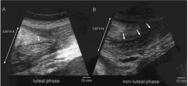
Figure 6: Ultrasound images of the cervix in an African elephant (in longitudinal view) (Source: Hermes et al., 2000). A – Luteal phase: the cervix presents a triangular shape and central cervical canal is less echogenic (arrow) B – Nonluteal phase: the cervical canal is well
distinguished as an anechogenic band (arrows). 11
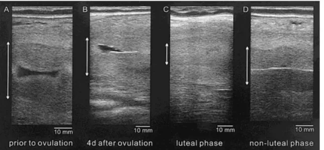
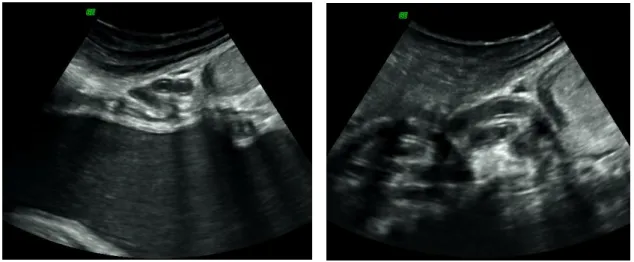
Figure 7: Ultrasound images of the uterus in an African elephant (longitudinal view). Endometrium height measures by the two-headed arrow; A) Before ovulation: the low echogenic endometrium is at its maximum height and fluid is observed in the uterus; B) Four days after ovulation: the endometrium is more echodense and although is still large in height, it is difficult to distinguish from the myometrium. The fluid can still observed in the uterine lumen; C) Luteal phase: characteristic flat endometrium; D) Non-luteal phase: the endometrium is less echogenic and increased in height. Endometrium and myometrium are well distinguished and blood vessels are observed in the myometrium. The central white line
indicates adjacent endometrial folds (Source: Hermes et al., 2000). 12
Figure 8: Ultrasound image of the uterus in early follicular phase (Courtesy of Dr. Thomas
Hildebrandt, Department of Reproduction Management – IZW, Germany). 12
Figure 9: Ultrasound image of the uterus horn, presenting thickened endometrium and mucous content in the lumen near ovulation (Courtesy of Dr Thomas Hildebrandt, Department of
Reproduction Management – IZW, Germany). 13
Figure 10: Ultrasound image of the uterus (longitudinal view), during luteal phase (Courtesy of Dr
Thomas Hildebrandt, Department of Reproduction Management – IZW, Germany). 13
Figure 11: Ultrasound image showing a partial view of the coaled oviduct (longitudinal view) (Courtesy of Dr Thomas Hildebrandt, Department of Reproduction Management – IZW,
Germany). 13
Figure 12: Ultrasound image of a dormant ovary (left side) although presenting normal size, in early follicular phase (Courtesy of Dr. Thomas Hildebrandt, Department of Reproduction
Management – IZW, Germany). 14
Figure 13: Ultrasound image of the ovary (right side) with a visible Graafian follicle in development (1.62 x 1.10 cm) (Courtesy of Dr Thomas Hildebrandt, Department of Reproduction
Management – IZW, Germany). 14
Figure 14: Ultrasound images of the ovary in an African elephant. A) Two follicles are visualized in luteinisation process, already starting to produce a wall and leading to the formation of an acCL (see below); B) One day after the first anLH surge, luteinisation process of the previously imaged follicles (arrows); C) Dominant follicle in growing process, before the second ovulatory LH (ovLH) surge; D) Graafian follicle on the day of ovLH surge and a CL in
formation (arrows) (Image source: Hermes et al., 2000). 15
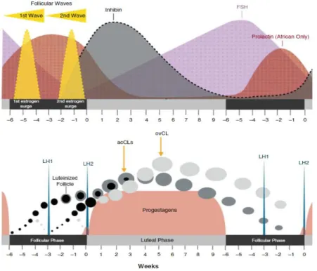
Figure 15: Scheme of the ovarian cycle, representing hormones secreted during the follicular and lutheal phase (estrogen, progestogens, LH, FSH, prolactin and inhibin). The development of the two follicular waves culminate at the 1st and 2nd LH peak and the formation of follicles, leading to the two types of corpora lutea: ovulatory and accessory CL. In this image prolactin
ix
curve is presented only for the African elephant, since the Asian species is not influenced by
any stage of the cycle (Adapted from Brown, 2014). 18
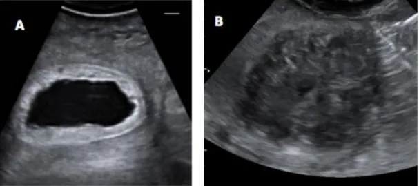
Figure 16: Examples of pathological uterine findings during ultrasound in female Asian elephant. A) Pyometra: fluid content in the uterine horn (black) and inflamed endometrium (white). (This condition needs treatment before GnRH vaccination may be considered – see chapter 3); B) Leiomyoma in uterine horn with destruction of normal appearance; no distinction of myometrium and endometrium is possible (Source: Luerdes & Oerke, 2016). 21 Figure 17: The male elephant reproductive system schematic representation (image source:
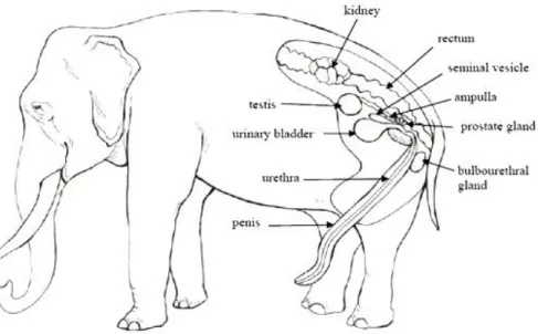
http://www.asianelephantresearch.com/about-elephant-anatomy-and-biology-p4.php) 24 Figure 18: Illustration of the main parts of the urogenital tract: testis (TE); ductus deferens (DD);
ampulla (AM); seminal vesicle SV); prostate (PR); bulbo-urethral gland (BU); urinary bladder UB); urethra (UR); penis (PE); pelvic bone (PB), modified from Hildebrandt et al. (2006). 24 Figure 19: Ultrasound image of the bulbourethral gland (left side), with irregular cavity (Courtesy of
Dr Thomas Hildebrandt, Department of Reproduction Management – IZW, Germany). 25 Figure 20: Ultrasound images of the urethra in a male African elephant; A – penial aspect of the
urethra, with corpus cavernosum, B – Urethra in the pelvic cavity (Courtesy of Dr Thomas
Hildebrandt, Department of Reproduction Management – IZW, Germany). 26
Figure 21: Ultrasound image of an inactive prostate (right side) of an African elephant. Due to the absent internal cavities it has a similar aspect as normal functional prostate of Asian elephant males (Courtesy of Dr Thomas Hildebrandt, Department of Reproduction Management –
IZW, Germany). 27
Figure 22: Ultrasound image of the prostate presenting irregular-shaped internal cavities in an African elephant (Courtesy of Dr Thomas Hildebrandt, Department of Reproduction
Management – IZW, Germany). 27
Figure 23: Ultrasound image of the ampulla (left) before ejaculation (Courtesy of Dr Thomas
Hildebrandt, Department of Reproduction Management – IZW, Germany). 28
Figure 24: Ultrasound image of the ampulla (left) after ejaculation (Courtesy of Dr Thomas
Hildebrandt, Department of Reproduction Management – IZW, Germany). 28
Figure 25: Ultrasound images of the left and right ampullae showing different densities; image on the right presents higher spermatozoa concentration (can concentrate over 1 billion sperm cells per millilitre) (Courtesy of Dr Thomas Hildebrandt, Department of Reproduction
Management – IZW, Germany). 29
Figure 26: Ultrasound image of an ampulla (left side) presenting two different densities, that is a sing of long lasting storage, leading to a gravity based separation, with the death of sperm cells (Courtesy of Dr Thomas Hildebrandt, Department of Reproduction Management – IZW,
Germany). 29
Figure 27: Ultrasound image of the ampullae showing different echogenicity: ampulla presented on the right side contains a anechoic fluid, due to non-functional right testicle which is a very unusual situation (Courtesy of Dr Thomas Hildebrandt, Department of Reproduction
Management – IZW Germany). 29
Figure 28: Ultrasound image of the left seminal vesicle (Courtesy of Dr Thomas Hildebrandt,
Department of Reproduction Management – IZW, Germany). 30
Figure 29: Ultrasound image of the ampullae and seminal vesicle (left side) (Courtesy of Dr Thomas Hildebrandt, Department of Reproduction Management – IZW, Germany). 30 Figure 30: Ultrasound image of the ductus deferens with visible lumen (Courtesy of Dr Thomas
Hildebrandt, Department of Reproduction Management – IZW, Germany). 31
Figure 31: Ultrasound image of the right testicle (Courtesy of Dr. Thomas Hildebrandt, Department
of Reproduction Management – IZW, Germany). 32
Figure 32: Temporal gland secretion in African bull in musth (Source:
http://www.bbc.com/earth/story/20160614-the-people-who-ate-elephant-heads) 33 Figure 33: Scheme of the hormonal changes in testosterone, LH, cortisol, inhibin, TSH and T4
during musth in elephant bulls (Brown, 2014). 34
Figure 34: Electroejaculation probes used in large animals. On the left back we can see the one used for semen collection in elephants; the probe in the front is normally used in humans, for
comparison.(Image source:
http://michaelmanyak.com/wp-content/uploads/2014/05/5probes.jpg). 38
Figure 35: Exteriorized penis during electroejaculation with condom for semen collection (Courtesy
of Dr Thomas Hildebrandt, IZW, Berlin). 38
Figure 36: Asian elephant sperm morphology (using a magnification of 1000x) A) Normal; B) Proximal cytoplasmic droplet; C) Abnormal mid-piece; D) Tightly coiled tail; E) Bent tail with cytoplasmic droplet; F) Spermac staining (solid arrow: Spermac positive; dotted arrow:
x
Figure 37: Spermac staining of elephant spermatozoa. Observations should be made using a light microscope (1000x) using oil immersion (Image Source: https://elephantconservation. org/programs/captive-programs). Stain explanation: Acrosome – Dark green, Nucleus – Red,
Equatorial region – Pale green, Midpiece and tail – Green. 40
Figure 38: Scheme of semen processing after collection (Original, information source Hermes et
al., 2013). 41
Figure 39: Artificial insemination in Woodland Park Zoo. Note that on the elevated structure the veterinary is controlling the process with ultrasonography to check if the insertion of the small insemination catheter and the deposition of semen is in the correct place. On the ground the other operator has access to the vestibule and manipulates the endoscope (Credit: Ryan Hawk; accessible at
http://voices.nationalgeographic.com/2011/08/05/elephant-artificial-insemination/). 44
Figure 40: Scheme of secretion of progestagens, LH, FSH, prolactin, relaxin, cortisol and inhibin during the gestation, and during a prior oestrous cycle in the elephant (Brown, 2014). 50 Figure 41: Elephant conceptus at 11 month of gestation; zonary placenta, passing in the equatorial
region and presenting a narrow hilus, which attaches the embryo to the endometrium (Allen
et al., 2002). 52
Figure 42: Schematic graphic of embryonic and foetal development from ovulation until birth.
(Adapted from Drews et al., 2008) 53
Figure 43: Ultrasonographic stages during the first third of gestation (Hildebrandt et al., 2007).
Scale bars – 1 cm 54
Figure 44: Ultrasonographic images of elephant conceptus (adapted from Drews et al., 2008).
Scale bars – 1 cm. 55
Figure 45: Illustration of a pregnant African elephant presenting the foetus in the most common
posterior position, inside the uterine horn (Image source:
http://www.dkfindout.com/uk/animals-and-nature/mammals/mammals-and-their-young/). 63 Figure 46: Malformation of the foetus presenting ankylosis caused by excessive abdominal lipoma
of the cow, making this case impossible to deliver, not even with foetotomy (Hermes et al.,
2008). 63
Figure 47: Surgical extraction of foetus during episiotomy, with detail on the direction of the ropes
during forced extraction (Hermes et al., 2008). 64
Figure 48: In case of a very small calf/very tall dam, a stool may be used to help nursing (Image
source: Olson, 2004b). 68
Figure 49: Milk collection by hand and desensitization of the mammary gland and nipple manipulation in order to stimulate the calf’s initial attempts to nurse and milking (Olson,
2004d). 68
Figure 50: Electric human breast pump being used to collect milk from an elephant cow (Olson,
2004d). 69
Figure 51: The ultrasound examinations were performed with “Voluson i” and an abdominal probe
of 2-5 MHz (original picture). 77
Figure 52: Extension used to access the four animals, measuring approximately 45 cm. The US
probe is adapted to the extremity of the extension (original picture). 78
Figure 53: Manual removal of faeces, using a hose without sharp endings and presenting a small diameter (1 – 2 cm). Note the need of help from the keepers to maintain access to the rectum
in the bull, handled in “Protected contact” (original picture). 79
Figure 54: Ultrasound examination on Delhi, in a “Free Contact” system, with no sedation and in
standing position (original picture). 79
Figure 55: Erected penis achieved by rectal manual stimulation. Note that the four limbs are
chained and the bull is restrained standing in a chute (original picture). 80
Figure 56: Exteriorized penis with condom for semen collection, during the rectal manual stimulation approach. Note that the keeper holding the device is physically protected from the
bull (Original picture). 81
Figure 57: Semen collection device - sleeve (condom) to place underneath the penis tip. A clean plastic bag will funnel the semen into a 50 mL tube, and is adapted to the end of a pole/extender, to lengthen the safety distance from the animal (the bag should reach the
entire surface of the metal ring) of the (original picture). 81
Figure 58: Device commonly used at IZW to transport fresh semen to be used for AI –
EQUITAINER® (original picture). 82
Figure 59: Ultrasonographic finding of the reproductive tract examination of the bull “Calvin”: A – urethra; B – prostate with visible lobes (left side); C – blood vessels, lobes, and bridge, creating a “butterfly” sitting in the urethra with cuniculos seminalis inside (left side); D – Left ampulla filled with snow fluid; and ureter entering the bladder (in cross-section); E – Both
xi
ampullae are visible and the left seminal vesicle can be seen; F – Left seminal vesicle
presenting folds. 84
Figure 60: Ultrasonographic findings of the reproductive tract examination of the bull “Tembo”: A – urethra; B – prostate without content, visible lobes (right side); C – empty prostate (left side), D – empty ampulla and bladder (right side), E – empty ampulla with seminal vesicle on top (right side); F – empty ampulla, seminal vesicle, urethra (left side); G – seminal vesicle nicely filled (right side); H – seminal vesicle (left side), I – seminal vesicle with deposit of protein (right side); J – bladder with hyperechoic deposits, visible blood vessels. 85 Figure 61: Ultrasonographic findings of the reproductive tract examination of the female “Delhi”: A
– vestibule, in its horizontal portion (cross section); B – urethra presenting a stone/calcification; C – vagina (longitudinal view) with small mucous volume at the lumen; D – uterine endometrium with its visible coiled arteries (arteria spiralis); E – right ovary; F – right ovary with an old CL; G – ovarian cyst of 2,18 cm in diameter (right ovary); H – tip of uterus in cross section; I – left ovary with a small follicle of 0,99cm (in diameter); J – uterine
endometrial cysts. 87
Figure 62: Ultrasonographic findings of the reproductive tract examination of the female “Kala”: A – vestibule, with a visible muscular part; B – stone in the beginning of the urethra and several blood vessels; C – longitudinal view of the urethra with calcification; D – vagina with a white line in lumen, bladder with clear urine; E – vagina, presenting with white line; F –mucous in the vagina and the detail of the ureter entering the bladder; G – cyst in vagina; H – same; I – cyst in front of the fornix, and another cyst in the cervix; J – cercyst; K – cheese like
endometrium (cysts) and leiomyoma; L – ovarian cyst. 89
Figure 63: Most commonly used drugs and dosages in African and Asian elephants for setation
xii
List of Tables
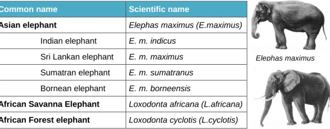
Table 1: Common name and scientific name of the existent elephant species and subspecies. (Adapted from Blanc, 2008; Rohland et al., 2010; AZA, 2011). Representative image of E. maximus (top) and L. Africana (below) for comparison (Images source: British & Irish
Association of Zoos and Aquariums (BIAZA), 2010). 5
Table 2: Demographic distribution for the African Elephant EEP population, regarding their breeding population status on February 15th of 2013; (information from Schwammer &
Fruehwirth, 2013). 6
Table 3: Average semen characteristics, analysed from samples collected by rectal massage; mean volume, concentration and pH for the first and second ejaculatory responses (Schmitt
& Hildebrandt, 1998). 39
Table 4: African elephant semen (Hähnel, 2007) 39
Table 5: List of the general supplies, medical supplies (which include many of those commonly found in a veterinary clinic) and some of the most important drugs to have available when expecting an elephant parturition (Original table. Information source: Olson, 2004d;
Schaftenaar and Hildebrandt, 2005; Emanuelson, 2006). 58
Table 6: Pre-birth preparations needed for expected captive elephant parturition (Original table, Information sources: Olson, 2004d; Schaftenaar & Hildebrandt, 2005; Emanuelson, 2006;
Hermes et al., 2008; Weber & Miller, 2012). 59
Table 7: Summary of impending parturition signs and behavioural manifestations (Schaftenaar &
Hildebrandt, 2005; Hildebrandt et al., 2008). 60
Table 8: Possible causes of dystocia (Hermes et al., 2008). 62
Table 9: Initial after birth and ongoing ideal calf health examination (Emanuelson, 2006; Weber &
Miller, 2012; Wiedner, 2015). 67
Table 10: Hand Rearing nutrition required for zoo animals specifically elephant calves, proposed
by MERCK veterinary manual. 70
Table 11: Identification of the previous offspring from the bull "Calvin" (adapted from
http://www.elephant.se/database2.php?elephant_id=212). 74
Table 12: Identification of the previous offspring from bull “Tembo” (adapted from
http://www.elephant.se/database2.php?elephant_id=6). 75
Table 13: Table presenting the minimum nutrient concentrations (DM basis) proposed for elephant diets (based largely on extrapolation from nutrient requirements of horses) (Adapted from:
http://www.zutrition.com/elephant-nutrition-guide; Olson, 2004c). 101
Table 14: Sedation protocol used in Lisbon Zoo, during the semen collection attempt of the male
“Jasa” (Courtesy of Dr Teresa L. Fernandes, Lisbon Zoo). 103
Table 15: Elephant captive herd of Lisbon Zoo, ZIMS Data on the Taxon Report Loxodonta africana, acceded on 9 December 2013 (Courtesy of Dr Rui Bernardino, Lisbon Zoo,
xiii
List of Abbreviations and Symbols
% - Percentage °C – Degrees Celsius 3D – Three-dimensional reconstruction acCL – Accessory CL AI – Artificial Insemination Al – Allantois Am – AmnionanLH – Anovulatory LH peak AV – Artificial vagina
Av – Allantoic vessels or arteria vertebralis AZA – Association of Zoos and Aquariums BC – Before Christ
BIAZA - British & Irish Association of Zoos and Aquariums bpm – Beats per minute
CITES – Convention on International Trade in Endangered Species of Wild Fauna and Flora CL – Corpora Lutea or Corpus Luteum
cm – Centimetres DM – Dry matter Dr – Doctor
e.g. – exempli gratia Ea – Ear
EAZA – European Association of Zoos and Aquariums
EAZWV – European Association of Zoo and Wildlife Veterinarians ECZM – European College of Zoological Medicine
EEHV - Elephant Endotheliotropic Herpesvirus EEP – European Endangered species Programmes EIA – Enzymimmunoassay
ELISA – Enzyme-linked immunosorbent assay Em – Embryo Ev – Embryonic vesicle Ey – Eyes Fl – Front legs FSH – Follicle-stimulating Hormone GnRH – Gonadotropin-releasing hormone
GnRH-Ag – Gonadotropin-releasing hormone Agonist h – Hour(s)
He – Heart Hl – Hind legs
iCa – Ionized serum calcium
ICSI – Intra-Cytoplasmatic Sperm Injection Ir- Inhibin – Immunoreactive Inhibin
IUCN – International Union for Conservation of Nature IV – Intravenous
IVF – In Vitro Fertilisation
IZW – Leibniz Institute for Zoo and Wildlife Research kg – Kilogram
L – Liter(s)
LH – Luteinizing Hormone mg – Milligram
mg/kg – Milligram per kilogram Mh – Midgut herniation
min – Minute(s)
xiv nm – Nanometers
ng/mL – Nanograms per millilitre ovCL – Ovulatory CL
ovLH – Ovulatory LH peak PhD – Philosophiae Doctor Pl – Placenta
PRL – Prolactin
RIA – Radio Immuno Assay SSP – Species Survival Plan T3 – Triiodothyronine
T4 – Tiroxina
TAG – Taxon Advisory Group Tr – Trunk
TSH – Thyroid-stimulating hormone Ul – Uterine lumen
US – Ultrasound Uv –Umbilical vessels
VLDL – Very light-density lipoprotein x – Time(s)
Ys – Yolk sac
Z 7-12:Ac – Z-7-dodecenyl acetate µm – Micrometer
1
Internship activities
Training activities report
The curricular internship was accomplished at the Leibniz Institute for Zoo and Wildlife research (IZW) in Berlin. I had the opportunity to participate in various veterinary activities and side projects which involved the handling of animals kept at the facility. The practical part with a duration of 8 months, from September 2013 until May 2014, comprised 800 hours.
During that period, my main focus was to gain a deeper understanding of how reproductive management aids conservation efforts in captive elephants. In this context, under the guidance of Prof Dr Thomas Hildebrandt, I was able to assist four veterinary reproductive examinations, which will be further detailed in this work. With all the preparations and travels to the Zoos, around 50 hours were spent in these assessments.
Regarding elephants, I participated also in one non-reproductive examinations performed at Leipzig Zoo (German), in which I could improve my knowledge about endoscopy technique, during a trunk exploration to collect bronchial lavage fluid, to be further tested for tuberculosis.
Besides elephants, I was able to assist other veterinary reproductive assessments in a variety of species (around 50 hours for clinical activities in other zoo):
- Ultrasonography of male and female gorillas and chimpanzees, during which the practical tasks included opening an intravenous route, monitoring anaesthesia and collection of blood samples, guided by Prof Dr Thomas Hildebrandt,
- Ultrasonography of hyenas for gender determination and collection of saliva and hair samples, performed by Prof Dr Thomas Hildebrandt,
- Ultrasonography and artificial insemination of two female white rhinos, thereby assisting with the processing of frozen semen samples, under the supervision of Dr Robert Hermes,
- Ultrasonography and electroejaculation for semen collection of a wolf, learning the main important steps to achieve a good sample for cryopreservation with Dr Robert Hermes and Dr Frank Göritz.
Furthermore, I was able to follow the routine activities performed by the veterinarians at the IZW, and to participate in numerous projects during the course of the internship. These activities included assisting the daily management of animals kept and bred at the institute for research purposes, for example guinea pigs and naked mole rats, in which I spent most of my internship time (around 350 hours), since the infrastructures are allocated at IZW. Some of the common tasks consisted in feeding, health checks and application of simple
2
treatments, temperature monitoring and weighting. In the case of naked mole rats, I performed behavioural observations regarding their social hierarchy, and assisted in ultrasound examinations, micro-chipping, semen collections with the use of electroejaculation, tissue sampling for genetic testing and necropsy procedures.
In guinea pigs, research involved recording of ultrasonographic images of embryonic and foetal development during several phases of pregnancy. After learning the technique and milestones with Dr Susanne Holtze, I was able to independently perform the examinations in the pregnant females. Further, I performed the castration of one male guinea-pig, under supervision.
The IZW holds a field station (Niederfinow, Germany), which houses European roe deer for scientific purposes, mainly regarding the reproductive system. There, I participated in the capture, anaesthesia (by blow darting) and monitoring, endotracheal intubation, and sample collection, and further observed the ultrasonographic assessments of the reproductive status, as well as testicular biopsies. Vaccinations, the monitoring of post-vaccination immune reactions, as well as tissue and hair sampling were also performed in hares at this place. I was for several weeks at this field station, comprising around 200 hours of my curricular practical teaching.
During my internship I also assisted researchers from other departments in smaller tasks. In a PhD project that involved collaring wild boars for GPS tracking, I collected samples (hair and blood), measured temperature and monitored anaesthesia under the supervision of Dr Frank Göritz. For a PhD-project on bat metabolism, I learned how to perform brown fat biopsies in hibernating bats. I spent around 50 hours in other projects training activities. Organizing and packing the travel equipment needed for examinations of the “Department of Reproduction Management” abroad, transmitted a great knowledge of the available and necessary veterinary equipment and the essential steps of various reproductive techniques performed in many wildlife species; e.g., tigers, rhinos, elephants, chimpanzees, gorillas, bears, etc.
I was also able to acquire more profound knowledge and to assist in Computed Tomography examinations of live and dead animals, which are regularly performed at the research institute.
Moreover, blood collection and subsequent processing to obtain serum or plasma samples, as well as biochemical and haematological analyses were frequently performed as part of my work at the department’s laboratory.
In 2014, I participated in the International Conference on Diseases of Zoo and Wildlife Animals in Warsaw, Poland, which is an annual event where European zoo veterinarians and wildlife researchers meet, present and debate interesting clinical cases and new discoveries in this area.
3
Although the gross amount of cases seen was in the field of reproduction management, the experience of contacting with diverse and interesting species, the literature research regarding the cases at hand and the newly acquired clinical knowledge added considerable value to this practical curricular internship. A total amount of 100 hours was spent on research activities during the masters’ internship.
4
Chapter1 Introduction
1.1 Use of elephants by manThe interaction between humans and elephants started long ago, during the Stone Age, when elephants were being hunted alongside their extinct relatives, the mammoths. Elephants appear in art work dating back to 30.000 BC in Europe, and also in Neolithic paintings in Africa (approximately 4.500 - 2.000 BC). However, the earliest evidence of elephants in captivity starts only at around 4.500 years ago, long after evidence of most other livestock species (10.000 BC, Csuti, 2006).
Since then, elephants have been used as war machines by several empires on different continents and were considered to have had an important influence on the course of several battles (Sukumar, 2003). Elephants also served as valuable offers for many European Kings, and Portugal was no exception; “Hanno”, an Asian white elephant was offered to Pope Leo X at his coronation by King Manuel I of Portugal. Hanno arrived in Rome in 1514 and became the Pope's favourite animal, astounding the papal court and being the star of many processions, festivals, paintings, and sculptures for the Roman people. Nowadays, ceremonial elephants are still present in human culture, especially in India, alongside with working elephants, which are used for their capacity of working in steep logging terrain as well as for tourist entertainment in Africa and Asia. The first evidence of the exhibition of elephants in circuses dates back to the ancient Indian princes and to the Romans (Csuti, 2006).
1.2 Taxonomy
The elephant belongs to the Kingdom Animalia, Phylum Chordata, Class Mammalia and Order Proboscidea, which contains only one extant family - the Elephantidae, including the generae Loxodonta, with two described species (Rohland et al., 2010) and Elephas, with four extant subspecies (Blanc, 2008) (Table 1).
5
Table 1: Common name and scientific name of the existent elephant species and subspecies. (Adapted from Blanc, 2008; Rohland et al., 2010; AZA, 2011). Representative image of E. maximus (top) and L. Africana (below) for comparison (Images source: British & Irish Association of Zoos and Aquariums (BIAZA), 2010).
Common name Scientific name
Asian elephant Elephas maximus (E.maximus)
Indian elephant E. m. indicus Sri Lankan elephant E. m. maximus Sumatran elephant E. m. sumatranus Bornean elephant E. m. borneensis
African Savanna Elephant Loxodonta africana (L.africana) African Forest elephant Loxodonta cyclotis (L.cyclotis)
1.3 Current status context of African elephant
In this thesis, I have mainly focused on the African elephant as this is the species currently maintained in captivity at Lisbon Zoo. However, analogies with the closely related Asian elephant are made use of whenever literature is lacking for Loxodonta.
The African elephant was classified in 1986 as Vulnerable by the International Union for Conservation of Nature (IUCN), maintaining this status until 1996, when it acquired the Endangered classification. In 2004, this species was again rated as Vulnerable by the IUCN Red List of Threatened Species, and has been keeping this category until today. At the moment, the main threat faced by the African elephant is poaching for ivory and meat, constituting the major cause for the species’ decline. The increase of human-elephant conflicts further aggravates the threat to elephant populations within their distribution range. Loxodonta africana are now regionally extinct from Burundi, Gambia and Mauritania, and were reintroduced to Swaziland (Blanc, 2008).
The Convention on International Trade in Endangered Species of Wild Fauna and Flora (CITES) listed the African elephant as one of the most endangered species, threatened with extinction. It “prohibits international trade of all specimens of these species “except when the purpose of the import is not commercial (…) for instance for scientific research”. In these exceptional cases, trade may take place, “provided it is authorized by the granting of both an import and an export permit”. These regulations exclude the African elephant populations in Botswana, Namibia, Zimbabwe and South Africa, countries in which the species is considered to be at a lower risk of extinction (http://cites.org).
Loxodanta africana Elephas maximus
6 1.4 Zoos, conservation and major concerns
Elephants have been kept in royal animal collections for at least 3,500 years. One of the most famous examples is the menagerie at Versailles (1665 - 1681). Also Schönbrunn Zoo, in Vienna, once an imperial collection, is the world's oldest zoo, and has been keeping elephants since 1752 (wwww.zoovienna.at).
According to the European Association of Zoos and Aquaria (EAZA), a new strategic Masterplan for European Endangered species Programmes (EEP) which was released in 2013, the captive African elephant population is considered to be in a highly critical situation. The captive birth rate increased over the years; however numbers are still not sufficient to counterbalance the death rate (Schwammer & Fruehwirth, 2013). Likewise, the population contains a large group of females between their mid-twenties to early thirties, which is expected to cause a peak in mortality in the future. Also, the majority of these females are considered not to be able to breed again (Hermes et al., 2004; Schwammer & Fruehwirth, 2013), while proven breeders constitute the minority of the EEP population (Table 2).
Table 2: Demographic distribution for the African Elephant EEP population, regarding their breeding population status on February 15th of 2013; (information from Schwammer & Fruehwirth, 2013).
Females Males Total
Current EEP population 139 46 185
Excluded from breeding population 60 3 63
*Potential breeders 79 43 122
**Proven breeders 25 8 33
* Animals at breeding age
**Individuals from the potential breeding group already confirmed as breeders after reproductive examination
Therefore, reproduction management of captive elephants becomes one of the major conservation efforts that zoos can provide to create self-sustaining populations of African elephants, both under human care and in the wild.
1.5 New aid technologies
Captive elephant groups constitute highly fragmented isolated populations. As such, inbreeding and the consequential loss of genetic diversity and fitness (including reproductive fitness) of the population are of constant concern regarding population management (Hermes et al., 2009).
Recent advances in technologies and methodologies have allowed for a better understanding of the reproductive biology of elephants. Through the use of ultrasound examinations, for example, Hildebrandt (2000a, 2000b) and his team were able to identify morphological abnormalities and to reveal the importance of this non-invasive method for identifying potential breeders that are healthy and suitable for natural or assisted breeding efforts. Therefore, among the captive elephant population, ultrasound examinations are
7
considered to be an essential tool by the Association of Zoos and Aquariums (AZA) elephant Species Survival Plan (SSP) and EAZA studbook breeding programme (Schwammer & Fruehwirth, 2013).
Likewise, further methodologies concerning the development of sperm cryopreservation or the study of hormonal cycles will advance our knowledge of assisted reproduction in elephants. Routine endocrine monitoring of the oestrous cycle is achieved through sampling of faeces, blood, and urine and is nowadays considered an indispensable tool for the successful management of elephant breeding groups in captivity (Hildebrandt et al., 2012).
1.6 Main Goals
This thesis aims at reviewing the current knowledge on the African elephant reproductive biology and management. For this purpose, I have collected bibliographic information comprising publications of the last fifteen years, compiled to a comprehensive literature review.
The main goal is to present the latest discoveries, empirical results, as well as, methodologies employed to study reproduction in elephants, and to assemble this information as a consultation manual for orientation within the management facilities. Therefore, I have structured this thesis into seven relevant chapters:
Chapter I – Introduction of the general situation of captive elephants and the importance of this dissertation;
Chapter II - Reproductive biology of both female and male elephants, spanning anatomy, physiology and associated pathologies;
Chapter III - Management techniques, including semen collection, as well as hormonal contraception and estrous cycle induction;
Chapter IV - Pregnancy and parturition, including management techniques of the gestating cow and known cases of dystocia;
Chapter V - Neonatal care in captivity and the most important milestones and interventions during the first months of the calves;
Chapter VI - African elephant population at the Lisbon Zoo, focussing on the current breeding status of the herd and reproduction management recommendations;
Chapter VII – Clinical cases assisted by the author, to enhance understanding on the conservation efforts being done from a reproductive point of view to aid the elephant population.
8
Chapter 2 Reproductive Biology
2.1 Female elephant2.1.1 Reproductive Anatomy of the Female
Female elephants present unique reproductive characteristics. For many years, these constituted a challenge and an obstacle for breeding programs, given that techniques developed for domestic or laboratory species were not applicable to this mega vertebrate mammal. Concerning anatomy, techniques for organ visualisation by rectal ultrasonography had to be developed, including the design of a suitable probe extension (Figure 3), which is normally required to examine the most cranial portions of the 3 m long urogenital tract of the female elephant (Hildebrandt et al. 2006) (Figure 1 and 2).
Figure 1: Schematic representation of the female elephant reproductive tract (image
source:http://www.asianelephantresearch.com/about-elephant-anatomy-and-biology-p4.php).
Figure 2: Illustration of the female urogenital tract: ovaries (OV); uterus bicornis (UT); cervix (CE); vagina (VA); vaginal os (VO) with two bilateral blind pouches; vestibule (VE); urinary bladder (UB); urethra (UR); pelvic bone (PB) (modified from Hildebrandt et al., 2006).
9
Figure 3: Example of extension used for US examination; adapting the US probe to the extremity of the extension allows the operator to scan deeper organs. Video glasses to monitor the ultrasound exam are shown on the bottom (Courtesy of Dr Thomas Hildebrandt, Department of Reproduction Management – Leibniz Institute for Zoo and Wildlife Research (IZW), Germany).
Vestibule
Female elephants possess a vestibule, or canalis urogenitalis, which is positioned ventrally between the hind legs, opens at the vulva, and has a length of 1 – 1.4 m. It is a tube-like structure, which runs vertically up to underneath the anus, where it curves cranially. At this point, it acquires a horizontal position, running near the pelvic bone (Schmitt, 2006; Lueders & Hildebrandt, 2012). There it creates a sac (20 – 40 cm) and meets the urethra (with a transurethral fold) and the vaginal opening (Hildebrandt et al., 2000a; Schmitt, 2006) (Figure 4).
The glans clitoris (7 – 12 cm) is located close to the external opening of the vestibule, protruding from it during urination and relaxation (Hildebrandt et al., 2000a). The clitoris continues through the vestibule, being integrated into the muscular wall of the vertical part and believed to help directing the penis into the vestibular canal (Hildebrandt et al., 2000a; 2012).
Figure 4: Ultrasound image of the vestibule in cross section (Courtesy of Dr Thomas Hildebrandt, Department of Reproduction Management – IZW, Germany).
10 Vagina
In nulliparous females, a hymen-like structure is found at the cranial end of the vestibule, leaving only a small orifice of approximately 0.4 × 0.2 cm to the vagina. (Hildebrandt et al., 2011; 2012; Lueders & Hildebrandt, 2012; Wiedner, 2015).This opening at the hymeneal membrane is flanked by two blind pouches, thought to be the vestigial remains of the Wolffian ducts (Hildebrandt et al., 2000a).
The penis is introduced only into the cranial part of the urogenital canal during copulation, and not directly into the vagina. Therefore, this structure will not rupture during mating, but only during the first parturition (Hildebrandt et al., 2011), after which the diameter of the vaginal orifice increases to approximately 2 - 3 cm in diameter (Lueders & Hildebrandt, 2012; Wiedner, 2015). Hildebrandt et al. (2000a) reports that one year postpartum the opening measures around 1 x 1 cm and the blind pouches disappear.
The vagina possesses longitudinal folds and measures approximately 30 x 15 x 10 cm, (Hildebrandt et al., 2000a; Wiedner, 2015). It may extend to a total length of 50 cm (Hildebrandt et al., 2011; 2012) and contains mucous. The accumulation of mucous varies according to the phase of the estrous cycle (Hildebrandt et al., 2000a) (Figure 5). The amount of mucous increases markedly during the transition from luteal to follicular phase, and gradually dissolves between the two Luteinizing Hormone (LH) peaks, being excreted during urination (Hildebrandt et al., 2011). This mixture of urine and mucous contains specific sexual pheromones, which for breeding bulls serves as an attractant towards receptive females (Rasmussen et al., 2008). During pregnancy the vaginal mucous increases in thickness, functioning as a protective barrier against mechanical damage and infectious agents (Hildebrandt et al., 2000a; Schmitt, 2006).
Figure 5: Ultrasound images in longitudinal view of the vagina in an African elephant, at different stages of the oestrous cycle: A) Mid-luteal phase with small mucous volume (arrow); B) Late luteal phase, showing a large volume of vaginal mucus (arrow); C) Follicular phase phase, with a reduced echogenicity of the vaginal mucosa (arrows) and D) Before ovulation, with echodense vaginal mucus, showing a broken, shell-like appearance (arrow) (Source: Hermes et al., 2000).
11 Cervix
The cervix exhibits longitudinal folds (as found in the mare) and measures approximately 15 cm in length. It holds a prominent cone-shaped portio cervicalis (Hildebrandt et al., 2011; Lueders & Hildebrandt, 2012) of approximately 9 x 7 x 5 cm that protrudes into the vagina, resembling a rosebud (Hildebrandt et al., 2000a; 2012; Schmitt, 2006). Cervical mucosa thickness ranges from 8 - 15 mm (Hildebrandt et al., 2000a). The cervical canal can also be visualized via sonography; its aspect changes during the different stages (Hermes, 2000). During luteal phase this structure appears less echogenic, while during follicular phase it is well distinguished as an anechogenic band (Figure 6).
Figure 6: Ultrasound images of the cervix in an African elephant (in longitudinal view) (Source: Hermes et al., 2000). A – Luteal phase: the cervix presents a triangular shape and central cervical canal is less echogenic (arrow) B – Nonluteal phase: the cervical canal is well distinguished as an anechogenic band (arrows).
Uterus
The bicorneal uterus measures between 0.8 – 1.5 m in length. It comprises a short uterine body, corpus uteri, of 5 – 10 cm (Lueders & Hildebrandt, 2012) and long uterine horns of approximately 50 – 70 cm with a lumen of about 12 – 45 mm (Hildebrandt et al., 2006). The endometrial lining of the uterine body and horns presents longitudinal rugae (Schmitt, 2006) and both possess a ciliated mucous membrane (Lueders & Hildebrandt, 2012). Pronounced changes in the appearance of the endometrium in ultrasound examinations occur over the course of the estrous cycle (Figure 7) (Hermes et al., 2000; Hildebrandt et al., 2006). Endometrial thickness is variable (Figures 8, 9 and 10); the uterine body has a thinner endometrial lining, which may explain why implantation does not occur in this structure, but only in the uterine horns (Lueders & Hildebrandt, 2012). Hildebrandt et al., (2000a) mention that the phenomenon of twin pregnancy in elephants may rarely occur due to the wide separation between the two horns. In such cases, calves are delivered
12
independently, with a long inter-birth interval (up to several months), with neither offspring being adversely affected.
With each pregnancy, damage to the endometrium occurs where the placenta has been attached, leaving permanent circularly shaped scars (Allen et al., 2003). These scars can be detected during post-mortem or ultrasound examinations, and indicate the number of previous pregnancies in animals of unknown reproductive history (Hildebrandt et al., 2006). Additionally, according to their appearance, the time interval between the last birth and a sonography examination can be estimated (Hildebrandt et al., 2000a).
Figure 7: Ultrasound images of the uterus in an African elephant (longitudinal view). Endometrium height measures by the two-headed arrow; A) Before ovulation: the low echogenic endometrium is at its maximum height and fluid is observed in the uterus; B) Four days after ovulation: the endometrium is more echodense and although still large in height, becomes difficult to distinguish from the myometrium. Fluid is observed in the uterine lumen; C) Luteal phase: characteristic flat endometrium; D) Non-luteal phase: the endometrium is less echogenic and increased in height. Endometrium and myometrium are well distinguished and blood vessels are observed in the myometrium. The central white line indicates adjacent endometrial folds (Source: Hermes et al., 2000).
Figure 8: Ultrasound image of the uterus in early follicular phase (Courtesy of Dr. Thomas Hildebrandt, Department of Reproduction Management – IZW, Germany).
13
Figure 9: Ultrasound image of the uterus horn, presenting thickened endometrium and mucous content in the lumen near ovulation (Courtesy of Dr Thomas Hildebrandt, Department of Reproduction Management – IZW, Germany).
Figure 10: Ultrasound image of the uterus (longitudinal view), during luteal phase (Courtesy of Dr Thomas Hildebrandt, Department of Reproduction Management – IZW, Germany).
Oviducts
The oviducts are approximately 10 cm long (Schmitt, 2006) and their mucosa appears hypoechoic when compared to the surrounding tissue. In ultrasound examinations, the oviducts are considered important landmarks for locating the ovaries within the abdominal cavity (Hildebrandt et al., 2000a) (Figure 11).
Figure 11: Ultrasound image showing a partial view of the coaled oviduct (longitudinal view) (Courtesy of Dr Thomas Hildebrandt, Department of Reproduction Management – IZW, Germany).
14 Ovaries
At three to four years of age, the ovaries develop a convoluted, brain-like surface, which is detectable by ultrasonography (Hildebrandt et al., 2000a). The ovaries are completely enveloped in a double pouch. The inner serosal pouch forms the oviductal infundibulum and the external pouch encapsulates the ovary. Additional fat inclusions can complicate the ultrasound examination of the ovarian structures (Lueders & Hildebrandt, 2012). In juveniles, the ovary is divided into the echoic central medulla ovarii and a less echogenic peripheral cortex ovarii (Hildebrandt et al., 2000a). Until puberty, no large follicles or corpora lutea (CL) are visible on the ovaries (Schmitt, 2006).
The ovarian size of an adult female elephant is small. Ovaries weigh approximately 60 g and measure ca. 7 x 5.5 x 2.5 cm3 (Lueders & Hildebrandt, 2012) (Figure 12). For healthy females, the ovary always presents functional structures, except for during the late lactational anestrus (Lueders & Hildebrandt, 2012).
Lueders et al. (2010) measured the size of the dominant follicle on the day of ovulation, in a study conducted on 11 Asian female elephants and has found that, on average, mature follicles measure 20.2 ± 0.8 mm, are of round shape with anechoic appearance, and a typical white line can be seen below these fluid-filled structures (Hildebrandt et al., 2000a) (Figure 13).
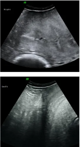
Figure 12: Ultrasound image of a dormant ovary (left side) although presenting normal size, in early follicular phase (Courtesy of Dr Thomas Hildebrandt, Department of Reproduction Management – IZW, Germany).
Figure 13: Ultrasound image of the ovary (right side) with a visible Graafian follicle in development (1.62 x 1.10 cm) (Courtesy of Dr Thomas Hildebrandt, Department of Reproduction Management – IZW, Germany).
15
An interesting feature of elephants is the development of multiple corpora lutea (on average 4 – 6 and a reported maximum of 42), measuring between 10 - 50 mm. These appear as a protrusion from the ovarian cortex in cycling as well as in pregnant cows (Lueders & Hildebrandt, 2012). Two different types of CL can be found in the elephants: accessory CL (acCL) and the ovulatory CL (ovCL). Both types are moderately echogenic, sometimes with an elongated echogenic center and a homogeneous parenchyma. The acCL appears fluid-filled and differs from the ovCL in shape, location within the ovary and the absence of stigmata (Hildebrandt et al., 2000a) (Figure 14).
In ultrasound examinations, around 2 - 10 acCL can be found on each ovary of a cycling or pregnant female elephant (Lueders & Hildebrandt, 2012). It is proposed that these acCL will help to maintain a (future) pregnancy (Lueders et al., 2012).
Figure 14: Ultrasound images of the ovary in an African elephant. A) Two follicles are visualized in luteinisation process, already starting to produce a wall and leading to the formation of an acCL (see below); B) One day after the first anLH surge; luteinisation process of the previously imaged follicles (arrows); C) Dominant follicle in growing process, before the second ovulatory LH (ovLH) surge; D) Graafian follicle on the day of ovLH surge and a CL in formation (arrows) (Image source: Hermes et al., 2000).
2.1.2 Physiology of the oestrous cycle
Female elephants present poliestric spontaneous ovulation and are considered non-seasonal breeders. Although peaks of births have been observed in the wild (Lueders & Hildebrandt, 2012) this phenomenon has yet not been reported in a more stable captive environment. There are however, some differences between the responsiveness of Asian and African elephants: Brown (2014) states that Asian elephants seem to show more consistent cycles, even when facing management changes, in contrary to African elephant females, for which temporary or permanent suppression of the oestrous cycle has been
16
repeatedly reported (e.g., after translocations, changes in blood collection frequency, changes in herd dynamics or exchange of keepers).
Elephants possess the longest oestrous cycle known among all mammals, lasting 13 - 17 weeks. It includes a luteal phase of 8 - 10 weeks and a 4 - 7 weeks inter-luteal, i.e. follicular period (Brown, 2014). Therefore, captive females may express only three to four fertile cycles per year (Hildebrandt et al., 2011). The oestrous cycle length may vary between individuals but seems to be extremely consistent for each individual cow (Brown, 2000). Synchronisation of oestrous cycles has been reported by Weissenböck et al. (2009) in a captive group of four African female elephants, and similar observations have been made in human care (T. Hildebrandt, personal communication, 2014).
Wild elephants are continuous breeders, and cycles with no conception seem to be rare, as females are usually found either gestating or lactating (Hildebrandt et al., 2006). Unfortunately, this is not the reality for most captive elephants.
Luteinizing Hormone, Progestagens, Inhibins, and Follicle-stimulating Hormone
The remarkable difference of elephants’ oestrous is the occurrence of two luteinizing hormone (LH) peaks, with comparable concentrations and separated by 3 weeks, during the follicular phase. Each LH peak coincides with a follicular wave. The first LH surge constitutes the anovulatory peak (anLH), and the second the ovulatory peak (ovLH) (Lueders et al., 2010). At the first anLH surge only follicles that presented with a diameter larger than 7.5 mm are luteinized. This occurs ca. 7 ± 2.4 days before the second LH peak and serves to create the acCL. The follicles smaller than 7.5 mm consequently start regressing (Lueders et al., 2010; 2011).
Simultaneously with the luteinization of this first wave of follicles, immunoreactive inhibin (ir-inhibin) concentrations rise and stay elevated for 41.8 ± 5.8 days after ovulation (Lueders et al., 2011). Another finding is that inhibin α, βA and βB subunits are imunnolocalised in both granulosa cells of antral follicles and luteal cells in the ovary. These findings strongly suggest that the corpus luteum is one of the sources of inhibin, as well as the follicles themselves (demonstrated in an Asian elephant, Kaewmanee et al., 2011). As a consequence, the luteinized follicles should be of importance in the selection of the dominant follicle (Lueders et al., 2011). The concentrations of ir-inhibin return to baseline 20-35 days before the end of the luteal phase and it starts to decline after the acCL have reached their maximum size (Lueders et al., 2011). The serum ir-inhibin levels are positively correlated with progesterone throughout the oestrous cycle and negatively correlated with Follicle-stimulating Hormone (FSH) during the late non-luteal and early luteal phases (Kaewmanee et al., 2011). There is still an elevation in FSH-levels during the luteal phase, even with high inhibin concentrations, suggesting that inhibin alone is not capable of suppressing FSH completely (Lueders et al., 2011). The inverse relationship between FSH