Universidade de Aveiro Secção Autónoma das Ciências da Saúde 2014
RUI PEDRO
TRATAMENTO DA LEUCEMIA LINFOCÍTICA
GARCIA DE
COM CARs PARA O ANTIGÉNIO CD19
OLIVEIRA BENTO
CAR-MODIFIED T CELLS TARGETED TO
CD19 ANTIGEN FOR LYMPHOCYTIC
Universidade de Aveiro
Secção Autónoma das Ciências da Saúde 2014RUI PEDRO
TRATAMENTO DA LEUCEMIA LINFOCÍTICA
GARCIA DE
COM CARs PARA O ANTIGÉNIO CD19
OLIVEIRA BENTO
CAR-MODIFIED T CELLS TARGETED TO
CD19 ANTIGEN FOR LYMPHOCYTIC
LEUKEMIA
Dissertação apresentada à Universidade de Aveiro para cumprimento dos requisitos necessários à obtenção do grau de Mestre em Biomedicina Farmacêutica, realizada sob a orientação científica do Professor Doutor Miguel Forte, Professor Associado Convidado da Universidade de Aveiro e Professor Doutor Bruno Gago, Professor Associado Convidado da Universidade de Aveiro.
Dissertation presented to the University of Aveiro in partial fulfillment of the requirements for the degree of Master of Science in Pharmaceutical Medicine, under the supervision of Professor Miguel Forte, Invited Associate Professor at the University of Aveiro and Professor Bruno Gago, Invited Associate Professor at the University of Aveiro.
In memoriam
Susana Ferrão
Thesis Jury
President Professor Doctor Luís Almeida
Invited Associate Professor, University of Aveiro
Professor Doctor Miguel Forte
Invited Associate Professor, University of Aveiro
Doctor Cláudia Silva
Head of Research at Bluepharma
Acknowledgements Foremost, I would like to express my sincere gratitude to both my supervisors, Professor Miguel Forte and Professor Bruno Gago for the continuous support of my MSc study and research.
My sincere thanks also goes for my fellow colleagues at Novartis Pharmaceuticals, for their encouragement, insightful comments and patience, in particular to Luís Rocha, Teresa Guerreiro, Paula Jesus and Sandra Amaral.
I am grateful to the MSc colleagues for all the support, stimulating discussions, sleepless nights before deadlines and for all the fun we have had in the last two years. In particular to Joaquim Fonseca, Márcio Barra, André Andrade and Eduardo Ribeiro. To the three pillars of my life: God, my Family and my beloved Rita. This thesis would not have been written without their immense strength, support and consistency. Thank you for the invaluable love and understanding. We made it…
Last but not least, to Susana Ferrão - an example of hope, faith, indelible strength, courage and love – and to the warriors that do not give up their fight against cancer.
Keywords Leukemia, Chimeric Antigen Receptors, T Cells, Cell Therapy, CTL019, immunotherapy, Clinical, Cancer.
Abstract Cellular immunotherapies, or Advanced Therapy Medicinal
Products (ATMPs), are emerging as novel and specific therapeutic approaches to treat diseases, such as certain types of leukemias, which are difficult or impossible to treat with today’s biopharmaceutical products. Breakthroughs in basic, preclinical, and clinical science spanning cellular immunology, and cell-processing technologies has allowed clinical applications of chimeric antigen receptor–based therapies. A recent example is CTL019, a lentivirus-based gene therapy for autologous T cells, acquired by Novartis in 2012 through a global alliance with the University of Pennsylvania. Although this technology is still in its infancy, clinical trials have already shown clinically significant antitumor activity in chronic lymphocytic leukemia and acute lymphocytic leukemia. Trials targeting a variety of other adult and pediatric malignancies are under way. The potential to target essentially any tumor-associated cell-surface antigen for which a monoclonal antibody can be made opens up an entirely new arena for targeted therapy of cancer. The regulatory environment for these Advanced Therapies Medicinal Products is complex and in constant evolution. Many challenges lie ahead in terms of manufacturing process, non-conventional supply chain logistics, business models, intellectual property, funding and patient access.
Disclosure Rui Bento is a full-time employee of Novartis Farma – Produtos Farmacêuticos SA. The contents and views expressed in this manuscript are the views of the author and not necessarily the views of Novartis. The scientific information may include data/information on investigational uses of compounds/drugs/healthcare technologies whose efficacy, safety and indications have not been established nor approved by regulatory authorities. CTL019 is an investigational compound. Efficacy and safety have not been established. There is no guarantee that CTL019 will become commercially available. The author has no other relevant affiliations or financial involvement with any organization or entity with a financial interest in or financial conflict with the subject matter or materials discussed in the manuscript apart from those disclosed.
Table of Contents
1 Introduction to Leukemia ... 11
1.1 Introduction to Dysregulation of Normal Blood Cell Development ... 11
1.2 Mechanisms of Leukemia Development ... 11
1.3 Overview of Risk Factors ... 11
1.4 Genetic Risk Factors ... 11
1.5 Leukemia Classifications ... 12 1.5.1 Acute vs. Chronic ... 13 1.5.2 Lymphocytic vs. Myelogenous ... 13 1.5.3 Types of Leukemia ... 14 1.6 Epidemiology ... 16 1.6.1 Incidence ... 16 1.6.2 Mortality ... 17
2 The Diagnosis of Leukemia ... 19
2.1 Diagnostic Steps ... 19
2.1.1 General Symptoms ... 19
2.1.2 Types of Tests and Interpretation of Results ... 20
2.1.3 Stages, Phases, and Classifications of Leukemia ... 21
2.2 Diagnosis by Leukemia Subtype – ALL and CLL ... 23
2.2.1 ALL ... 23
2.2.2 CLL ... 24
3 The Treatment of Leukemia ... 27
3.1 Management strategies ... 27
3.1.1 Management Types ... 27
3.1.2 Treatment Phases ... 28
3.2 Treatment Recommendations – ALL and CLL ... 29
3.2.1 General Factors That Impact Therapy ... 29
3.2.2 ALL ... 29
3.2.3 CLL ... 32
4 Chimeric Antigen Receptors (CARs) for Cancer ... 37
4.1 Introduction to CARs ... 37
4.1.2 Principles of T Cell Transfer ... 38
4.2 Strategies for T Cell Culture and Engineering ... 38
4.2.1 Approaches for T Cell Culture ... 39
4.2.2 Approaches for T Cell Engineering ... 39
4.2.3 Strategies Using Synthetic Biology with Engineered T Cells ... 40
4.2.4 Strategies with CAR T Cells ... 40
4.3 Current Status of Chimeric Antigen Receptor T Cell Therapy ... 41
4.3.1 First Generation CARs ... 42
4.3.2 Second- and Third-Generation CARs ... 42
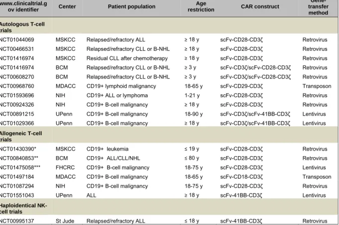
4.3.3 CAR Trials Targeting B Cell Malignancies ... 43
4.4 Issues and Future Directions for CAR T Cells ... 45
4.4.1 Additional target antigens in cell therapy of leukemia ... 45
4.4.2 The Next Generation of CAR T Cells ... 46
4.4.3 Does Dose matter? ... 46
4.4.4 CAR T Cells and Allogeneic Stem Cell Transplant ... 47
5 Toxicity with CAR T Cells ... 49
5.1 Introduction to Toxicity in T Cell Therapies ... 49
5.2 Cytokine Release Syndrome with CAR T Cells ... 51
5.2.1 Clinical Manifestations of CRS ... 51
5.2.2 Differentiation of CRS in CAR-T ... 53
5.2.3 Precautions to avoid severe CRS ... 53
5.2.4 Treatment of CRS ... 55
5.2.5 Challenges for the management of CRS ... 56
6 Chimeric Antigen Receptor–Modified T Cells in Lymphocytic Leukemia ... 57
6.1 Chimeric Antigen Receptor–Modified T Cells in CLL ... 57
6.2 Chimeric Antigen Receptor–Modified T Cells in ALL ... 60
7 R&D alliances in CAR-based cellular therapies ... 61
7.1 Novartis and University of Pennsylvania broad-based R&D alliance ... 63
7.2 Other partnerships ... 64
8 Regulatory Environment of Advanced Therapy Medicinal Products in the EU and US ... 67
8.1 Introduction to R&D Activities on ATMPs in the EU: current landscape ... 67
8.2 Market Authorizations and Overview of the ATMP Regulation ... 67
8.2.1 Marketing Authorizations ... 68
8.3 Requirements for the marketing authorization of ATMPs ... 69
8.3.1 The case of autologous ATMPs ... 69
8.4 EU versus US Regulations ... 70
8.5 Intellectual Property for ATMPs ... 71
8.5.1 Several trade secrets may help protect ATMPs ... 72
9 Pricing and Patient Access to Cellular Therapies ... 73
9.1 Reimbursement and Funding of ATMPs ... 73
9.2 Value Demonstration and Pricing ... 75
10 Business Model Considerations for Development of Cell Therapies ... 77
10.1 Introductory Development and Commercialization of Cell Therapies ... 77
10.1.1 Attributes and Challenges of Development by Design ... 78
10.1.2 Contract Manufacturing Option ... 79
10.2 Assessing Commercial Opportunities for Autologous and Allogeneic Cell Therapies ... 80
10.2.1 Autologous versus allogeneic business models ... 80
10.2.2 A hybrid Autologous Model ... 81
10.2.3 Which Cell-based Approach is More Likely to Succeed? ... 82
10.2.4 Cost of Goods Comparison ... 83
10.2.5 Finding the ‘sweet spot’ for each cell-based approach ... 84
10.3 Summary of Challenges for Commercial Manufacturing of Cell Therapies ... 86
10.4 Points to Consider for a Successful Commercialization ... 87
10.4.1 Summary of Technical Challenges for CTL019 ... 88
11 Future Directions and Conclusion ... 89
List of Tables
Table 1 Genetic risk factors influencing the incidence of leukemia ... 12
Table 2 Different in acute vs. chronic leukemia ... 13
Table 3 Three Main ALL Classification Subtypes. ... 15
Table 4 Diagnostic Tests for Leukemia ... 21
Table 5 Prognosis Varies Greatly by Clinical Stage ... 22
Table 6 Factors that Contribute to ALL Prognosis ... 30
Table 7 Overall Outcomes for Adult ALL Patients Substantially Worse than for Pediatric Patients ... 30
Table 8 Treatment Strategies for ALL Patients ... 32
Table 9 Various Prognostic Markers Complicate Staging but May Influence Patient Selection for Transplantation ... 35
Table 10 Treatment Strategies for CLL Patients ... 35
Table 11. Summary of actively recruiting clinical trials with CD19-targeted CARs.. ... 44
Table 12. The cytokines and symptoms involved in CRS in the CAR-T cell clinical trials. ... 52
Table 13. Selected deals and partnerships in the adoptive T cell immunotherapeutic space from 2008 onward. ... 61
Table 14. The European Commission’s regulation 1394/2007 establishes the legal and regulatory framework for ATMPs in Europe. ... 69
Table 15. Regulatory requirements for ATMPs in EU and US. ... 71
List of Figures
Figure 1. Cause of common symptoms associated with leukemia.. ... 19
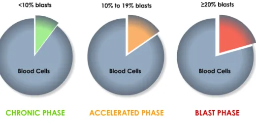
Figure 2. Phases of CML are defined by the number of blasts in the blood and bone marrow.. ... 23
Figure 3. Antibodies can bind to surface antigens expressed on tumor cells. ... 37
Figure 4. Chimeric antigen receptor (CAR) therapy is similar to an autologous bone marrow transplantation procedure. ... 40
Figure 5. CAR technology evolution through the generation of more potent CARs. ... 41
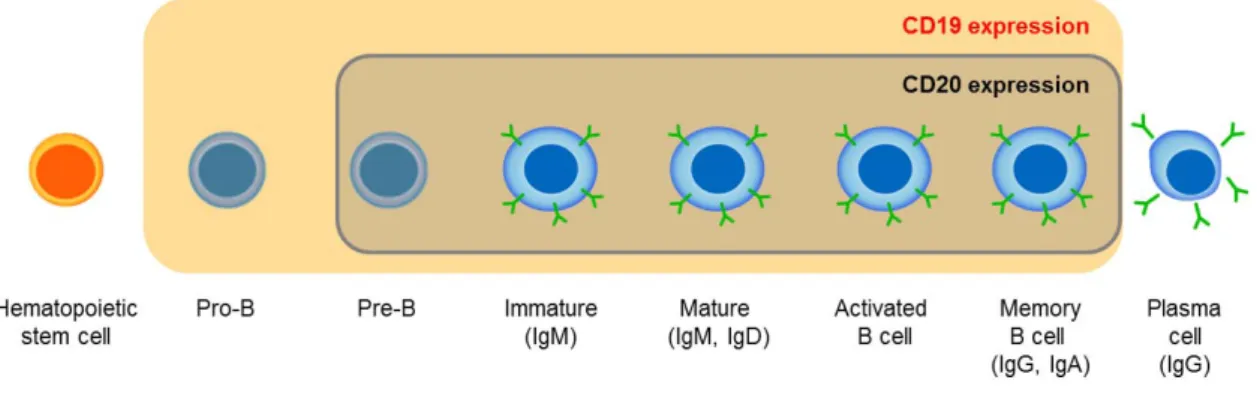
Figure 6. CD19: An Ideal Tumor Target in B-Cell Malignancies. ... 43
Figure 7. Variables in clinical trial design. ... 45
Figure 8. The classic chimeric antigen receptor (CAR) therapy (CAR-T) approach. ... 50
Figure 9. Contrast-enhanced CT scans obtained before the patient was enrolled in the study and 31 days and 104 days after the first infusion. ... 59
Figure 10. Bone marrow–biopsy specimens obtained 3 days after chemotherapy and 23 days and 6 months after CART19-cell infusion. ... 59
Figure 11. Preclinical and clinical studies that drove CAR-based T cell therapeutic development. ... 62
Figure 12. Spectrum of ATMPs and likely evaluations. ... 73
Figure 13. The life cycle of a T-cell-based process.. ... 74
Figure 14. The payer dilemma when funding products that purport to cure disease. ... 75
Figure 15. Staggering payment, the intervention is paid only for patients continuing to benefit. ... 76
Figure 16. Development and commercialization path for cell therapies. ... 77
Figure 17. Target product profile (TPP) and commercial manufacturing vision. ... 78
Figure 18. Early development quality realization. ... 79
Figure 19. Autologous and allogeneic cell-based business models compared.. ... 81
Figure 20. Hybrid autologous cell-based business model.. ... 82
Figure 21. Comparison of autologous versus allogeneic therapies and potential commercial advantages/disadvantages. ... 83
Figure 22. Relative cost of goods versus profitability by type of therapy. ... 84
List of Abbreviations
ALL Acute Lymphocytic Leukemia
AML Acute Myelogenous Leukemia
AMNOG Arzneimittelmarkt-Neuordnungsgesetz ARDS Acute Respiratory Distress Syndrome
ATMPs Advanced Therapy Medicinal Products
BCR B-Cell Receptor
CACT Center for Advanced Cellular Therapies
CAR Chimeric Antigen Receptor
CAT Committee for Advanced Therapies
CBER Center for Biologics Evaluation and Research CD19 Cluster of Differentiation 19
CLL Chronic Lymphocytic Leukemia
CML Chronic Myelogenous Leukemia
CNS Central Nervous System
CoGS Cost of Goods Sold
CRS Cytokine Release Syndrome
CsA Cyclosporine
CT Computer Tomography
CTCAE Common Terminology Criteria for Adverse Events CTL019 T Cells Engineered to Express CAR which Targets CD19
CTMP Somatic Cell Therapies
DbD Development by Design
DNA Deoxyribonucleic Acid
DRGs Diagnostic Related Groups
EBV Epstein–Barr Virus
EC European Commission
EGF Epidermal Growth Factor
EMA European Medicines Agency
EU European Union
EudraCT European Union Drug Regulating Authorities Clinical Trials
FcRγ Fc-alpha Receptor
FDA US Food and Drug Administration
FISH Fluorescence In Situ Hybridization FLT3 Fms-like tyrosine kinase 3
GM-CSF Granulocyte Macrophage Colony Stimulating Factor GMP Good Manufacturing Practice
GTMP Gene Therapies
GVHD Graft-Versus-Host Disease
HCT/Ps Human Cells, Tissues, or Cellular or Tissue based products
HIV Human Immunodeficiency Virus
HLH Hemophagocytic Lympohistiocytosis
HMMR Hyaluronan-Mediated Motility Receptor
HSCT Hematopoietic Stem Cell Transplantation HSV-TK Thymidine Kinase from Herpes Simplex Virus icasp9 inducible caspase 9
IFN-γ Interferon Gamma
mAb monoclonal Antibody
MDS Myelodysplastic Syndrome
MF Myelofibrosis MHC Major Histocompatibility Complex
MLL Mixed-Lineage Leukemia gene
MRI Magnetic Resonance Imaging
mRNA messenger Ribonucleic Acid
MSCs Mesenchymal Stem Cells
MSKCC Memorial Sloan-Kettering Cancer Center NCI National Cancer Institute
NK Natural Killer cells
PCR Polymerase Chain Reaction
Ph1 Philadelphia Chromosome
PLL Prolymphocytic Leukemia
QbD Quality by Design
RNA Ribonucleic Acid
ROR1 Tyrosine kinase-like Orphan Receptor 1 scFv single-chain antibody Fragment
SLL Small Lymphocytic Lymphoma
SME's Small and Medium sized Enterprises SOFA Sequential Organ Failure Assessment T regs regulatory T cells
TCM Central Memory T Cells
TCR T Cell Receptor
TEP Tissue Engineered Products
TLS Tumor Lysis Syndrome
TNF-α Tumor Necrosis Factor alpha
TPP Target Product Profile
Tscm memory stem T cell
1 Introduction to Leukemia
1.1 Introduction to Dysregulation of Normal Blood Cell Development
The development of leukemia is a multiple-step process that requires the normal blood cells to be susceptible at different stages. The production of normal blood cells markedly decreases, which results in varying degrees of symptoms, increased proliferation of the transformed cancer cells with reduced programmed cell death, and increased ability of these cells to proliferate [1]. There are many distinct causal mechanisms that can initiate the development of leukemia, including specific molecular abnormalities and exposures to carcinogens in the environment.
1.2 Mechanisms of Leukemia Development
Leukemia is thought to occur when precursors of mature blood cells in the bone marrow acquire mutations in their DNA [2] and as a consequence, the bone marrow makes abnormal white blood cells [3]. Changes in the DNA code can lead to rapid, aberrant growth of the cells as well as survival of the cells beyond their normal lifespan. Over time, the mutated cells can accumulate and overcome the population of normal, healthy cells in the bone marrow, enter the bloodstream, and invade other parts of the body (including lymph nodes, spleen, liver, and central nervous system), resulting in the signs and symptoms of leukemia [2].
The presence of leukemia stem cells is thought to contribute to the development and relapse of certain types of leukemia. Leukemia stem cells are a rare subset of stem cells that have specific developmental, cellular, and molecular properties that are distinct from leukemic blast cells, including the ability to self-renew. However, leukemia stem cells still retain key features of normal stem cells. They are also quiescent, meaning they are inactive or at rest. This property is thought to contribute to relapse because treatments may target and kill active leukemic blast cells but may not effectively target the leukemia stem cell population. A deeper understanding of these cells will help develop improved leukemia therapies [4].
1.3 Overview
of
Risk
Factors
The causes of leukemia are complex and sometimes unexplainable. It does, however, usually develop from a combination of initiating factors. Certain environmental factors explain some cases, while other causes of leukemia can be traced to familial genetic abnormalities or acquired changes in genes. In studying potential causes, several leukemia risk factors have been identified.
1.4 Genetic Risk Factors
The rapid advances in human genomics and molecular techniques have helped define the role of genetic traits in the development of leukemia. Genetic factors have been shown to significantly influence the incidence of leukemia, and certain populations are known to be more susceptible to the disease.
These genetic factors include:
• Family history of leukemia and other malignant blood disorders • Genetic chromosomal abnormalities like Down’s syndrome • Subtle genetic disorders and polymorphisms
Environmental Risk Factor Explanation Exposure to high levels of
radiation
Prolonged exposure to radiation has been known to be linked with leukemia. Early radiologists, before the use of appropriate shielding, were found to have an increased likelihood of developing leukemia. Smoking and second-hand
smoke
Smoking is considered a risk factor for leukemia. Many people who have never smoked develop leukemia, which suggests that secondhand smoke may also play a role.
Long-term exposure to certain chemicals
Long-term exposure to chemicals such as benzene or formaldehyde has been associated with leukemia. Inhalation is the predominant route of exposure to these exogenous chemicals, and upon inhalation, these chemicals rapidly react with molecules in the body and are swiftly metabolized by erythrocytes to form adducts with DNA and proteins.
‐ Formaldehyde is a simple one-carbon molecule found in most cells at varying concentrations as a normal product of metabolism.
‐ materials, glues, and fabrics as well as other consumer products like medicines and beauty aids.
‐ Benzene is a colorless and extremely flammable organic hydrocarbon that is one of the most used chemicals in the United States. Numerous studies have confirmed a link between exposure to benzene and leukemia, especially occupational exposures. Benzene is also found in car emissions and cigarette smoke and in certain foods.
Previous chemotherapy Chemotherapeutic drugs like alkylating agents (i.e., cyclophosphamide), topoisomerase II inhibitors, and anthracyclines (i.e., doxorubicin) that are used to treat cancers in adults or children may induce secondary leukemias. These agents cause DNA damage and aberrant cell growth as a mechanism of their clinical activity, which may in turn introduce mutations and rearrangement of genes, leading to cancer development.
Certain viral infections The human T-cell lymphoma virus type 1 (HTLV-1) was the first human retrovirus to be discovered and has been recognized as the cause of specific types of leukemia. During infection of cells with the HTLV-1, the virus incorporates its genetic information and hijacks the cellular machinery to produce an oncoprotein, Tax, which promotes hyperactivation of survival and growth pathways.
Table 1 Genetic risk factors influencing the incidence of leukemia (Adapted from [5])
1.5 Leukemia
Classifications
The classification of leukemia has evolved significantly over the past few decades as a result of increased understanding of the development of normal immune cells. Classifications of leukemias have developed from two distinct clinical needs—to understand the natural history of these diseases in order to predict outcomes and to make treatment decisions in a rational fashion. In these modern classifications, distinct disease entities are defined based on the combination of morphology, immunological and molecular techniques, and clinical features. Leukemias are classified two different ways—by the speed of disease progression and by the type of cells that have been transformed. Therefore, the classification of leukemias takes into consideration the aggressiveness of the cancer
and how quickly it progresses. Another factor to consider is how mature the cancer cells are compared to the stem cells from which they are derived.
1.5.1 Acute
vs.
Chronic
Acute
Acute refers to a disorder of rapid onset. Abnormal cells grow rapidly from their immature states, are unable to perform their functions, and do not differentiate [6]. This type of leukemia usually occurs in children and requires immediate therapeutic intervention [7].
Chronic
Chronic refers to an onset that tends to be slower. Cells in chronic disease are generally mature and functional, though abnormal, and typically accumulate in various organs gradually [6]. Conversely, chronic leukemias mostly affect adults, as it may take some time for the disease to progress to the point when effects are noted [7].
Leukemia Features Acute Chronic
Onset Rapid Slower
Cell Type Immature Mature
Disease Characteristics Occurs in children, requires
immediate therapy Occurs in adults, slow progression
Table 2 Different in acute vs. chronic leukemia
1.5.2 Lymphocytic vs. Myelogenous
Another way to classify the type of leukemia is by the white blood cells from which the cancer is derived. This section will describe how cancer can occur in either the lymphoid (lymphocytic leukemia) or myeloid (myelogenous leukemia) white blood cells.
Lymphocytic
Lymphocytic leukemia arises from the transformation of normal lymphoid cells to malignant cancerous cells. Since this subgroup of leukemias is derived from cells from the lymphoid cellular pathway, lymphocytic leukemias can either be B-cell leukemias or T-cell leukemias. Lymphocytic leukemia may be distinguished from other malignant lymphoid disorders by the immunophenotype of the cells, which is similar to B- or T-precursor cells [1]. Although B-cell and T-cell leukemias may superficially resemble one another, they have distinct clinical and pathologic features and must be distinguished from one another by a pathologist. The majority of lymphocytic leukemias are B-cell type [8].
Myelogenous
Myelogenous, or myeloid, leukemia develops from the early myeloid cell pathway [9]. Depending on whether the disease is acute or chronic, this can lead to:
• An abnormal development of a type of immature white blood cell called myeloblasts, which do not differentiate into normal, healthy white blood cells [10],
• An increased proliferation of white bloods cells called granulocytes [11].
1.5.3 Types
of
Leukemia
Taking into account both the speed of disease progression and the type of cells that have been transformed leads to the division of four main types of leukemias. These common types include acute myelogenous leukemia (AML), chronic myelogenous leukemia (CML), acute lymphocytic leukemia (ALL), and chronic lymphocytic leukemia (CLL). [7]
Acute Myelogenous Leukemia
Acute myelogenous leukemia (AML) is an aggressive disease in which the myeloid stem cells usually become a type of immature white blood cell called myeloblasts. The myeloblasts in AML are abnormal and do not differentiate into normal, healthy white blood cells [10]. Additionally, too many of these abnormal cells can be found in the bone marrow and blood and sometimes, too many stem cells become abnormal red blood cells or platelets [12].
Epidemiological and genotypic studies have demonstrated that AML cells have more than one recurring mutation that is shared within the group of diseases. Fms-like tyrosine kinase 3 (FLT3), a receptor tyrosine kinase expressed by immature hematopoietic cells and important for the normal development of stem cells, is the most commonly mutated protein in AML. FLT3 is constitutively activated by acquired mutations in approximately 30 to 35% of AML [4]. These mutations ultimately provide survival and growth advantage to the cells [4]. Because of this, the presence of the FLT3 mutation is often associated with a poorer prognosis for AML patients. Polymorphisms of NQO1 are also closely associated with increased risk of AML [13]. Most AML subtypes are distinguished from other leukemias and blood disorders by the presences of more than 20% blasts in the bone marrow [13].
Most patients who present with newly arisen AML have no identifiable risk factors [13]. There is, however, a documented progression from hematologic disorders like myelodysplastic syndrome (MDS) and myelofibrosis (MF) to AML [13]. Patients with low-risk MDS generally do not develop AML, while patients with high-risk MDS do. MDS is typically a disease that has an increased incidence with age that can contribute to and explain the high incidence of AML in the elderly [14]. AML is more common in men than in women, which especially becomes apparent with increased age and may be due to the fact that MDS, which tends to develop into AML, is also more common in men [13]. Other risk factors in developing AML include race and ethnicity, the father’s age at conception (with an increased risk over age 35), and the time since the mother’s last live birth (with greater risk associated with over seven years since the last childbirth). [14]
Chronic Myelogenous Leukemia
Chronic myelogenous leukemia (CML) is a myeloproliferative disorder characterized by increased proliferation of the myeloid cell line, without the loss of the ability to differentiate [11]. The result is the overproduction of abnormal granulocytes. CML progresses through three phases:
• The chronic phase, where mature cells proliferate,
• The accelerated phase, where additional abnormal genetic events occur, and • The blast phase, where immature cells grow rapidly.
Approximately 85% of patients are diagnosed in the chronic phase and then progress to the other phases within three to five years [11]. The hallmark of this leukemia is the presence of the Philadelphia chromosome (Ph1), a fusion chromosome that is the result of abnormal genetic translocations between chromosomes 22 and 9 resulting in a shortened chromosome 22. This translocation creates a hyperactive protein kinase product called BCR-ABL that drives the proliferation of the cancer, making growth factors often unnecessary [15]. Because BCR-ABL is the major molecular event in the development of CML, this protein is a great target for diagnosis, treatment, and disease monitoring. CML was the first cancer to be associated with a chromosomal translocation and a single, specific genetic mutation that drives the cancer. [15] [11]
Acute Lymphocytic Leukemia
Acute lymphocytic leukemia (ALL) is the most common childhood leukemia and the most common childhood cancer in developed countries [4]. ALL is thought to arise from malignant transformation of B- or T-cell progenitor cells [16]. Most adults with ALL have no identifiable risk factors [1]. However, 80% of the cases of infant ALL are associated with genetic abnormalities in the MLL gene, leading to the increase in survival factors that causes this disease to be resistant to chemotherapy and other treatments [4]. ALL also often presents in infants with features associated with poor outcome, including diseases of the central nervous system and poor response to treatment. In terms of inherited chromosomal abnormalities, children with Down’s syndrome have an increased risk of developing both ALL and AML. The cumulative risk is approximately 2.1% by age 5, with most of the cases being ALL [17]. Cases of chromosomal abnormalities following treatment with topoisomerase II inhibitors have been linked with the development of ALL, but these patients are more likely to develop AML [1]. Also, the Philadelphia chromosome occurs in about 20% of adults and a small percentage of children with ALL [16]. In the majority of children and in more than 50% of adults with Ph1-positive ALL, the molecular abnormality is different from that in Ph1-positive CML.
Immunologic Subtype
Percentage
of Cases FAB Subtype Cytogenic Abnormalities Pre-B ALL 75% L1, L2 t(9;22), t(4;11), t(1;19)
T-cell ALL 20% L1, L2 14q11 or 7q34
B-cell ALL 5% L3 t(8;14), t(8;22), t(2;8)
FAB: French American British L1: small uniform cells L2: large varied cells
L3: large varied cells with vacuoles
Table 3 Three Main ALL Classification Subtypes. Adapted from [18]
Chronic Lymphocytic Leukemia
Chronic lymphocytic leukemia (CLL) is the most common form of leukemia found in adults in Western countries [8]. The cells of origin in most patients with CLL are clonal B cells. These cells are arrested in the differentiation pathways between immature blasts and mature B cells, though they may look similar to mature lymphocytes [8]. Much progress has been made in differentiating subsets of CLL and
predicting disease progression. CLL was once grouped together as subtypes under the broad term of non-Hodgkin’s lymphoma, but after extensive refinement of the classifications of blood cancers to better distinguish and group the lymphoid disorders by their clinical and biological characteristics, a separate and distinct disease group was created [19]. The exact cause of CLL is uncertain, but it is known that CLL is an acquired disorder, with familial cases being extremely rare [8]. The identification and use of molecular and cellular markers like B-cell receptor (BCR), a protein that plays a key role in signal transduction, has helped define CLL [20]. While low expression of the BCR is the hallmark of the B-CLL lymphocyte, increased expression correlates with mutated cells [20]. Another important genetic parameter in defining pathogenic and prognostic subgroups of CLL is the mutation of the VH genes, which is observed in about half of all CLL cases [21].
1.6 Epidemiology
Leukemia and the consequences of this group of diseases represent a substantial worldwide concern. This section describes the incidence and mortality of leukemia and explains the risk factors associated with this blood disorder.
1.6.1 Incidence
The American Cancer Society estimates that 31,500 individuals in the US will be diagnosed with leukemia every year [14]. From 2005 to 2009, the median age of leukemia diagnosis was 66 years of age; 10% were diagnosed under the age of 20, 4.9% between the ages of 20 and 34, 5.3% between the ages of 35 and 44, 26.7% between the ages of 45 and 64, and 52.6% 65 years of age and over [22].
Childhood Leukemias
Leukemia is the second most common malignancy in the first year of life [23]. About 85% of leukemias in children are acute [6], and ALL accounts for 65% of the acute leukemias in children [6]. Approximately 4.1 of every 100,000 young people under 20 years of age in the US are diagnosed with leukemia [24], and infant leukemia is more common in females than males, with a ratio of 1.17, female to male [23]. Leukemia is also the most common cancer diagnosis in children less than 15 years old, and ALL is approximately five times more common than AML in this age group [4].
Around 2000, the average incidence for this age group in the European Region was 46.7 cases per million per year, with a slightly lower level in eastern than in western European countries. European population-based cancer registries show an average increase in the incidence of childhood leukemia of 0.7% per year between 1970 and 1999 [25].
Adult Leukemias
As adult leukemias are more common in men than women, approximately 0.48% of men will develop leukemia between their 50th and 70th birthdays compared with 0.30% of women [22]. CLL is the most common form of leukemia in Western countries and mainly affects elderly individuals, with the median age of presentation being 72 years [8]. More than 17,000 new cases of CLL are reported each year in the US [8]. CLL is almost twice as common as CML, which makes up only 20% of all leukemias affecting adults, typically middle-aged adults [13] [6]. AML is the most common acute leukemia in adults, accounting for about 25% of all leukemias in the Western world. The incidence of acute
leukemias accounts for less than 3% of all cancers, but is still the leading cause of death to individuals under 40 who have developed the disease [14].
1.6.2 Mortality
Epidemiological studies of leukemia have focused on mortality to determine how specific populations and age groups are more susceptible to succumb to the disease. The progression of many types of leukemia is extremely variable, with survival ranging from months to decades in some cases.
Statistical reports show that the median age of death for leukemia is 75 years. Approximately 2.8% of leukemia deaths occur in patients that are under the age of 20 years. About 3.1% leukemia deaths occur in those aged 20 and 34 years, 3.1% occur between 35 and 44, 19.3% occur in those aged 45 to 64 years, and 71.8% occur in those 65 years and over. It was estimated that, while 47,150 men and women would be diagnosed with leukemia in 2012, 23,540 men and women would die from it [22].
2 The Diagnosis of Leukemia
2.1 Diagnostic Steps
Symptoms develop based on how quickly the disease progresses. As acute disease is associated with a rapid onset, symptoms develop fairly quickly when a person is inflicted with acute leukemias. Individuals are typically diagnosed right after they become ill. Conversely, for chronic disease, which has a more gradual onset, symptoms are slow to develop. It is not uncommon for some people with chronic leukemia to not even present with symptoms at diagnosis [26].
2.1.1 General Symptoms
Some of the symptoms associated with leukemia are due to the increase of abnormal blood cells that have replaced the population of normal, healthy cells. Because this is a general characteristic of all leukemias, there are a number of symptoms that can be common to all four of the major types of leukemia [26].
These symptoms include [26]: unexplained fevers, frequent infections, night sweats, fatigue, weight loss, easy bleeding or bruising, as showed below:
Figure 1. Cause of common symptoms associated with leukemia. Adapted from Stoppler M, 2014.
Leukemia patients are commonly referred to as having anemia, leukopenia, neutropenia, and thrombocytopenia, which cause generalized symptoms [27]. Other symptoms are due to the accumulation of leukemia cells in vital tissues and organ systems of the body. Some of the most common sites of leukemia cell accumulation are the lymph nodes, liver, spleen, kidney, lungs, and skin [26].
Many of these symptoms can also occur across the leukemia spectrum [26]: headache, confusion, balance problems, blurred vision, painful swellings in the neck, under the arms, or in the groin, shortness of breath, nausea or vomiting, abdominal pain or swelling, testicular pain or swelling, pain in the bones or joints, weakness or loss of muscle control and seizures.
2.1.2 Types of Tests and Interpretation of Results
In order to properly diagnose leukemia, healthcare professionals utilize a number of diagnostic tests. Some of these tests are not needed to establish the diagnosis of leukemia but are sometimes performed at diagnosis to predict the prognosis or to assess tumor burden.
When a clinician is confronted with an individual who is suspected of having leukemia, there are a number of tests that are available at initial diagnosis. The tests include [26] [27] [7]: blood tests, organ function tests, biopsy, genetic tests, lumbar puncture, lymph node excision and imaging techniques. Diagnostic
Tests
Description
Blood tests Blood is drawn from a vein and tested in order to check blood cell count, size, and maturity.
In most cases of leukemia, the overall white blood cell count will be high, while platelet, red blood cell, and neutrophil counts will be low (thrombocytopenia, anemia, and neutropenia).
Organ function
tests Leukemia cells often accumulate in the liver and kidneys and affect normal organ function. The function of these organs may be checked by examining specific markers that may indicate damage or stress to these organs (i.e., albumin, cholesterol, creatinine).
Biopsy In a biopsy, a small sample of the relevant tissue from the bone marrow is taken, usually from the hip bone, and analyzed for the presence of abnormal cancerous cells under a microscope. The procedure is brief but does require a pre-injection of anesthesia.
Because leukemia may cause an increase in white blood cell count, a biopsy of the bone may be taken to examine blood cell composition. Genetic tests As the presence of genetic abnormalities may be a cause of the
development of leukemia, chromosomes of the cancerous cells are examined to look for potential genetic changes that also may help to classify the specific type of leukemia.
There exist a number of individual genetic tests that can be conducted: o Conventional cytogenetics: analysis of the chromosomes under
a microscope to find any significant changes, a process known as karyotyping; chromosomes are best seen when the cells are undergoing division, so a sample of the blood or bone marrow is often grown in a laboratory
o Fluorescent in situ hybridization (FISH): test that uses special fluorescent dyes designed to attach to specific portions of chromosomes to help identify the presence of specific changes attributed to cancer
o Polymerase chain reaction (PCR): a sensitive test that amplifies DNA or RNA from blood or bone marrow to allow for the detection of even small amounts of genetic abnormalities
Lumbar puncture (Spinal tap)
Leukemia cells can also find their way into the fluid surrounding the brain and spinal cord of the central nervous system, also known as the cerebrospinal fluid. This often causes individuals’ mental processing to be affected.
In this procedure, a small injection is performed first in the site of the spinal tap to minimize the discomfort of the procedure. A hollow needle is then inserted in the back between the bones in the spine around the
waist area to remove a small amount of fluid for analysis.
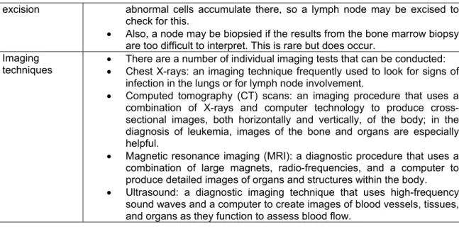
excision abnormal cells accumulate there, so a lymph node may be excised to check for this.
Also, a node may be biopsied if the results from the bone marrow biopsy are too difficult to interpret. This is rare but does occur.
Imaging
techniques There are a number of individual imaging tests that can be conducted: Chest X-rays: an imaging technique frequently used to look for signs of infection in the lungs or for lymph node involvement.
Computed tomography (CT) scans: an imaging procedure that uses a combination of X-rays and computer technology to produce cross-sectional images, both horizontally and vertically, of the body; in the diagnosis of leukemia, images of the bone and organs are especially helpful.
Magnetic resonance imaging (MRI): a diagnostic procedure that uses a combination of large magnets, radio-frequencies, and a computer to produce detailed images of organs and structures within the body. Ultrasound: a diagnostic imaging technique that uses high-frequency
sound waves and a computer to create images of blood vessels, tissues, and organs as they function to assess blood flow.
Table 4 Diagnostic Tests for Leukemia
2.1.3 Stages, Phases, and Classifications of Leukemia
With the exception of CLL, leukemia is not staged numerically (I, II, III, or IV) as many solid tumors are [28]. Instead, descriptors like acute or chronic and time-to-progression mostly indicate the severity of the disease. In addition, treatment status gives another level of disease classification.
Staging is the way cancer is classified based on certain criteria, including the size and extent of spread. Leukemias like CLL may be classified by a staging system based on the part of the body that is affected. In terms of CLL, the most commonly used staging system in the US is the Rai classification system [29] and in Europe, the Binet staging system is used.
The Rai system is separated into five stages [29]:
Stage 0: The blood lymphocyte count is too high, usually defined as over 15,000 lymphocytes/mm3 of blood (lymphocytosis) and > 40% lymphocytes in the bone marrow. The lymph nodes, spleen, and liver are not enlarged, and the red blood cell and platelet counts are near normal. This is considered a low-risk group.
Stage I: Lymphocytosis plus enlarged lymph nodes—The spleen and liver are not enlarged, and the red blood cell and platelet counts are near normal. This is considered an intermediate-risk group.
Stage II: Lymphocytosis plus an enlarged spleen and possibly an enlarged liver, with or without enlarged lymph nodes—The red blood cell and platelet counts are near normal. This is considered an intermediate-risk group.
Stage III: Lymphocytosis plus anemia, with or without enlarged lymph nodes, spleen, or liver— Platelet counts are near normal. This is considered a high-risk group.
Stage IV: Lymphocytosis plus thrombocytopenia, with or without anemia, enlarged lymph nodes, spleen, or liver—This is considered a high-risk group.
Europe’s Binet staging system can be summarized in [30]:
Stage A: Hemoglobin ≥ 10 g/dL, platelets ≥ 100,000/mm3, and < 3 enlarged areas Stage B: Hemoglobin ≥ 10 g/dL, platelets ≥ 100,000/mm3, and ≥ 3 enlarged areas
Stage C: Hemoglobin < 10 g/dL, platelets < 100,000/mm3, and any number of enlarged areas
System Stage Definition Median
Survival Rai Staging System (Common in US)
0 lymphocytosis (>5 G/L) > 10 years
I lymphocytosis + lymphadenopathy > 8 years
II lymphocytosis + splenomegaly +/-lymphadenopathy 6 years III ymphocytosis + anemia (Hb <11g%) +/-lymphadenopathy or
splenomegaly
2 years IV lymphocytosis + thrombocytopenia
(Plt < 100 G/L) +/- anemia +/-lymphadenopathy +/- splenomegaly
< 2 years Binet Staging (Common Ex US)
A < 3 involved areas, Hb > 10g%, Plt > 100 G/L > 10 years B > 3 involved areas, Hb > 10g%, Plt > 100 G/L 7 years C Any number of involved area, Hb < 10g% & Plt < 100 G/L 2 years
Table 5 Prognosis Varies Greatly by Clinical Stage [31]
Certain leukemias like CML are classified by phases, defined by the number of immune leukemia stem cells, or blasts, in the blood and bone marrow [27].
These phases are [32]:
Chronic phase: fewer than 10% of blood cells are blasts. Accelerated phase: 10% to 19% of blood cells are blasts. Blast phase: 20% or more of blood cells are blasts.
Figure 2. Phases of CML are defined by the number of blasts in the blood and bone marrow. Adapted from Duke Cancer Institute, 2011.
In general leukemias are classified rather than staged in order to determine the most appropriate therapy. Acute lymphoblastic leukemia has no standard staging system. Instead it is classified as follows [32]:
Untreated: Leukemia was recently diagnosed and has not yet been treated.
In remission: There exist no signs, symptoms, or medically defined presence of the disease (less than 5% leukemia cells).
Recurrent: Signs, symptoms, and presence of the disease have returned.
2.2 Diagnosis by Leukemia Subtype – ALL and CLL
Each of the major types of leukemia (ALL, AML, CLL, and CML) may have unique signs and symptoms that help to diagnose the disease state. In addition, results of diagnostic tests and aspects of their clinical manifestations may point to a particular leukemia subtype.
2.2.1 ALL
A minority of cases of ALL are detected during blood counts and blood examinations performed for investigation of unexplained fever, pallor, and bleeding.
Symptoms
Fever is one of the most common symptoms of ALL. Patients with ALL often have fevers without any other evidence of infection [1]. ALL patients may also present with symptoms from the increased amount of abnormal, cancerous cells in the peripheral circulation (leukostasis), including respiratory distress and altered mental status. This is much less common in ALL than AML patients, however, and only occurs in patients with the highest WBC counts [1].
ALL dominates as a hematological malignancy in children, though it can present at any age. The onset may be present as fatigue, persistent fever, bleeding, and/or bone and joint pain. Young children may present with problems with walking or even the total inability to walk. In addition, 2% to 3% of children
may first have anemia and CNS system involvement in their disease [33]. A small percentage may have skeletal symptoms due to widespread osteoporosis. In children, many of these symptoms present without the presence of enlarged organs (organomegaly), swollen lymph nodes (lymphadenopathy), or leukocytosis.In adults, the symptoms of ALL are similar to those in children. Almost 50% may have fever at presentation. However, adult onset ALL is commonly associated with an enlarged liver and spleen (hepatomegaly and splenomegaly, respectively) and lymphadenopathy at diagnosis. About 10% to 20% of ALL patients present with left upper quadrant fullness and early satiety due to splenomegaly [33].
Clinical Presentation
ALL patients often have decreased neutrophil counts, regardless of whether their total white blood cell count is low, normal, or elevated, putting them at increased risk of infection. Because of this, infections are still the most common cause of death in ALL patients undergoing care for their disease [1]. Finding blasts raises the suspicion of acute leukemia, and a bone marrow exam is then suggested. A bone marrow biopsy usually confirms the diagnosis of ALL [33].
A diagnosis of ALL may be easily confused with other hematological conditions like AML, hairy cell leukemia, and malignant lymphoma. Malignant cells are often sent for conventional cytogenetic studies, such as detection of the Philadelphia chromosome and MLL gene rearrangements, to add important information for treatment decision making. Flow cytometry is usually also performed to characterize expression of lineage-defining antigens and allow determination of the specific ALL subtype [16].
2.2.2 CLL
It is not unusual for CLL to be discovered incidentally after a blood cell count is performed for another reason, as 25% to 50% of patients will be asymptomatic at the time of presentation [8]. CLL is predominately a disease of older individuals—most patients are older than 50 at the time of diagnosis. Because of this and because disease progression is often slow, patients often succumb to other medical problems rather than CLL [34].
Symptoms
Enlarged lymph nodes and node swelling are the most common presenting symptoms, seen in 87% of the patients who are symptomatic at the time of diagnosis. A smaller number of patients report less severe symptoms like fever, weight loss, and night sweats. People with CLL may also be predisposed to repeated infections, such as pneumonia. Early satiety and/or abdominal discomfort, related to an enlarged spleen, may also be noted in this patient population [8].
Clinical Presentation
The diagnosis of CLL requires the presence of at least 5x10⁹ B lymphocytes/L (or 5,000/μL) in the peripheral blood. The tests necessary to give a diagnosis of CLL are blood count evaluations, blood smear, and the immune phenotype of the circulating lymphoid cells. Using fluorescent in situ hybridization (or FISH) during cell division, cytogenetic lesions can be identified in more than 80% of all CLL cases, with the most common being deletions in the long arm of chromosome 13 [35]. A bone marrow aspirate and biopsy generally are not required at diagnosis, but can help evaluate for factors that might contribute to symptoms like anemia and thrombocytopenia that may or may not be directly
related to organ infiltration. Bone marrow findings in CLL include normal to high cellularity, or when the bone marrow has more blood-forming cells than expected [34]. In CLL, more than 30% of the nucleated cells in the aspirate will be lymphoid [35].
Mantle cell lymphoma, hairy cell leukemia, and prolymphocytic leukemia (PLL) can have a clinical presentation very similar to CLL but are more aggressive. Additionally, CLL may also present as a lymph-node-based disease called small lymphocytic lymphoma (SLL). A combination of factors including antigen presentation, cytogenetics of chromosome translocations, intracellular mediator expression, and morphology are used to differentiate the diseases [34]. For example, CLL cells coexpress the T-cell antigen CD5 and B-cell surface antigens CD19, CD20, and CD23. Additionally, according to the WHO classification, CLL is only distinguishable from SLL by its leukemic appearance [35].
3 The Treatment of Leukemia
3.1 Management strategies
Development of treatment strategies is driven by the need to reduce toxicities associated with current therapies, overcome the onset of drug resistance, and improve clinical efficacy. Unprecedented efforts are currently underway to define molecular mechanisms that are important in the development of leukemia and determine alternatives to conventional approaches to disease management.
3.1.1 Management Types
A Watch and Wait
In certain clinical situations, treatment is not indicated until the disease is further along. With the “watch and wait” strategy, the disease is monitored with regular physical exams and lab tests until a time when decisions can be made on the most appropriate therapy to avoid therapy-related side effects. This is more likely the case when a person has no symptoms and a disease has been diagnosed by chance [36].
Chemotherapy
Chemotherapy is treatment using cytotoxic, non-specific agents to stop the growth and spread (metastasis) of cancerous cells. Chemotherapeutic agents usually accomplish this by either inhibiting cell division or promoting cell death. Chemotherapy can be taken by mouth (oral) or is injected in the vein or muscle (intravenous or subcutaneous, respectively) in order to enter the bloodstream and reach the site of the cancer (systemic chemotherapy). Chemotherapy can also be placed directly into the cerebrospinal fluid (intrathecal chemotherapy). The main types of chemotherapy include [37]:
Alkylating agents: drugs that inhibit DNA production by inducing DNA crosslinks
Antimetabolites: drugs that inhibit the cell’s utilization of natural metabolites like folic acid, needed for cell growth
Plant alkaloids: drugs that block cell division, often by inhibiting the normal function of microtubules, fibrous proteins in the cells important for cell structure and mitosis
Antibiotics: agents that interfere with cellular processes, including DNA or protein synthesis Purine analogs: agents that inhibit DNA synthesis by mimicking natural purines and
intercalating into DNA chains
Hypomethylators: agents that inhibit DNA methyltransferase, the cellular enzyme that methylates DNA, necessary for many cellular functions
Radiation Therapy
Radiation therapy uses high energy X-rays or other types of radiation to kill cancerous cells. There are generally two types of radiation. The choice of which is utilized depends primarily on how advanced the cancer is [37]:
External radiation: A machine outside of the body is utilized to send radiation toward to the portion of the body where the cancer resides.
Internal radiation: A radioactive agent is packaged in a carrier mechanism (such as a catheter) and placed directly inside the body in or near the site of the tumor.
Targeted Therapy
Targeted therapy uses agents that have been designed to specifically target cancer cells, theoretically leaving normal, non-cancerous cells unharmed. For instance, these agents may target specific proteins that are only expressed or are overexpressed on the surface of cancer cells but not healthy cells [37]. Immunotherapy
Immunotherapy uses the body’s own immune system to inhibit cancer growth and disease progression by directly or indirectly boosting the normal immune response against the disease. Because of the required involvement of the immune system to the activity of immunotherapeutic agents, this type of therapy usually involves some component of the normal immune system. Though this type of therapy may carry with it its own particular side effects, immunotherapy is typically less invasive and less toxic than conventional treatment strategies like chemotherapy [38].
Surgery
Surgery as a management strategy in leukemia usually involves the removal of swollen lymph nodes (lymphadenectomy) to confirm the diagnosis of leukemia or the removal of the spleen (splenectomy) if it has begun to destroy components of the bone marrow [39].
Stem Cell Transplantation
Stem cell transplantation (SCT) is a method of getting rid of the blood-forming cells that are no longer healthy by replacing the bone marrow. To begin the process, normal stem cells are removed from the bone marrow. After specific points in the treatment plan for a patient, the stem cells are infused into the patient. There are generally two major types of SCT [39]:
Autologous stem cell transplant (autoSCT): The non-cancerous stem cells are derived from the bone marrow of the leukemia patient.
Allogeneic stem cell transplant (alloSCT): The non-cancerous stem cells are taken from a donor to be infused in the patient. Before referral for alloSCT, a suitable donor must be identified, ideally a fully HLA-matched sibling. If this is not possible, as is the case for many patients, alternatives for a donor include a matched unrelated person or the patient’s own cord blood.
3.1.2 Treatment Phases
Induction
Induction therapy is the initial therapy given to a patient. The goals of induction therapy are to empty the bone marrow of all hematopoietic elements both healthy and cancerous and to allow the repopulation of the bone marrow with normal, functioning cells, yielding remission [40].
Postremission
Once remission is achieved, additional therapy is often required to reduce the undetectable burden of cancerous cells so that long-term disease-free survival (DFS) may be possible. Postremission therapy may involve two types of therapy:
Consolidation therapy: therapy designed to “consolidate” the gains made with induction therapy
Maintenance therapy: less intensive regimens used in order to “maintain” remission Relapse/Refractory
After induction and postremission therapy, further therapy is necessary if a patient relapses or develops refractory disease. In this setting, careful analysis is conducted of a patient’s risk factors, response to prior therapy, and duration of this response.
Relapse refers to the return of cancerous cells in the bone marrow after remission is achieved, while refractory disease refers to when these cells remain even after treatment, usually a consequence of being unresponsive to therapy (drug resistance). This resistance can take place initially or over time. Often relapse and refractory disease are grouped together in one setting for the design of treatment regimens.
3.2 Treatment Recommendations – ALL and CLL
Treatment plans may incorporate multiple drugs and a number of combinations and sequences of time and dose, with an objective of restoring normal hematopoietic processes, preventing the further expansion of the abnormal, cancerous cells, and giving supportive and palliative care to relieve symptoms associated with the disease.
3.2.1 General Factors That Impact Therapy
Specific treatment for leukemia is usually determined by specific characteristics of patients [7]. These characteristics can include:
Medical history Age at diagnosis Extent of the disease
Tolerance for specific therapeutic agents/procedures Specific goals and objectives for disease management Pre-existing conditions (co-morbidities)
Cytogenetics
These characteristics generally help to identify a treatment for a specific patient, with the goal of minimizing disease symptoms while avoiding excessive risk of treatment-related toxicity.
3.2.2 ALL
Specific Treatment for ALL consists of a combination of bone marrow control and systemic treatments, as well as prevention of associated involvement of the CNS [41]. The average length of treatment for ALL varies from 1.5 years to 3 years in the effort to eradicate the leukemic population [41].
Prognostic Factors
Up to 75% of adults with ALL are considered to be poor-risk with an expected Disease-Free Survival (DFS) rate of 25%. The remaining 25% are considered standard-risk, with a DFS rate greater than
50%. There are a number of factors associated in risk-adapted therapy and prognostic evaluations for ALL [41]:
Age: In general, prognosis is better in younger patients (<25 years), possibly due to the increased incidence of Ph1-associated disease in older patients, a subgroup with poor prognosis. Children with low-risk disease have survival rates as great as 95%. Adolescent young adults have intermediate disease characteristics and prognosis.
CNS involvement: ALL patients, regardless of age, are at risk of developing CNS involvement during the course of the disease, which can influence treatment.
Cellular morphology: Patients with certain morphologies (such as, L3[Burkitt] morphology) may require aggressive, rapidly cycling chemotherapy.
Chromosomal abnormalities: Patients with Ph1-positive ALL, as well as other Bcr-Abl-associated diseases have a poor prognosis, comprising over 30% of adult cases. Other chromosomal abnormalities with poor prognosis include ALL characterized by the rearrangements of the gene MLL:
o TEL-AML1 gene fusion is associated with good outcomes o Hyperdiploidy associated with good outcomes
o Evaluation of TPMT gene polymorphism – predicts patients at high risk of hematopoietic toxicity
Parameters Good Poor
White blood cell count Low High (>50 x 109 /L)
Gender Female Male
Age Child Adult or infant
Cytogenetics Normal, hyperdiploid Ph+, 11q23 rearrangements Time to clear blasts from blood < 1 week > 1 week
Time to remission < 4 weeks > 4 weeks
CNS disease at presentation Absent Present
Minimal residual disease Negative at 1-3m Still positive at 3-6m
Table 6. Factors that Contribute to ALL Prognosis [42]
5 year event free survival
Child Adult Pre-B > 80% 30-40% T-cell 75-85% 45-55% Ph+ 20-25% < 10% MLL 40-50% 20% TEL/AML1 90% N/A
Table 7. Overall Outcomes for Adult ALL Patients Substantially Worse than for Pediatric Patients [43]
Standard induction therapy for adults has been modeled after pediatric programs and was originally developed when supportive care options were inferior to the agents available today [44]. Most induction therapy is based on intensive approaches, including hyper-CVAD (a four-drug regimen of cyclophosphamide, vincristine, an anthracycline like doxorubicin, and dexamethasone) given over four to six weeks, based on the success achieved with short-term dose-intensive chemotherapeutic regimens in children [44]. Complete remission is obtained in 65% to 85% of patients with hyper-CVAD, with the time to reach complete remission (CR) correlated to treatment outcome [44]. In keeping with this, studies have shown that patients whose disease is in CR within four weeks of therapy have longer disease-free survival and overall survival (OR) than others who enter remission after four weeks or more of treatment.
Newer modifications to the hyper-CVAD regimens include the addition of novel agents like imatinib for patients whose leukemia is Ph1-positive (a group with poor responses to traditional chemotherapeutic regimens) and the incorporation of rituximab for patients whose leukemia is CD20-positive, with both of these approaches resulting in DFS [44]. Imatinib is an orally available tyrosine kinase inhibitor (TKI) of the Bcr-Abl kinase first investigated in CML. Imatinib has been shown to also have clinical activity as a single agent in Ph1-positive ALL, leading to 90% CR rates when included in standard induction regimens. Imatinib is now often incorporated into the therapeutic plan for ALL patients, especially the younger population. For those patients with Ph1-positive ALL who are resistant to imatinib, ponatinib, a pan Bcr-Abl tyrosine kinase inhibitor may be used [45].
Since myelosuppression in leukemia management can be both disease- and treatment-related, patients must be closely monitored during induction treatment [45]. The use of growth factors during induction may alleviate this myelosuppression and allow for timely administration of dose-intensive therapy [46].
The benefits of consolidation therapy in ALL management have been supported by a number of clinical investigations. Therapy with daunorubicin and cytosine arabinoside (Ara-C) versus no consolidation therapy has demonstrated a 38% three-year DFS rate. Because most studies have shown a benefit to consolidation therapy, regimens using the standard four- or five-drug induction regimens usually include consolidation therapy with an Ara-C-based combination with other chemotherapeutic agents [44].
The effectiveness of maintenance chemotherapy in adults has not been studied in controlled clinical trials, though several clinical studies without maintenance therapy have shown inferior results compared with controls. Although maintenance may be necessary, using a more intensive versus less intensive regimen does not appear to be beneficial. Intensification of maintenance therapy from a 12-month course of a four-drug regimen compared with a 14-12-month course of a seven-drug regimen did not show a difference in DFS between the two groups in clinical trials [44].
ALL patients, in contrast to patients with AML, frequently have meningeal leukemia at the time of relapse. This can present at the time of initial diagnosis, though rare (less than 10% of cases), or in the majority of cases (50% to 70%) at one year in the absence of CNS-directed therapy or at the time of relapse. For this reason, CNS prophylaxis is an important part of both induction and postremission therapy [46]. Four consecutive clinical trials have found that high-dose systemic chemotherapy reduces CNS relapse, but early intrathecal (IT) chemotherapy is necessary to achieve the lowest risk of CNS relapse [44]. This makes CNS prophylaxis with intrathecal chemotherapy essential. Current
recommendations for CNS prophylaxis therapy include cranial radiation therapy plus IT chemotherapy, a mix of high-dose systemic and IT chemotherapy or IT chemotherapy alone [45].
Few studies have compared transplantation with chemotherapy in ALL. Some studies have shown that allogeneic transplantation can also be effective therapy for patients who have experienced relapse after chemotherapy. Most suggest that allogeneic transplantation should be offered to young patients with high-risk features whose disease is in first remission. For instance, SCT seems to be a valuable option for a subgroup of infants with MLL+ ALL carrying poor prognosis. In young patients without adverse features, transplantation is reserved for relapse [44]. Older patients whose disease is in CR may be considered for investigational approaches including alloSCT. Because of the current high treatment-related morbidity and mortality associated with the use of matched unrelated donors, it is reserved for patients in second remission or beyond [47].
Patients with relapsed disease have extremely poor prognosis and are unlikely to be cured with further chemotherapy alone [45]. These patients are normally referred for investigational therapy and clinical trials or are given reinduction therapy with supportive care options, including palliative radiation, a chemotherapy combination (such as hyper-CVAD and Ara-C-based regimens), and/or novel TKI (imatinib mesylate for instance) when appropriate [45].
Disease status Treatment Strategy
Untreated Induction: Hyper-CVAD or Ara-C-based therapy
CNS prophylaxis: IT chemotherapy alone with systemic chemotherapy or cranial radiation
In Remission Postremission: Ara-C-based therapy or other chemotherapeutic combination, imatinib or novel TKI, stem cell transplantation
CNS prophylaxis: IT chemotherapy alone with systemic chemotherapy or cranial radiation
Recurrent/Relapse Reinduction: Hyper-CVAD/Ara-C-based therapy Palliative radiation
Investigational agent: Novel TKIs
Table 8. Treatment Strategies for ALL Patients
3.2.3 CLL
Treatment of CLL ranges from periodic observation with treatment of complications, like infections, to a variety of therapeutic options, including alkylating agents, purine analogs, combination chemotherapy, monoclonal antibodies, and transplantation. The disease is generally not curable, occurs primarily in an elderly patient population, and often progresses slowly. The management of CLL is typically conservative [48].
Prognostic Factors
Several factors have been associated with prognosis of CLL. Some of these include [34]:
Lymphocyte doubling time: The doubling of peripheral blood absolute lymphocyte count in less than 12 months is associated with decreased survival.
Expression of biologic markers: Serum beta-2 microglobulin (B2M) and CD38 expression known to be expressed on leukemia cells early in CLL progression may affect prognosis.
![Table 3 Three Main ALL Classification Subtypes. Adapted from [18]](https://thumb-eu.123doks.com/thumbv2/123dok_br/15883626.1089572/29.892.119.654.681.915/table-main-classification-subtypes-adapted.webp)

![Table 9 Various Prognostic Markers Complicate Staging but May Influence Patient Selection for Transplantation [51]](https://thumb-eu.123doks.com/thumbv2/123dok_br/15883626.1089572/49.892.117.623.291.575/various-prognostic-markers-complicate-staging-influence-selection-transplantation.webp)