MUSEU PARAENSE EMÍLIO GOELDI UNIVERSIDADE FEDERAL DO PARÁ
PROGRAMA DE PÓS-GRADUAÇÃO EM ZOOLOGIA CURSO DE MESTRADO EM ZOOLOGIA
Revisão taxonômica do grupo rubripes do gênero Corinna Koch, 1842
(Araneae; Corinnidae)
BRUNO VINICIUS BASTOS RODRIGUES
MUSEU PARAENSE EMÍLIO GOELDI UNIVERSIDADE FEDERAL DO PARÁ
PROGRAMA DE PÓS-GRADUAÇÃO EM ZOOLOGIA CURSO DE MESTRADO EM ZOOLOGIA
Revisão taxonômica do grupo rubripes do gênero Corinna Koch, 1842
(Araneae; Corinnidae)
BRUNO VINICIUS BASTOS RODRIGUES
Orientador: Dr. Alexandre Bragio Bonaldo
Belém 2013
ADVERTÊNCIA
Esta dissertação não é valida como publicação, conforme
capítulo 3 do CÓDIGO INTERNACIONAL DE NOMENCLATURA
ZOOLOGICA. Portanto, os novos nomes e mudanças
taxonômicas propostos aqui não têm validade para fins de
nomenclatura ou prioridade.
WARNING
BRUNO VINICIUS BASTOS RODRIGUES
Revisão taxonômica do grupo rubripes do gênero Corinna Koch, 1842 (Araneae; Corinnidae)
________________________________________________
Dr. Alexandre Bragio Bonaldo (Orientador) Museu Paraense Emílio Goeldi
________________________________________________
Dr. Gustavo Rodrigo Sanches Ruiz Universidade Federal do Pará
________________________________________________
Dr. Adalberto José dos Santos Universidade Federal de Minas Gerais
________________________________________________
Dr. Antônio Domingos Brescovit Instituto Butantan São Paulo
________________________________________________
Dr. Arno Antônio Lise
Pontifícia Universidade Católica do Rio Grande do Sul
________________________________________________
i AGRADECIMENTOS
Agradeço inicialmente ao meu orientador Dr. Alexandre Bragio Bonaldo pela paciência e incentivo que me permitiram finalizar esse trabalho. Sua orientação foi de grande valia para o meu conhecimento científico.
À CAPES pela concessão da bolsa que possibilitou o andamento do projeto. Ao Programa de Pós-graduação em Zoologia da Universidade Federal do Pará (UFPA), em convênio com o Museu Paraense Emílio Goeldi (MPEG) por todo apoio e suporte necessário durante os anos de desenvolvimento dessa dissertação.
Aos amigos do laboratório de Aracnologia Emanuel Cafofo, Manoel Barros, Regiane Saturnino, Laura Miglio, Nayane Bastos e Erika Larissa pelas conversas e discussões que contribuíram para minha dissertação, bem como aos momentos de descontração e diversão que tornaram meus dias no laboratório mais agradáveis.
Aos meus amigos do Conjunto Itaúba e do Colégio Santa Rosa por todo momento de diversão nesse período. Em especial à amiga Ellen Caroline Couto Vilanova pelo apoio, compreensão, companheirismo e pelos vários momentos agradáveis nesses anos de estudo.
SUMÁRIO
Introdução Geral ... 1
Referências Bibliográficas ... 8
Abstract ... 1
Introduction ... 1
Materials and Methods ... 6
Taxonomy ... 8
Acknowledgments ... 77
1 INTRODUÇÃO GERAL
2 O gênero Corinna foi proposto por C.L. Koch (1842), e como salientado por
Bonaldo (1996), logo foi transformado em um depositário artificial de espécies, no sentido de que várias espécies de Corinninae sem características derivadas óbvias, descritas nos séculos XIX e XX, foram incluídas no gênero. Bonaldo (1996) propôs uma sinapomorfia putativa para Corinna, a presença de um condutor esclerotizado,
indicando que todas as espécies com condutor hialino (um caráter provavelmente primitivo) deveriam ser transferidas para outros gêneros. Segundo Bonaldo (2000),
Corinna é caracterizado pela combinação dos seguintes caracteres: carapaça
sub-retangular, região cefálica bem diferenciada, com bordas laterais sinuosas, infladas e projetadas. Palpo do macho com apófise tibial retrolateral única; processo tegular triangular, virguliforme ou digitiforme; condutor esclerotinizado, com margem prolateral dobrada ventralmente, formando uma calha onde se aloja o êmbolo; êmbolo longo, filiforme ou achatado; epígino com uma abertura de copulação, geralmente anterior ao ducto de copulação. Atualmente, Corinna é o quarto gênero mais diverso de
Corinnidae com 69 espécies válidas (Platnick 2013). Destas, 58 ocorrem na região Neotropical. Entretanto, Bonaldo (2000) indicou que as espécies africanas atualmente alocadas em Corinna, não pertencem a este gênero. Na América do Sul, o gênero está
distribuído em quase todos os países, exceto Chile, Bolívia e Suriname. A ausência do gênero na Bolívia e Suriname, provavelmente é consequência de um baixo esforço amostral.
A descrição original da espécie-tipo, Corinna rubripes C. L. Koch, 1842, foi
baseada somente em espécimes machos provenientes do estado da Bahia, Brasil. Bonaldo (1996) redescreveu o macho dessa espécie e descreveu a fêmea pela primeira vez. Bonaldo (2000) dividiu as espécies neotropicais do gênero em quatro grupos: grupo
3
capito (Lucas 1857) e C. colombo Bonaldo 2000; o Grupo kochi as espécies C. kochi
(Simon 1898) e C.ducke Bonaldo 2000; e o Grupo aenea as espécies C. aenea Simon
1896 e C. recurva Bonaldo 2000. O grupo rubripes atualmente compreende três
espécies, Corinna nitens (Keyserling 1891) do sul e sudeste do Brasil, norte da
Argentina e Paraguai, C. mourai Bonaldo 2000do sul e sudeste do Brasil e a
espécie-tipo C. rubripes do nordeste do Brasil e Guiana (Bonaldo 1996). Desses, somente o
grupo capito é claramente monofilético, caracterizado por uma inserção embolar
articulada, enquanto nos outros grupos de Corinninae o êmbolo é contínuo ao tégulo (Bonaldo 2000).
O grupo rubripes é diagnosticado pela combinação dos seguintes caracteres:
carapaça e quelíceras com granulações finas, região cefálica alta com rebaixamento posterior abrupto; fila de olhos posteriores procurva. Palpo do macho com apófise tibial retrolateral robusta, sem processo ventral; tégulo amplo e ovóide com reservatório de orientação elipsóide; processo tegular triangular pouco desenvolvido ou ausente; conductor não estendido prolateralmente, alojando parcialmente ou não o êmbolo; êmbolo fusionado ao tégulo. Fêmeas apresentam uma placa vulvar posterior bem desenvolvida com dobras laterais cobrindo parcialmente as espermatecas primárias e secundárias e fenestras latero-mediana no nível das espermatecas primárias.
Como salientado por Bonaldo (1996), o gênero Corinna, serviu por muito tempo
como depositário artificial de espécies. Apesar dos esforços de Bonaldo (2000), o gênero Corinna ainda não foi revisado e apenas nove espécies (do total de 69 do
4 Portanto, é preciso que seja feita a revisão taxonômica dos grupos de espécies, definindo-os com base em hipóteses filogenéticas.
Neste trabalho apresentamos uma revisão taxonômica do grupo rubripes do
gênero Corinna re-diagnosticando as três espécies do grupo. Um exame cuidadoso de
evidências disponíveis sugere que nenhuma das demais espécies atualmente em
Corinna, pertencem ao grupo rubripes. Entretanto, a diversidade do grupo é grande e
são propostas aqui dezessete espécies novas, todas do Brasil. Além disso, novos registros de distribuição do grupo para o Peru e o primeiro registro do gênero para a Bolívia são apresentados.
As espécies deste grupo distribuem-se principalmente pelo sul e sudeste da Mata Atlântica brasileira. Dessas espécies, Corinna nitens apresenta a maior
distribuição, do norte da Argentina ao nordeste do Brasil, com registros isolados para o Peru, o primeiro registro do grupo para o país. Além disso, um macho e uma fêmea foram registrados para a Bolívia, o primeiro do gênero para o país. Um macho da espéci-tipo, C. rubripes, foi registrado para a Guiana por Bonaldo (1996), entretanto
esse material não foi examinado neste trabalho. Sete espécies são registradas para o nordeste brasileiro. Entre essas, C. caatinga sp. n. foi a única registrada somente na
Caatinga, tipo de vegetação exclusiva do Brasil.
O grupo rubripes apresenta um conjunto de sinapomorfias putativas, que inclui o
palpo do macho com uma apófise tibial retrolateral robusta sem processo ventral (Figs. 47, 50, 63, 78), processo tegular de Corinna triangular pouco desenvolvido (Figs. 43,
45, 54, 74, 83, 87) ou ausente (Figs. 90, 94); fêmeas com uma placa vulvar posterior dobrada lateralmente, cobrindo parcialmente as espermatecas primárias com fenestras laterais (Figs. 53, 66, 77, 81, 86, 97, 110). Nos outros grupos de Corinna a apófise tibial
5 segundo Bonaldo (2000), provavelmente é uma simplesiomorfia em Corinninae, presente em Ianduba Bonaldo, um gênero considerado Corinnidae insertae sedis.
Assim, a ausência desse processo nas espécies do grupo rubripes pode indicar uma
perda sinapomórfica, relacionada ou não com a robustez da apófise tibial retrolateral. O processo tegular de Corinna é pouco desenvolvido e triangular na maioria das espécies,
diferentemente dos outros grupos que apresentam esse processo bem desenvolvido, digitiforme (grupo capito) ou virguliforme (grupo aenea e kochi). Além disso, a posição
desse processo pode ser considerada filogeneticamente informativa, pois no grupo
rubripes geralmente é prolateral, enquanto nos outros grupos esse processo é inserido
apicalmente em relação à base do condutor. Entretanto, não está claro se esse processo no grupo rubripes representa uma redução de uma estrutura bem desenvolvida. As
espécies C. demersa sp. n e C. maracas sp. n. compartilham a ausência desse processo.
As fêmeas do grupo rubripes apresentam uma placa vulvar posterior bem desenvolvida
cobrindo parcialmente as espermatecas primárias com suas dobras laterais, além das fenestras laterais e da borda posterior dessa placa acima das espermatecas primárias, enquanto os grupos capito e kochi apresentam essa placa pouco desenvolvida e sua
borda fica ao nível da base das espermatecas primárias. No grupo aenea essa placa é
ausente. Segundo Bonaldo (2000), essa placa esclerotizada é um caráter comum em Corinninae.
Algumas características são promissoras como informação para a definição das relações filogenéticas entre as espécies do grupo rubripes, como a presença, em
algumas espécies, de um processo mediano na superfície prolateral da apófise tibial retrolateral. Esse processo é pouco evidente em C. balacobaco sp. n., C. telecoteco sp.
n. e C. ziriguidum sp. n. Entretanto, em C. aechmea sp. n. e C. jecatatu sp. n. é
6 subquadrado. A presença dessa estrutura pode suportar um grupo composto pelas espécies citadas acima, enquanto o maior comprimento e largura pode ser considerado uma sinapomorfia para as últimas três espécies. Outra estrutura presente somente em algumas espécies do grupo é uma projeção tegular geralmente retrolateral, mas apical em C. aechmea sp. n. Essa projeção é pouco desenvolvida em C. rubripes e C. nitens,
enquanto em C. maracas sp. n., C. vesperata sp. n. e C. vilanovae sp. n. é bem
desenvolvida. As espécies C. demersa sp. n. e C. maracas sp. n. além de
compartilharem a ausência de processo tegular, também compartilham uma incisão ventro-apical na apófise tibial retrolateral e uma área não esclerotizada bem delimitada no ápice do condutor. Esses caracteres indicam uma forte relação filogenética entre essas espécies. As espécies C. vesperata sp. n. e C. hyalina sp. n. compartilham a
apófise tibial retrolateral fortemente arrendondada retrolateralmente. A maioria das
fêmeas do grupo compartilha um ducto copulatório em forma de “T” invertido, ligando
a abertura copulatória média às espermatecas localizadas lateralmente. Na maioria dessas espécies, a abertura é anterior e a placa epiginal não é projetada posteriormente. Entretanto, as espécies C. rubripes, C. nitens e C. kuryi sp. n. apresentam o epígino
projetado além do sulco epigástrico e a abertura é posterior, fazendo com que o ducto
copulatório não assuma um formato de “T” invertido. Esse caráter é considerado uma sinapomorfia putativa para o grupo formado por essas três espécies.
Dados de história natural referente às espécies aqui abordadas são escassos. Entretato, dados de etiquetas e observações pessoais revelam que algumas espécies pertencem à guilda de caçadoras aéreas ou terrestres (Dias et. al. 2010). Corinna nitens
e C. mourai podem ser encontradas em pequenas aberturas de troncos vivos (A.B.
7 arquitetura que permite criar um microhábitat que fornece às espécies uma área de forrageamento, proteção contra predadores e berçários (Romero & Vasconcellos-Neto 2004, 2005). Espécimes de C. aechmea sp. n. foram coletados em bromélias da espécie Aechmea distinchantha Lem. em paredões rochosos de arenito, onde essa espécie
geralmente ocorre, enquanto que vários indivíduos de C. demersa sp. n. foram coletados
em bromélias da espécies Quesnelia arvensis (Vell.) Mez, sendo que neste caso foi
registrado que esta espécie é capaz de mergulhar no reservatório da planta (Piccoli, 2011). Ambas as espécies apresentam uma alta densidade de pelos plumosos no abdômen, os quais poderiam auxiliar na manutenção de uma bolha de ar entre o abdômen e as pernas IV, de modo similar ao registrado em Argyroneta aquatica
(Clerck, 1757), espécie que vive submersa na água (Ehlers 1939). Entretanto, C. demersa sp. n. visivelmente apresenta uma quantidade desse pelos mais significativa
que C. aechmea sp. n., permitindo essa espécie submergir em fitotelmata de bromélias,
provavelmente para escapar de seus predadores (Piccoli, 2011). Entre outros caracteres morfológicos, a presença desses pelos em grande densidade no abdômen de C. demersa
sp. n. é compartilhado com C. maracas sp. n., indicando que possivelmente essa espécie
também habita bromélias.
A seguir são apresentados os resultados da revisão taxonômica do grupo
rubripes do gênero Corinna, em formato de artigo científico a ser submetido ao
8 REFERÊNCIAS BIBLIOGRAFICAS
Bonaldo, A.B. (1996) On the identity of the type species Corinna rubripes Kock, 1842,
with remarks on the taxonomy of the genus (Araneae, Corinnidae). Revue Suisse Zoologie, hors série, 79–86.
Bonaldo, A.B. (2000) Taxonomia da subfamília Corinninae (Araneae, Corinnidae) nas regiões Neotropical e Neártica. Iheringia, 89, 3–148.
Coddington, J.A. & Levi, H.W. (1991) Systematics and evolution of spiders (Araneae).
Annual Review of Ecology and Systematics, 22, 565–592.
Dias, S.C., Carvalho L.S., Bonaldo A.B. & Brescovit A.D. (2010) Refining the establishment of guilds in Neotropical spiders (Arachnida: Araneae). Journal of Natural
History, 44, 219–239.
Ehlers M. (1939) Untersuchungen uber Formen aktiver Lokomotion bei Spinnen.
Zoologische Jahrbücher für Systematik, 72, 373–499.
Kasch, F. (1880) Arachnologische Blätter (Decas I). Zeitschrift für die gesammten Naturwissenschaften, 53, 373–409.
Koch, C.L. (1842) Die Arachniden. Numberg, 9, 17–19.
9 Lehtinen, P.T. (1996) The ultrastructure of leg skin in the phylogeny of spiders. Revue Suisse Zoologie, hors série, 399–421.
Penniman, A.J. (1985) Revision of the britcheri and pugnata groups of Scotinella
(Araneae, Corinnidae, Phrurolithinae) with a reclassification of phrurolithine spiders.
PhD dissertation, The Ohio State University, Columbus, Ohio. Available through
University Microfilms International (n° 8510623). (Unpublished).
Piccoli, G.C.O. (2011) História natural da aranha Corinna sp. nov. (corinnidae):
interações com bromélias e comportamento de submersão em fitotelmata. Dissertação de Mestrado, Universidade Estadual Paulista, São José do Rio Preto, São Paulo. (Unpublished).
Platnick, N.I. (2013) The world spider catalog, version 13.5. American Museum of Natural History. Available from http://research.amnh.org/iz/spiders/catalog. (acessed February 2013).
Romero, G.Q. & Vasconcellos-Neto, J. (2004) Spatial distribution patterns of juping spiders associated with terrestrial bromeliads. Biotropica, 36, 596–601.
Romero, G.Q. & Vasconcellos-Neto, J. (2005) The effects of plant structure on the spatial and microspatial distribution of a bromeliad-living jumping spider (Salticidae).
Taxonomic revision of the group rubripes of Corinna Koch, 1842 (Araneae;
Corinnidae)*
BRUNO V. B. RODRIGUES* & ALEXANDRE B. BONALDO
Museu Paraense Emílio Goeldi, Departamento de Zoologia, Laboratório de
Aracnologia. Av. Perimetral, n° 1901, CEP 66077-830, Belém, Pará, Brazil:
brunovbr@yahoo.com.br; bonaldo@museu-goeldi.br
* Corresponding author.
Table of contents
Abstract ... 1
Introduction ... 1
Material and Methods ... 6
Taxonomy ... 8
Corinna C.L. Koch, 1842... 8
Group rubripes ... 9
Identification key for species of Corinna gr rubripes ... 17
Corinnarubripes Koch, 1842 ... 21
Corinnanitens (Keyserling, 1891)... 22
Corinnamourai Bonaldo, 2000. ... 26
Corinnatelecoteco n. sp... 29
Corinnaziriguidum n. sp. ... 30
Corinnaescalvada n. sp. ... 32
Corinnaaechmea n. sp... 35
Corinnajecatatu n. sp. ... 38
Corinnazecarioca n. sp. ... 41
Corinnacaatinga n. sp. ... 46
Corinnavesperata n. sp. ... 51
Corinnahyalina n. sp. ... 53
Corinnatranquilla n. sp. ... 55
Corinnavilanovae n. sp ... 60
Corinnademersa n. sp ... 63
Corinnamaracas n. sp ... 66
Corinnaloiolai n. sp. ... 71
Corinnaregi n. sp. ... 73
Corinnakuryi n. sp... 74
Acknowledgements ... 77
1 Abstract
The species of the group rubripes, which harbors the type species of the genus Corinna,
are revised, including 20 Neotropical species. Three previously known species were re-diagnosed: Corinna rubripes C. L. Koch, 1842, Corinna nitens (Keyserling, 1891) and Corinna mourai Bonaldo, 2000. New records of Corinna nitens are provided, including
the first ones from Peru and Bolivia. Seventeen new species, all from Brazil, were described: C. aechmea n. sp., C. balacobaco n. sp., C. caatinga n. sp., C. demersa n.
sp., C.escalvada n. sp., C.hyalina n. sp., C.jecatatu n. sp., C.kuryi n. sp., C.loiolai n.
sp., C. maracas n. sp., C. regi n. sp., C. telecoteco n. sp., C. tranquilla n. sp., C. vesperata n. sp., C.vilanovae n. sp., C. zecarioca n. sp. and C.ziriguidum n. sp. A key
for all twenty species of the group is presented.
Key words: New species, Neotropical Region.
Introduction
The genus Corinna was proposed by C. L. Koch (1842) and was soon
transformed into a “dump genus”, since most species of Corinninae described in the 19th and 20th centuries were included there. Bonaldo (1996) presented the first modern redescription of the type species Corinna rubripes C. L. Koch, proposing a putative
synapomorphy for the genus, the presence of a sclerotized conductor. Bonaldo (2000)
excluded from Corinna several species presenting hyaline conductors, but still the
2 all South American countries, except in Chile, Bolivia and Suriname. These absences, at least in Bolivia and Suriname, are probably undersampling artifacts.
The original description of the type species, Corinna rubripes C. L. Koch, 1842,
was based only on a male specimen from the state of Bahia, Brazil. The description was poorly illustrated, making the recognition of the species impossible without examining the types. Bonaldo (1996) provided illustrations of the male and described the female for first time, but a more general view of the genus diversity was provided by Bonaldo (2000) who proposed four informal species groups defined by combinations of characters, most of them from the male palp: group rubripes, group capito, group kochi
and group aenea. Bonaldo (2000) included only a few species in each group, without
attempting to allocate in these groups all other species he did not transferred to other genera. Besides, he did not propose putative sinampomorphies for three of his species groups, stating that only the group capito appeared to be clearly monophyletic since it
gathers species with a uniquely articulated embolar insertion, while the embolus is continuous to the tegulum in all other Corinninae.
The group rubripes currently includes three species, Corinna nitens (Keyserling
1891) from Southeastem and Southem Brazil, Northem Argentina and Paraguay, C. mourai Bonaldo 2000, from Southeastem and Southem Brazil and the type species C. rubripes, known from Northeastem Brazil and Guyana. In this paper we present a
taxonomic review of the group rubripes, accessing a larger sample of the group’s
diversity andrediagnosing the three species studied by Bonaldo (1996, 2000). Carefull examination of the available evidence based in original descriptions, illustrations and type material suggests that none of the species currently in Corinna, other than these
three species, belongs to the group rubripes. However, the richness of the group is
3 paper we describeseventeen new species, all from Brazil. Corinna nitens have a wide
distribution, occuring from Northern Argentina to Northeastern Brazil. This species is newly recorded from Peru, which represents the first record of the group rubripes for
that country. Besides, one male and one female were recorded from Bolivia, representing the first record of the entire genus to that country. Most species described below occurs in Brazilian Atlantic Forest, including six species recorded from Northeastern Brazil, which appear to be related to relicts of this forest formation. However, at least one additional Northeastern species, C. caatinga n. sp., is apparently
restricted to the semi-arid phytophysionomy called Caatinga.
Additionally, we propose a set of putative synapomorphies for the group
rubripes, which must be tested in a future formal phylogenetic analysis of the
subfamily. The male palp of all species here addressed presents an extremely robust retrolateral tibial apophysis (Figs. 47, 50, 63, 78), which is devoided of a ventral process, and a poorly developed or even absent tegular process of Corinna (Figs. 43, 45,
54, 74, 83, 87, 90, 94). In females, the posterior vulval plate is folded, enveloping most
of the spermathecae surface, but is fenestrated laterally, revealing partially the primary spermathecae and the fertilization ducts (Figs. 53, 66, 77, 81, 86, 97, 110). In other groups of Corinna, the retrolateral tibial apophysis is not as developed as in the group
rubripes and the ventral process is recognizable in several species (Bonaldo, 2000: figs.
127, 145, 149). According to Bonaldo (2000), the presence of the ventral process is
probably a symplesiomorphy for Corinninae, present at least in Ianduba Bonaldo, a
genus of Corinnidae insertae sedis. Thus, its absence in all known species of the group
may indicate an instance of synapomorphic loss, connected or not with the event of enlargement of the retrolateral tibial apophysis itself, which also may constitute
4 by Bonaldo (2000) as a synapomorphy of Corinna. In all species groups but the group rubripes, this process is well developed, being digitiform (group capito) or virguliform
(group aenea and kochi) and is loosely inserted on the tegulum at an apical position. In
the group rubripes, when present, it is small, triangular, fused to the tegulum and is
generally prolaterally located instead of apically inserted in relation to the conductor base. It is not clear at this time whether this state represents a reduction of a well-developed structure, and therefore a putative synapomorphy for the group rubripes, or
merely a symplesiomorphy shared with other groups, such as Abapeba Bonaldo.
According to Bonaldo (2000), most Corinninae present a sclerotized dorsal plate in the vulva. In Corinna, this character is extremely variable, from poorly developed
(groups capito and kochi) or even absent (group aenea) to sclerotized, well developed in
the group rubripes. In this group the vulvar plate is uniquely modified, and its lateral
folds and latero-median fenestrae are here hypothetized as synapomorphies for the group.
As for the relationships between the species of the group, a few characters from the male palp and female epigynum with potential phylogenetic information can be discussed here. The prolateral surface of the retrolateral tibial apophysis has a median process of which size and shape may be informative. This process is small in C.
balacobaco n. sp., C. telecoteco n. sp. and C. ziriguidum n. sp., but in C. aechmea n. sp.
and C. jecatatu n. sp. itis large and long while in C. zecarioca n. sp.this process is also
large, but stout and sub-squared. The presence of this structure can support a group composed by all species listed above. The two species that share the absence of the tegular process (C. maracas n. sp. and C. demersa n. sp.) also share a sub-apical notch
5 sister-species relationship between these two species. Two additional species, C. vesperata n.sp. and C. hyalina n. sp., may be united by a unique modification of the
retrolateral tibial apophysis, which is almost rounded, inflated retrolaterally. The
majority of known females in the group share an inverted “T” shaped copulatory duct,
linking the single median copulatory openig to the laterally located pair of spermathecae. In most of these species, the copulatory opening is anteriorly located and the epigynal plate is not projected posteriorly. This configuration is common in those Corinninae with a single copulatory opening. However, in a group of three species (C.
rubripes, C. nitens and C. kuryi n. sp.), the epigynum is projected far beyond the
epigastric furrow and the copulatory opening is shift posteriorly, causing the copulatory duct to assume a non-inverted “T”shaped configuration (compare figs. 108 and 110). This character is here considered as a putative synapomorphy of the group formed by these three species.
The natural history data for most of the species treated below are scarse or inexistent. Data from labels and reports from collectors indicated that at least some species of the group are trunk-drilling ambushers, belonging to the guild of aereal or ground hunters (Dias et. al. 2010). At least Corinna nitens and Corinna mourai can be
found occupying burrows in hardpan bounds or living trunks (A. B. Bonaldo personal observation). However, recent observations of two species here described as new, C. demersa n. sp. and C. aechmea n. sp., indicated that the natural history of the group is
much more diverse. Several individuals of C. demersa n. sp. and C. aechmea n. sp. were
collected inside bromeliads, and at least C. demersa n. sp. was reported to be able to submerge into the bromeliad’s phytotelmata and stay there for several minutes (Piccoli, 2011). Specimens of C. aechmea n. sp.were collected in Aechmea distinchantha Lem.,
6 individuals of C. demersa n. sp. were collected in Quesnelia arvensis (Vell.) Mezat Ilha
do Cardoso State Park, State of São Paulo. Both species have highly dense covering of feathery hairs, along with long single hairs, on the abdomen, which could represent an adaptation to retain air boobles. The density of such hairs appears to be greater in C. demersa n. sp. than in C. aechmea n. sp. On the other hand, the presence of highly
dense abdominal hair covering is also shared by C. maracas n. sp., the putative sister
species of C. demersa n. sp., indicating that this species could have the same
capabilities that were observed in C. demersa n. sp.
Material and methods
The material examined belong to the following institutions (acronyms and curators in parentheses): American Museum of Natural History, New York (AMNH, N. I. Platnick); California Academy of Sciences, San Francisco (CAS, C.E. Griswold); Instituto de Biología Neotropical, Tucumán (IBN, P. Globoff); Instituto Butantan, São Paulo (IBSP, D.M. Barros Battesti); Museu de Ciências e Tecnologia da Pontifícia Universidade Católica do Rio Grande do Sul, Porto Alegre (MCTP, A.A. Lise); Museu Nacional do Rio de Janeiro, Rio de Janeiro (MNRJ, A.B. Kury); Museu Paraense Emílio Goeldi, Belém (MPEG, A.B. Bonaldo); Museu de Zoologia da Universidade de São Paulo, São Paulo (MZSP, R. Pinto da Rocha); Universidade de Brasília, Brasília (UNB, P.C. Motta); Coleção de História Natural de Universidade do Piauí, Piauí (CHNUFPI, L.S. Carvalho); Museu de Ciências Naturais, Fundação Zoobotânica do Rio Grande do Sul, Porto Alegre (MCN, R. Otti); Museu de História Natural “Capão da
Imbuia”, Curitiba (MHCI, M. Arzua); Universidade Federal de Minas Gerais, Belo
7 (SMNK, H. Höfer); Museo de Historia Natural, Universidad Nacional de San Marcos, Lima (MUSM, D. Silva).
The material was examined submersed in alcohol 80%, on a ZEISS Discovery V8 stereomicroscope. Measurements are expressed in millimeters and were obtained with the use of an ocular with micrometric ruler. The format of the descriptions and morphological terms follows Bonaldo (2000). All illustrations were made in the same equipment, with the aid of a camera lucida. The left palps of males were illustrated in
8 apophysis; PS—Primary spermathecae; PVP—Posterior vulvar plate; r—Retrolateral; RTA—Retrolateral tibial apophysis; SS—Secondary spermathecae; T—Tegulum; TPC—Tegular process of Corinna; TPr—Tegular projection; v—Ventral; vp—Ventral
prolateral; vr—Ventral retrolateral.
Taxonomy
Corinna C. L. Koch, 1842
Corinna C. L. Koch, 1842: 17. Type species by original designation, Corinna rubripes
C. L. Koch, 1842; Bonaldo, 2000: 37; Bosselaers & Jocqué, 2002: 250.
Diestus Simon, 1898: 199. Type species by original designation, Diestus kochi Simon,
1898; Synonymized with Corinna by Bonaldo, 2000: 37.
Lausus Simon, 1898: 199. Type species by original designation, Corinna aenea Simon,
1896; Synonymized with Corinna by Bonaldo, 2000: 37.
Tranquilinus Mello-Leitão, 1915:140. Type species by original designation and
monotypy, Tranquilinus benefaciens Mello-Leitão, 1915; Synonymized with Corinna by Mello-Leitão, 1925: 445.
Diagnosis and Description. See Bonaldo (2000).
9 event or if the various shapes of the sclerotized conductor across the Corinna species
groups represent different instances of transformation. The solution to this problem requires a formal phylogenetic analysis that may lead to the redefinition of the genus to include fewer groups of species or even only the species here addressed.
Group rubripes
Diagnosis. Species of the group rubripes of Corinna can be recognized by the finely
granulated carapace and chelicerae (Figs. 4–6, 9–10), high cephalic region, abruptly depressed posteriorly (Figs. 7–8, 37–40); posterior eye row procurved (Figs. 2–3, 6). Male palp with retrolateral tibial apophysis robust, without ventral process (Figs. 47, 50, 68, 74, 99); tegulum wide and ovoid (Figs. 45, 54, 78, 87); reservoir with ellipsoid orientation (Figs. 43, 58, 63, 90); tegular process triangular, poorly developed or absent (Figs. 43, 54, 74, 83, 94); conductor not extended prolaterally, generally accommodating the distal third of the embolus (Figs. 47, 58, 69, 90); embolus fused to tegulum (Figs. 45, 63, 70, 94). Posterior vulval plate well developed, with lateral folds embracing the primary and secondary spermatecae and latero-median fenestrae at the level of primary spermatecae (Figs. 53, 66, 77, 81, 86, 97, 110).
Description. Total length (males and females) 7.25–16.5. Carapace suboval in dorsal view, with thin granulations (Figs. 1, 4–5, 33–36), longer than wide, widest at coxae II, cephalic area high (Figs. 7–8, 37–40), well delimited, specially in C. caatinga (Fig. 40);
thoracic area with abrupt posterior depression, thoracic groove short; clypeus relatively low (variation 0.27–0.65); posterior and anterior eye rows procuverd in frontal view (Figs. 2–3, 6); MOQ wider than long (C. regi n. sp.and C. ziriguidum n. sp., longer than
10 AME separated by approximately an AME diameter; AME–ALE by one to two AME diameter (in C. loiolai n. sp. separated by more two times the AME diameter); PME–
PME by one to two PME diameter (females of C. balacobaco n. sp. and C. caatinga n.
sp. separated by more two times the PME diameter); PME–PLE by two to four PME diameter; ALE–PLE separated by approximately a PLE diameter. Chilum glabrous and entire (reduced in C. tranquilla n. sp.). Chelicerae voluminous, strongly geniculate, with
thin granulations and conspicuous basal condylus (Figs. 9–10, 13, 37–40); cheliceral promargin with 3 teeth, retromargin mostly with 4 teeth (Fig. 12), C. demersa n. sp., C.
maracas n. sp. and C. loiolai n. sp. with 5 teeth (Fig. 14) and C. caatinga n. sp. with 6;
fang strong (Fig. 11), cheliceral length approximately equal to height of carapace (C. caatinga sp. n., females of C. zecarioca n. sp and males of C. maracas n. sp.,
approximately twice times or more the carapace height). Endites convergent, promargin concave, retromargin with discrete internal excavation (Fig. 16), serrula in single row; labium longer than wide, with laterally projected posterior margin (Fig. 16); sternum longer than wide, entirely rebordered, especially on anterior margin (Fig. 15). Legs long, Leg formula variable, generally I, IV, II, III; in C. rubripes, C. demersa n. sp., C. maracas n. sp., C. caatinga n. sp., C. tranquilla n. sp., males of C. vesperata n. sp.,
females of C. jecatatu n. sp. and C. mourai, IV, I, II, III; femur I and II generally with 2
11 one pectinate claw (Fig. 25). Abdomen generally oval, with long sparse simple and feathery hairs, except in C. aechmea n. sp., C. demersa n. sp. and C. maracas n. sp. with
highly dense hair covering (Figs. 29–32); male dorsal scutum placed on anterior distal third or anterior half of abdomen, generally elongate (Figs. 33–36); scutum absent in females; tracheal tubercle absent; colulus inconspicuous, with few hairs.
Male palp: femur unmodified, generally with two posterior dorsal spines (C. jecatatu n.
sp. with one; C. balacobaco n. sp. and C. zacarioca n. sp. with three spines). Patella
unmodified. Tibia with single RTA, robust, without ventral process (Figs. 47, 54, 63, 78, 90); ppRTA small in C. telecoteco n. sp., C. ziriguidum n. sp. and C. balacobaco n.
sp. (Figs. 43, 45, 83), large in C. aechmea n. sp., C. jecatatu n. sp. and C. zecarioca n.
sp. (Figs. 50, 54, 58); C. demersa n. sp. and C. maracas n. sp. with ventro-apical
incision (Figs. 91, 95); C. vesperata n. sp. and C. hyalina n. sp. with RTA retrolaterally
enlarged (Figs. 75, 79). Cymbium with distal dorsal scopulae; in C. caatinga n. sp.,
cymbium with a large spurn-like basal dorsal projection, tapering towards the dorsum of tibia (Figs. 64, 68). T wide and ovoid, reservoir with ellipsoid orientation, with four folds visible ventrally (Figs. 47, 50, 63, 78); TPC triangular, fused to tegulum, pooly developed, generally inserted prolaterally (Figs. 43, 58, 74, 87), except in C. caatinga n.
sp. inserted retrolaterally (Figs. 63, 70), absent in C. demersa n. sp. and C. maracas n.
sp. (Figs. 90, 94); TPr inserted retroapically in C. vesperata n. sp., C. vilanovae n. sp.
and C. maracas n. sp. (Figs. 74, 87, 94), inserted apically in C. aechmea n. sp. (Fig. 50).
C sclerotized, not extended prolaterally, with retrolateral margin forming a groove which accommodates the distal third of embolus (Figs. 47, 58, 69, 90); groove absent in
C. vilanovae n. sp. and C. maracas n. sp. (Figs. 87, 94). E fused to tegulum, curved
retrolaterally, generally filiform (Figs. 45, 63, 78, 90); in C. vilanovae n. sp., flattened,
12 FIGURES 1–8. Corinna aechmea n. sp., male, carapace: 1) dorsal; 2) frontal; 3) eyes,
frontal; 4) eyes, dorsal; 5) cephalic region granulations, dorsal. Corinna ziriguidum n.
sp., male, carapace: 6) frontal 7) lateral. Corinna maracas n. sp., female, carapace: 8)
13 FIGURES 9–16. Corinna ziriguidum n. sp., male: 9) chelicerae, frontal; 10) chelicerae,
lateral. Corinna aechmea n. sp., male: 11) chelicerae, fang; 12) chelicerae, teeth. Corinna maracas n. sp., female: 13) chelicerae, lateral; 14) chelicerae, ventral; 15)
14 FIGURES 17–24. Corinna demersa n. sp., male: 17) thricobothria on RTA,
retrolateral. Corinna aechmea n. sp., male, tarsus I: 18) thricobothria; 19) thricobothria
base; 20) tarsal organ; 21) thricobothria and tarsal organ; 22) thricobotrhial cluster; 23) scopula. Corinna ziriguidum n. sp., male, metatarsus IV: 24) ventral distal stout hairs
15 FIGURES 25–32. Corinna maracas n. sp., female: 25) palpal claw. Corinna ziriguidum n. sp., male, tarsus I: 26) claws, dorsal; 27) claws, lateral. Corinna demersa
16 Epigynum: ventral plate generally not projected posteriorly (Figs. 56, 60, 76, 85, 96, 105); moderatelly projected in C. rubripes and C. kuri n. sp. (Fig. 109), strongly
projected in C. nitens and C. caatinga n. sp. (Fig. 65). CO generally median (Figs. 52,
80, 107), anteriorly placed in C. demersa n. sp. and C. maracas n. sp. (Figs. 92, 96,
103), posteriorly placed in C. rubripes, C. nitens and C. kuryi n. sp. (Fig. 109). CD
ventrally visible in C. rubripes, C. demersa n. sp. and C. caatinga n. sp. (Figs. 65, 92).
SS ventrally visible, except in C. caatinga n. sp. (Fig. 65). PS and SS partially covered
by PVP (Figs. 53, 77, 86, 106), in C. caatinga n. sp. PVP covers only primary
spermathecae (Fig. 66); PVP well developed. SS generally globular, with unsclerotized apex; ovoid in C. tranquilla n. sp. (Fig. 81) and in irregular C. caatinga n. sp. (Fig. 66);
PS globular, smalller than SS, dorsally visible through PVP fenestrae (Figs. 61, 77, 81, 108). FD small, generally curved inward toward anterior end of epigynum (Figs. 53, 77, 97, 110).
17 FIGURES 37–40. Corinna spp., males, carapace and chelicerae, lateral view: 37) Corinna balacobaco n. sp.; 38) Corinna vesperata n. sp.; 39) Corinna demersa n. sp.;
40) Corinna caatinga n. sp.
Key to species of the group rubripes
18 - Tegular apical margin without retrolateral projection; embolus partially covered by an apical groove of conductor (Figs. 90, 102) ... C. demersa n. sp.
4(2) – Cymbium with tapered basal projection (Fig. 64, 68) ... C. caatinga n. sp.
- Cymbial projection absent ... 5 5(4) – Prolateral surface of RTA with a median projection (Figs. 43, 45, 50, 54, 58, 83). ... 6 - RTA without such projection... 11 6(5) – RTA median projection large (Figs. 50, 54, 58). ... 7 - RTA median projection small (Figs. 43, 45, 83). ... 9 7(6) – Tegulum with a rounded apical projection (Fig. 50) ... C. aechmea n. sp.
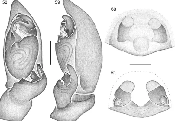
- Tegulum otherwise. ... 8 8(7) – Median process in the prolateral surface of RTA stout, sub-squared (Fig. 59); tegular process close to embolar base (Fig. 58). ... C. zecarioca n. sp.
- Median process in the prolateral surface of RTA longer than wide (Fig. 55); tegular process far from embolar base (Fig. 54)………..C. jecatatu n. sp.
9(6) – Embolus short, bent in straight angle (Fig. 83)…...C. balacobaco n. sp.
- Embolus longer, gently bent . ... 10 10(9) – Tegular process curved prolaterally, FR1 restricted to midlle of tegulum (Fig. 43) ... C. telecoteco n. sp.
- Tegular process directed apically, FR1 large, expanded to retro-apical sector of tegulum (Fig. 45) ... C.ziriguidum n. sp.
19 12(11) – Apex of RTA pointed, conductor with a quadrangular basal process (Bonaldo, 1996: fig. 16) ... C. rubripes
- Apex of RTA obtuse, conductor without basal process (Bonaldo 2000: fig. 119) ... …….C. nitens
13(11) – Embolus stout and spatulated, not covered by conductor (Fig. 87) ... ...C. vilanovae n. sp.
- Embolus filiform, partially cover by groove of conductor ... 14 14(13) – RTA enlarged retrolaterally (Figs. 75, 79). ... 15 - RTA otherwise. ... 16 15(14) – Apex of RTA not projected ventrally, with small hyaline process, tegular process inseted closely to embolar base (Figs. 78, 79) ... C. hyalina n. sp.
- Apex of RTA strongly projected ventrally, without hyaline process (Fig. 75); tegular process inseted far from embolar base (Fig. 74). ... C. vesperata n. sp.
16(14) – Apex RTA rounded, directed prolaterally (Bonaldo, 2000: fig. 123)……….C. mourai
- Apex of RTA pointed, directed apically (Fig. 47). ... C. escalvada n. sp.
17(1) – Epigynal plate projected posteriolly, beyond epigastric groove (Figs. 65, 109)... ... 18 - Epigynal plate not projected posteriolly. ... 21 18(17) – Copulatory opening medially placed (Fig. 65) ... C. caatinga n. sp.
- Copulatory opening posteriorly placed. ... 19 19(18) – Copulatory ducts visible ventrally (Bonaldo, 1996: fig. 16) ... C. rubripes
20 -Copulatory opening placed in undifferentiated area; posterior epigynal margin medially truncated (Fig. 109) ... C. kuryi n. sp.
21(17) – Copulatory opening inserted in a ventral triangular epigynal projection (Fig. 85) ...C. balacobaco n. sp.
- Triangular epigynal projection absent ... 22 22(21) – Copulatory opening small, “v”-shaped and anteriorly placed, in the level of secondary spermatecae (Figs. 92, 96, 103) ... 23 - Copulatory opening otherwise (Figs. 52, 56, 60, 76, 80, 105, 107)... ... 24 23(22) – Copulatory ducts long, inverted T-shaped and visible ventrally, epigynal surface not grooved (Fig. 92) ... C. demersa n. sp.
- Copulatory ducts not visible ventrally, epigynal surface with wide longitudinal median groove (Fig. 96) ... C. maracas n. sp.
24(22) – Copulatory opening defined both anteriorly and posteriorly by well defined margins (Figs. 105, 107) ... 25 - Copulatory opening defined only posteriorly by a well defined margin ... 26 25(24) – Secondary spermatecae separated from each other (Fig. 106); posterior margin of copulatory opening nearly straight (Fig. 105) ... C. loiolai n. sp.
- Secondary spermatecae touching each other (Fig. 108); posterior margin of copulatory opening procurve (Fig. 107) ... C. regi n. sp.
26(24) – Posterior margin of copulatory opening large, “V”- shaped (Figs. 76) ... ...C. vesperata n. sp.
21 - Secondary spermatecae touching each other (Bonaldo, 2000: fig. 125) or separated from each other by less than one diameter (Figs. 53, 57) ... 29 28(27) – Secondary spermatecae globular (Fig. 60, 61) ... C. zecarioca n. sp.
- Secondary spermatecae oblong (Fig. 80, 81) ... C. tranquilla n. sp.
29(27) – Secondary spermatecae touching each other ... C. mourai
- Secondary spermatecae separated from each other ... 30 30(29) – Posterior margin of copulatory opening “u”- shaped (Fig. 52); PVP with lateral rounded protuberances in the internal margin (Fig. 53) ... C. aechmea n. sp.
- Posterior margin of copulatory opening straigth (Fig. 56); PVP without lateral protuberances (Fig. 57) ... C. jecatatu n. sp.
Corinna rubripes C. L. Koch, 1842 Fig. 41
Corinna rubripes C. L. Koch, 1842: 17, pr. 293, fig. 702 (male holotype from Bahia,
Brazil, Gomez col., ZMB 2134, not re-examined); Karsch, 1880: 375, est. 12, fig. 1; Simon, 1898: 198; Petrunkevitch, 1911: 469; 1928: 177; Roewer, 1954: 600; Bonnet, 1956: 1216; Moritz & Fischer, 1988: 137; Bonaldo, 1996: 80 (figs. 1–19); Bonaldo, 2000: 40 (figs. 21, 22).
Sparassus rubripes; Walckenaer, 1847: 561.
Diagnosis. Males of Corinna rubripes resemble those of C. nitens by the tip of
22 can be recognized by the copulatory duct visible ventrally and epigynal plate only slightly projected posteriorly (Bonaldo, 1996: figs. 18–19).
Description. See Bonaldo, 1996: figs. 1–19. Distribution. Guyana and northeastern Brazil.
New records: BRAZIL. Bahia: Jequié (Brejo Novo) [13º56'41"S 40º06'33.9"W], 1 female, 6-7.VI.2009, Chagas Jr, A. Kury, D. Pedroso, A. Giupponi and V. Dill (MNRJ 6123); Uruçuca (Fazenda Santa Tereza), 2 males, 3.VI.1970 (MNRJ 13192); (Fazenda Santo Antonio), 1 female, 24.X.1978, J.S. Santos (CPDC-4225); Ilhéus (Reserva Zoobotânica) 14°46'22.07"S 39° 13'13.8"W, 2 males, 8-9.XII.2010, G.H.F. Azevedo and A.J. Santos (UFMG 9442); Camacan (RPPN Serra Bonita) [15°23’25'' S 39°34’05''W], 1 female, 11-13.VI.2009, Chagas Jr, A. Kury, D. Pedroso, A. Giupponi and V. Dill (MNRJ 6120); (Fazenda Esperança), 1 male, 18.VI.1969 (MNRJ 13202); Itapebi [15°57'9.18"S 39°32'1.69"W], 1 female, T. Bernabé (MNRJ 6234); Porto Seguro [16°27'4.04"S 39° 3'52.79"W], 1 female (MNRJ 6444); (Fazenda Nossa Senhora da Conceição), 1 female, 21.III.1971 (MNRJ 1899); Itamarajú (Fazenda Pau
Brasil ) [17°04’S 39°31’W], 1 male, 8.VII.1968 (MNRJ 3304); 1 female, 10.I.1969
(MNRJ 13237); Minas Gerais: Águas Vermelhas [15°44'50.92"S 41°27'39.37"W], 1 male (MNRJ 2051); Marliéria (Parque Estadual do Rio Doce) 19°42'58.62" S 42°44'1.69" W, 1 male, 5-6.I.2010, W. H. Thomassen et. al. (UFMG 7512).
Corinna nitens (Keyserling, 1891)
Fig. 41
Hypsinotus nitens Keyserling, 1891: 57, est. 2, fig. 30 (lectotype and paralectotype from
23
Corinna nitens; Simon, 1897: 192; Petrunkevitch, 1911: 468; Mello-Leitão, 1923: 54;
1927: 398; Roewer, 1954: 598; Bonnet, 1956; 1214. Bonaldo, 2000 (figs. 119–
122): 41; Bosselaers & Jocqué, 2002: 250.
Diestus altifrons Mello-Leitão, 1945: 260 (holotype from Pindapoy, Misiones,
Argentina, II.1942, Bridarolli col., MLP 16575, not re-examined); Roewer, 1954: 602; Arrozpide, 1986: 17. Synonymized with Corinna nitens by Bonaldo,
2000.
Diagnosis. Males of Corinna nitens differs from those of C. rubripes by the obtuse apex
of RTA and by the absence of both the proapical tegular projection and the quadrangular process in conductor base (Bonaldo, 2000: figs. 119–120); females differ from those of C. rubripes and C. kuryi n. sp. by the epigynal plate extremely projected
posteriorly, copulatory ducts not visible ventrally and copulatory opening placed in a small posterior atrium (Bonaldo, 2000: figs. 121–122).
Description. See Bonaldo, 2000: figs. 13, 27, 33, 57, 67, 74, 77–82, 89, 119–122, 153. Distribuition. Peru, Bolívia, Paraguay, Northern Argentina, Southeastern and Southern Brazil.
25 Teresópolis (Parque Nacional da Serra dos Orgãos) [22°27'23.4''S 42°59'41.4''W], 1 male, X.2006, Exp. Arachné (MNRJ 6439); Jacarepaguá (Represa do Cigano) [22°56'24.84"S 43°17'55.12"W], 1 female, 12.VII.89, A. Kury (MNRJ 6433); Rio de Janeiro (Parque Nacional da Tijuca) [22°57'40.93"S 43°16'30.83"W], 1 female, 20.I.2001, R.L.C. Baptista (MNRJ 3943); 1 female, XI.1985 (MNRJ 3301); (Parque Nacional da Tijuca, Serra da Carioca) [22°57'38.79"S 43°14'45.25"W], 1 male, 18.XI.2001, E.H. Wienskoski (MNRJ 6448); (Parque Nacional da Tijuca, Mesa do Imperador) [22°58'13"S 43°15'28"W], 1 male, 9.V.1988, R.L.C. Baptista (MNRJ 3297); Niterói (Itaipú) [22°56'59.19"S 43° 2'2.62"W], 1 male, 9.X.1996, C.C. Ratto (MNRJ 6445); São Paulo: Botucatu [22°53'53"S 48°29'29"W], 1 female, X.2002, (MNRJ 6443); 1 female, VI.2001, E.H. Wienskoski (MNRJ 6430); 1 female, 25.V.1988, R.L.C. Baptista (MNRJ 3298); Paraná: Fernandes Pinheiro (Floresta Nacional de Irati), 25°23'2.22"S 50°34'55.07"W, 1 male, 7.IV.2012, J.Ricetti (MPEG 20261); Candói (Usina Hidrelétrica de Fundão, Rio Jordão), 25°43'30.43"S 52° 1'32.19"W, 2 males and 12 females, V.2005, J. Ricetti (MPEG 20262);Santa Catarina: (Reserva Biológica Marinha, Ilha do Arvoredo) [27°16'60’’S 48°22'W], 1 female, 3–
26 11°52'57"S 72°39'02"W, 1 male, 29.XI.1997, S. Cordova (MUSM 504355); Puno: Sandia [14°14'41.93"S 69°25'51.93"W], 1 female, 9.XII.1997, J.A. Ochoa (MUSM 504195). BOLIVIA. La Paz: Nor Yungas (Coroico) [16°11'39.04"S 67°43'43.72"W], 1 female, 1.III.1990, E. Penaranda (IBN); 1 male, 24.II.1990 (IBN).
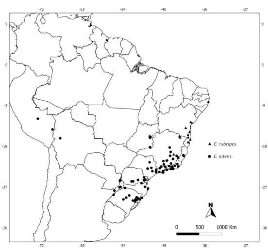
FIGURE 41. Known distribution of Corinna rubripes and C. nitens. Records from
Bonaldo (1996, 2000) included.
Corinna mourai Bonaldo, 2000
Fig. 42
Corinna mourai Bonaldo, 2000: 42, figs. 123–126 (holotype from Curitiba, Paraná,
27 Diagnosis. Males of Corinna mourai resemble those of C. escalvada n. sp. and C. balacobaco n. sp. by the wide base of RTA, occupying most of ventral tibial surface;
they differ from those of C. balacobaco n. sp. by the absence of a median process the
RTA prolateral surface and from those of C. escalvada n. sp. by the apex of RTA
rounded and directed prolaterally (Bonaldo 2000: figs. 123–124); females differ from those of other species with epigynal plate not projected posteriorly and with small, unconspicuous copulatory opening by the secondary spermathecae touching each other (Bonaldo, 2000: 125–126).
Description. See Bonaldo, 2000: figs. 123–126. Distribution. Southeastern to southern Brazil.
28 female, VII.2008, A.A. Lise (MCTP 22572); 1 female, 06.III.2011 (MCTP 29956); São Francisco de Paula [29°26'48.23"S 50°34'48.33"W], 1 male, 24.X.1996, R. Ott (MCTP 10668); (Barragem dos Bugres), 1 male and 2 females, L.A. Moura (MCN 30668); (Potreiro Velho), 2 female, 2–4.II.1999, A.A. Lise (MCTP 15841); 1 female, 02.VI.2000, A.A. Lise (MCTP 14633); 1 male, IV.2002, L.A. Bertoncello et. al.
(MCTP 23104); 1 male (MCTP 23107); 1 male (MCTP 23099); 1 male (MCTP 23100); Triunfo (Parque COPESUL) [29°53'21.14"S 51°23'4.67"W], 1 female, 14–15.I.1997, L.A. Moura (MCN 28248); Eldorado do Sul (Parque Estadual do Delta do Jacuí), 2 males and 2 females, 5–7.I.2000, A.B. Bonaldo (MCN 31977).
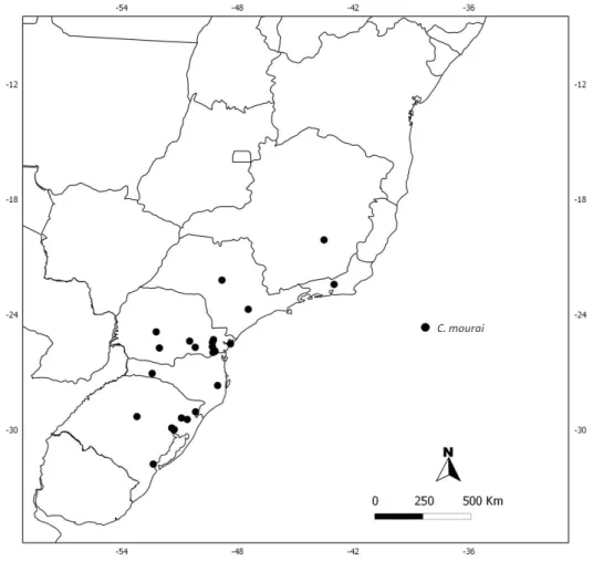
FIGURE 42. Known distribution records of Corinna mourai in Southeastern and
29 Corinna telecoteco n. sp.
Figs. 35, 43–44, 49
Type Material. Male holotype from Serra do Caraça [20°7'60.00"S 43°30'0.00"W], Minas Gerais, Brazil, 12–23.XI.1961, U. Martins-Lenko and R. Kloss, deposited in MZSP (6688). Paratypes: Brazil. Minas Gerais: (Serra do Cipó) [19°12'31.53"S 43°46'59.87"W], 1 male (MNRJ 1775).
Etymology. The specific name refers the classic samba tune “teleco-teco”, interpreted
by Cyro Monteiro in his Long Play “Sr. Samba” (Mr. Samba) released in 1961, the year in which the holotype was collected.
Diagnosis. Males of Corinna telecoteco n. sp. are similar to those of C. ziriguidum n.
sp. by the combined presence of a small median process on prolateral surface of RTA and tegular process inserted closely to the embolar base; differ by presenting the median projection on prolateral surface of RTA directed apically and the tegular process curved prolaterally (Figs. 43–44).
Description. Male (holotype). Carapace reddish brown, chelicerae black, endites and labium brown, with lightened posterior end, sternum yellow, legs I dark yellow, legs II, III and IV yellow; abdomen light yellow with dorsal scutum in the anterior half of abdomen. Total length: 10.2. Carapace 4.6 long; 3.95 wide; 2.65 high. Clypeus 0.43 high. Abdomen 5.3 long; 3.5 wide. Eyes: anterior row 1.75, posterior row 2.1. MOQ: 0.63 long, 0.68 anterior wide, 0.68 posterior wide. Eyes diameters and interdistances: AME 0.23; ALE 0.2; PME 0.2; PLE 0.2; AME–AME 0.25; AME–ALE 0.35; PME–
30 12.5; III 2.95/ 1.4/ 2.05/ 2.5/ 1.1/ 10.0; IV 3.8/ 1.5/ 3.25/ 3.3/ 1.2/ 13.05. Leg formula 1423. Leg spination: I – femur d1-0-0, p0-0-1; tibia v2-1r-2-2; metatarsus v2-2-0. II –
femur d1-1-0, p0-0-1; tibia vr-1-1-1; metatarsus v2-1p-1r. III – femur d1-1-0, p0-0-1, r0-1-0; tibia v2-2-0; metatarsus p0-1-0, r0-1-0, v2-2-1. IV – femur d1-1-0, r0-0-1; tibia r0-1-1, vp1-1-0; metatarsus r0-1-0, v2-1p-1. Palp: femur with two posterior dorsal spines; t without projection; E filiform partially covered by the conductor apical fold (Figs. 43–44).
Female. Unknown.
Variation. Length (2 males): total 7.8–10.2; carapace 3.6–4.6; femur I 3.25–4.0. Distribution. Minas Gerais, Brazil.
Other material examined. None
Corinna ziriguidum n. sp.
Figs. 6–7, 9–10, 16, 24, 26–27, 45–46, 49
Type Material. Male holotype from Torre da Telepar, Curitiba, [25°25'42.08"S 49°16'23.71"W] Paraná, Brazil, 01.IV.1987, J.G. Kastelic, deposited in MCN (17198). Paratypes: Brazil. Rio de Janeiro: Petropólis [22°30'16.70"S 43°10'56.38"W], 1 male (MNRJ 704); Paraná: [Guaratuba, 25°43'35.67"S 48°56'56.19"W] (Usina Hidrelétrica de Guaricana), 1 male, 27–31.I.1986, S.R. Malkowiski (MHCI); Paranaguá (Praia Grande, Ilha do Mel), [25°30'42.00"S 48°20'20.00"W], 1 male, 08.I.1989, R. Dutra (MCN 20553).
31 several samba tunes and is also used to qualify the skills of samba players or dancers, as synonym of expertise.
Diagnosis. Males of Corinna ziriguidum n. sp. resemble those of C. telecoteco n. sp. by
the presence of a small median projection on prolateral surface of RTA and tegular process inserted closely to the embolar base; differ by the course of reservoir in ventral view, with FR1 ample, reaching the retroapical region of tegulum and by the small median projection in the prolateral surface of the RTA directed prolaterally (Figs. 45–
46).
Description. Male (holotype). Carapace dark reddish brown, chelicerae black, endites and labium red with posterior extremity lightened, sternum brown, leg I brown, legs II, III and IV dark yellow, posterior femora, tibiae and patellae with dark spots; dorsum of abdomen gray with dorsal scutum extending to the median region, ventrally light gray. Total length 8.8. Carapace 4.4 long; 3.5 wide; 2.5 high. Clypeus 0.4 high. Abdomen 4.25 long; 2.75 wide. Eyes: anterior row 1.7, posterior row 2.0. MOQ: 0.65 long, 0.55 anterior wide, 0.53 posterior wide. Eyes diameters and interdistances: AME 0.2; ALE 0.18; PME 0.18; PLE 0.15; AME–AME 0.25; AME–ALE 0.32; PME–PME 0.3; PME–
32 spines; T without projection, course of reservoir in ventral view with a broad first loop; E filiform partly covered by apical groove of conductor (Figs. 45–46).
Female. Unknown.
Variation. Length (4 males): total 7.8–9.5, carapace 3.9–4.7, femur I 3.5–3.9. Distribution. Rio de Janeiro and Paraná, Brazil.
Other material examined. None
Corinna escalvada n. sp.
Figs. 34, 47
–
49Type Material. Male holotype from Fazenda Escalvada, Mucuri [18°3'19.97"S 39°32'58.78"W], Bahia, Brazil, 11.IX.1979, A. C. Niella, deposited in SMNK.
Etymology. The specific name is a noun in apposition taken from the type locality. In Portuguese, escalvada is an adjective and means devoid of vegetation.
Diagnosis. Males of Corinna escalvada n. sp. differ from those of other species with
tegular process inserted closely to the embolar base by the massive, wide base of RTA, without a median projection on its prolateral surface (Figs. 47–48).
33 interdistances: AME 0.28; ALE 0.2; PME 0.2; PLE 0.2; AME–AME 0.33; AME–ALE 0.43; PME–PME 0.38; PME–PLE 0.6; ALE–PLE 0.25. Chelicerae 2.55 long; 4 retromarginal teeth and 3 promarginal teeth. Sternum 2.5 long; 2.1 wide. Leg measurements: femur I 4.05/ patella 2.0/ tibia 3.5/ metatarsus 3.0/ tarsus 1.75/ total 14.3; II 3.7/ 1.85/ 3.05/ 3.05/ 1.55/ 13.2; III 3.3/ 1.65/ 2.3/ 2.65/ 1.3/ 11.2; IV 4.0/ 1.75/ 3.25/ 3.5/ 1.35/ 13.85. Leg formula 1423. Leg spination: I – femur d1-1-0, p0-0-1; tibia v1p-2-2-2-2; metatarsus v2-2-0. II – femur d1-1-0, p0-0-1; tibia vr-1-1-1; metatarsus v2-2-0. III – femur d1-1-1, p0-1-1, r0-1-0; tibia 0; metatarsus p1-0-0, r0-1-0, v2-2-1. IV – femur d1-1-1, r0-0-1; tibia r0-1-1, v2-1r-1p; metatarsus r0-1-0, v2-2-1. Palp: femur with two posterior dorsal spines; RTA basally wide, apex tapered; T without projection; E filiform partially covered by the distal groove of conductor (Figs. 47–48). Female: Unknown.
34 FIGURES 43–48. Corinna spp., male palp. Corinna telecoteco n. sp., 43) ventral; 44)
retrolateral. Corinna ziriguidum n. sp.: 45) ventral; 46) retrolateral. Corinna escalvada
35 FIGURE 49. Known distribution records of Corinna telecoteco n. sp., C. escalvada n.
sp. and C. ziriguidum n. sp. in Southeastern Brazil.
Corinna aechmea n. sp.
Figs. 1
–
5, 11,–12, 18–23, 31–
33, 50–53, 6236 Etymology. The specific name is a noun in apposition taken from the scientific name of the bromeliad genus in which the holotype was collected, Aechmea distichantha Lem.
Diagnosis. Males of Corinna aechmea n. sp. resemble those of C. jecatatu n.sp. and C. zecarioca n. sp. by presenting the RTA bifurcated, with a large median process in the
prolateral suface, but it is readly recognized by the rounded apical tegular projection (Figs. 50–51); females differ from those of C. zecarioca n. sp. by presenting the
secondary spermathecae closer to each other and from those of C. jecatatu n. sp. by the posterior margin of copulatory opening “u”- shaped and posterior vulval plate with lateral rounded protuberances in the internal margin (Figs. 52–53).
Description. Male (holotype). Carapace black with posterior end of thoracic region dark red, chelicerae black, endites red, posterior end paler; labium black, posterior end paler; sternum brown, legs with coxae, trochanters and femora dark brown, patellae, tibiae, metatarsi and tarsi dark yellow; dorsum of abdomen light gray, dorsal scutum with distal portion large tapering to the median region; abdomen ventrally light gray with large median brown band in longitudinal axis. Total length: 11.1. Carapace 5.0 long; 4.2 wide; 2.7 high. Clypeus 0.4 high. Abdomen 5.4 long; 3.3 wide. Eyes: anterior row 1.9, posterior row 2.2. MOQ: 0.68 long, 0.76 anterior wide, 0.73 posterior wide. Eyes diameters and interdistances: AME 0.27; ALE 0.21; PME 0.21; PLE 0.2; AME–
37 p0-1-0, r0-1-0, v2-2-1. IV – femur d1-1-0, 0-1; tibia 1-1, vp1-2-0; metatarsus r0-1-0, v2-1p-1. Palp: femur with two posterior dorsal spines; TPC inserted far from base of embolus; E filiform, partly covered by the apical groove of conductor; C with large unsclerotized area in the distal region (Figs 50, 51).
Female (paratype MPEG 20249). Coloration as in male, except dorsum of abdomen gray, without dorsal scutum. Total length: 11.3. Carapace 4.7 long; 3.8 wide; 2.4 high. Clypeus 0.38 high. Abdomen 6.5 long; 4.1 wide. Eyes: anterior row 1.77, posterior row 2.05. OQ: 0.62 long, 0.71 anterior wide, 0.68 posterior wide. Eyes diameters and interdistances: AME 0.23; ALE 0.2; PME 0.18; PLE 0.18; AME–AME 0.28; AME–
ALE 0.37; PME–PME 0.35; PME–PLE 0.44; ALE–PLE 0.14. Chelicerae 2.5 long. Sternum 2.3 long; 2.05 wide. Leg measurements: femur I 3.8/ patella 1.85/ tibia 3.25/ metatarsus 2.65/ tarsus 1.6/ total 13.15; II 3.45/ 1.7/ 2.8/ 2.55/ 1.45/ 11.95; III 2.85/ 1.4/ 2.2/ 2.4/ 1.1/ 9.95; IV 3.65/ 1.5/ 3.2/ 3.25/ 1.15/ 12.75. Leg formula 1423. Leg spination: I – femur d1-1-0, p0-0-1; tibia v2-2-2-2; metatarsus v2-2-0. II – femur d1-1-0, p0-0-1; tibia vr-1-1-1; metatarsus v2-3-0. III – femur d1-1-1, p0-1-1, r0-1-0; tibia v2-2-0, r0-1-1; metatarsus p0-1-0, r0-1-0, v2-2-1. IV – femur d1-1-1, r0-0-1; tibia r0-1-1, vp1-r1-p1; metatarsus r0-1-0, v2-2-1. Epigynum: epigynal plate not projected posteriolly; CO median, small and inconspicuous; CD not visible ventrally; PVP enveloping partly the PS and SS (Figs. 52–53).
Variation. Length (8 males): total 7.4–11.1; carapace 3.8–5.1; femur I 2.7–3.8; (6 females) total 8.0–12.2; carapace 3.8–5.1; femur I 3.2–4.1.
Distribution. Minas Gerais and Paraná, Brazil.
38 19°53'S 43°22'W, 3 males and 1 female, 05–06.VI.2010, A.J. Santos (UFMG 4460); 1 female, 07.IV.2004, E.T. Rodrigues; Belo Horizonte (Parque Municipal das Mangabeiras), 19°57'14.86"S 43°54'19.15"W, 1 male, 5–12.XII.2008, H.H. Santos et. al. (UFMG 8508); Paraná: Porto Rico (Planície de Inundação do Alto Rio Paraná), 22°44'38.78''S 53°12'59.85''W, 1 female, 15.IV.2010, F. Amadeo (MPEG 20251); Vila Alta [currently Alto Paraíso] (Ilha Grande), [23°45'0.00"S 54°2'60.00"W], 2 males, 08.XII.1995, M.L. Fischer (MPEG); Altônia (Ilha do Cristo), [23°52'21.25"S 53°53'48.30"W], 1 female, 08.X.1989, R.Pinto da Rocha (MHCI).
Corinna jecatatu n. sp.
Figs. 54–57, 62
Type Material. Male holotype from Barueri [23°30'40.69"S 46°52'36.39"W], São Paulo, Brazil, 06.XII.1964, K. Lenko, deposited in MZUSP (3821). Paratypes: Brazil. São Paulo: Barueri [23°30'40.69"S 46°52'36.39"W], 1 female, 16.I.1966, K. Lenko, (MZUSP 5591); 1 male, 06.XII.1964, K. Lenko (MZUSP 3821); Osasco [23°31'54.09"S 46°47'23.72"W], 1 male and 1 female, 15.IV.1938, F.Lane (MZUSP 12525); São Paulo [23°32'56.19"S 46°38'19.74"W], 1 female, 11.VII.1937, J.R. Mattos, (IBSP); 1 male, I.1960 (IBSP 1480).
Etymology. The specific name refers to classic hick character “Jeca Tatu”, created by
the Brazilian writer Monteiro Lobato for his masterpiece “Urupês”, first published in
39 Diagnosis. Males of Corinna jecatatu n. sp. resemble C. aechmea n. sp. and C. zecarioca n. sp by presenting the RTA bifurcated, with a large median process in the
prolateral suface; differ from those of C. aechmae n. sp. by the absence of a rounded
tegular apical projection and from those of C. zecarioca n. sp. by the longer RTA
median process (Figs. 54–55); as in C. aechmae n. sp., females present the secondary
spermathecae close to each other, but are readily recognized by posterior margin of copulatory opening straight and posterior vulval plate without internal protuberances (Figs. 56–57).
Description. Male (holotype). Carapace dark reddish brown with posterior end of thoracic region red, chelicerae black, endites red with posterior end paler; labium black with posterior end paler, sternum brown; legs I and II dark yellow except metatarsi and tarsi, dark red, leg III and IV yellow except metatarsi and tarsi, dark yellow; abdomen light yellow with dorsal scutum extending to the median region of abdomen. Total length: 10.2. Carapace 5.1 long; 4.3 wide; 3.1 high. Clypeus 0.5 high. Abdomen 4.7 long; 3.1 wide. Eyes: anterior row 1.9, posterior row 2.4. MOQ: 0.8 long, 0.87 anterior wide, 0.8 posterior wide. Eyes diameters and interdistances: AME 0.32; ALE 0.17; PME 0.2; PLE 0.22; AME–AME 0.22; AME–ALE 0.37; PME–PME 0.37; PME–PLE 0.6; ALE–PLE 0.15. Chelicerae 2.4 long; 4 retromarginal teeth and 3 promarginal teeth. Sternum 2.7 long; 2.1 wide. Leg measurements: femur I 4.3/ patella 2.05/ tibia 3.75/ metatarsus 3.3/ tarsus 1.7/ total 15.1; II 3.85/ 1.6/ 3.2/ 3.0/ 1.55/ 13.2; III 3.25/ 1.6/ 2.25/ 2.7/ 1.25/ 11.05; IV 4.1/ 1.8/ 3.5/ 3.75/ 1.35/ 14.5. Leg formula 1423. Leg spination: I –