ISSN 2317-6431 https://doi.org/10.1590/2317-6431-2016-1761
Association between temporomandibular disorders and
tinnitus in the elderly
Associação entre disfunção temporomandibular e zumbido em idosos
Julya Macedo1, Marcelo Yudi Doi2, Alyne Macedo3, Paula Vanessa Pedron Oltramari-Navarro4, Regina Célia Poli-Frederico5, Ricardo de Lima Navarro6, Luciana Lozza de Moraes Marchiori1
ABSTRACT
Purpose: To verify the association between tinnitus and temporomandibular dysfunction in the elderly. Methods: A cross sectional study was conducted with the inclusion of elderly individuals with independent living. Temporomandibular dysfunction was assessed by odontological evaluation and tinnitus was verified by medical history. Statistical analysis was performed using the chi-square test, relative risk and logistic regression. Results: Tinnitus was observed in 82.9% of individuals with temporomandibular dysfunction and through this analysis is shown that temporomandibular dysfunction as a risk factor for tinnitus. Conclusion: The results showed that there was association between tinnitus and temporomandibular dysfunction in the elderly population and emphasizes the importance of identifying risk factors for tinnitus that can be modified through specific interventions, since it is essential in the prevention of future episodes, as well as managing the process of treatment of elderly patients in general.
Keywords: Tinnitus; Temporomandibular joint disorders; Elderly; Audiology; Odontology
RESUMO
Objetivo: Verificar a associação entre zumbido e disfunção temporomandibular
em idosos. Métodos: Estudo transversal realizado com a inclusão de idosos
com vida independente. A disfunção temporomandibular foi avaliada por exame odontológico e o zumbido foi verificado pela história médica. A análise estatística foi realizada utilizando o teste Qui-quadrado, o risco relativo e a regressão logística. Resultados: O zumbido foi observado em 82,9% dos indivíduos com disfunção temporomandibular e, através desta análise, observou-se que a disfunção temporomandibular é um fator de risco para o zumbido. Conclusão: Houve associação entre zumbido e
disfunção temporomandibular na população idosa. Ressalta-se a importância de identificar fatores de risco para o zumbido, que podem ser modificados por meio de intervenções específicas, uma vez que esta prática é essencial na prevenção de episódios futuros, bem como na gestão do processo de tratamento de pacientes idosos, em geral.
Palavras-chave: Zumbido; Transtornos da articulação temporomandibular;
Idoso; Audiologia; Odontologia
Study carried out at Programa de Mestrado em Ciências da Reabilitação, a graduate study program affiliated with the Universidade Estadual de Londrina – UEL and Universidade Norte do Paraná – UNOPAR – Londrina (PR), Brasil.
1Programa de Pós-graduação em Ciências da Reabilitação, Departamento de Fonoaudiologia, Universidade Norte do Paraná – UNOPAR – Londrina (PR), Brasil. 2Programa de Pós-graduação em Ciências da Reabilitação, Departamento de Fisioterapia, Universidade Norte do Paraná – UNOPAR – Londrina (PR), Brasil. 3Curso de Medicina, Universidade de Marília – UNIMAR – Marília (SP), Brasil.
4Programa de Pós-graduação em Odontologia, Universidade Norte do Paraná – UNOPAR – Londrina (PR), Brasil.
5Programa de Pós-graduação em Ciências da Reabilitação, Departamento de Biologia Molecular, Universidade Norte do Paraná – UNOPAR – Londrina (PR),
Brasil.
6Programa de Pós-graduação em Odontologia, Universidade Estadual de Maringá – UEM – Maringá (PR), Brasil. Conflict of interests: No.
Authors’ contribution: JM: study design, scientific research, and writing of the manuscript; MYD: interpretation of data and statistical analysis; AM: interpretation of data and manuscript revision; PVPON: data collection and manuscript revision; RCPF: statistical analysis and manuscript revision; RLN: data collection and manuscript revision; LLMM: study design, scientific research, data collection, and manuscript revision.
Funding: None.
Corresponding author: Julya Macedo. E-mail: julyamacedo@hotmail.com
INTRODUCTION
Increased population aging in Brazil (10.8% of the total
population)(1), and in particular in the municipality of Londrina
(approximately 66,000 inhabitants aged >60 years), Parana
state(1), has aroused the interest of several sectors of society,
including that of health professionals, who aim to provide greater comfort and adaptations to the changes that occur with the aging process.
Speech-language Pathology (SLP) is one of the areas that
contributes to the prevention and treatment of aging disorders seeking to improve the quality of life of this population(2).
Subjective tinnitus, which is a sound heard only by the patient,
is presented with many distinct forms and is similar to chronic neuropathic pain. It is commonly observed in the elderly and is a symptom present in a group of diverse pathologies, thus
needing different treatments. The clinical approach of tinnitus
as a single disease has hindered progress in understanding its pathophysiology, and it is perhaps the most serious obstacle to
the development of effective treatments(3).
Tinnitus and other hearing symptoms found in the elderly population are commonly reported by patients with
temporomandibular disorders (TMD). Although there are
several theories regarding the pathophysiology of tinnitus, its precise mechanism has not been be elucidated yet, and may be associated with otological, neurological and traumatic causes,
adverse drug effects, nutritional deficiencies, metabolic and
eating disorders, depression, and TMD(4).
A study conducted with a sample of the elderly observed
prevalence of tinnitus of 42.77%, with difference between tinnitus and hearing loss and correlation between the side affected by
tinnitus and that affected by hearing loss(5).
TMD involve structural and functional changes in the
stomatognathic system and are characterized by pain in the
temporomandibular joint (TMJ) and masticatory muscles, limitations to mandibular movements, TMJ noise, functional
impairments, and otological symptoms(6).
Some authors(7) have added that signs and symptoms of
TMD may manifest on areas of the face and neck, on temporal,
occipital and frontal areas of the head, as well as on pre-auricular and auricular areas. It is estimated that 40-75% of the adult
population presents some sign of TMD, and that at least 5%
has symptoms(8).
In the case of tinnitus, it is known that such a symptom results from the dynamic interaction between several centers of the nervous and limbic systems, and that changes and/or lesions in the cochlea are the precursors of this process(9).
The same authors also stated that tinnitus is the perception of a sound, without there being production by an external source.
Tinnitus affects approximately 15% of the world’s population(10)
and this prevalence can reach 33% among individuals aged >60
years. Increasing age is directly proportional to the presence of multiple auditory symptoms, including tinnitus(11).
Prevalence of tinnitus in the population with TMD is higher
than that observed in the general population(7). Some authors
stated that the frequency of tinnitus among patients with TMD
has been reported with a range of 33 to 76%(10).
The specific scientific literature has documented a controversial
debate on the possible association of auditory symptoms
with craniomandibular dysfunction (CMD) since 1920(10).
This variation results mainly from the methods, instruments and
definitions of TMD used. However, there is need to elucidate
this correlation, because understanding the nature and causes of tinnitus is necessary in order to improve prevention and develop appropriate intervention and rehabilitation in view of their high prevalence in the elderly population(5).
Considering the lack of studies and the relevance of research on this theme to health science, this survey aimed to verify the
correlation between tinnitus and TMD in the elderly.
METHODS
This cross-sectional study is part of a project entitled Study on Aging and Longevity (EELO). The study population consisted of elderly individuals of both genders, aged >60 years, with independent living and levels 3 and 4 in the Functional Status
Score(12), who agreed to voluntarily participate in the survey
by signing an Informed Consent Form (ICF).
The study sample was calculated based on a population
of 43,610 elderly enrolled in the 38 Basic Health Units of the urban area of the municipality of Londrina, Paraná state, Brazil. The sample was randomly stratified considering the five regions of
the city as follows: 15% - central region, 27% - northern region, 23% - southern region, 19% - eastern region, and 16% - western
region. In addition, the EELO project included 519 individuals of both genders, aged >60 years, with independent living and
levels 3 and 4 in the Functional Status Score(12).
Of this population, a sample of 199 elderly was included in
this part of the study to verify the correlation between TMD and
tinnitus. The inclusion criteria were as follows: present natural
teeth or prostheses (total prosthesis; partial or removable fixed prosthesis) and acceptable functional occlusion. In addition,
volunteers should have been rehabilitated for at least one year prior to study commencement, considering that permanence
without prosthesis may influence the diagnostic process for TMD.
This study was approved by the Human Research Ethics
Committee of the aforementioned Institution (CEP-UNOPAR) according to the Brazilian National Health Council (NHC)
resolution no. 196/96 of October 10, 1996.
Information on tinnitus, gender, age, and cephalalgia was collected, based on protocol, by means of audiological anamnesis
at the Audiology Clinic of the Speech-language Pathology Department of a higher education institution in the north of
Parana state(13). Complaint of tinnitus including type and side
affected was verified.
Functional occlusion was assessed by requesting the patient
to perform lateral mandibular movements using cellophane paper to detect occlusal interferences on the non-working
side. Discrepancies between the centric relation and the usual
intercuspal position were also registered through bilateral manipulation. Individuals who presented marked discrepancies
(deviation >4 mm) or uncertain results were excluded from the study. Presence of anterior and lateral guides was verified to
classify the patient as with stable functional occlusion. Individuals who were edentulous and not rehabilitated by prostheses were
excluded. Moreover, there was concern about the educational
level of the individuals in the sample; to participate in the study, after a general explanation of the researchers, the elderly should be able to read, interpret, and respond independently to a questionnaire.
process was conducted by a standard examiner and the training activities were theoretical-practical. At the end of training,
calibration of the examiner was verified by the Kappa test(14),
in which the results of two evaluations were considered in the same group of 20 elderly.
The elderly selected for this study were interviewed using a questionnaire that aimed to collect information on their general
health status, signs and symptoms of TMD, and occlusal aspects.
The questionnaire(15) was applied to the patients without
interference of the examiner, so that no expectation was created, which could possibility divert the results of the clinical examination to be performed. The patients responded
to ten questions regarding symptoms of TMD, which enable classification of each individual with respect to the presence
and severity of these disorders. Anamnestic questionnaire:
1. Do you have difficulty in opening your mouth? 2. Do you have difficulty in moving your jaw sideways? 3. Do you feel discomfort or muscular pain when chewing? 4. Do you often have headaches?
5. Do you feel pain in your neck and/or shoulders? 6. Do you feel earaches or pain near your ears? 7. Do you notice any noise in your TMJ? 8. Do you regard your bite as normal?
9. Do you use only one side of your mouth to chew? 10. Do you feel any pain in your face when you wake up? Three response options were offered for the anamnestic
questionnaire: “yes”, “no”, or “sometimes”. Value “2” was attributed to every answer indicating the presence of a symptom; value “0”, for the absence thereof; and value “1”, for the “sometimes” answer. The total sum of the values obtained enabled
classification of the sample regarding TMD, as a TMD index: • Values from 0 to 3: free of TMD;
• Values from 4 to 8: mild TMD; • Values from 9 to 14: moderate TMD; • Values from 15 to 23: severe TMD.
Assessment of the presence of TMJ painful symptoms was accomplished first by guiding the patients as to the difference between pressure and discomfort in order to ensure
more trustworthiness to their responses. This examination was performed through bilateral digital palpation with the
examiner’s pointer fingers placed 10-20 mm before the external auditory conduct. The lateral aspect of the TMJ was
palpated with patients keeping their mouths closed, whereas the posterior aspect was palpated with patients maintaining their mouths open. These regions were pressed upon gently and continuously with 450-900 kgf(16).
For the examination of muscular palpation, patients were given the same instructions regarding the difference between pain and discomfort. Palpation of the masticatory muscles
was performed with digital, bilateral, constant pressure of approximately 1500 kgf(15). Presence of pain was verified
through the eyelid reflex and/or by questioning the patient.
Cervical muscles were palpated by clipping one’s fingers as
pincers on both sides.
Presence of joint noises, based on right and left TMJ inspection,
was also evaluated. This assessment was conducted by placing
the pointer fingers lightly upon the region corresponding to
the lateral pole of the condyle, before the external acoustic meatus, while the patient performed movements of mandibular opening and closing.
Statistical analysis was performed using the Chi-square and
Odds Ratio tests to determine possible correlations between
ear pain and TMD. P<0.01 was considered for the univariate
analyses, whereas p<0.05 was used for inclusion in the final
model for the Chi-square test and the relative risk value, with
a 95% confidence interval, in addition to logistic regression
analysis for age, gender and tinnitus; the Student’s t-test and
the Fisher’s exact test were applied for gender.
RESULTS
Of the 199 participants of this study, 75 (37.69%) were male and 124 (62.31%) were female. Mean age was 68.7 years, with
a minimum of 60 and a maximum of 85 years. Among them,
88 (44.2%) had tinnitus, 141 (71%) presented changes in the temporomandibular joint (TMJ), and 73 (36.7%) had both.
Mean age was similar for both males (69.9 years) and females (68 years). No difference was observed between genders for the
variable age, which evidences a fairly homogeneous sample in
this aspect. However, significant difference was found between the variables gender and temporomandibular disorder (TMD)
(p = 0.038) (Table 1).
Regarding the presence of tinnitus reported by the individuals
interviewed, no significant response was observed for gender
(p = 0.976). No correlation between genders was found for
tinnitus in the left and right ears (Table 2).
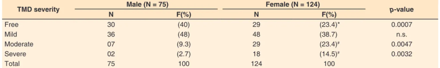
Regarding prevalence and severity of TMD in relation to gender, presence of TMD was higher in the female gender (77%)
compared with that in the male gender. As for the severity of
TMD presented, significant difference was observed in females for the moderate (23.4%) and severe (14.5%) levels, whereas no difference was found for the mild level. Assessment results were similar concerning participants free of TMD. Correlation between the results was observed regarding TMD severity and
presence of tinnitus, with predominance of the female gender
(Table 3).
TMD was observed in 45 (31.9%) males and 96 (68.1%) females, with female participants being 2.20 (95% CI: 1.18-4.11;
p = 0.018) times more likely to present TMD compared with
male participants. Tinnitus was observed in 73 individuals
(51.7%) with TMD and showed a relationship between both manifestations. At each occurrence of tinnitus, TMD were 3.71
(95% CI: 1.76-7.82; p = 0.001) times more likely occur (Table 4).
Table 1. Sample distribution by age and gender according to the temporomandibular disorder index
Variables Male (N = 75) Female (N = 124) Total (N = 199) p-value
Age (years)/Mean (± SD) 69.93 (± 6.05) 68.06 (± 5.48) 68.77 (± 5.76) 0.098
TMD Score 0.75 ± 0.73 1.30 ± 0.98* 1.09 ± 0.93 0.038
*Statistically significant difference between genders for p<0.05 (Student’s t- test for independent samples); Values shown in mean and standard deviation
DISCUSSION
In the present survey, severity of temporomandibular
disorders (TMD) was significantly higher in females than
in males (p = 0.038) (Table 1), corroborating the findings of
other studies(15,17,18). Similarly, a research(17) that associated
individuals from different age groups (aged 35 to 74 years)
with gender, reported sensitivity or pain in at least one muscle in 18.5% of the females and 9.5% of the males regarding the
temporomandibular joint (TMJ). In addition, the study found sensitivity or pain at palpation on TMJ in 7.3% of women and
3.4% of men. When addressing the higher incidence in the female gender, some authors(18) reported pain in general, and
could not determine a single factor responsible for the higher
prevalence of pain in women. Nevertheless, they pointed to some probable causes, such as differences in pain mechanisms and factors not yet identified for the craniofacial system, psychosocial and hormonal differences, and environmental
factors(18). In contrast, other authors(19) referred to TMD more
specifically, and found no differences between genders. It has
also been reported(19) that these differences could exist in
populations of Western countries, but not in Asian countries, or that they could be associated with cultural divergences in the perception and communication of symptoms.
Presence of tinnitus was higher in females and in the right
ear (Table 2). Regarding gender, some studies reported higher
prevalence in males(20) whereas other surveys showed the same
among females(21), as in the present research. Higher prevalence
of tinnitus in the female population may be due to presence
of hormonal or metabolic changes in this population(21,22),
considering that the changes that occur during the menstrual cycle, gestation, and menopause can lead to impairment of
homeostasis of the labyrinthine fluids, which acts directly in the enzymatic processes and neurotransmitters, and may influence
the basal metabolism of the inner ear(22).
As for the right ear, such findings may be in line with research on tinnitus theories. Such research has increasingly focused
on the exploration of brain-related mechanisms, and generally supposes that tinnitus presents a number of physiological causes(16,23), such as the cross-talk theory of memory capacity(23,24),
according to which nerve compression can be influenced by
auditory structures, as well as by stimulation of non-auditory structures, because the somatosensory system is the only non-auditory sensory system that seems to be associated with tinnitus, as temporomandibular joint syndrome, for instance. In this syndrome, somatic tinnitus can develop from activation of latent, ipsilateral, oto-somatic interaction(24). This may explain
why, in the present study, difference was observed regarding the side affected by tinnitus, with greater complaint about the right side, because TMJ problems can change this function
ipsilaterally, thus it is quite possible for the brain to interpret normal sounds as abnormal, and patients report tinnitus also ipsilaterally.
According to the cross-talk theory, the nerve fibers demyelinated due to lack of electrical insulation may increase
the spontaneous activity of different fibers(25). This probably
results from nerve compression, which can be influenced by
auditory structures, as well as by stimulation of non-auditory Table 4. Correlation between temporomandibular disorders and independent variables
Variables With TMD
N (%)
Without TMD
N (%) p-value OR (CI95%)
Gender
Male 45 (31.9) 30 (50.8) 0.018 2.20 (1.18 – 4.11)
Female 96 (68.1) 29 (49.2)
Tinnitus 73 (51.7) 15 (7.5) 0.001 3.71 (1.76 – 7.82)
Subtitle: TMD = temporomandibular disorder; OR = Odds Ratio with confidence interval (CI) of 95% and p-value
Table 3. Distribution of elderly individuals according to presence and severity of temporomandibular disorders
TMD severity Male (N = 75) Female (N = 124) p-value
N F(%) N F(%)
Free 30 (40) 29 (23.4)* 0.0007
Mild 36 (48) 48 (38.7) n.s.
Moderate 07 (9.3) 29 (23.4)# 0.0047
Severe 02 (2.7) 18 (14.5)# 0.0032
Total 75 100 124 100
*Statistically significant difference between genders for p<0.05 (X2 = Chi-square test); #Statistically significant difference between genders for p<0.05 (Fisher’s exact test)
Subtitle: TMD = temporomandibular disorder; N = sample size; F = frequency; n.s. = non-significant
Table 2. Distribution of elderly individuals according to presence of tinnitus by ear
Variables Male (n = 75) Female (n = 124) Total (n = 199) p-value
N % N % N %
Tinnitus 38 50.7 50 40.3 88 44.2 0.976
L Tinnitus 26 34.7 33 26.6 59 29.6
R Tinnitus 35 46.7 43 34.7 78 39.2
structures. Thus, the somatosensory system seems to be associated with tinnitus because of compression changes in
the TMJ. Therefore, tinnitus associated with TMD may also
be characterized by malfunctioning of the masticatory muscles
and TMJ, in addition to pain in the orofacial region, myoclonus
of the middle ear muscles, and palatal myoclonus(26).
Findings of this study show that there was correlation between
tinnitus and presence of TMD in the participants (Table 4). In a
population-based investigation, higher prevalence of tinnitus was observed in individuals who exhibited more than two symptoms
of TMD, and patients with tinnitus presented greater pain on TMJ
palpation and pain on opening the mouth than the individuals in the control group(27). The current analysis is in line with the
aforementioned investigation, because it evidenced correlation
between tinnitus and presence of TMD. Such correlation can be explained by the fact that TMD are part of a group of structural
and functional changes in the stomatognathic system that can impair the middle and inner ears, causing otological symptoms such as tinnitus(6-10). Nevertheless, there is still doubt about
the existence of any causal relationship between TMD and
otological changes(28).
Findings of the present study (Table 4) are also in agreement
with those of another research, because investigation of the
prevalence of TMD in patients with subjective tinnitus in
comparison with those in the control group, and the correlation
between TMD, tinnitus, and chronic pain symptoms, showed that of the 199 patients assessed, TMD were more prevalent
in those with tinnitus, and concluded that there is correlation
between TMD and subjective tinnitus(27). Furthermore, a study
conducted at the Department of Dental Prosthesis of the Medical Center at Regensburg University, Germany, to examine the prevalence of TMD and tinnitus in a sample of consecutive
patients found that prevalence of tinnitus was eight times higher
in participants with TMD (36.6%) than in participants without
TMD (4.4%)(29).
Despite the lack of studies investigating the correlation
between TMD and tinnitus in the elderly, one study(30) verified
prevalence of TMD in the elderly population. The authors highlighted that identification in this population is difficult
because the symptoms of these disorders are similar to those commonly found in some systemic disorders associated with aging. Another survey(28), which analyzed 11,745 participants in
the Korean national health system, observed higher prevalence of tinnitus in individuals with symptoms of TMD (31.2%), whose
occurrence of tinnitus was 1.6-fold that of individuals without
TMD. The study also observed that women, obese individuals, and individuals aged >65 years, presented higher prevalence of
these symptoms. The present research is in agreement with the
findings of these studies, considering that correlation between TMD and tinnitus was observed in the elderly.
Based on the present study, it is suggested that research on the improvement of tinnitus based on TMD therapy be
conducted with the elderly population considering the particular characteristics of each individual, such as biotype and greater
lability to changes in the ear and in the TMJ, metabolic and
circulatory changes, eating habits, and lifestyle.
CONCLUSION
Correlation between tinnitus and temporomandibular
disorders (TMD) was observed in the elderly population. Thus,
new prospective studies should be conducted in order to deepen
knowledge about other manifestations associated with tinnitus
and TMD, because this correlation in the population studied
demonstrates the importance of identifying risk factors for
tinnitus that could be modified by means of specific interventions.
REFERENCES
1. IBGE: Instituto Brasileiro de Geografia e Estatística [Internet]. Brasília: Instituto Brasileiro de Geografia e Estatística; 2011 [citado em 2017 Jan 30]. Disponível em: http:www.ibge.com.br
2. Kano CE, Mezzena LH, Guida HL. Estudo comparativo da classificação do grau de perda auditiva em indivíduos institucionalizados. Rev CEFAC. 2009;11(3):473-7. http://dx.doi.org/10.1590/S1516-18462009005000024. 3. Møller AR. Sensorineural tinnitus: its pathology and probable
therapies. Int J Otolaryngol. 2016;2016(3):1-13. http://dx.doi. org/10.1155/2016/2830157. PMid:26977153.
4. Camparis CM, Formigoni G, Teixeira MJ, Siqueira JT. Clinical evaluation of tinnitus in patients with sleep bruxism: prevalence and characteristics. J Oral Rehabil. 2005;32(11):808-14. http://dx.doi. org/10.1111/j.1365-2842.2005.01519.x. PMid:16202044.
5. Gibrin PC, Melo JJ, Marchiori LL. Prevalência de queixa de zumbido e prováveis associações com perda auditiva, diabetes mellitus e hipertensão arterial em pessoas idosas. CoDAS. 2013;25(2):176-80. http://dx.doi.
org/10.1590/S2317-17822013000200014. PMid:24408248. 6. Felício CM, Mazzetto MO, Silva MA, Bataglion C, Hotta TH. Preliminary
Protocol for Multi-Professional centers for determination of signs and symptoms of temporomandibular disorders. Cranio.
2006;24(4):258-64. http://dx.doi.org/10.1179/crn.2006.041. PMid:17086855. 7. Lam DK, Lawrence HP, Tenenbaum HC. Aural symptoms in
temporomandibular disorder patients attending a craniofacial pain unit. J Orofac Pain. 2001;15(2):146-57. PMid:11443826.
8. Okeson JP. American Academy of Orofacial Pain: guidelines for assessment diagnosis and management. Chicago: Quintessence; 1996. p. 113-84.
9. Pinto PCL, Sanchez TG, Tomita S. Avaliação da relação entre severidade do zumbido e perda auditiva, sexo e idade do paciente. Rev Bras Otorrinolaringol. 2010;76(1):18-21.
10. Rubinstein B. Tinnitus and craniomandibular disorders: is there a link? Swed Dent J Suppl. 1993;95:1-46. PMid:8503098.
11. Ganança FF, Gazzola JM, Ganança CF, Caovilla HH, Ganança MM, Cruz OLM. Quedas em idosos com vertigem posicional paroxística benigna. Braz J of Otorhinol. 2010;76(1):113-20. http://dx.doi. org/10.1590/S1808-86942010000100019.
12. Spirduso WW. Dimensões físicas do envelhecimento. 2. ed. São Paulo:
Manole; 2005.
13. Miller MH. A integração dos achados audiológicos. In: Katz J, editor.
Tratado de audiologia clínica. 3. ed. São Paulo: Manole; 1999. Capítulo
13; p. 268-70.
14. Fleiss J. Statistical methods for rates and proportions. 3rd ed. New
York: John Wiley & Sons; 1973.
15. Conti PC, Ferreira PM, Pegoraro LF, Conti JV, Salvador MC. A cross-sectional study of prevalence and etiology of signs and symptoms of temporomandibular disorders in high school and university students.
16. Austin DG, Pertes RA. Examination of the dysfunction patients. In: Pertes RA, Gross SG, editores. Clinical management of temporomandibular disorders and orofacial pain. Chicago: Quintessence; 1995. p. 123-60. 17. Mundt T, Mack F, Schwahn C, Bernhardt O, Kocher T, John U, Biffar R.
Gender differences in associations between occlusal support and signs of temporomandibular disorders: results of the population-based Study of Health in Pomerania (SHIP). Int J Prosthodont. 2005;18(3):232-9.
PMid:15945311.
18. Dao TT, LeResche L. Gender differences in pain. J Orofac Pain. 2000;14(3):169-84. PMid:11203754.
19. Pow EH, Leung KC, McMillan AS. Prevalence of symptoms associated with temporomandibular disorders in Hong Kong Chinese. J Orofac Pain. 2001;15(3):228-34. PMid:11575193.
20. Heller AJ. Classification and epidemiology of tinnitus. Otolaryngol Clin North Am. 2003;36(2):239-48.
http://dx.doi.org/10.1016/S0030-6665(02)00160-3. PMid:12856294.
21. Almeida TAS, Samelli AG, Mecca FDN, Martino E, Paulino AM. Sensação subjetiva do zumbido pré e pós intervenção nutricional em alterações metabólicas. Pro Fono. 2009 Dec;21(4):291-6. http://
dx.doi.org/10.1590/S0104-56872009000400005. PMid:20098946. 22. Schmidt PMS, Flores FT, Rossi AG, Silveira AF. Queixas auditivas
e vestibulares durante a gestação. Rev Bras Otorrinolaringol. 2010;76(1):29-33.
23. Kaltenbach JA, Zhang J, Finlayson P. Tinnitus as a plastic phenomenon and its possible neural underpinnings in the dorsal cochlear nucleus.
Hear Res. 2005;206(1-2):200-26. http://dx.doi.org/10.1016/j.
heares.2005.02.013. PMid:16081009.
24. Han BI, Lee HW, Kim TY, Lim JS, Shin KS. Tinnitus: characteristics, causes, mechanisms, and treatments. J Clin Neurol. 2009;5(1):11-9.
http://dx.doi.org/10.3988/jcn.2009.5.1.11. PMid:19513328. 25. Sanchez TG, Rocha CB. Diagnosis and management of somatosensory
tinnitus: review article. Clinics. 2011;66(6):1089-94. http://dx.doi.
org/10.1590/S1807-59322011000600028. PMid:21808880. 26. Won JY, Yoo S, Lee SK, Choi HK, Yakunina N, Le Q, Nam EC.
Prevalence and factors associated with neck and jaw muscle modulation of tinnitus. Audiol Neurootol. 2013;18(4):261-73. http://dx.doi. org/10.1159/000351685. PMid:23881235.
27. Saldanha AD, Hilgenberg PB, Pinto LM, Conti PC. Are temporomandibular disorders and tinnitus associated? Cranio. 2012;30(3):166-71. http://
dx.doi.org/10.1179/crn.2012.026. PMid:22916668.
28. Kim YH, Park YG, Han KD, Vu D, Cho KH, Lee SY. Prevalence of tinnitus according to temporomandibular joint disorders and dental pain: The Korean National Population-based study. J Oral Rehabil. 2018;45(3):198-203. http://dx.doi.org/10.1111/joor.12604. PMid:29314140.
29. Buergers R, Kleinjung T, Behr M, Vielsmeier V. Is there a link between tinnitus and temporomandibular disorders? J Prosthet Dent. 2014;111(3):222-7. http://dx.doi.org/10.1016/j.prosdent.2013.10.001. PMid:24286640.