Role of non-coding RNAs in non-aging-related
neurological disorders
A.S. Vieira
1,3, D.B. Dogini
2,3and I. Lopes-Cendes
2,31Departamento de Biologia Estrutural e Funcional, Instituto de Biologia, Universidade Estadual de Campinas, Campinas, SP, Brasil 2Departamento de Genética Médica, Faculdade de Ciências Médicas, Universidade Estadual de Campinas, Campinas, SP, Brasil 3Instituto Brasileiro de Neurociência e Neurotecnologia, Campinas, SP, Brasil
Abstract
Protein coding sequences represent only 2% of the human genome. Recent advances have demonstrated that a significant portion of the genome is actively transcribed as non-coding RNA molecules. These non-coding RNAs are emerging as key players in the regulation of biological processes, and act as "fine-tuners" of gene expression. Neurological disorders are caused by a wide range of genetic mutations, epigenetic and environmental factors, and the exact pathophysiology of many of these conditions is still unknown. It is currently recognized that dysregulations in the expression of non-coding RNAs are present in many neurological disorders and may be relevant in the mechanisms leading to disease. In addition, circulating non-coding RNAs are emerging as potential biomarkers with great potential impact in clinical practice. In this review, we discuss mainly the role of microRNAs and long non-coding RNAs in several neurological disorders, such as epilepsy, Huntington disease, fragile X-associated ataxia, spinocerebellar ataxias, amyotrophic lateral sclerosis (ALS), and pain. In addition, we give information about the conditions where microRNAs have demonstrated to be potential biomarkers such as in epilepsy, pain, and ALS.
Key words: microRNA; Gene regulation; Molecular biomarkers
Introduction
Recent developments have indicated that numerous non-coding sequences present in the human genome are actively transcribed as non-coding RNA (ncRNA) mole-cules (1). These ncRNAs may be grouped into different classes and classified according to size and function. They have emerged as key players in the regulation of many biological processes and the fine-tune control of gene expression (2).
It is not surprising that the complexity of neurological disorders is determined by different molecular mecha-nisms, including genetic mutations and epigenetic factors. In this context, changes in ncRNA gene expression regula-tion have emerged as a putative mechanism in a variety of neurological disorders such as epilepsy, neurodegenera-tive disorders, and autoimmune conditions (3,4). Specific processes by which ncRNAs may influence disease vary widely and include quantitative changes in coding and ncRNA expression, induction of abnormal RNA species, and others (2,5). Furthermore, circulating ncRNAs may act as disease biomarkers, contributing to early disease diagnosis and treatment follow-up (6).
In this review, we discuss the classification, biogenesis, and mechanisms of action of ncRNAs. We also review key studies that show associations between microRNA
(miRNA) and long non-coding RNA (lncRNA) dysregula-tion and different early and adult onset neurological disorders, as well as the use of circulating miRNAs as biomarkers and potential therapeutic strategies based on manipulating ncRNAs. The role of ncRNAs in aging-related neurological disorders, such as Alzheimer’s or Parkinson’s disease, are thoroughly reviewed elsewhere and are not the focus of the present review (7–9).
Structure, function, and classi
fi
cation of
non-coding RNAs
ncRNAs are defined as RNA molecules transcribed from genomic DNA that are not translated into proteins (10). The earliest recognized members of this category of RNA molecules were transfer RNAs (tRNAs) and riboso-mal RNAs (rRNAs) (10). More recently, an increasing number of other ncRNAs have been detected and char-acterized, leading to the discovery that at least two thirds of the mammalian genome is actively transcribed (1).
ncRNAs are, in a broader sense, classified as long or small RNAs. lncRNAs are molecules ranging from
B200 nucleotides (nt) to more than 20 kilobases. The major components of this category are rRNAs, tRNAs,
Correspondence: I. Lopes-Cendes<[email protected]>
X-chromosome inactivation RNAs (XIST RNAs) and regulatory lncRNAs (2). However, lncRNAs are an ever-increasing category, with more components than the four mentioned above (2). Small ncRNAs have lengths ranging from 20 to 200 nt, including small regulatory miRNAs, small nucleolar RNAs (snoRNAs), and piwi interacting RNAs (piRNAs) (11,12).
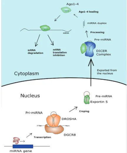
The molecular machinery responsible for miRNA bio-genesis and interaction with mRNAs (Figure 1) is better elucidated than that underlying the activity of other ncRNAs. miRNA genes are transcribed by RNA polymerase II or III. This process generates a molecule, the pri-miR, that folds itself into a hairpin conformation and is 50 capped and 30 polyadenylated (13,14). The pri-miR molecule is recog-nized by the DROSHA RNAse III enzyme and cleaved, forming a 60- to 100-nt hairpin molecule, the pre-miR, that is exported from the nucleus to the cytoplasm (14,15). In the cytoplasm, the pre-miR is cleaved by the DICER enzyme, yielding a double-strandedB22nt RNA molecule (16). One of the strands of the formed 22-nt miRNA molecule is loaded into an RNA-induced silencing complex (RISC) protein to serve as the template for target mRNA recognition (17).
Mature miRNA molecules loaded into RISCs have two mechanisms of action. Perfect or near-perfect base pairing of the entire miRNA molecule to a complementary region within an mRNA leads to mRNA degradation by RISC (18). Perfect base pairing of almost all 22 nt is an uncommon scenario in animals. The more common scenario involves imperfect pairing, or pairing of a 5–8 nt
‘seed’ region of the miRNA, which leads to reduced translation or destabilization of the target mRNA (19). A single miRNA molecule may regulate multiple genes that contain a sequence complementary to the miRNA seed, and a given mRNA may be regulated by different miRNAs (20). Notably, the administration of exogenous nucleic acid sequences can mimic miRNA action (mimic-miRs), and employ the endogenous cellular machinery for miRNA-mediated gene silencing (21). Another possibility is the administration of stabilized exogenous nucleic acid sequences that are complementary to endogenous miRNAs, such as antagomirs, resulting in the inhibition of target cellular miRNAs (22).
miRNAs are also present and enriched in the plasma and serum. Furthermore, these RNAs are especially
Figure 1.Main processes involved in the
resistant to degradation (23). Blood circulating miRNAs are contained in microvesicles known as exosomes or are associated with Argonaute 2 complexes and, as a consequence, are protected from degradation (6,24). Because circulating miRNAs may originate from many different tissues throughout the body and may reflect normal function, changes in the circulating levels of these miRNAs may constitute a useful and easily accessible biomarker of many different pathological conditions. More-over, it is feasible to quantify the levels of such circulating miRNAs by RT-PCR or even high throughput techniques such as micro-arrays or RNA-sequencing. The dysregula-tion of miRNA expression is well established in some tumors, and circulating miRNAs are indeed emerging as promising biomarkers in thisfield (23,25). The search for circulating miRNAs as biomarkers is also being applied to neurological disorders.
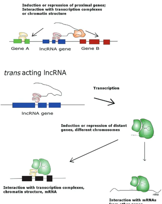
lncRNAs boast distinct and diverse molecular machinery involved in the regulation of gene expression (Figure 2). Most of these ncRNAs are RNA polymerase II products that lack open reading frames but are generally 50capped and 30 polyadenylated (26,27). lncRNAs are numerous, with estimates in the range of thousands of lncRNA coding genes (28). Briefly, lncRNAs may act in cis, silencing or enhancing the expression of proximal genes on the same chromosome. For example, the lncRNA HOTTIP gene is present in the HOXA gene cluster, and its expression enhances the expression of other component genes in the same cluster (20). lncRNAs may also act intrans, silenc-ing or enhancsilenc-ing the expression of genes on different chromosomes. One example of an lncRNA acting intrans is Six3OS. This lncRNA was shown to activate the targets of the retinal development involving the Six3 transcription factor (29). Another mechanism of action for lncRNAs is the regulation of other ncRNAs. lncRNA can act as a
‘sponge’or decoy target. The lncRNA lincRNA-RoR mech-anism of action illustrates this mechmech-anism: this lncRNA has a binding site for miR-145, and the presence of lincRNA-RoR inhibits miR-145 action by interacting directly with lncRNA miRNA (30). The mechanisms of lncRNA-mediated regulation of protein-coding gene transcription are explored in more detail in the current literature (26,27).
Role of non-coding RNAs in disease
Table 1 presents a list of ncRNAs associated with mechanisms underlying selected neurological disorders.
Epilepsy. Epilepsy is a neurological condition with a high prevalence in the population (1.5–2%). A common feature of different epileptic conditions is the occurrence of seizures (31,32). The mechanism responsible for epilep-togenesis (the process by which normal nervous tissue becomes epileptic) is complex and multifactorial (33). Evidence in the literature, as reviewed below, indicates that ncRNAs may have critical roles in the molecular mechanisms associated with epilepsy (34).
Hippocampal tissue from patients with mesial temporal lobe epilepsy (MTLE) who underwent temporal lobe resection for the control of seizures has been shown to have a reduction in the overall expression of miRNAs when compared with normal hippocampus from autopsy controls (35). Moreover, MTLE is associated with infl am-mation, and changes in the expression of miRNAs involved in the regulation of inflammation have been demonstrated in samples from MTLE patients (36,37). For example, miR-146-a, a miRNA involved in inflammation, is upregu-lated in resected hippocampus from MTLE patients (37).
In animal models of epilepsy, the dysregulation of miRNAs has been explored more extensively. miRNA expression studies were performed, using high-throughput platforms, in the animal model induced by lithium-pilocarpine, systemic kainic acid, and by intra-amygdalar kainic acid injection (38–40). Based on such studies, an extensive list of candidate miRNAs was found, but relatively few miRNAs were consistent among different studies. One example of replicable findings is mir-34a, which was found to be differentially expressed in two independent studies (38,41). mir-134 is another promising miRNA that may be involved in the molecular mechanisms of epilepsy. mir-134 was found to be differentially expressed in an epilepsy animal model, and the reduction in its expression by antagomir administration was shown to reduce cell death and seizure severity (42). In addition, downregulation of mir-132 in an animal model reduced seizure-induced neuronal death (40).
More recently, Jimenez-Mateos et al. (3) demonstrated that miR-22 downregulates the purinergic P2X7 receptor, a key component of the inflammatory response, in a mouse model of focal onset status-epilepticus. Further-more, an increase in miR-22 activity by the administration of a Mir-22 mimic molecule reduced spontaneous seizures in these mice (3).
found four upregulated and two downregulated blood circulating miRNAs when comparing epilepsy patients to healthy controls. Among the differentially expressed miRNAs, miR-106b-5p had the highest sensitivity and specificity (46). Furthermore, in a subsequent study, there were five circulating miRNAs identified as potential bio-markers of drug-resistant epilepsy, and miR-301a-3p had the highest sensitivity and specificity (47). We have identified
that miR-134 is a circulating biomarker for patients with mesial temporal lobe epilepsy regardless of their response to treatment, which may help in the diagnosis of this type of epilepsy (48).
In focal cortical dysplasia, a cortical malformation frequently associated with refractory seizures, miR-4521 has been shown to be upregulated in the plasma of patients compared to control subjects (49).
Neurodegenerative and neuromuscular disorders. Neurodegenerative disorders are associated with a wide range of genetic mutations and epigenetic and environ-mental factors. Among genetic mutations, trinucleotide repeat expansion is increasingly recognized as the cause of a large subset of these conditions. Trinucleotide repeat expansions account for more than 30 neurological and neuromuscular diseases that are categorized into coding and non-coding repeat expansion disorders, depending on the genetic location of their causative mutations (50–52). Disorders such as Huntington’s disease (HD), spinocer-ebellar ataxia (SCA) types 1, 2, 3, 6, 7, 8, and 17, dentatorubral-pallidoluysian atrophy, and spinal and bulbar muscular atrophy are typically associated with a protein gain-of-function mechanism (53). In contrast, diseases such as myotonic dystrophy type 1 (DM1) (54,55), fragile X-associated tremor ataxia syndrome (FXTAS), myotonic dystrophy type 2 (DM2), SCA31, SCA10, SCA8, and, more recently, amyotrophic lateral sclerosis and frontotemporal sclerosis have been associated with an RNA gain-of-function mechanism in which the trinucleotide expansion leads to the formation of nuclear RNA foci that sequester specific RNA-binding proteins (5,56,57).
Studies of FXTAS have established that the seques-tration of RNA-binding proteins due to the expression of pathogenic RNA with expanded repeats is involved in disease pathogenesis (58) (Figure 3). A recent study
identified that the double-stranded RNA-binding protein DGCR8 binds to expanded CGG repeats, resulting in the partial sequestration of DGCR8 and its partner, DROSHA, within CGG RNA aggregates. Consequently, the process-ing of miRNAs is reduced, resultprocess-ing in decreased levels of mature miRNAs in neuronal cells expressing expanded CGG repeats such as in brain tissue from patients with FXTAS (59).
SCA8 is a dominantly inherited, slowly progressive neurodegenerative disorder caused by a CTG CAG repeat expansion (60). In pathological samples from SCA8 patients, bidirectional (sense and antisense) expression of the SCA8 CTGCAG expansion produces toxic non-coding CUG expansion in RNAs from the Ataxin 8 opposite strand (ATXN8OS) and a nearly pure polyglutamine expan-sion protein encoded by ATXN8 (61,62). In SCA7, the tissue-specific alterations caused by CAG repeat expres-sion in the ATXN7 gene seems to be related to cross-talk between the lncRNA lnc-SCA7, the ATXN7 mRNA, and mir-124. Mutant ATXN7 disrupts this crosstalk and is itself upregulated, since it is not repressed by ncRNAs (63).
Recent studies have suggested that alterations in small regulatory ncRNAs, such as miRNAs, could contribute to the pathogenesis of several neurodevelopmental disor-ders. Some studies have found a relationship between miRNAs and DM1 (64). Alterations in the miRNA expres-sion patterns have been observed in muscle-specific
Table 1.List of ncRNAs associated with different mechanisms underlying selected neurological disorders.
Disorder Gene Affected Proposed mechanisms associated with Noncoding RNAs
References
FXTAS FMR1; FMR4 Sequestration of RNA binding protein;
antisense transcript
Tassone et al. 2004 (58)
DM1 DMPK Sequestration of RNA binding protein;
antisense transcript
Rau et al. 2011 (66)
SCA1 ATXN1 Altered miRNA pathway Galka-Marciniak et al. 2012 (56)
SCA3 ATXN3 An auxiliary toxic long CAG repeat RNA;
altered miRNA pathway
Galka-Marciniak et al. 2012 (56)
SCA7 ATXN7 Antisense transcript repress sense ataxin-7 Tan et al. 2014 (63)
SCA8 ATXN8OS; ATXN8 Sequestration of RNA binding protein;
antisense transcript
Daughters et al. 2009 (61); Moseley et al. 2006 (62)
HDL2 JPH3 Antisense transcript; polyQ toxicity Wojciechowska and
Krzyzosiak, 2011 (5)
MTLE P2X7 Down-regulation by miR-22 Jimenez-Mateos et al. 2015 (3)
HD HTT An auxiliary toxic long CAG repeat RNA;
altered miRNA pathway
Wojciechowska and Krzyzosiak, 2011 (5)
MTLE Genes involved with
inflammation
Up-regulation of miR-146a expression Aronica et al. 2010 (37)
ALS SOD1and others An artificial microRNA may extend survival and delays paralysis; Up regulation of miR-206.
Stoica et al. 2016 (79); Takahashi et al. 2015 (81)
Cortical dysplasia Lis1 Dysregulation of miR-139-5p Huang et al. 2014 (90)
Pain Inflammation,
neural processing
miRNAs (myomiRs). Given the small distance between the seed binding sites of miR-206 and 148a in the DMPK 30UTR, Koscianska et al. (65) analyzed the binding mech-anism of both miRNAs. They discovered cooperative binding; the joint binding of miRs 206 and 148a increased the negative regulation of DMPK mRNA. Thesefindings provide mechanistic insights into the miRNA-mediated regulation of the DMPK transcript. In this regard, the dysregulation of DM1-associated miRNAs has also been linked to altera-tions in their predictive target expression, showing that miRNA dysregulation in DM1 is functionally relevant and may contribute to disease pathology (66,67). Furthermore, RNA toxicity has been confirmed in transgenic mice har-boring long triplet repeats in thedmpkgene. Seznec et al. (68) showed that mice develop multi-system abnormalities mimicking the human DM phenotype, with predominant involvement of muscles and the central nervous system (CNS). Pathway and function analysis highlighted the involvement of the miRNA-dysregulated mRNAs in multi-ple aspects of DM2 pathophysiology as well (4,69).
Huntington’s disease is characterized by wide-spread mRNA dysregulation, especially in the striatum
and cortical regions and alterations in miRNA-mediated post-transcriptional regulation could be an important mech-anism contributing to mRNA dysregulation in HD (70). In addition, there is evidence that abnormal neurodevel-opment might also have a critical role in HD (71). These emerged from studies using mouse embryonic stem cells and patient-derived induced pluripotent stem cells (The HD iPSC Consortium, 2012) showing that chromatin modifi ca-tions and DNA methylation status support the hypothesis that wild-type and mutant Huntingtin might affect key chromatin regulators such as DNA and histone methyl-transferases, and demethylases (72–74). In fact, a growing body of evidence suggests that alterations of epigenetic modifications constitute a basic molecular mechanism caused by the HD mutation and are responsible for early features of the pathological process (75). Furthermore, a recent genome-wide screen of miRNAs inpost mortem brains highlighted miRNAs that were differentially expressed in HD patients, especially miRNAs in the HOX family, which have been associated with early brain development (76). Indeed, there are several classes of lncRNAs that are potentially involved in developmental processes and that
Figure 3.Mechanism involved in microRNA machinery sequestration by aberrant RNA species produced in a triplet repeat disease,
were found to be dysregulated in brain tissue from patients with HD such as TUG1, NEAT1, MEG3, and DGCR5 (77). Amyotrophic lateral sclerosis (ALS) is a widespread motor neuron disorder causing injury and death of lower and upper motor neurons. Familial ALS (B10% of all ALS cases) is inherited as a dominant trait, and 20% of these cases have mutations in the gene encoding Cu/Zn cyto-solic superoxide dismutase 1 (SOD1) (78). A recent study demonstrated that an AAV9-delivered SOD1-specific arti-ficial miRNA is an effective and translatable therapeutic approach to ALS (79). Another promising miRNA with a possible therapeutic use in ALS is mir-155. It was demonstrated that this inflammation-associated miRNA is upregulated in the mutant SOD1 mouse model and that reduction in the expression of mir-155 significantly extended the life span of this mouse (80).
In addition, expression levels of certain miRNAs, such as miR-4649-5p and hsa-miR-4299, were significantly correlated with disease progression and might be useful as prognostic biomarkers (81). Another potential bio-marker was mir-206, found to be upregulated in the plasma of SOD1-G93A mice, an experimental ALS model, and in patients with confirmed ALS (82). In addition, there is evidence of dysregulation of miRNAs extracted from leukocytes from sporadic ALS patients (83). More recently, we have demonstrated that among 11 miRNAs identified as differently expressed in muscle of patients with ALS, only two, miR-214 and miR-424, correlated with clinical deterioration over time in these patients (84).
Pain. Conditions leading to chronic pain are related to multiple etiologic factors, ranging from maladaptive neuronal plasticity to diverse inflammatory pathways (85). Due to the complexity of chronic pain, some studies have explored the possible role of ncRNAs in different experi-mental pain models. Kusuda et al. (86) observed a change in the expression of three miRNAs, miRs 1, 16, and 206, in different pain conditions such as peripheral inflammation, nerve ligation, or axotomy. Other studies have employed low-density TaqMan arrays to profile the expression pattern of miRNAs after spinal nerve ligation in rats and found 63 altered miRNAs (87).
A possible role for lncRNAs has been explored in experimental models of neuropathic pain. A microarray analysis demonstrated hundreds of differentially expressed lncRNAs and mRNAs in the spinal cords of mice sub-jected to spinal nerve ligation. As demonstrated in other experiments, 35 differentially regulated lncRNAs were in genomic regions proximal to differentially regulated genes from the same dataset (88).
Non-coding RNAs as target treatments for
neurologic disorders
The use of ncRNAs as therapeutic tools in human disorders is still in its early stages. To date, there is only one therapeutic use of human miRNA for the treatment of hepatitis C (HCV) that has passed phase IIa clinical trials (89). The clinical trial data showed the efficacy of the employed anti-miRNA in reducing viral load and showed good treatment tolerability, thus indicating the feasibility of similar strategies for other clinical uses such as in the case of neurological conditions.
Animal experiments already indicate some promising targets for the use of ncRNAs as therapeutic tools in disorders affecting the CNS. In epilepsy, the use of miR antagonists for miR-134 or mimic-miRs for miR-22 was capable of reducing neuronal death and seizure severity in animal models (3,42). These and other examples of pre-clinical uses of miRNAs for the treatment of neurological conditions need further study; however, due to the good tolerability already shown in the existing human clinical trial for HCV, there is optimism about the possible utility of ncRNAs in the treatment of neurological conditions in the future. However, several challenges remain for the efficient delivery of ncRNA molecules into the CNS, thus most of the pre-clinical studies still use invasive tech-niques for administering these molecules (4,44)
Conclusions
In conclusion, ncRNAs are emerging as key players in thefield of neurological disorders. ncRNAs are involved in many conditions, either as part of the molecular mecha-nisms underlying disease or as biomarkers that may be used for improved diagnosis or assessment of disease progression. ncRNAs are also promising targets for new therapeutic strategies to be employed in the treatment of neurological conditions.
Acknowledgments
A.S.V. is supported by a grant from Fundac¸ão de Amparo a Pesquisa do Estado de São Paulo (FAPESP; #2016/22447-5), Brazil. D.B.D. is supported by a fel-lowship from Coordenac¸ão de Aperfeic¸oamento de Pessoal de Nível Superior (CAPES), Brazil. I.L-C is supported by grants from FAPESP (#2011/50680 and #2013/07559-3) and from Conselho Nacional de Pesquisa (CNPq), Brazil.
References
1. Djebali S, Davis CA, Merkel A, Dobin A, Lassmann T, Mortazavi A, et al. Landscape of transcription in human cells.
Nature2012; 489: 101–108, doi: 10.1038/nature11233.
3. Jimenez-Mateos EM, Arribas-Blazquez M, Sanz-Rodriguez A, Concannon C, Olivos-Ore LA, Reschke CR, et al. micro-RNA targeting of the P2X7 purinoceptor opposes a con-tralateral epileptogenic focus in the hippocampus.Sci Rep
2015; 5: 17486, doi: 10.1038/srep17486.
4. Greco S, Perfetti A, Fasanaro P, Cardani R, Capogrossi MC, Meola G, et al. Deregulated microRNAs in myotonic dystrophy type 2. PloS One 2012; 7: e39732, doi: 10.1371/journal. pone.0039732.
5. Wojciechowska M, Krzyzosiak WJ. Cellular toxicity of expanded RNA repeats: focus on RNA foci.Hum Mol Genet
2011; 20: 3811–3821, doi: 10.1093/hmg/ddr299.
6. Arroyo JD, Chevillet JR, Kroh EM, Ruf IK, Pritchard CC, Gibson DF, et al. Argonaute2 complexes carry a population of circulating microRNAs independent of vesicles in human plasma. Proc Natl Acad Sci USA2011; 108: 5003–5008,
doi: 10.1073/pnas.1019055108.
7. Femminella GD, Ferrara N, Rengo G. The emerging role of microRNAs in Alzheimer’s disease.Front Physiol2015; 6: 40, doi: 10.3389/fphys.2015.00040.
8. Zhang Z. Long non-coding RNAs in Alzheimer’s disease.
Curr Top Med Chemy 2016; 16: 511–519, doi: 10.2174/
1568026615666150813142956.
9. Majidinia M, Mihanfar A, Rahbarghazi R, Nourazarian A, Bagca B, Avci CB. The roles of non-coding RNAs in Parkinson’s disease. Mol biol rep 2016; 43:1193–1204, doi: 10.1007/s11033-016-4054-3.
10. Huttenhofer A, Schattner P, Polacek N. Non-coding RNAs: hope or hype? Trends Genet 2005; 21: 289–297, doi:
10.1016/j.tig.2005.03.007.
11. Mattick JS, Makunin IV. Non-coding RNA.Hum Mol Genet
2006; 15 Spec No 1: R17–R29, doi: 10.1093/hmg/ddl046.
12. Megosh HB, Cox DN, Campbell C, Lin H. The role of PIWI and the miRNA machinery in Drosophila germline determi-nation.Curr Biol2006; 16: 1884–1894, doi: 10.1016/j.cub.
2006.08.051.
13. McNeill E, Van Vactor D. MicroRNAs shape the neuronal landscape. Neuron 2012; 75: 363–379, doi: 10.1016/
j.neuron.2012.07.005.
14. Kim VN. MicroRNA biogenesis: coordinated cropping and dicing.Nat Rev Mol Cell Biol2005; 6: 376–385, doi: 10.1038/
nrm1644.
15. Lee Y, Ahn C, Han J, Choi H, Kim J, Yim J, et al. The nuclear RNase III Drosha initiates microRNA processing. Nature
2003; 425: 415–419, doi: 10.1038/nature01957.
16. Hutvágner G, McLachlan J, Pasquinelli AE, Bálint E, Tuschl T, Zamore PD. A cellular function for the RNA-interference enzyme Dicer in the maturation of the let-7 small temporal RNA. Science2001; 293: 834–838, doi: 10.1126/science.
1062961.
17. Du T, Zamore PD. microPrimer: the biogenesis and func-tion of microRNA. Development 2005; 132: 4645–4652,
doi: 10.1242/dev.02070.
18. Gu S, Kay MA. How do miRNAs mediate translational repression?Silence1, 11, doi: 10.1186/1758-907X-1-11. 19. Cannell IG, Kong YW, Bushel M. How do microRNAs
regulate gene expression?Biochem Soc Trans 2008; 36: 1224–1231, doi: 10.1042/BST0361224.
20. Wang KC, Yang YW, Liu B, Sanyal A, Corces-Zimmerman R, Chen Y, et al. A long noncoding RNA maintains active
chromatin to coordinate homeotic gene expression.Nature
2011; 472: 120–124, doi: 10.1038/nature09819.
21. Wang Z. The guideline of the design and validation of MiRNA mimics. Methods Mol Biol 2011; 676: 211–223,
doi: 10.1007/978-1-60761-863-8.
22. Krutzfeldt J, Rajewsky N, Braich R, Rajeev KG, Tuschl T, Manoharan M, et al. Silencing of microRNAs in vivo with
‘antagomirs’. Nature 2005; 438: 685–689, doi: 10.1038/ nature04303.
23. Chen X, Ba Y, Ma L, Cai X, Yin Y, Wang K, et al. Characterization of microRNAs in serum: a novel class of biomarkers for diagnosis of cancer and other diseases.Cell Res2008; 18: 997–1006, doi: 10.1038/cr.2008.282.
24. Valadi H, Ekstrom K, Bossios A, Sjostrand M, Lee JJ, Lotvall JO. Exosome-mediated transfer of mRNAs and microRNAs is a novel mechanism of genetic exchange between cells.
Nat Cell Biol2007; 9: 654–659, doi: 10.1038/ncb1596.
25. Toiyama Y, Takahashi M, Hur K, Nagasaka T, Tanaka K, Inoue Y, et al. Serum miR-21 as a diagnostic and prognostic biomarker in colorectal cancer.J Nat Cancer Inst2013; 105: 849–859, doi: 10.1093/jnci/djt101.
26. Kornienko AE, Guenzl PM, Barlow DP, Pauler FM. Gene regulation by the act of long non-coding RNA transcription.
BMC Biol2013; 11: 59, doi: 10.1186/1741-7007-11-59. 27. Ulitsky I, Bartel DP. lincRNAs: genomics, evolution, and
mechanisms. Cell 2013; 154: 26–46, doi: 10.1016/j.cell. 2013.06.020.
28. Derrien T, Johnson R, Bussotti G, Tanzer A, Djebali S, Tilgner H, et al. The GENCODE v7 catalog of human long noncoding RNAs: analysis of their gene structure, evolution, and expression.Genome Res 2012; 22: 1775–1789, doi:
10.1101/gr.132159.111.
29. Rapicavoli NA, Poth EM, Zhu H, Blackshaw S. The long noncoding RNA Six3OS acts in trans to regulate retinal development by modulating Six3 activity.Neural Dev2011; 6: 32, doi: 10.1186/1749-8104-6-32.
30. Wang Y, Xu Z, Jiang J, Xu C, Kang J, Xiao L, et al. Endogenous miRNA sponge lincRNA-RoR regulates Oct4, Nanog, and Sox2 in human embryonic stem cell self-renewal.
Dev Cell2013; 25: 69–80, doi: 10.1016/j.devcel.2013.03.002.
31. Engel J Jr. Mesial temporal lobe epilepsy: what have we learned?The Neuroscientist2001; 7: 340–352, doi: 10.1177/
107385840100700410.
32. Annegers JF, Rocca WA, Hauser WA. Causes of epilepsy: contributions of the Rochester epidemiology project.Mayo Clin Proc1996; 71: 570–575, doi: 10.4065/71.6.570. 33. Pitkänen A, Lukasiuk K. Mechanisms of epileptogenesis and
potential treatment targets.Lancet Neurol2011; 10: 173–186,
doi: 10.1016/S1474-4422(10)70310-0.
34. Dogini DB, Avansini SH, Vieira AS, Lopes-Cendes I. MicroRNA regulation and dysregulation in epilepsy. Front Cell Neurosci2013; 7: 172, doi: 10.3389/fncel.2013.00172. 35. McKiernan RC, Jimenez-Mateos EM, Bray I, Engel T, Brennan GP, Sano T, et al. Reduced mature microRNA levels in association with dicer loss in human temporal lobe epilepsy with hippocampal sclerosis. PloS One 2012; 7: e35921, doi: 10.1371/journal.pone.0035921.
37. Aronica E, Fluiter K, Iyer A, Zurolo E, Vreijling J, van Vliet EA, et al. Expression pattern of miR-146a, an infl ammation-associated microRNA, in experimental and human temporal lobe epilepsy. Euro J Neurosci 2010; 31: 1100–1107,
doi: 10.1111/j.1460-9568.2010.07122.x.
38. Hu K, Xie YY, Zhang C, Ouyang DS, Long HY, Sun DN, et al. MicroRNA expression profile of the hippocampus in a rat model of temporal lobe epilepsy and miR-34a-targeted neuroprotection against hippocampal neurone cell apoptosis post-status epilepticus. BMC Neurosci 2012; 13: 115, doi: 10.1186/1471-2202-13-115.
39. McKiernan RC, Jimenez-Mateos EM, Sano T, Bray I, Stallings RL, Simon RP, et al. Expression profiling the microRNA response to epileptic preconditioning identifies miR-184 as a modulator of seizure-induced neuronal death.
Exp Neurol2012; 237: 346–354, doi: 10.1016/j.expneurol.
2012.06.029.
40. Jimenez-Mateos EM, Bray I, Sanz-Rodriguez A, Engel T, McKiernan RC, Mouri G, et al. miRNA Expression profile after status epilepticus and hippocampal neuroprotection by targeting miR-132. Am J Pathol 2011; 179: 2519–2532,
doi: 10.1016/j.ajpath.2011.07.036.
41. Sano T, Reynolds JP, Jimenez-Mateos EM, Matsushima S, Taki W, Henshall DC. MicroRNA-34a upregulation during seizure-induced neuronal death. Cell Death Dis 2012; 3: e287, doi: 10.1038/cddis.2012.23.
42. Jimenez-Mateos EM, Engel T, Merino-Serrais P, McKiernan RC, Tanaka K, Mouri G, et al. Silencing microRNA-134 produces neuroprotective and prolonged seizure-suppressive effects.Nat Med2012; 18: 1087–1094, doi: 10.1038/nm.2834.
43. Lee DY, Moon J, Lee ST, Jung KH, Park DK, Yoo JS, et al. Dysregulation of long non-coding RNAs in mouse models of localization-related epilepsy. Biochem Biophys Res Com-mun2015; 462: 433–440, doi: 10.1016/j.bbrc.2015.04.149.
44. Liu DZ, Tian Y, Ander BP, Xu H, Stamova BS, Zhan X, et al. Brain and blood microRNA expression profiling of ischemic stroke, intracerebral hemorrhage, and kainate seizures.
J Cereb Blood Flow Metab2010; 30: 92–101, doi: 10.1038/
jcbfm.2009.186.
45. Roncon P, Soukupova M, Binaschi A, Falcicchia C, Zucchini S, Ferracin M, et al. MicroRNA profiles in hippocampal granule cells and plasma of rats with pilocarpine-induced epilepsy--comparison with human epileptic samples. Sci Rep2015; 5: 14143, doi: 10.1038/srep14143.
46. Wang J, Yu JT, Tan L, Tian Y, Ma J, Tan CC, et al. Genome-wide circulating microRNA expression profiling indicates biomarkers for epilepsy.Sci Rep2015; 5: 9522, doi: 10.1038/ srep09522.
47. Wang J, Tan L, Tan L, Tian Y, Ma J, Tan CC, et al. Circulating microRNAs are promising novel biomarkers for drug-resistant epilepsy.Sci Rep2015; 5: 10201, doi: 10.1038/srep10201. 48. Avansini SH, de Sousa Lima BP, Secolin R, Santos ML,
Coan AC, Vieira AS, et al. MicroRNA hsa-miR-134 is a circulating biomarker for mesial temporal lobe epilepsy.
PLoS One2017; 12:e0173060, doi: 10.1371/journal.pone. 0173060.
49. Wang X, Sun Y, Tan Z, Che N, Ji A, Luo X, et al. Serum MicroRNA-4521 is a Potential Biomarker for Focal Cortical Dysplasia with Refractory Epilepsy.Neurochem Res2016; 41: 905–912, doi: 10.1007/s11064-015-1773-0.
50. Lopez Castel A, Cleary JD, Pearson CE. Repeat instability as the basis for human diseases and as a potential target for therapy. Nat Rev Mol Cell Biol 2010; 11: 165–170,
doi: 10.1038/nrm2854.
51. Mirkin SM. Expandable DNA repeats and human disease.
Nature2007; 447: 932–940, doi: 10.1038/nature05977.
52. Ranum LP, Day JW. Dominantly inherited, non-coding microsatellite expansion disorders. Curr Opin Genet Dev
2002; 12: 266–271, doi: 10.1016/S0959-437X(02)00297-6. 53. Orr HT, Zoghbi HY. Trinucleotide repeat disorders.Ann Rev
Neurosci2007; 30: 575–621, doi: 10.1146/annurev.neuro.
29.051605.113042.
54. Wang LC, Chen KY, Pan H, Wu CC, Chen PH, Liao YT, et al. Muscleblind participates in RNA toxicity of expanded CAG and CUG repeats in Caenorhabditis elegans.Cell Mol Life Sci2011; 68: 1255–1267, doi: 10.1007/s00018-010-0522-4.
55. Mykowska A, Sobczak K, Wojciechowska M, Kozlowski P, Krzyzosiak WJ. CAG repeats mimic CUG repeats in the misregulation of alternative splicing. Nucleic Acids Res
2011; 39: 8938–8951, doi: 10.1093/nar/gkr608.
56. Galka-Marciniak P, Urbanek MO, Krzyzosiak WJ. Triplet repeats in transcripts: structural insights into RNA toxicity.
Biol Chem2012; 393: 1299–1315, doi: 10.1515/hsz-2012-0218.
57. Gendron TF, Belzil VV, Zhang YJ, Petrucelli L. Mechanisms of toxicity in C9FTLD/ALS. Acta Neuropathol 2014; 127: 359–376, doi: 10.1007/s00401-013-1237-z.
58. Tassone F, Iwahashi C, Hagerman PJ. FMR1 RNA within the intranuclear inclusions of fragile X-associated tremor/ataxia syndrome (FXTAS).RNA Biol2004; 1: 103–105, doi: 10.4161/
rna.1.2.1035.
59. Sellier C, Freyermuth F, Tabet R, Tran T, He F, Ruffenach F, et al. Sequestration of DROSHA and DGCR8 by expanded CGG RNA repeats alters microRNA processing in fragile X-associated tremor/ataxia syndrome. Cell Rep 2013; 3: 869–880, doi: 10.1016/j.celrep.2013.02.004.
60. Ikeda Y, Shizuka-Ikeda M, Watanabe M, Schmitt M, Okamoto K, Shoji M. Asymptomatic CTG expansion at the SCA8 locus is associated with cerebellar atrophy on MRI.
J Neurol Sci2000; 182: 76–79, doi: 10.1016/S0022-510X
(00)00446-9.
61. Daughters RS, Tuttle DL, Gao W, Ikeda Y, Moseley ML, Ebner TJ, et al. RNA gain-of-function in spinocerebellar ataxia type 8.PLoS Genet2009; 5: e1000600, doi: 10.1371/ journal.pgen.1000600.
62. Moseley ML, Zu T, Ikeda Y, Gao W, Mosemiller AK, Daughters RS, et al. Bidirectional expression of CUG and CAG expansion transcripts and intranuclear polyglutamine inclusions in spinocerebellar ataxia type 8.Nat Genet2006; 38: 758–769, doi: 10.1038/ng1827.
63. Tan JY, Vance KW, Varela MA, Sirey T, Watson LM, Curtis HJ, et al. Cross-talking noncoding RNAs contribute to cell-specific neurodegeneration in SCA7. Nat Struct Mol Biol
2014; 21: 955–961, doi: 10.1038/nsmb.2902.
64. Turner C, Hilton-Jones D. The myotonic dystrophies: diagnosis and management.J Neurol Neurosurg Psychiatry2010; 81: 358–367, doi: 10.1136/jnnp.2008.158261.
66. Rau F, Freyermuth F, Fugier C, Villemin J. P, Fischer M. C, Jost, B, et al. Misregulation of miR-1 processing is associated with heart defects in myotonic dystrophy.Nature Struct Molec Biol2011, 18, 840–845, doi: 10.1038/nsmb.2067.
67. Perbellini R, Greco S, Sarra-Ferraris G, Cardani R, Capogrossi MC, Meola G, et al. Dysregulation and cellular mislocalization of specific miRNAs in myotonic dystrophy type 1.Neuromuscul Disorder2011; 21: 81–88, doi: 10.1016/ j.nmd.2010.11.012.
68. Seznec H, Agbulut O, Sergeant N, Savouret C, Ghestem A, Tabti N, et al. Mice transgenic for the human myotonic dys-trophy region with expanded CTG repeats display muscular and brain abnormalities.Hum Mol Genet2001; 10: 2717–
2726, doi: 10.1093/hmg/10.23.2717.
69. Deng JH, Deng P, Lin SL, Ying SY. Gene silencing in vitro and in vivo using intronic microRNAs.Meth Molecular Biol
12015; 1218: 321–340, doi: 10.1007/978-1-4939-1538-5.
70. Hoss AG, Lagomarsino VN, Frank S, Hadzi TC, Myers RH, Latourelle JC. Study of plasma-derived miRNAs mimic differences in Huntington’s disease brain. Mov Disorder
2015; 30: 1961–1964, doi: 10.1002/mds.26457.
71. Humbert S. Is Huntington disease a developmental disorder?
EMBO Rep2010; 11: 899, doi: 10.1038/embor.2010.182. 72. HD iPSC Consortium. Induced pluripotent stem cells
from patients with Huntington’s disease show CAG-repeat-expansion-associated phenotypes.Cell Stem Cell2012; 11: 264–278, doi: 10.1016/j.stem.2012.04.027.
73. Ng CW, Yildirim F, Yap YS, Dalin S, Matthews BJ, Velez PJ, et al. Extensive changes in DNA methylation are associated with expression of mutant huntingtin. Proc Natl Acad Sci USA2013; 110: 2354–2359, doi: 10.1073/pnas.1221292110.
74. Biagioli M, Ferrari F, Mendenhall EM, Zhang Y, Erdin S, Vijayvargia R, et al. Htt CAG repeat expansion confers pleiotropic gains of mutant huntingtin function in chromatin regulation. Hum Mol Genet 2015; 24: 2442–2457, doi:
10.1093/hmg/ddv006.
75. Kerschbamer E, Biagioli M. Huntington’s disease as neuro-developmental disorder: altered chromatin regulation, coding, and non-coding RNA transcription.Front Neurosci2015; 9; 509, doi: 10.3389/fnins.2015.00509.
76. Hoss AG, Kartha VK, Dong X, Latourelle JC, Dumitriu A, Hadzi TC, et al. MicroRNAs located in the Hox gene clusters are implicated in huntington’s disease pathogenesis.PLoS Genet2014; 10: e1004188, doi: 10.1371/journal.pgen.1004188. 77. Johnson R. Long non-coding RNAs in Huntington’s disease neurodegeneration. Neurobiol Dis 2012; 46: 245–254, doi: 10.1016/j.nbd.2011.12.006.
78. Rosen DR, Siddique T, Patterson D, Figlewicz DA, Sapp P, Hentati A, et al. Mutations in Cu/Zn superoxide dismutase gene are associated with familial amyotrophic lateral sclerosis.Nature1993; 362; 59–62, doi: 10.1038/362059a0.
79. Stoica L, Todeasa SH, Toro Cabrera G, Salameh JS, ElMallah MK, Mueller C, et al. Adno associated virus delivered artificial microRNA extends survival and delays paralysis in an amyotrophic lateral sclerosis mouse model.
Ann Neurol2016; 79: 687–700, doi: 10.1002/ana.24618.
80. Butovsky O, Jedrychowski MP, Cialic R, Krasemann S, Murugaiyan G, Fanek, et al. Targeting miR-155 restores abnormal microglia and attenuates disease in SOD1 mice.
Ann Neurol2015; 77: 75–99, doi: 10.1002/ana.24304. 81. Takahashi I, Hama Y, Matsushima M, Hirotani M, Kano T,
Hohzen H, et al. Identification of plasma microRNAs as a biomarker of sporadic amyotrophic lateral sclerosis. Mol Brain2015; 8; 67, doi: 10.1186/s13041-015-0161-7. 82. Toivonen JM, Manzano R, Olivan S, Zaragoza P,
Garcia-Redondo A, Osta R. MicroRNA-206: a potential circulating biomarker candidate for amyotrophic lateral sclerosis.PloS One2014; 9: e89065, doi: 10.1371/journal.pone.0089065. 83. De Felice B, Guida M, Guida M, Coppola C, De Mieri G,
Cotrufo R. A miRNA signature in leukocytes from sporadic amyotrophic lateral sclerosis. Gene 2012; 508: 35–40,
doi: 10.1016/j.gene.2012.07.058.
84. de Andrade HM, de Albuquerque M, Avansini SH, de S Rocha C, Dogini DB, Nucci A, et al. MicroRNAs-424 and 206 are potential prognostic markers in spinal onset amyo-trophic lateral sclerosis. J Neurol Sci 2016; 368: 19–24, doi: 10.1016/j.jns.2016.06.046.
85. Basbaum AI, Bautista DM, Scherrer G, Julius D. Cellular and molecular mechanisms of pain.Cell 2009; 139: 267– 284, doi: 10.1016/j.cell.2009.09.028.
86. Kusuda R, Cadetti F, Ravanelli MI, Sousa TA, Zanon S, De Lucca FL, et al. Differential expression of microRNAs in mouse pain models. Mol Pain2011; 7: 17, doi: 10.1186/ 1744-8069-7-17.
87. von Schack D, Agostino MJ, Murray BS, Li Y, Reddy PS, Chen J, et al. Dynamic changes in the microRNA expression profile reveal multiple regulatory mechanisms in the spinal nerve ligation model of neuropathic pain.PloS One2011; 6: e17670, doi: 10.1371/journal.pone.0017670.
88. Jiang BC, Sun WX, He LN, Cao DL, Zhang ZJ, Gao YJ. Identification of lncRNA expression profile in the spinal cord of mice following spinal nerve ligation-induced neuropathic pain.Mol Pain2015; 11: 43, doi: 10.1186/s12990-015-0047-9. 89. van der Ree MH, van der Meer AJ, de Bruijne J, Maan R, van Vliet A, Welzel TM, et al. Long-term safety and efficacy of microRNA-targeted therapy in chronic hepatitis C patients.
Antiviral Res2014; 111: 53-9, doi: 10.1016/j.antiviral.2014. 08.015.