This Accepted Author Manuscript is copyrighted and published by Elsevier. It is posted here by agreement between Elsevier and University of Brasilia. Changes resulting from the publishing process - such as editing, corrections, structural formatting, and other quality control
mechanisms - may not be reflected in this version of the text. The definitive version of the text was subsequently published in [Theriogenology, Volume 54, Issue 5, 15 September 2000, Pages 809–822, doi:10.1016/S0093-691X(00)00392-7].You may download, copy and otherwise use the AAM for non-commercial purposes provided that your license is limited by the following
restrictions:
(1) You may use this AAM for non-commercial purposes only under the terms of the CC-BY-NC-ND license.
(2) The integrity of the work and identification of the author, copyright owner, and publisher must be preserved in any copy.
(3) You must attribute this AAM in the following format: [agreed attribution language, including link to CC BY-NC-ND license + Digital Object Identifier link to the published journal article on Elsevier’s ScienceDirect® platform].
________________________________________________________________________
Este Manuscrito do Autor Aceito para Publicação (AAM) é protegido por direitos autorais e publicado pela Elsevier. Ele esta disponível neste Repositório, por acordo entre a Elsevier e a Universidade de Brasília. As alterações decorrentes do processo de publicação - como a edição, correção, formatação estrutural, e outros mecanismos de controle de qualidade - não estão refletidas nesta versão do texto. A versão definitiva do texto foi posteriormente publicado em [Theriogenology, Volume 54, Número 5, 15 de Setembro de 2000, Páginas 809–822,
doi:10.1016/S0093-691X(00)00392-7]. Você pode baixar, copiar e utilizar de outra forma o AAM para fins não comerciais , desde que sua licença seja limitada pelas seguintes restrições: (1) Você pode usar este AAM para fins não comerciais apenas sob os termos da licença CC- BY- NC-ND.
(2) A integridade do trabalho e identificação do autor, detentor dos direitos autorais e editor deve ser preservado em qualquer cópia.
(3) Tem de atribuir este AAM no seguinte formato: [acordo na linguagem atribuída, incluindo o link para CC BY-NC-ND licença Digital + DOI do artigo publicado na revista Elsevier ScienceDirect ® da plataforma].
Effect of coconut water and Braun-Collins solutions at different temperatures and
incubation times on the morphology of goat preantral follicles preserved in vitro
J.R.V. Silva C.M. Lucci F.C.A. Carvalho S.N. Báo S.H.F. Costa R.R. Santos J.R. Figueiredo Abstract
Preservation of preantral follicles becomes very important to ensure follicle quality at the onset of cryopreservation or in vitro culture. However, for domestic animals, the ovarian donor of preantral follicles for in vitro studies is commonly encountered far away from reproduction laboratories. We investigated the effectiveness of coconut water and Braun-Collins solutions on the preservation of goat preantral follicles. At the slaughterhouse, the ovarian pair of each animal was divided into 19 fragments. One ovarian fragment was immediately fixed (Control — Time 0). The other 18 fragments were randomly distributed into tubes containing 2 mL of coconut water or Braun-Collins solution at 4 °, 20 ° or 39 °C and then stored for 4, 12 or 24 h. Histological analysis showed that the storage of ovarian fragments in coconut water and Braun-Collins solutions at 20 ° or 39 °C for 12 or 24 h significantly reduced (P < 0.05) the percentage of morphologically normal preantral follicles when compared with the control. However, storage in coconut water at 20 °C for 4 h and in both solutions at 4 °C kept the percentage at control values. Ultrastructural analysis of follicles exposed to the stated conditions confirmed the integrity of preantral follicles stored at 4 °C in Braun-Collins and coconut water solutions for up to 12 and 24 h, respectively. Reduced cellular metabolism at 4 °C may explain why the best preservation of preantral follicles was at 4 °C, which may suggest a useful method for ovary transport in the future.
Key words: goat; preantral follicle; preservation; ultrastructural; morphology
INTRODUCTION
The ovarian preantral follicles represent 90% of the follicular population (31). It is well known that the ovaries contain many thousands of follicles, and that the vast majority (99.9%) become atretic during their growth and maturation (8). Methods for manipulation of oocytes enclosed in preantral follicles have been developed, with the aim to rescue preantral follicles from the ovaries before they become atretic, and culture these follicles in vitro through the maturation stages by preventing follicular atresia. Offspring from preantral follicles isolated and cultured in vitro have been obtained in mice (8, 11, 12). In farm animals the donor of the ovarian follicles is usually far away from the laboratory where the research is done. Therefore, preservation of the follicles during transport from the field to the laboratory becomes a major concern. Various media have been successfully used in several studies of preservation of
bovine cumulus oocyte complexes enclosed in antral follicles, PBS (34,38), saline (33) and TCM 199 (36). Coconut water solution is a medium that reportedly has been successful in the preservation of ram (17), human (27), goat (28), and pig (35) semen, while the Braun-Collins solution is a medium which has been used for preserving liver (l), heart (lo), lungs (16), and kidney (32) tissue. Neither one of these media have been tested as a transport medium for preantral follicles. Our aim was to evaluate the effect of coconut water and Braun-Collins solutions on the preservation of goat preantral follicles, at different temperatures and times of storage, and to investigate the histologic and ultrastructural morphology of the preserved preantral follicles.
MATERIALS AND METHODS
Source of Ovaries
Ovaries (n=lO) from 5 adult mixed breed goats were collected at a local slaughterhouse. The ovaries were stripped of surrounding fat tissue and ligaments and washed in 70% alcohol for approximately 10 seconds and then twice in saline solution.
Media
The media tested were: 1) commercial Braun-Collins solution (osmolarity: 401 mOsm/L; pH: 7.2 [Table l] - Laboratory B.Braun S.A, Rio de Janeiro, Brazil) and 2) coconut water solution composed of two parts of filtered coconut water, one part of purified water and one part of 5% sodium citrate - final osmolarity: 300 mOsm/L and pH: 6.8 (27). Coconut water was obtained from six months old fruit collected from the Green Beach variety (Cocus nucifera).
Experimental Protocol
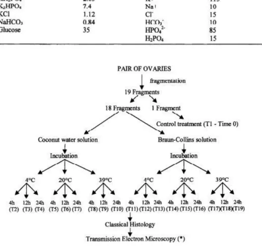
In the slaughterhouse, the ovarian pair from the same animal was divided with a scalpel into 19 fragments approximately 5 x 5 mm (3 mm thick). One ovarian fragment was taken randomly, a small piece removed for electron microscopy, and the remainder immediately futed for histological examination (Control, Treatment 1, Time 0). The other 18 tiagments were randomly distributed into tubes containing 2 mL of coconut water or Braun-Collins solution at 4’, 20 ° or 39°C and stored for 4, 12 or 24 h (Treatments 2 to 19) as shown in Figure 1. The temperatures were maintained using thermosflasks tilled with water at 4 °, 20 ° or 39°C. For each treatment, parameters such as temperature, osmolarity and pH of the
solutions were monitored at the beginning and at the end of the treatments. Each treatment was repeated 5 times.
Table 1. Composition of the Braun-Collins solution
Figure 1. Experimental protocol for preservation of caprine preantral follicles in vitro.
Qualitative Analysis of Caprine Preantral Follicles
In order to evaluate the morphology of the caprine preantral follicles at the end of the treatments, the ovarian fragments were processed as follows. Small pieces of the ovarian fragment horn each treatment, including the control, were removed for electron microscopy with the remainder being fixed individually in Bouins fluid for 12 h. Following this, they were dehydrated in a graded series of ethanol, clarified with xylol and embedded in paraffin wax. The tissue was sectioned serially at a thickness of 7pm and every 10th section was stained with
hematoxylin and eosm, each section was deparaffmized with xylol and rehydrated in graded alcohol. Sections were examined by light microscopy (Zeiss, Jena, Germany).
Preantral follicles were classified according to the stage of development as primordial (one layer of flattened or flattened-cuboidal granulosa cells around the oocyte), primary (a single layer of cuboidal granulosa cells around the oocyte), or secondary (oocyte surrounded by two or more layers of cuboidal granulosa cells (20). Follicle morphology was evaluated based on the integrity of the basement membrane, density of the granulosa cells, presence of pycnotic bodies, and integrity of the oocyte, using the nucleus as a marker. The proportion of atretic follicles in the control samples was considered as a baseline value for that seen in the stored samples, changes seen due to storage. Based on these parameters, preantral follicles were classified as normal (containing an intact oocyte and well-organized granulosa cells without a pycnotic nuclei; Figure 2A), degenerated Type 1 follicles (containing a somewhat shrunken oocyte with a pycnotic nucleus and strongly eosinophilic cytoplasm, but normal granulosa cells; Figure 2B) and degenerated Type 2 follicles (containing a shrunken oocyte with somewhat pycnotic nuclei, and disorganized granulosa cells and low cellular density; Figure 2C). These three classifications were assigned on a basis of atresia observed in the control and combined with changes that occurred as result of storage. Criteria for follicular cell and oocyte degeneration were identified using ovaries fixed at Time 0.
Figure 2. Histological section of ovarian fragment after staining with hematoxylin-eosin, showing A) normal preantral follicles (x 1160), B) degenerated Type 1 follicles (x 1160) and C) degenerated Type 2 follicles (x 1160). 0: oocyte, Nu: nucleus of oocyte, GC: granulosa cells, do: degenerated oocyte (* marks pycnotic nucleus) and dGC: degenerated granulosa cells. Bars = 20pm.
To better evaluate follicular morphology, ultrastructural analysis was performed using preantral follicles from the control treatment, as well as from the treatment that did not differ horn control. Briefly, small pieces of ovarian cortex were fixed in a solution containing 2% paraformaldehyde and 2.5% glutaraldehyde in 0.1 M cacodylate buffer pH 7.2. After fixation,
the specimens were rinsed in buffer and post-fixed in 1% osmium tetroxide, 0.8% potassium ferricyanide, and 5 mM CaClr in 0.1 M sodium cacodylate buffer. Subsequently the samples were dehydrated in acetone and embedded in Spurr. Semi-thin sections (3um) were stained with toluidine blue. Thin sections (65nm) were contrasted with uranyl acetate and lead citrate, and examined using Jeol 100 C and a Zeiss 912 transmission electron microscopes.
Statistical Analysis of Data
For each treatment, data of normal and degenerated preantral follicles Tom five ovarian fragments were pooled. The effect of preservation solution (coconut water solution or BraunCollins solution), temperatures and incubation times on the percentage of normal follicles were analyzed using a Chi-square test (Instat for Macintosh). The mean values of osmolarity and pH between the control and other treatments were compared using ANOVA and Fisher PLSD test (Stat View for Macintosh). Values were considered statistically significant when P < 0.05.
RESULTS Morphological Aspects of Goat Preantral Follicles in the Control and After
Storage In Vitro
Primordial (3,423), primary (293) and secondary (129) follicles were examined. Of the total of follicles analyzed in each category, degenerated primordial, primary and secondary follicles represented 53.6%, 63.4% and 64.3%, respectively. The distribution of degenerated Type 1 and Type 2 follicles in primordial, primary and secondary follicles was 7.2% and 92.8%, 5.1% and 94.9% and 2.4% and 97.6%, respectively. It is important to note that neither pycnotic bodies in the granulosa cells nor rupture of the basement membrane were observed in the control nor in other in vitro stored follicles.
Storage of Goat Preantral Follicles in Coconut Water Solution or in Braun-Collins
Solution
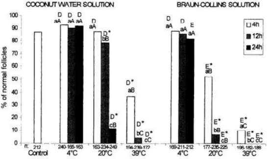
Figure 3 shows the effect of temperature and time of storage on the percentage of morphologically normal preantral follicles stored in coconut water or in Braun-Collins solution. We observed that storage of preantral follicles in Braun-Collins solution at 20 ° or 39°C at all incubation times tested, decreased significantly the percentage of normal follicles when compared with the control. In contrast, the percentage of normal follicles after storage in BraunCollins solution at 4°C for up to 24 h was not significantly affected in relation to control values (P > 0.05). Similar results were obtained with the use of coconut water, except for the
treatment in which the ovarian fragment was stored at 20°C for 4 h, this treatment presented percentages of normal follicles similar to those obtained in the control (P > 0.05). Comparisons between coconut water and Braun-Collins solution at the same temperature and incubation time showed a significantly higher percentage of normal follicles after storage in coconut water at 4°C for 24 h, 20°C (all incubation times) and at 39°C for 4 and 12 h. After storage in both solutions at 39°C for 24 h we found no normal follicles (Figure 3).
There was no effect of incubation time on the percentage of normal follicles, using both solutions at 4°C. However, in the fragments stored at 20 ° or 39°C in both solutions, there was a progressive reduction of percentage of normal follicles with the increase of incubation time (P < 0.05). With regard to the effect of temperature at the same incubation time, the results showed that for the Braun-Collins solution there was a significant effect of temperature on the percentage of normal follicles at all incubation times tested, with a progressive reduction of the percentage of normal follicles with the increase of temperature from 4’ to 39°C. Similar results were obtained for the coconut water except for the storage time of 4 h, at which the temperature of 20°C did not decrease the percentage of normal follicles when compared with storage at 4°C.
Figure 3. Effect of temperature and storage time on the percentage of normal follicles preserved in coconut water or Braun-Collins solution. n: number of preantral follicles analyzed per treatment. h: hours of incubation. * - Differs significantly from control. a, b, c - Different letters at the same preservation temperature show significant difference (P < 0.05). A, B, C - Different letters at the same preservation time show significant difference (P < 0.05). D, E - Different letters show significant difference among solutions for the same temperature and incubation time (P < 0.05).
Distribution of Follicular Degeneration Types in the Control and Other Treatments Figure 4 shows the distribution of degenerated Type 1 and 2 preantral follicles in the control and after storage in the different treatments: in coconut water (Figure 4A) and in BraunCollins
solution (Figure 4B). There was a predominance of degenerated Type 1 in the control as well as in tiagments stored in both solutions at 4°C at all incubation times tested, except in the fragments stored in Braun-Collins solution for 24 h. Degenerated Type 2 follicles were not observed in the control and after storage in both solutions at 4°C for 4 h. In contrast. at 20o and 39°C a significant predominance of degenerated Type 2 follicles was observed. In both solutions, a significantly lower percentage of degenerated Type 1 follicles compared with the control was observed at 20 ° and 39°C at all incubation times. At 4°C significant differences were not observed. A significantly higher percentage of degenerated Type 2 follicles compared with control values was observed in follicles kept in Braun-Collins solution at 4’C for 12 or 24 h, and at 20 ° and 39°C at all incubation times. Similar results were observed for coconut water, except in follicles stored at 4’C for 24 h.
Figure 4. Percentage distribution of the degenerated preantral follicles Type 1 and 2, from control and after preservation in coconut water (A) and Braun-Collins solution (B). * - denotes a significant difference of degeneration types within each treatment (P < 0.05). a, b - different letters show significant differences between the percentage of degenerated Type 1 follicles (Deg Tl F) found in different treatments and in control (P < 0.05). A, B - different letters show significant differences between the percentage of degenerated Type 2 follicles (Deg T2 F) found in different treatments and in control (P < 0.05)
Values of pH and Osmolarity of Coconut Water Solution and Braun-Collins Solution
with Preservation Time
The Figure 5 shows the mean values of pH (A) and osmolarity (B) of coconut water and Braun-Collins solution in the control (fresh medium) and after storage of ovarian fragments. The mean values of pH and osmolarity in the l?esh media (before the addition of the tkagment) were 6.62*0.071 and 293.2k5.23 mOsm/L for coconut water solution and 7.5*0.084 and 367.8h3.96 mOsm/L for Braun-Collins solution. The storage of ovarian fragments at 4°C for up to 24 h and at 20°C for 4 h did not result in significant changes of pH in both solutions. In contrast, alter preservation at 20°C for 12 and 24 h or at 39’C (all incubation times) there was a significant decrease of pH when compared with i?esh medium. In the ovarian tkagments stored in both solutions at 39°C for 4 or 12 h there was a significant increase of the osmolarity when compared with control values. In contrast, after storage of kagments in Braun-Collins solution at 20°C for 4 h there was significant decrease of the osmolarity when compared to the control.
Figure 5. The pH (A) and osmolarity (B) of coconut water and Braun-Collins solution along preservation times. h: hours of incubation. * - shows a significant difference in relation to the fresh medium (P < 0.05).
Ultrastructural Analysis of Caprine Preantral Follicles Preserved In Vitro
To better evaluate follicular morphology, ultrastructural analysis was performed using preantral follicles from the control treatment as well as from the treatment that did not differ from the control (i.e., ovarian fragments maintained in both solutions at 4°C [longer preservation time] and in coconut water solution at 20°C for 4 h). The preantral follicles stored in Braun-Collins solution at 4°C for 24 h (Figure 6A) or in coconut water at 20°C for 4 h (data not shown) were considered morphologically normal after staining semi-thin sections with toluidine blue, but ultrastructural analysis showed a great number of vacuoles in the ooplasm varying greatly in size. Many vacuoles are confluent and their accumulation appears to be associated with rupture of the nuclear membrane (Figure 6A). This enormous number of vacuoles was not observed in the control (Figure 6B). However, ultrastructural analysis of preantral follicles cold-stored at 4°C in Braun-Collins solution for up to 12 h (Figure 6C) or in coconut water for up to 24 h (Figure 6D) showed the integrity of the oocyte, the granulosa cells, and the basement membrane, thus confirming the results obtained by classical histology
DISCUSSION
This work shows for the first time that goat preantral follicles can be stored successfully for long time in coconut water solution or Braun-Collins solution. However, the effectiveness of each preservation solution is dependent on the temperature and storage time.
Histological analysis of fresh preantral follicles (control) showed that in vivo a pycnotic nucleus of the oocyte is the first sign of degeneration. This kind of degeneration also predominates after preservation at 4°C. Similar results were also observed after analysis of fresh caprine (3,25) and ovine (23) or cold-stored feline preantral follicles at 4°C (37). Jorio et al. (23) described that degeneration of the oocyte is the mode of atresia more frequently observed in preantral follicles. Hirshtield (19) also reported that in secondary follicles, between 75 pm and 150 pm of diameter, oocyte degeneration was usually the only sign of atresia. In vitro studies have shown that in certain preantral follicles the oocyte degenerates or completely disappears while granulosa cells appear healthy and continue to proliferate, showing that the oocyte is much more sensitive to degenerative events than granulosa cells (6, 14). In contrast, in the treatments where the ovarian fragments were stored at 20 ° or 39’C, a significantly higher percentage of preantral follicles with degenerated oocyte and granulosa cells were observed. These degenerated follicles showed a retracted oocyte with or without a pycnotic nucleus, with the granulosa cells exhibiting disorganization and low cellular density,
probably because they were enlarged in volume and the oocyte was retracted. It is important to note that in this study no pycnotic bodies in granulosa cells were observed, This result may be due to the short incubation time to which the ovarian fragments were submitted. Moreover, according to Ingran (21) the pycnosis during atresia is a process of unknown duration. Cahill et al. (7) and Jorio et al. (23) have described that while the pycnosis of granulosa cells occurs in antral follicles it is almost absent in preantral follicles.
The storage of ovarian fragments at 4’C in both solutions keeps the percentage of normal preantral follicles equivalent to control. The preservation at 4°C provided higher rates of reduction of cellular metabolism, consequently minimizing the metabolic need and thus increasing the resistance of follicles to the reduction of nutrients and oxygen during preservation in vitro (30). Studies involving goat preantral follicles stored in vitro are not currently available.
Figure 6. Electron micrographs of an initial degeneration of an apparently normal preantral follicle stored at 4°C for 24 hours in Braun-Collins solution (x 5000 - A), showing vacuolated cytoplasm oocyte (arrow marks nuclear membrane rupture); and morphologically normal follicle Tom control (x 2600 - B) and after storage at 4°C for 12 hours in Braun-Collins solution (x 5000 - C) or for 24 hours in coconut water (x 3800 - D). 0: oocyte; Nu: nucleus of oocyte; V: vacuoles. M: mitochondria and GC: granulosa cells. Bars = 2pm.
However, bovine cumulus oocyte complex has being preserved in situ at 4° C for up to 24 h, although the production of blastocysts after preservation was unsuccessful (38). The low percentage of blastocysts may be due the great sensitivity of microtubules of the meiotic spindles of oocytes to cooling (2, 29). In these studies. the quality of preantral follicles was not evaluated.
The oocytes enclosed in preantral follicles may be less susceptible to microtubule disruption, because most of the microtubular systems remain unorganized, and the chromatin is in a condensed form protected by the nuclear membrane (26).
The present study shows that preservation at 20° or 39°C significantly reduced the percentage of morphologically normal preantral follicles when compared to the control. In contrast, the preservation of bovine oocyte cumulus complexes in situ using different solutions (i.e. at 25’C for up to 11 h in PBS [38], at 21’C for up to 24 h in saline [33], at 37°C for up to 4 h in PBS [38] or at 38.5° C for up to 6 h in TCM199 [36]) neither reduced the fertilization ability of oocytes nor the capacity of early embryos to develop into blastocysts. This success may be due to the lesser sensitivity of microtubules of the oocyte at temperatures of 20° or 39°C (2). However, preantral follicles seem to be more sensitive to storage at these temperatures. These follicles are quiescent or in early growth, possessing low nutrient reserves and may thus be more sensitive to adverse conditions in vitro. The normal (39°C) or subnormal (20%‘) metabolism associated with low oxygen tension in vitro could result in a higher rate of follicular degeneration found in the treatments where the ovarian fragments were stored at 20°C and 39°C. Jennings et al. (22) suggested that changes in the cellular membrane permeability, induced by lack of oxygen, caused changes at a level of intracellular Na+, K+, and Cl, which were associated with changes in the distribution of Ca ° and increase of intracellular water. This may lead to increased cellular volume and consequently cellular degeneration. Degenerated cells can exhibit rupture of cellular membranes, which induces release of acid contents within the dead cell (13). The cellular death during preservation at 20°C and 39°C could have provoked an alteration in the values of pH and osmolarity of the media.
The coconut water solution was more effective than Braun-Collins solution in the preservation of goat preantral follicles at 4°C for 24 h, at 20°C (for all incubation times) and at 39°C for 4 and 12 h. While coconut water solution is lightly acidic, this medium is rich in nutrients (24) isosmotic, and has been successfully used for semen preservation (17, 27, 28, 35). Coconut water solution was also as effective as TCM 199 during oocyte maturation (4) and embryo culture (5). The Braun-Collins solution at 4°C has been successfully used in preservation of lung (16), liver (l), kidney (32) and heart (10) tissue. The Braun-Collins solution has as its principle hypothermic preservation in hyperosmotic solution, followed by a low
cellular dehydration, avoiding consequent cell swelling. At higher temperatures, our results were similar to those obtained by Savioz et al. (32) with kidney preservation at 14’C. These authors showed a low effectiveness of Brats-Collins solution when it was not used at 4°C. Davey and Skegg (9) showed that after 2 h of incubation at 26°C in hyperosmotic solution kidney cells recovered normal volume and swelled. The early cellular dehydration associated with normal (39°C) or subnormal (20°C) metabolism may have caused high rates of follicular degeneration found in the treatments where the fragments were stored at 20°C or 39°C in Braun-Collins solution under our conditions.
The transmission electron microscopy revealed the presence of numerous small vacuoles in the ooplasm of preantral follicles after preservation in coconut water at 20°C for 4 h or in BraunCollins solution at 4’C for 24 h. These vacuoles were not detected afler staining with toluidine blue. According to Hay et al. (18), the cytoplasmic vacuoles are a characteristic sign of early atresia in granulosa cells and represent endoplasmic reticulum swelling. On the other hand, these vacuoles may be altered mitochondria, as observed by Fuku et al. (15) in cryopreserved bovine oocytes. In this work, abnormal mitochondria exhibited extensive vacuohzation, disappearance of most of the cristae, and significant damage to the mitochondrial membrane. Moreover, these authors suggest that the migration and fusion of vacuoles along the oolemma may be a result of exposure to low temperatures (1.5). In contrast, preantral follicles preserved at 4°C in coconut water for up to 24 h or in Braun-Collins solution for up to 12 h were normal by ultrastructural analysis. These results showed that ultrastructural analysis is very important to evaluate the quality of preantral follicles after preservation.
In conclusion, our study shows for the first time that goat preantral follicles may be stored successfully at 4°C in coconut water solution for up to 24 h or in Braun-Collins solution for up to 12 h. These storage conditions are of obvious importance for the maintainance of follicular morphology during transportation of ovaries to the laboratory, which is necessary to provide healthy oocytes for in vitro growth and maturation, as well as for the formation of banks of cryopreserved oocytes from valuable animals.
REFERENCES
[1] Adam R, Astarcioglu I, Raccuia JS, Ducot B, Reynes M, Bismuth H. Beneficial effects of Eurocollins as aortic flush for the procurement of human lives. Transplantation 1996;61:705-709.
[2] Aman RR, Parks JE. Effects of cooling and rewarming on the meiotic spindle and chromosomes of in vitro-matured bovine oocytes. Biol Reprod 1994; 50: 103- 110.
[3] Bezerra MB, Rondina D, Lima AKF, Oliveira LC, Cecchi R, Lucci CM, Giorgetti A, Figueiredo JR. Aspectos quantitativos e qualitativos da foliculogCnese na fase prenatal na especie caprina. Ciencia Animal 1998; 8:47-56.
[4] Blume H, Vale Filho-VR, Marques Jr AP, Saturnino HM Avaliqao da água de coco na maturação de oocitos bovinos. Rev Bras Reprod Anim 1997; 21:72-75.
[5] Blume H, Vale Filho VR, Marques Jr AP, Saturnino HM. Use da agua de coca no cultivo de embrioes bovinos. Rev Bras Reprod Anim 1997; 21:78-81.
[6] Braw-Tal R, Yossefi S. Studies in vivo and in vitro on the initiation of follicle growth in the bovine ovary. JReprod Fertill997; 109:165-171.
[7] Cahill LP, Mariana JC, Mauleon P. Total follicular populations in ewes of high and low ovulation rates. J Reprod Fertil 1979; 55:27-36.
[8] Carroll J, Whittingham DG, Wood MJ, Telfer E, Gosden RG. Extra-ovarian production of mature viable mouse oocytes from frozen primary follicles. J Reprod Fertil 1990; 90:321- 327. [9]Davey KJ, Skegg DCG. The effects of high concentrations of an electrolyte on the swelling of non-metabolizing tissue slices, J Physioll971; 212:641-653.
[10] Demmy TL, Biddle JS, Bennett LE, Walls JT, Schmaltz RA, Curtis JJ. Organ preservation solutions in heart transplantation - Patterns of usage and related survival. Transplantation 1997; 63:262-269.
[11] Eppig JJ, O’Brien MJ. Development in vitro of mouse oocytes from primordial follicles. Biol Reprod 1996; 54: 197-207.
[12] Eppig JJ. Schroeder AC. Capacity of mouse oocytes from preantral follicles to undergo embryogenesis and develop to live young after growth, maturation and fertilization in vitro. Biol Reprod 1989; 41:268-276.
[13] Farber JL. Membrane injury and calcium homeostasis in the pathogenesis of coagulative necrosis. Lab Invest 1982; 47:114-123
[14] Figueiredo JR, Hulshof SCJ, Van den Hurk R, Nusgens B, Bevers MM, Ectors FJ, Beckers JF. Preservation of oocyte and granulosa cell morphology in bovine preantral follicles cultured in vitro. Theriogenology 1994; 41:1333-1346.
[15] Fuku E, Xia L, Downey BR. Ultrastructural changes in bovine oocytes cryopreserved by vitrification. Cryobiology 1995, 32:139-156.
[16] Fukuse T, Hirata T, Ueda M, Hitomi S, Wada, H. Effects of Euro-Collins, University of Wisconsin, and new extracellular-type trehalase-containing Kyoto Solutions in an ex vivo rat lung preservation model. Transplantation 1996; 62:1212-1217.
[17] Guerra FFA, Nunes JF. Fertilidade in vivo e avalia@o in vitro do dmen ovino resfiiado e conservado em agua de coca por 72 horas. Rev Bras Reprd Anim 1999; 23:287-289.
[18] Hay MF, Cran DG, Moor RM. Structural changes occurring during atresia in sheep ovarian follicles. Cell Tiss Res 1976; 169: 515-529.
[19] Hirshfield AN. Compensatory ovarian hypertrophy in the long term hemicastrate rat: size distribution of growing and atretic follicles. Biol Reprod 1983; 28:271-278.
[20] Hulshof SCJ, Figueiredo JR, Beckers JF, Bevers MM, Van den Hurk R. Isolation and
characterization of preantral follicles from foetal bovine ovaries. Vet Quartely 1994; 2:78- 80. [21]Ingram DL. Atresia. In: Zuckerman S (ed), The Ovary. New York: Academic Press, 1962; 247-273.
[22] Jennings RB, Ganote CE, Reimer KA. Ischemic tissue injury. Amer J Path01 1975; 81: 179- 198.
[23] Jorio A, Mariana JC, Lahlou-Kassi A. Development of the population of ovarian follicles during the prepubertal period in D’man and Timahdite sheep. Anim Reprod Sci 1991; 26:239-250.
[24] Laguna LE, Nunes JF. Avaliação fisico-química da água de coco proveniente de frutos das variedades da praia e anão. Rev Bras Reprod Anim 1997; 2 1: 156.
[25] Lucci CM, Amorim CA, Rodrigues APR, Figueiredo JR, Bfio SN, Silva JRV, Goncalves PBD. Study of preantral follicles population in situ and after mechanical isolation i?om caprine ovaries at different reproductive stages. Anim Reprod Sci 1999; 56:223-236.
[26] Matson BA, Albertini DF. Oogenesis: Chromatin and microtubule dynamics during meiotic prophase. Mol Reprod Devell990; 25:374-383.
[27] Nunes JF. Utilização da água de coca coma diluidor do sêmen de animais domésticos e do homem Rev Bras Reprod Anim 1998; 22:109-112.
[28] Nunes JF, Salgueiro CCM. Utilização da água de coca coma diluídor do sêmen de caprinos e ovinos. Rev Cient Prod Anim 1999; 1: 17-26.
[29] Pickering SJ, Braude PR, Johnson MH, Cant A, Currie J. Transient cooling to room
temperature can cause irreversible disruption of the meiotic spindle in the human oocyte. Fert Steril 1990; 54:102-108.
[30] Roy SK, Treaty BJ. Isolation and long-term culture of human preantral follicles. Fert Steril 1993; 59:783-790.
[31] Saumande J. La folliculogCnese chez les ruminants. Ret M&l Vet 1991; 167:205-218. [32] Savioz D, Bolle JF, Graf JD, Jeanjacquot A, Savioz M, Dietler G, Favre H, Leski M, Morel D, Morel P. Kinetics of cellular viability in warm versus cold ischemia conditions of kidney preservation. Transplantation 1996; 62:414-417.
[33] Schemthaner W, Schmoll F, Brem G, Schellander K. Storing bovine ovaries for 24 hours between 15° and 21°C does not influence in vitro production of blastocysts. Theriogenology 1997; 47:297 abstr.
[34] Solano R, Armas R, Pupa CA, Castro FO. Short term preservation of intrafollicular oocytes at 4°C. Theriogenology 1994; 41:299 abstr.
[35]Toniolli R, Mesquita DSM, Cavalcante SG. Avaliaqb in vitro do .sêmen suíno diluido em BTS e na água de coco in natura e estabilizada. Rev Bras Reprod Anim 1998; 22:198-201
[36] Twagiramungu H, Morm N, Guilbault LA, Sirard MA, Bousquet D. Media and time of oocytes transport influence their developmental competence for in vitro production of bovine embryos. Theriogenology 1998; 49:299 abstr
[37]Wood TC, Montali RI, Wildt DE. Follicle-oocyte atresia and temporal taphonomy in coldstored domestic cat ovaries. Mol Reprod Devel1997; 46:190-200.
[38]Yang NS, Lu RI-I, Gordon I. In vitro fertilization (IVF) and culture (IVC) of bovine oocytes Tom stored ovaries. Theriogenology 1990; 33:352 abstr.