U
NIVERSIDADE DE
L
ISBOA
F
ACULDADE DE
C
IÊNCIAS
D
EPARTAMENTO DE
B
IOLOGIA
V
EGETAL
IDENTIFICATION
OF
NF-
K
B
AND
MYC
BINDING
MOTIFS
IN
HERPESVIRUS
PROTEIN
ORF73
Sofia Isabel Arriaga Mimoso Cerqueira
M
ESTRADO EM
M
ICROBIOLOGIA
A
PLICADA
U
NIVERSIDADE DE
L
ISBOA
F
ACULDADE DE
C
IÊNCIAS
D
EPARTAMENTO DE
B
IOLOGIA
V
EGETAL
IDENTIFICATION
OF
NF-
K
B
AND
MYC
BINDING
MOTIFS
IN
HERPESVIRUS
PROTEIN
ORF73
Dissertação orientada pelo Prof. Doutor João Pedro Simas (IMM)
e pela Prof. Doutora Maria Filomena Caeiro (FCUL)
Sofia Isabel Arriaga Mimoso Cerqueira
M
ESTRADO EM
M
ICROBIOLOGIA
A
PLICADA
IDENTIFICATION
OF
NF-
K
B
AND
MYC
BINDING
MOTIFS
IN
HERPESVIRUS
PROTEIN
ORF73
Sofia Isabel Arriaga Mimoso Cerqueira
M
ASTER
T
HESIS
2011
This thesis was fully performed at the Viral Pathogenesis Unit of Instituto de
Medicina Molecular (IMM) under the direct supervision of Prof. Doutor João Pedro
Simas.
Prof. Doutora Maria Filomena Caeiro was the internal supervisor designated in the
scope of the Master in Applied Microbiology of the Faculty of Sciences of the
University of Lisbon.
iii
ACKNOWLEDGEMENTS
First, I would like to thank my supervisor at Instituto de Medicina Molecular, Prof. Doutor João Pedro Simas, for giving me the opportunity to develop my project in his laboratory, and for showing interest in my results and concern about my well-being. His enthusiasm about science has really inspired me. I would also like to thank my internal supervisor at the Faculty of Sciences of the University of Lisbon, Prof. Doutora Maria Filomena Caeiro, for her advice and for always encourage me. Her questions about my work definitely influenced the way I wrote this thesis. I also thank the Faculty of Sciences of the University of Lisbon for accepting me as a master student, and Instituto de Medicina Molecular for providing adequate conditions for the development of my project.
I would like to give my special thanks to Lénia Rodrigues, from whom I learned not only how to perform my experiments and interpret my results, but also how work and friendship can be partners. Her support and, above all, her friendship, were very important throughout this year. I also wish to express my gratitude to all the other members of the Viral Pathogenesis Unit, who kindly welcomed me and helped me whenever I needed.
Further thanks should go to Prof. Doutor Rogério Tenreiro, coordinator of the Master in Applied Microbiology, for his patience and willingness, but also for being a demanding teacher, who made me want to be a better apprentice scientist.
Agradeço à minha família, por todo o apoio e carinho demonstrados, e por estar sempre presente na minha vida. Em especial, agradeço à minha irmã, pelo companheirismo e amizade, e à minha mãe, a quem devo aquilo que sou.
Gostaria ainda de agradecer aos meus amigos, Catarina, Elisa, Mariana, Sara, Vanessa, Henrique, Rui, José e Pedro, companheiros de aventuras dos últimos cinco anos. À Nádia, Miguel e Rita, com quem partilho alegrias e tristezas desde criança.
Finalmente, um agradecimento especial à Professora Aida Duarte, com quem aprendi a gostar de investigação, e a todos os membros do Laboratório 125 da FFUL.
iv
RESUMO
O herpesvírus murídeo tipo 4, MuHV-4, é um gama-herpesvírus e, enquanto tal, estabelece infecções latentes em tecidos linfóides. Durante a infecção latente, o genoma de MuHV-4 persiste sob a forma de um epissoma no núcleo das células latentemente infectadas, e várias proteínas são expressas, entre as quais a proteína ORF73. Além do seu papel putativo na manutenção do epissoma viral, ORF73 modula os factores de transcrição celulares NF-kB e MYC, através do recrutamento das proteínas celulares Elonguina C e Culina 5, o que leva à formação do complexo de ligase de ubiquitina EC5S
ORF73 (ElonguinaC–Culina5–SOCS). O complexo EC 5S
ORF73
interage com p65/RelA, um dos membros da família NF-kB, promovendo a sua poli-ubiquitinação e, consequentemente, a sua degradação pelo proteossoma. O complexo EC5S
ORF73
promove ainda a poli-ubiquitinação de MYC, o que resulta na estabilização desta proteína em vez da sua degradação, como se verifica para p65/RelA. Deste modo, ORF73 tem efeitos contrários sobre NF-kB e MYC, inibindo a actividade transcricional de NF-kB, mas activando a actividade transcricional de MYC. Recentemente, foi identificado um motivo na proteína ORF73, o motivo SOCS-box (resíduos 199–215), que é responsável pelo recrutamento das proteínas Elonguina C e Culina 5 e, consequentemente, pela formação do complexo EC5S
ORF73
. A delecção funcional do motivo SOCS-box de ORF73, originando a proteína mutante ORF73–SOCS, impede a proteína viral de mediar tanto a poli-ubiquitinação de p65/RelA como a poli-ubiquitinação de MYC, comprometendo simultaneamente a sua capacidade de inibir a actividade transcricional de NF-kB e de activar a actividade transcricional de MYC, embora o mutante ORF73–SOCS mantenha intacta a capacidade de se ligar a ambos os factores celulares. Esta mutação suprime ainda a expansão do vírus MuHV-4 em células B e impede a infecção persistente in vivo. No entanto, como a mutação do motivo SOCS-box impede ORF73 de inibir NF-kB e de activar MYC em simultâneo, não é possível distinguir, em termos de fenótipo de infecção latente, o fenótipo que é devido à inibição de NF-kB e o fenótipo que resulta da activação de MYC. Além disso, a possibilidade de ORF73 mediar a poli-ubiquitinação de outros alvos celulares, ainda não identificados, não deve ser excluída. Portanto, é importante identificar regiões da proteína ORF73 cuja mutação permita segregar as suas funções modulatórias sobre os substratos celulares. A mutação dos motivos de ORF73 envolvidos na ligação a p65/RelA e a MYC constitui uma boa alternativa à mutação do motivo SOCS-box. Contudo, estes motivos de ligação ao substrato ainda não foram identificados, tendo sido apenas demonstrado que a região N-terminal de ORF73 (resíduos 1–140) é indispensável para a interacção de ORF73 com p65/RelA e MYC. A identificação dos motivos de ligação ao substrato na proteína ORF73 será importante para, em futuras experiências, construir vírus recombinantes que sejam incapazes de interagir e, consequentemente, de modular a actividade transcricional de NF-kB e de MYC, em separado. Deste modo, será possível distinguir os efeitos da inibição de NF-kB dos efeitos da activação de MYC sobre o fenótipo de MuHV-4, durante a infecção latente.
O principal objectivo deste projecto foi a identificação de resíduos de aminoácidos específicos na proteína ORF73 necessários para a sua ligação a MYC ou a p65/RelA. Para tal, quinze
v
proteínas ORF73 mutantes foram construídas, através de mutagénese por scanning de alanina na porção N-terminal de ORF73. Esta técnica consiste na substituição dos resíduos-alvo por resíduos de alanina, os quais permitem eliminar as cadeias laterais para além do carbono β, sem alterar a conformação da cadeia principal e sem impor efeitos electrostáticos extremos. A interacção dos mutantes de ORF73 com MYC e p65/RelA foi depois testada em experiências de co-imunoprecipitação. Os mutantes que interagiram fracamente com estas proteínas celulares foram sujeitos a ensaios funcionais, de modo a confirmar a diminuição da sua capacidade de modular MYC ou NF-kB. Deste modo, realizaram-se ensaios de gene repórter para testar se a perda de ligação de ORF73 a MYC ou a p65/RelA resultou na abolição da sua acção modulatória sobre a actividade transcricional destes factores celulares, e ainda ensaios de poli-ubiquitinação para investigar se essa perda de ligação se traduziu na perda da actividade de ligase de ubiquitina de ORF73. A capacidade destes mutantes para recrutarem os componentes do complexo EC5S
ORF73
também foi avaliada, através de experiências de co-imunoprecipitação com as proteínas Elonguina C e Culina 5.
As experiências de co-imunoprecipitação com MYC ou p65/RelA e os mutantes de ORF73 que foram construídos permitiram identificar dois mutantes com uma capacidade comprometida para interagirem com estes factores celulares. O mutante ORF73141KKY143, com os resíduos 141KKY143 substituídos por alaninas,demonstrou uma severa diminuição na sua capacidade de
interagir com MYC e p65/RelA, sugerindo que os resíduos 141KKY143 são importantes para a
ligação de ORF73 a MYC e a p65/RelA. Por outro lado, o mutante ORF73138PLP140, com os
resíduos 138PLP140 substituídos por alaninas,revelou uma diminuição parcial da sua capacidade de
interagir com MYC, tendo permanecido intacta a sua capacidade de interagir com p65/RelA, o que indica que os resíduos 138PLP140 têm um papel na ligação de ORF73 a MYC, mas não na ligação a
p65/RelA. Contudo, os mutantes ORF73141KKY143 e ORF73138PLP140 comportaram-se da mesma
forma nos ensaios de gene repórter, sendo incapazes de activar a actividade transcricional de MYC e apresentando uma diminuição parcial do seu efeito inibitório sobre NF-kB, o que indica que tanto a mutação dos resíduos 141KKY143 como a mutação dos resíduos 138PLP140 afectam a acção
modulatória de ORF73 sobre MYC e sobre NF-kB. Os resultados dos ensaios de poli-ubiquitinação mostraram estar de acordo com as observações dos ensaios de gene repórter, uma vez que ambos os mutantes foram incapazes de promover a poli-ubiquitinação de MYC e de p65/RelA, reforçando a ideia de que os resíduos 141KKY143 e 138PLP140 são importantes para a modulação de
MYC e de NF-kB pela proteína ORF73. De seguida, a capacidade dos mutantes ORF73141KKY143
e ORF73138PLP140 para recrutarem a Elonguina C e a Culina 5, componentes do complexo
EC5SORF73, foi avaliada através de experiências de co-imunoprecipitação. Ambos os mutantes
recrutaram a Culina 5, mas foram incapazes de recrutar a Elonguina C, comprometendo assim a formação do complexo EC5S
ORF73
e, consequentemente, suprimindo a função de ligase de ubiquitina de ORF73.
O facto de os mutantes ORF73141KKY143 e ORF73138PLP140 não conseguirem recrutar a
Elonguina C, embora não tivessem mutações no motivo SOCS-box, sugere que as mutações introduzidas nos resíduos 141KKY143 e 138PLP140 tiveram um impacto severo na estrutura
vi
tridimensional de ORF73, possivelmente afectando outras funções da proteína viral, como o seu papel putativo na manutenção do epissoma. Este trabalho demonstrou ainda que a actividade de ligase de ubiquitina dos mutantes ORF73141KKY143 e ORF73138PLP140 estava comprometida, à
semelhança do que se verificou com o mutante ORF73–SOCS, acima referido. Deste modo, não será possível clarificar, através da utilização destes mutantes em estudos in vivo, a contribuição independente da activação de MYC e da inibição de NF-kB pela proteína viral ORF73, durante a infecção latente de MuHV-4. Portanto, é necessário desenvolver outras estratégias para identificar os motivos de ligação de ORF73 a MYC e a p65/RelA, de modo a tornar possível a construção de mutantes de ORF73 que sejam incapazes de modular cada uma destas proteínas celulares separadamente. A resolução da estrutura tridimensional de ORF73, através de técnicas de cristalografia, pode dar um importante contributo para estes estudos.
Palavras-chave: ORF73, NF-kB, MYC, motivo de ligação ao substrato, mutagénese por scanning
vii
ABSTRACT
Murid herpesvirus-4 (MuHV-4) establishes latent infections in lymphoid tissues. During latency, ORF73 protein is expressed. ORF73 modulates the cellular transcription factors NF-kB and MYC, by assembling an EC5S
ORF73
(ElonginC-Cullin5-SOCS) ubiquitin-ligase complex. EC5S
ORF73
interacts with NF-kB family member p65/RelA, promoting its poly-ubiquitination and subsequent proteasomal degradation. EC5S
ORF73
also mediates MYC poly-ubiquitination, but this promotes MYC stabilization. Thus, ORF73 inhibits NF-kB whereas it activates MYC transcriptional activity. SOCS-box motif in ORF73 recruits ElonginC and Cullin5, assembling EC5S
ORF73
complex. However, the motifs of ORF73 involved in binding to p65/RelA and MYC are unidentified. Identification of ORF73 substrate-binding motifs is important to segregate NF-kB inhibition effects from MYC activation effects in MuHV-4 phenotype, during latency.
The aim of this project was to identify specific amino acid residues, within N-terminus of ORF73, required for binding to MYC and p65/RelA. To this end, fifteen ORF73 mutants were generated by alanine-scanning mutagenesis, and tested on their ability to interact with those cellular substrates. ORF73141KKY143 mutant showed impaired ability to interact with MYC and
p65/RelA, while ORF73138PLP140 mutant exhibited partially impaired ability to interact with MYC
and intact ability to interact with p65/RelA. ORF73141KKY143 and ORF73138PLP140 mutants were
unable to activate MYC transcriptional activity, had partially impaired inhibitory effect upon NF-kB, andwere unable to promote MYC and p65/RelA poly-ubiquitination. Both mutants recruited Cullin5, but were unable to recruit ElonginC, thereby compromising the assembly of EC5S
ORF73
complex and consequently preventing ORF73 to function as an ubiquitin-ligase. This suggested that mutations at 141KKY143 and 138PLP140 residues had severe consequences in ORF73 three
dimensional structure, possibly affecting several of its functions.
Altogether, this work showed that ORF73141KKY143 and ORF73138PLP140 mutants are not
useful to study the biological role of ORF73 modulatory function towards MYC and NF-kB. Hence, other strategies must be developed to identify MYC and p65/RelA binding motifs in ORF73.
viii
ABBREVIATIONS
Cul5 Cullin5
DMEM Dulbecco’s modified Eagle’s medium
EBNA1 Epstein-Barr virus nuclear antigen 1
EBV Epstein-Barr virus
ECS ElonginC–Culllin2/5–SOCS-box ubiquitin-ligase complex EloC ElonginC
FBS Foetal bovine serum
GC Germinal centre
HCMV Human cytomegalovirus
HEK Human embryonic kidney
HHV-6 Human herpesvirus-6
HHV-7 Human herpesvirus-7
HHV-8 Human herpesvirus-8
HSV-1 Herpes simplex virus-1
HSV-2 Herpes simplex virus-2
IKK IkB kinase
KSHV Kaposi’s sarcoma-associated herpesvirus LANA Latency-associated nuclear antigen
MuHV-4 Murid herpesvirus-4
NF-kB Nuclear factor-kappa B
Ni-NTA Nickel-nitrilotriacetic acid
NLS Nuclear localisation signal
PBS Phosphate buffered saline
SCF Skp1–Cullin1–F-box ubiquitin-ligase complex
SDS-PAGE Sodium dodecyl sulfate-polyacrylamide gel electrophoresis
SOCS Suppressors of cytokine signalling
TNF Tumor necrosis factor
VZV Varicella-zoster virus
ix
TABLE OF CONTENTS
ACKNOWLEDGEMENTS ... iii RESUMO ... iv ASBTRACT ... vii ABBREVIATIONS ... viii TABLE OF CONTENTS ... ix1. INTRODUCTION ... 1
1.1. Herpesviruses ...1 1.1.1.Gamma-herpesviruses ...11.2. Kaposi’s sarcoma-associated herpesvirus ...1
1.2.1. KSHV latency-associated protein ...2
1.2.2.Pathogenesis studies ...2
1.3. Murid herpesvirus-4 ...2
1.3.1. MuHV-4 latency-associated protein ...3
1.3.1.1.Modulation of the NF-kB family member p65/RelA by ORF73 ...3
1.3.1.2. Modulation of MYC by ORF73 ...4
1.3.1.3. p65/RelA and MYC binding motifs in ORF73 ...5
2. OBJECTIVES ... 7
3. MATERIALS AND METHODS... 8
3.1. Plasmids ...8
3.2. Immunological reagents ...8
3.3. Cells and transfections ...8
3.4. Immunoprecipitations ...9
3.5. Reporter gene assays ...9
3.6. Immunofluorescence analysis ... 10
3.7. Poly-ubiquitination assays ... 10
4. RESULTS ... 12
4.1. Alanine-scanning mutagenesis: ORF73141KKY143 weakly co-immunoprecipitates MYC and p65/RelA ... 12
4.2. Mutation at 141KKY143 in ORF73 impairs its ability to activate MYC transcriptional activity ... 16
4.3. Mutation at 141KKY143 in ORF73 partially impairs its inhibitory effect on NF-kB transcriptional activity ... 18
x
4.5. Mutation at 138PLP140 in ORF73 partially impairs its ability to co-immunoprecipitate MYC ... 21
4.6. Mutation at 138PLP140 in ORF73 impairs its ability to activate MYC transcriptional activity... 24
4.7. Mutation at 138PLP140 in ORF73 does not affect its ability to co-immunoprecipitate p65/RelA 26 4.8. Mutation at 138PLP140 in ORF73 partially impairs its inhibitory effect on NF-kB transcriptional activity ... 27
4.9. Mutation at 141KKY143 or 138PLP140 in ORF73 compromises its ability to promote MYC and p65/RelA poly-ubiquitination ... 28
4.10. Mutation at 141KKY143 or 138PLP140 in ORF73 compromises its ability to assemble an E3 ubiquitin-ligase complex ... 31
5. DISCUSSION ... 34
6. CONCLUSION ... 36
1
1. INTRODUCTION
1.1. Herpesviruses
Herpesviridae is a family of enveloped viruses that have large double-stranded DNA genomes
(>100 kb) with a vast coding capacity. Herpesviruses have biphasic life cycles that consist of a lytic and a latent phase; to achieve this, they have alternative programs of gene expression. During latency, only a limited subset of genes is expressed and viral particles are not produced. Thus, the virus escapes the host immune control and maintains its genome in the infected cells (Wu et al., 2010). In fact, the hallmark of herpesvirus infections is the establishment of lifelong latent infections that can reactivate, causing one or more rounds of disease (Flint et al., 2004). Herpesviruses account for many persistent mammalian infections, and they are well adapted to their hosts, as demonstrated by their high human infection rates in most populations. Although many herpesvirus infections are asymptomatic or cause a mild illness, they can be devastating in immunocompromised hosts (Wu et al., 2010).
The family Herpesviridae is divided into three subfamilies: Alphaherpesvirinae,
Betaherpesvirinae and Gammaherpesvirinae (Flint et al., 2004). Alpha-herpesviruses establish
latency mainly in neurons, beta-herpesviruses in monocytes, and gamma-herpesviruses in lymphocytes (Stevenson et al., 2009). Humans are the natural hosts of eight herpesviruses: herpes simplex virus (HSV)-1, HSV-2, and varicella-zoster virus (VZV) (Alphaherpesvirinae); human cytomegalovirus (HCMV), human herpesvirus (HHV)-6, and HHV-7 (Betaherpesvirinae); Epstein-Barr virus (EBV), and Kaposi’s sarcoma-associated herpesvirus (KSHV), also known as HHV-8 (Gammaherpesvirinae) (Flint et al., 2004; Wu et al., 2010).
1.1.1.
Gamma-herpesviruses
Gamma-herpesviruses infect a variety of mammalian species, from mice to man. However, each gamma-herpesvirus is host-range specific. These viruses establish latent infections within lymphocytes, induce lymphoproliferative disease and are associated with a variety of lymphoid and non-lymphoid cell tumours (Nash et al., 2001; Simas & Efstathiou, 1998).
Gamma-herpesviruses are divided into gamma-1-herpesviruses and gamma-2-herpesviruses, according to their genomic organization and gene content. EBV belongs to the gamma-1 subgroup (Simas & Efstathiou, 1998), while KSHV is included in the gamma-2 subgroup (Chang et al., 1994).
1.2. Kaposi’s sarcoma-associated herpesvirus
KSHV infection is uncommon (~3%) in Western countries, but it has a high prevalence (ranging from 20 to 80%) in sub-Saharan Africa (Dedicoat & Newton, 2003). KSHV has an etiologic role in Kaposi’s sarcoma, and also in primary effusion lymphoma and multicentric Castleman’s
2
disease (Chang et al., 1994; Jones et al., 1998; Soulier, et al., 1995). Although Kaposi’s sarcoma has low malignant potential in immunocompetent individuals, it can be lethal in immunocompromised hosts (Verma & Robertson, 2003) and it has become one of the most common AIDS malignancies worldwide (Cai et al., 2010; Wu et al., 2010). Therefore, it is essential to understand the mechanisms by which KSHV induces cellular proliferation, evades immune-mediated clearance and maintains viral genome during latency, in order to develop strategies that allow the control of KSHV-associated tumorigenesis.
1.2.1. KSHV latency-associated protein
During latent infection, KSHV expresses a latency-associated nuclear antigen (LANA or ORF73) (Dupin et al., 1999). KSHV genome persists as a multicopy, non-integrated, circular form (episome), in the nucleus of latently infected cells. LANA mediates episome persistence by tethering viral genomes to mitotic chromosomes, which allows the segregation of viral DNA to daughter nuclei, and thus episome persistence in proliferating cells (Ballestas et al., 1999; Decker
et al., 1996). Furthermore, LANA also regulates transcription and cell growth through interactions
with cellular proteins, including through E3 ubiquitin-ligase activity, which enables it to mediate the poly-ubiquitination of cellular targets, namely VHL and p53 proteins (Cai et al., 2006).
1.2.2.
Pathogenesis studies
In spite of the progress in molecular virology of KSHV, many aspects of its in vivo biology remain unknown. This is due to the fact that KSHV is a species-specific virus, so it does not infect laboratory mice or rats, limiting in vivo pathogenesis studies (Nash et al., 2001). For this reason, research has been done on the development of animal model systems to investigate the pathogenesis of KSHV infection. Infection of laboratory mice with murid herpesvirus-4 (MuHV-4) has proved to be a good model system (Simas & Efstathiou, 1998).
1.3. Murid herpesvirus-4
Murid herpesvirus-4 (MuHV-4; archetypal strain MHV-68) was originally isolated from bank voles (Clethrionomys glariolus) in Slovakia (Blaskovic et al., 1980), and it is widespread among rodent populations in Northern Europe (Simas & Efstathiou, 1998). MuHV-4, like KSHV, is a gamma-2-herpesvirus. Its genome consists of 118 kb of unique sequence DNA with a G+C content of 46%, flanked by a variable number of 1,2 kb terminal repeat regions (Efstathiou et al., 1990). It is estimated that MuHV-4 genome contains approximately 80 protein-coding open reading frames (ORFs), the majority of which are collinear and homologous to those of other gammaherpesviruses, namely KSHV (Virgin et al., 1997). Besides, several MuHV-4 genes are functionally homologous to cellular genes (Nash et al., 2001).
3
MuHV-4 establishes a lifelong latent infection in lymphoid tissues (Sunil-Chandra et al., 1992) and, like KSHV, its genome persists as a multicopy episome in latently infected cells. Latency is established predominantly in B cells (Marques et al., 2003), but can also be established in macrophages, dendritic cells and epithelial cells (Flaño et al., 2000; Stewart et al., 1998). MuHV-4 induces proliferation of latently infected B cells in germinal centre (GC)-like reactions, which allows the virus to gain access to the long-lived memory B cell pool, the major reservoir of MuHV-4 latency (Flaño et al., 2002; Stevenson, 2004; Stevenson et al., 2009).
1.3.1. MuHV-4 latency-associated protein
MuHV-4, like KSHV, encodes a latency-associated protein, which is the product of ORF73 gene. ORF73 protein of MuHV-4, with 314 amino acid residues, is homologous in sequence and function to ORF73 protein of KSHV (LANA) (Fowler et al., 2003). To easily distinguish these two proteins throughout this thesis, ORF73 of KSHV will be named LANA, while ORF73 of MuHV-4 will be simply denominated ORF73.
It has been shown that ORF73 is required for the establishment and maintenance of latency, particularly for in vitro maintenance of viral episome (Fowler et al., 2003). In addition, recent studies have shown that ORF73 regulates cellular transcription by modulating the cellular transcription factors NF-kB (Rodrigues et al., 2009) and MYC (Rodrigues et al., unpublished data). ORF73, like LANA, has E3 ubiquitin-ligase activity, promoting the poly-ubiquitination of cellular substrates and interfering with their turnover. The mechanisms by which ORF73 modulates NF-kB and MYC are described below.
Since ORF73 and LANA are functionally homologous, particularly in episome maintenance and E3 ubiquitin-ligase functions, and an excellent murine model of MuHV-4 latency exists (Simas & Efstathiou, 1998), ORF73 provides a potential model to investigate LANA in vivo. Moreover, recombinant genetics for MuHV-4 is robust and allows ready introduction of ORF73 mutations.
1.3.1.1.
Modulation of the NF-kB family member p65/RelA by ORF73
Nuclear factor-kappa B (NF-kB) is a family of ubiquitously expressed transcription factors. In mammalian cells, the NF-kB family comprises five members: p65/RelA, RelB, c-Rel, p105/p50 and p100/p52 (Blank et al., 1992). NF-kB family members can form homo- or heterodimers (Saccani et
al., 2003) and bind to specific DNA sequences (kB sites) in the promoter region of a variety of
genes, thereby modulating their transcription. NF-kB target genes are involved in critical biological functions, including immune response, inflammation, apoptosis, cell proliferation and differentiation (Baldwin, 2001; Ghosh et al., 1998). Deregulation of NF-kB signalling is associated with oncogenesis and cancer malignancies (Karin et al., 2002).
Under homoeostasis, NF-kB dimers are normally sequestered in the cytoplasm by the inhibitory IkB proteins. These proteins bind to NF-kB and mask its nuclear localization signal, thereby preventing nuclear uptake of NF-kB. In the presence of an extracellular stimulus, such as
4
tumor necrosis factor (TNF), an IkB kinase (IKK) complex is activated. IKK phosphorylates IkB molecules, leading to their poly-ubiquitination and proteasomal degradation. Free NF-kB is then translocated to the nucleus, where it binds to kB sites in DNA, activating target gene transcription (Karin & Ben-Neriah, 2000).
However, once the stimulus stops, NF-kB activity must be terminated. De novo expression of IkB molecules, whose genes are themselves NF-kB target genes, allows resynthesised IkB to enter the nucleus and to dissociate NF-kB dimers from kB sites, transporting NF-kB back to cytoplasm (Arenzana-Seisdedos et al., 1997). Furthermore, NF-kB activity can also be terminated through direct poly-ubiquitination and subsequent proteasomal degradation of promoter-bound p65/RelA (Saccani et al., 2004). This mechanism is regulated through the activation of E3 ubiquitin-ligase proteins, which accept ubiquitin from E2 ubiquitin-conjugating enzymes, and transfer it to the target protein (Weissman, 2001). The addition of successive molecules of ubiquitin to p65/RelA leads to its poly-ubiquitination; poly-ubiquitinated p65/RelA is then degraded by the proteasome. Traditionally, poly-ubiquitination has been viewed as a mechanism that targets proteins for degradation by the proteasome (Hershko & Ciechanover, 1998); however, it has been shown that poly-ubiquitination can also enhance the stability of target proteins (Popov et el., 2010), as will be described for MYC further on. The outcome of poly-ubiquitination depends on the kind of linkage between the individual ubiquitin molecules in a poly-ubiquitin chain.
E3 ubiquitin-ligase proteins are multisubunit complexes. The ECSSOCS1 (ElonginC–Culllin2/5– SOCS [suppressors of cytokine signalling]-box) E3 ubiquitin-ligase complex has been implicated in the poly-ubiquitination of p65/RelA (Ryo et al., 2003). In this complex, ElonginC bridges the scaffold proteins Cullin2 or Cullin5 to the SOCS-box-containing adaptor protein, the SOCS1 protein (Cai et al., 2006; Kamura et al., 2001; Kamura et al., 2004), which functions as the substrate recognition component of the ECSSOCS1 complex. Recently, it has been shown that the ORF73 viral protein has a SOCS-box motif (amino acid residues 199-215), which allows it to recruit ElonginC and Cullin5, thereby assembling an EC5SORF73 (ElonginC–Culllin5–SOCS-box) ubiquitin-ligase
complex (Rodrigues et al., 2009). EC5S ORF73
interacts with nuclear p65/RelA, promoting its poly-ubiquitination and subsequent proteasomal-dependent degradation, which in turn results in a strong termination of NF-kB activity. Therefore, ORF73 has an inhibitory effect upon NF-kB by promoting p65/RelA degradation.
1.3.1.2. Modulation of MYC by ORF73
MYC proteins include c-MYC, N-MYC and L-MYC, and are encoded by the MYC-family proto-oncogenes (Adhikary & Eilers, 2005; Eilers & Eisenman, 2008). MYC is essential in normal cell proliferation, growth, survival and differentiation. However, its deregulation can originate an enlarged pool of proliferating, self-renewing cells, which are vulnerable to further oncogenic events that accelerate tumorigenesis (Larsson & Henriksson, 2010). MYC is a global transcriptional regulator: it binds to approximately 10–15% of the genome and it can both activate and repress cellular transcription. Furthermore, MYC must form a heterodimer with Max in order to bind to DNA.
5
MYC-Max heterodimers bind to a consensus sequence (CACGTG), known as E-box (Meyer & Penn, 2008).
MYC is tightly regulated both at the transcriptional and post-transcriptional levels, including through poly-ubiquitination. It has been shown that the SCFβ-TrCP (Skp1–Cullin1–F-box) E3 ubiquitin-ligase complex mediates MYC poly-ubiquitination, thereby regulating its turnover. However, SCFβ-TrCP-mediated poly-ubiquitination enhances MYC stability, instead of targeting it to proteasomal degradation (Popov et al., 2010). In SCFβ-TrCP complex, β-TrCP is the adaptor protein that recognises the substrate. Recent studies suggest that ORF73 acts like β-TrCP protein, binding to MYC and promoting its poly-ubiquitination and subsequent stabilization (Rodrigues et al., unpublished data). Thus, ORF73 activates MYC transcriptional activity, which is in contrast with its inhibitory effect upon NF-kB. ORF73-mediated MYC ubiquitination, like p65/RelA poly-ubiquitination, involves the recruitment of ElonginC and Cullin5, through ORF73 SOCS-box motif, in order to assemble the EC5S
ORF73
ubiquitin-ligase complex.
1.3.1.3. p65/RelA and MYC binding motifs in ORF73
Rodrigues et al. have generated an ORF73 protein with four point mutations at its SOCS-box motif (ORF73-SOCS mutant), which result in disruption of SOCS-box and, consequently, in loss of ORF73 E3 ubiquitin-ligase activity (Rodrigues et al., 2009). In vivo studies with ORF73-SOCS mutant have shown that this functional deletion of SOCS-box motif in ORF73 abrogates its inhibitory effect upon NF-kB, suppresses MuHV-4 expansion in GC B cells, and prevents MuHV-4 persistent infection in mice, suggesting that inhibition of NF-kB signalling by ORF73 is essential for MuHV-4 latency. Recently, it has been shown that functional deletion of SOCS-box motif in ORF73 also abrogates its ability to activate MYC (Rodrigues et al., unpublished data). These findings indicate that mutations at SOCS-box disrupt not only the ability of ORF73 to terminate NF-kB activity, but also its ability to modulate MYC and even other possible cellular targets that remain to be identified. Therefore, mutations must be introduced at regions of ORF73 other than SOCS-box motif, in order to segregate the role of ORF73-mediated NF-kB inhibition and MYC activation in MuHV-4 latent infection, thereby clarifying the contribution of modulation of each transcription factor.
ORF73 protein is the substrate recognition component of the EC5S ORF73
complex, which promotes the poly-ubiquitination of p65/RelA and MYC. Therefore, ORF73 must have a motif that allows it to bind to p65/RelA, and a motif that allows it to bind to MYC. Since it has been shown that the SOCS-box is not required for binding of ORF73 to p65/RelA (Rodrigues et al., 2009) or MYC (Rodrigues et al., unpublished data), it is predicted that there are distinct functional motifs in ORF73: a motif implicated in the recruitment of ElonginC and Cullin5 (SOCS-box), a motif involved in p65/RelA interaction (p65/RelA binding motif), and a motif involved in MYC interaction (MYC binding motif). However, p65/RelA and MYC binding motifs in ORF73 remain to be identified. These substrate binding motifs may be either linear (specific amino-acid sequence) or structural (small local arrangement of secondary structure elements which then coalesce to form domains)
6
(Petsko & Ringe, 2004). Recent studies have shown that ORF73 interacts with p65/RelA and MYC through its proline-rich N-terminal region (amino acid residues 1-140) (Rodrigues et al., unpublished data). If linear motifs are involved in substrate binding, and the motif involved in p65/RelA binding is different from the motif required for MYC binding, then the mutation of specific residues within the N-terminus of ORF73 will abrogate its ability to interact and modulate either p65/RelA or MYC, separately. This will allow the construction of recombinant MuHV-4 viruses, with either p65/RelA or MYC binding-deficient ORF73. Since recombinant viruses will have disrupted ability to modulate NF-kB or MYC, separately, the phenotype analysis of these viruses upon infection of mice will segregate the biological significance of termination of NF-kB activity and activation of MYC, promoted by ORF73.
7
2. OBJECTIVES
The aim of this project was to identify specific amino acid residues within the N-terminus of ORF73 required for binding to p65/RelA or MYC. Identified motifs would be of extreme importance in future experiments, to construct recombinant viruses with disrupted ability to modulate NF-kB or MYC transcriptional activities, separately.
To achieve this, ORF73 proteins with point mutations were generated by alanine-scanning mutagenesis, and then tested on their ability to interact with p65/RelA and MYC. Binding-deficient ORF73 mutants were further characterised by functional assays to confirm their impaired ability to act upon NF-kB or MYC. Thus, reporter gene assays were performed to test if loss of binding of ORF73 to p65/RelA or MYC abrogated its ability to modulate transcriptional activity of NF-kB or MYC. Furthermore, poly-ubiquitination assays were performed to assess if loss of binding of ORF73 to these cellular substrates prevented ORF73 to function as an E3 ubiquitin-ligase.
8
3. MATERIALS AND METHODS
3.1. Plamids
pCMV-Myc construct encoding wild-type ORF73 (wtORF73) (pCMV-Myc-ORF73) was previously described (Rodrigues et al., 2009). p65/RelA expression plasmid and pC45 plasmid containing kB consensus sequences that drive the expression of firefly luciferase (NF-kB reporter plasmid) were also earlier described (Anrather et al., 1999; Winkler et al., 1996). MYC expression plasmid was kindly provided by Neil Perkins. pMYC-TA-Luc plasmid with six tandem copies of the E-box consensus sequence driving the expression of firefly luciferase (MYC reporter plasmid) was purchased from Clontech. Renilla plasmid was a kind gift by Xosé R. Bustelo. Histidine-tagged ubiquitin plasmid (His6-Ubiq) was kindly offered by Dr. D. Bohmann. Flag-tagged ElonginC and
Myc-tagged Cullin5 expression plasmids were kindly provided by Dr. E. Burstein and Dr. X.F. Yu, respectively.
pCMV-Myc constructs encoding ORF7311TTS13, ORF7321KRR23, ORF7335RRR37,
ORF7346PPP48, ORF7363PNP65, ORF7376TLP78, ORF7391PPS93, ORF73102SPD104,
ORF73121PPP123, ORF73134NPS136, ORF73141KKY143, ORF73141K, ORF73142K, ORF73143Y, and
ORF73138PLP140 mutants were generated by site-directed mutagenesis, using QuikChange II XL
Site-Directed Mutagenesis Kit (Agilent) according to the manufacturer’s instructions. The oligonucleotides designed to introduce alanine substitution mutations in pCMV-Myc-ORF73 constructs are listed in Table 1. All constructs were verified by DNA sequencing.
3.2. Immunological reagents
Anti-ORF73 serum obtained from rabbit was previously described (Rodrigues et al., 2009). Anti-p65, anti-EloC and anti-Cul5 antibodies were purchased from Santa Cruz Biotechnology. Mouse antibodies directed to Myc, HA and Flag epitopes were from Clontech, Covance and Sigma, respectively. Actin was detected with a rabbit anti-actin polyclonal antibody (Sigma). Horseradish peroxidase-conjugated secondary antibodies were from Amersham Biosciences. The fluorochrome-labelled secondary antibody used in immunofluorescence analysis (Alexa 594 anti-mouse) was from Jackson Immunoresearch.
3.3. Cells and transfections
Human embryonic kidney (HEK) 293T cells were cultured in Dulbecco’s modified Eagle’s medium (DMEM) supplemented with 10% foetal bovine serum (FBS), 2 mM glutamine and 100 U/ml penicillin–streptomycin. Transiently transfection of HEK 293T cells with plasmidic DNA was performed with Fugene 6 (Roche), according to the manufacturer’s instructions. In all transfections, an empty vector was used to normalise the total amount of plasmidic DNA.
9
3.4. Immunoprecipitations
For co-immunoprecipitation experiments with MYC and ORF73 mutants, or with p65/RelA and ORF73 mutants, HEK 293T cells (3x10⁶) were transiently transfected with expression plasmids encoding MYC (2 µg) or p65/RelA (2 µg), and wild-type or the mutant versions of ORF73 described above (2 µg). After 48 h in culture, cells were rinsed in ice-cold phosphate buffered saline (PBS) and disrupted in 500 µl of ice-cold lysis buffer containing 10 mM Tris–HCl (pH 7.5), 150 mM NaCl, 1% Triton X-100, 1 mM NaF, 100 mM Na3VO4 and a cocktail of protease inhibitors (Cϕmplete;
Roche). Lysates were clarified by centrifugation, and incubated with 1 µg of antibodies to ORF73 (anti-ORF73 serum) for 2 h at 4ºC. In p65/RelA experiments, lysates were precleared with protein G-conjugated Sepharose beads (GE Healthcare) for 1 h at 4ºC, prior to incubation with anti-ORF73 serum. Immunecomplexes were recovered by incubation with protein G-conjugated Sepharose beads (50 µl/sample) for 1 h at 4ºC. After three washes with ice-cold lysis buffer, proteins were eluted in reducing Laemmli’s buffer, resolved by sodium dodecyl sulfate-polyacrylamide gel electrophoresis (SDS-PAGE), transferred to nitrocellulose, and immunoblotted with the appropriate antibodies. In addition, representative aliquots of the total cellular lysates, not subjected to immunoprecipitation, were used to detect appropriate expression of MYC or p65/RelA, ORF73, and actin.
For co-immunoprecipitation experiments with ElonginC and ORF73 mutants, or with Cullin5 and ORF73 mutants, HEK 293T cells (3x10⁶) were transiently transfected with expression plasmids encoding ElonginC (2 µg) or Cullin5 (4 µg), and wild-type or mutant versions of ORF73 (2 µg). After 48 h in culture, cells were rinsed in ice-cold PBS and disrupted in 500 µl of ice-cold lysis buffer. Lysates were clarified by centrifugation and incubated with 1 µg of antibodies to either ElonginC or Cullin5 for 2 h at 4ºC. Immunecomplexes were recovered and processed as described above. Representative aliquots of the total cellular lysates, not subjected to immunoprecipitation, were used to detect appropriate expression of ElonginC or Cullin5, ORF73, and actin.
3.5. Reporter gene assays
For reporter gene assays with MYC, HEK 293T cells (2x10⁵) were transiently transfected with 500 ng of a firefly luciferase reporter plasmid (pMYC-TA-Luc), 1 µg of ORF73 expression plasmid (either wild-type or mutant versions) and, in experiments with MYC overexpression, 1 µg of MYC expression plasmid. In all transfections, a Renilla luciferase expression plasmid (20 ng) was used to normalise luciferase values. After 48 h in culture, cells were washed in PBS and lysedin 200 µl of passive lysis buffer (Promega). Firefly luciferase and Renilla luciferase activities were assayed using beetle luciferin and coelenterazine (Promega), respectively. Light emission in each sample was quantified in a luminometer. Results are shown as the fold induction relative to luciferase activity measured in cells with endogenous levels of MYC that did not express ORF73.
For reporter gene assays with p65/RelA, HEK 293T cells (2x10⁵) were transiently transfected with 150 ng of a firefly luciferase reporter plasmid (pC45) and 1 µg of ORF73 expression plasmid (either wild-type or mutant versions). A Renilla luciferase expression plasmid (20 ng) was used to
10
normalise luciferase values. After 48 h in culture, cells were left unstimulated or stimulated with TNF-α (50 ng/ml) for 7 h. Cells were washed in PBS and lysedin 200 µl of passive lysis buffer. Firefly luciferase and Renilla luciferase activities were assayed as described above. Results are shown as the fold induction relative to luciferase activity measured in unstimulated cells.
In all reporter gene assays, representative aliquots of the total cellular lysates were used to detect appropriate expression of ORF73.
3.6. Immunofluorescence analysis
HEK 293T cells grown on poly-L-lysine-coated coverslips were transiently transfected with 1 µg of ORF73 expression plasmid (either wild-type or mutant versions). After 24 h, cells were washed in PBS and incubated in fixative solution (2% sucrose and 5% formaldehyde in PBS) for 20 min. After three washes in PBS, cells were permeabilised (0.1% Triton X-100 in PBS) for 15 min, and then washed once in PBS. Immunostaining was performed with the appropriate antibodies diluted in PBS. DNA was stained with DAPI solution (5 µg/ml) to visualise the nucleus. Following staining, cells were fixed again in fixative solution for 5 min. After three washes in PBS, coverslips were rinsed in distilled water and mounted onto microscope slides with Mowiol.
3.7. Poly-ubiquitination assays
Levels of ubiquitinated overexpressed MYC or p65/RelA were determined by pull-down of histidine-tagged ubiquitin (His6–ubiquitin) with nickel-nitrilotriacetic acid (Ni-NTA) agarose beads
(Qiagen). HEK 293T cells (3x10⁶) were transfected with expression plasmids encoding MYC (3 µg) or p65/RelA (3 µg), wild-type or mutant versions of ORF73 (3 µg), and His6–ubiquitin (3 µg). After
48 h in culture, cells were rinsed in ice-cold PBS and lysed in 750 µl of ice-cold urea buffer containing 8 M urea, 50 mM Tris–HCl (pH 7.5), 300 mM NaCl, 1% Triton X-100, 10 mM imidazole, 1 mM Na3VO4 and a cocktail of protease inhibitors (Cϕmplete; Roche). Lysates were clarified by
centrifugation and incubated with Ni-NTA beads (50 µl/sample) for 3 h at 4ºC. After incubation, beads were collected by centrifugation and washed three times with urea buffer. Proteins were eluted and denatured by boiling in Laemmli’s buffer, resolved by SDS-PAGE, transferred to nitrocellulose, and immunoblotted with the appropriate antibodies. In addition, representative aliquots of the total cellular lysates, not subjected to Ni-NTA pull-down, were used to detect appropriate expression of MYC or p65/RelA, and ORF73.
11
Mutant a Primer sequence
ORF7311TTS13 CACCGACTACACGCAACGCAGCCGCAGGCAAAACCAGATCA ORF7321KRR23 CAGGCAAAACCAGATCAGGGTGCGCAGCTGCGTGCTTCAACAAACCAGCAGCCA ORF7335RRR37 AACAAACCAGCAGCCATGCCTCCTAAAGCAGCCGCCGCTCCAAAAAGACCAG ORF7346PPP48 GACCAGCCCCTCCTGCAGCAGCGGGATGCC ORF7363PNP65 CCAGGGAACTCAAACGGCAGCCGCCCCATCACCACCAGT ORF7376TLP78 TGCCCCCTTCATCACCAGCAGCTGCCTCATCCCCCGTCC ORF7391PPS93 CACCAGTACATGAGGCAGCAGCTCCTTCCCCCCCA ORF73102SPD104 CCCCACCAGCCCCACCAGCAGCAGCTGTTGATGTTGAAG ORF73121PPP123 GACGATCCCGGTCCCGCTGCAGCAAAAAGATACTCCAG ORF73134NPS136 AGATACTCCAGGTATCAAAAACCGCATGCTGCAGCTGATCCATTGCCTAAAAAATATCAG
ORF73141KKY143 AGGTATCAAAAACCGCATAATCCATCTGATCCATTGCCTGCAGCAGCTCAGGGAATGCGAAGACACCT
ORF73141K ATAATCCATCTGATCCATTGCCTGCAAAATATCAGGGAATGCGAAGAC
ORF73142K CCATCTGATCCATTGCCTAAAGCATATCAGGGAATGCGAAGAC
ORF73143Y GCATAATCCATCTGATCCATTGCCTAAAAAAGCTCAGGGAATGCGAAGA
ORF73138PLP140 TCAAAAACCGCATAATCCATCTGATGCAGCGGCTAAAAAATATCAGGGAATGCGAAG
Table 1. Oligonucleotides designed to generate ORF73 mutants.
a
4. RESULTS
4.1. Alanine-scanning mutagenesis: ORF73
141KKY
143weakly co-immunoprecipitates
MYC and p65/RelA
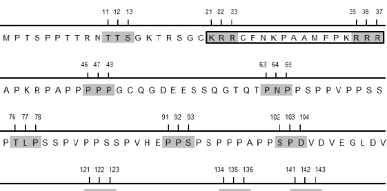
It has been shown that ORF73 interacts with MYC and p65/RelA through its proline-rich N-terminal region (amino acid residues 1-140), as observed in co-immunoprecipitation experiments (Rodrigues et al., unpublished data). To identify specific amino acid residues in ORF73 that were required for binding to MYC and p65/RelA, alanine-scanning mutagenesis was performed in the N-terminus of ORF73, essentially as described for LANA (Barbera et al., 2006). Alanine-scanning mutagenesis is a simple technique that is widely used to determine the functional role of protein residues; alanine is the substitution residue of choice because it eliminates the side chains beyond the β carbon, without altering the main-chain conformation, or imposing extreme electrostatic or steric effects (Cunningham & Wells, 1989; Lefèvre et al., 1997). Potentially, every amino acid in the N-terminus of ORF73 could be involved in binding to MYC and p65/RelA. Thus, triple-alanine substitutions were introduced at approximately every 10 residues contained within the N-terminus of ORF73 (Figure 1). In some cases, the sites of triple substitutions were adjusted, in order to include the beginning and the end of the putative nuclear localisation signal (ORF7321KRR23 and
ORF7335RRR37 mutants), proline-rich regions (ORF7346PPP48 and ORF73121PPP123 mutants) or
proline/serine-rich regions (ORF7391PPS93 and ORF73134NPS136 mutants). Residues 141-143 were
not within the region which was previously identified as being important for substrate binding (residues 1-140); however, as they were in the neighbourhood, they were still mutated (ORF73141KKY143mutant). Altogether, eleven ORF73 mutants were generated, each one of them
with only one triple-alanine substitution in its N-terminus.
The ability of ORF73 mutants to interact with MYC and p65/RelA was then tested by co-immunoprecipitation experiments. Co-co-immunoprecipitation is one of the most commonly used methods to study protein-protein interactions. This technique allows the isolation of multiprotein complexes in a cell lysate, by using an antibody that recognises one of the proteins present in the complex (bait protein). Next, Sepharose beads, which are covalently coupled to a protein (e.g. protein G) that stably binds to the constant region of the antibody, are added. Thus, the antibody-bait protein complex will immunoprecipitate. If the antibody-bait protein establishes either direct or indirect interactions with other proteins in a multiprotein complex, then all the proteins that are part of the complex will co-immunoprecipitate. The presence of target proteins can then be assessed by Western blotting with the appropriate antibodies, thereby identifying specific protein-protein interactions (Masters, 2004). Co-immunoprecipitation experiments with ORF73 mutants and MYC or p65/RelA were performed in HEK 293T cells overexpressing either MYC or p65/RelA, and co-expressing ORF73. The first ten ORF73 mutants (ORF7311TTS13, ORF7321KRR23, ORF7335RRR37,
ORF7346PPP48, ORF7363PNP65, ORF7376TLP78, ORF7391PPS93, ORF73102SPD104,
ORF73121PPP123 and ORF73134NPS136) were able to efficiently co-immunoprecipitate MYC and
13
MYC (Figure 2, first panel, lane 4) and p65/RelA (Figure 3, first panel, lane 4), suggesting that
141KKY143 residues are important for binding of ORF73 to these substrates. Furthermore, it was
previously shown that, in total cellular lysates, MYC levels are increased in the presence of wild-type ORF73 (wtORF73), because binding of ORF73 to MYC promotes its stabilization (Rodrigues
et al., unpublished data). This increase of MYC levels in the presence of wtORF73 was also
observed in this experiment (Figure 2, third panel, lane 3); however, MYC levels in the presence of ORF73141KKY143 (Figure 2, third panel, lane 4) were the same observed in cells not transfected
with ORF73 protein (Figure 2, third panel, lane 1), which reinforces that 141KKY143 residues are
important for ORF73 binding to MYC and, consequently, to ORF73 modulatory effect.
Figure 1. Alanine-scanning mutagenesis strategy. The N-terminal region of ORF73 is shown. To identify
specific amino acid residues in ORF73 that were required for binding to MYC and p65/RelA, triple-alanine substitutions were introduced in its N-terminal region. The residues that were replaced with alanines are shaded. Eleven ORF73 mutants were generated, each one of them with only one triple-alanine substitution.
14
Figure 2. ORF73141KKY143 mutant weakly co-immunoprecipitates MYC. HEK 293T cells were transiently
transfected with plasmids encoding HA-tagged MYC and Myc-tagged ORF73 (either wtORF73 or ORF73141KKY143), as indicated on top. After 48 h, cells were lysed and total cellular lysates were subjected to immunoprecipitation using an anti-ORF73 serum. Immunoprecipitates were analysed by Western blotting using the indicated antibodies (first and second panels). In addition, representative aliquots of the total cellular lysates were used to detect the appropriate expression of MYC (third panel), ORF73 (fourth panel), and actin (fifth panel). -, without; +, with; α, anti; IP, immunoprecipitation; TCL, total cellular lysates; WB, Western blotting.
15
Figure 3. ORF73
141KKY143 mutant weakly co-immunoprecipitates p65/RelA. HEK 293T cells were
transiently transfected with plasmids encoding Myc-tagged versions of p65/RelA and ORF73 (either wtORF73 or ORF73141KKY143), as indicated on top. After 48 h, cells were lysed and total cellular lysates were subjected to immunoprecipitation using an anti-ORF73 serum. Immunoprecipitates were analysed by Western blotting using an anti-Myc antibody (first and second panels). In addition, representative aliquots of the total cellular lysates were used to detect the appropriate expression of p65/RelA (third panel) and ORF73 (fourth panel). -, without; +, with; α, anti; IP, immunoprecipitation; TCL, total cellular lysates; WB, Western blotting.
16
4.2. Mutation at
141KKY
143in ORF73 impairs its ability to activate MYC
transcriptional activity
It has been shown that binding of wtORF73 to MYC stabilizes the latter, leading to an increase in MYC transcriptional activity in cells transfected with ORF73 (Rodrigues et al., unpublished data). ORF73141KKY143 mutant weakly interacted with MYC (Figure 2, first panel, lane
4), so it was expected that this mutant had impaired ability to activate MYC transcriptional activity. To investigate whether ORF73141KKY143 was able to modulate MYC transcriptional activity, reporter
gene assays were performed, using a MYC reporter plasmid with six tandem repeats of MYC consensus sequence (E-box) that drove the expression of firefly luciferase, whose activity was then measured. Luciferase activity was correlated with the amount of luciferase produced, which in turn depended on the amount of MYC molecules that bound to the consensus sequences, driving luciferase expression. Therefore, MYC transcriptional activity could be quantified by measuring luciferase activity in each experimental condition. To this end, HEK 293T cells were transiently transfected with the MYC reporter plasmid, in the presence or absence of ORF73 (either wtORF73 or ORF73141KKY143). The experiments were performed either with endogenous levels or
overexpression of MYC. In experiments with endogenous levels of MYC, cells expressing wtORF73 presented a 3,8-fold increase in MYC transcriptional activity (Figure 4, second bar) relative to control cells, which did not express ORF73. In contrast, cells expressing ORF73141KKY143 showed only a 1,4-fold increase in MYC transcriptional activity (Figure 4, third
bar), an indication that mutation at 141KKY143 impairs the ability of ORF73 to activate MYC
transcriptional activity. Since MYC cellular levels are tightly regulated, overexpression of MYC is the prototypical positive control for MYC transcriptional activity in reporter assays. Cells not expressing ORF73 showed a 8,7-fold increase in MYC transcriptional activity when MYC was overexpressed, as expected (Figure 4, fourth bar). Cells expressing wtORF73 exhibited a 42-fold increase in MYC transcriptional activity (Figure 4, fifth bar), whereas cells expressing ORF73141KKY143 showed only a 19-fold increase (Figure 4, sixth bar), reinforcing that mutation at 141KKY143 impairs the ability of ORF73 to activate MYC transcriptional activity, both with
17
Figure 4. ORF73
141KKY143 mutant is unable to efficiently activate MYC transcriptional activity, both
with endogenous levels and with overexpression of MYC. HEK 293T cells were transiently transfected
with a MYC luciferase reporter vector, and with plasmids encoding MYC protein and Myc-tagged ORF73 (either wtORF73 or ORF73
141KKY143), as indicated on bottom. MYC transcriptional activity associated with
each sample was assayed using a luminometer. Results are shown as the fold induction relative to luciferase activity measured in cells with endogenous levels of MYC that did not express ORF73. Error bars represent standard deviations of the mean in two independent experiments. In addition, representative aliquots of the total cellular lysates were used to detect appropriate expression of ORF73 (bottom panel). -, without; +, with; α, anti; WB, Western blotting.
18
4.3. Mutation at
141KKY
143in ORF73 partially impairs its inhibitory effect on NF-kB
transcriptional activity
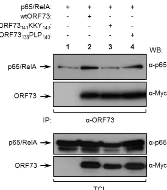
The interaction between wtORF73 and the NF-kB family member p65/RelA leads to the inhibition of the transcriptional activity of NF-kB, through proteasomal degradation of nuclear p65/RelA (Rodrigues et al., 2009). The interaction between ORF73141KKY143 mutant and p65/RelA
was weak, since the levels of p65/RelA that co-immunoprecipitated with ORF73141KKY143 were low
(Figure 3, first panel, lane 4), so it was expected that this mutant could not inhibit NF-kB transcriptional activity as efficiently as the wild-type protein. To test this hypothesis, a reporter gene assay was performed as described above for MYC. HEK 293T cells were transiently transfected with an NF-kB reporter plasmid with three copies of kB consensus sequences that drove the expression of firefly luciferase (Winkler et al., 1996), in the presence or absence of ORF73 (either wtORF73 or ORF73141KKY143). TNF-α was used as a stimulus leading to NF-kB activation. As in
MYC reporter assay, NF-kB transcriptional activity could be quantified by measuring luciferase activity. In control cells, which did not express ORF73, the luciferase activity increased by 5,4-fold in response to TNF (Figure 5, first open bar), meaning that, as expected, NF-kB transcriptional activity increases in the presence of a stimulus. In contrast, cells expressing wtORF73 were unable to respond effectively to TNF (Figure 5, second open bar), confirming that the viral protein abrogates TNF-driven NF-kB activation. On the other hand, cells expressing ORF73141KKY143
showed a 2,4-fold increase in NF-kB transcriptional activity in response to TNF (Figure 5, third open bar), indicating that mutation at 141KKY143 partially impairs the inhibitory effect of ORF73 upon
NF-kB transcriptional activity. However, ORF73141KKY143 did not completely lose its inhibitory
effect, since the increase of NF-kB transcriptional activity in cells expressing ORF73141KKY143 was
lower than in cells not expressing ORF73 at all. This result suggests that although 141KKY143
residues may be important for ORF73 to bind to p65/RelA, their mutation is not sufficient to completely prevent ORF73 inhibitory effect upon NF-kB. Therefore, ORF73141KKY143 mutant
cannot be used in further studies to investigate the in vivo role of termination of NF-kB transcriptional activity by ORF73. However, ORF73141KKY143 mutant was not able to activate MYC
transcriptional activity (Figure 4, third and sixth bars), so it is a good candidate to clarify the biological significance of ORF73-mediated MYC activation.
19
Figure 5. ORF73141KKY143 mutant has partially impaired ability to inhibit TNF-driven NF-kB
transcriptional activity. HEK 293T cells were transiently transfected with an NF-kB luciferase reporter vector
and a plasmid encoding Myc-tagged ORF73 (either wtORF73 or ORF73
141KKY143), as indicated on bottom.
Transfected cells were either stimulated with 50 ng/ml of TNF (open bars) or left unstimulated (filled bars). NF-kB transcriptional activity associated with each sample was assayed using a luminometer. Results are shown as the fold induction relative to luciferase activity measured in unstimulated cells. Error bars represent standard deviations of the mean in two independent experiments. In addition, representative aliquots of the total cellular lysates were used to detect the appropriate expression of ORF73 (bottom panel). -, without; +, with; α, anti; WB, Western blotting.
20
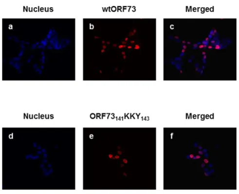
4.4. Mutation at
141KKY
143in ORF73 does not affect its nuclear localisation
ORF73 has a putative nuclear localisation signal (NLS) in residues 21 to 37 (KRRCFNKPAAMPPKRRR) (Figure 1), so it localises primarily to the nucleus when transiently expressed in HEK 293T cells, as assessed by immunofluorescence (Rodrigues et al., 2009). Although the mutations introduced in ORF73141KKY143 were not in the NLS, they could modify the
global structure of the protein in such a way that the NLS was masked. To analyse whether the introduced mutations affected the subcellular localisation of ORF73, HEK 293T cells were transiently transfected with either wtORF73 or ORF73141KKY143, and immunofluorescence
experiments were performed. Results showed that, similarly to wtORF73, ORF73141KKY143
localises to the nucleus of transfected cells (Figure 6, panel f).
Figure 6. ORF73141KKY143 localises to the nucleus. HEK 293T cells were transiently transfected with a
plasmid encoding Myc-tagged wtORF73 (panels a, b and c) or ORF73
141KKY143 (panels d, e and f). After 24 h,
cells were fixed and subjected to immunostaining with anti-Myc antibody, followed by incubation with Alexa 594 anti-mouse secondary antibody (red). Nuclear DNA was stained with DAPI solution (blue) to visualize the nucleus.
21
4.5. Mutation at
138PLP
140in ORF73 partially impairs its ability to
co-immunoprecipitate MYC
ORF73141KKY143 protein had three mutated amino acid residues; however, it was possible that
a mutation in a single residue was enough to lose MYC binding. If this hypothesis was true, then a mutant ORF73 protein with only one mutated residue could be used in further experiments, decreasing the probability that the introduced mutation modified the global structure of the protein, since a smaller number of original residues were mutated. Therefore, to assess if single mutations at 141KKY143 residues had the same effect that the triple mutation upon the ability of ORF73 to bind
to MYC, single-alanine substitutions were introduced at 141K, 142K, and 143Y residues, generating
ORF73141K, ORF73142K, and ORF73143Y mutants, respectively (Figure 7). 138PLP140 residues were
adjacent to 141KKY143 residues, and they were contained within the region that was previously
identified as being important for MYC binding (residues 1-140; Rodrigues et al., unpublished data). To test if these residues also had a role in MYC binding, a mutant with a triple-alanine substitution at 138PLP140 residues was generated (Figure 7). The ability of ORF73141K, ORF73142K, ORF73143Y
and ORF73138PLP140 mutants to interact with MYC was then tested by co-immunoprecipitation
experiments, in HEK 293T cells overexpressing MYC and co-expressing ORF73 (either wild-type or mutants). ORF73141K and ORF73143Y (Figure 8, first panel, lanes 4 and 6, respectively)
co-immunoprecipitated MYC as efficiently as wtORF73 (Figure 8, first panel, lane 2); in contrast, the levels of MYC that co-immunoprecipitated with ORF73142K (Figure 8, first panel, lane 5) were lower
than the levels with wtORF73, but higher than with ORF73141KKY143 (Figure 8, first panel, lane 3).
Accordingly, MYC levels in total cellular lysates were increased in the presence of ORF73141K,
ORF73142K, and ORF73143Y mutants (Figure 8, third panel, lanes 4, 5 and 6, respectively), relative
to MYC levels in cells not transfected with ORF73 protein (Figure 8, third panel, lane 1). This result indicated that triple-alanine substitution at 141KKY143 residues caused a more severe deficit in the
ability of ORF73 to bind to MYC than each of the single-alanine substitutions. Therefore, there was no advantage in using single mutants instead of the ORF73141KKY143 triple mutant to study the role
of ORF73-MYC interaction, so ORF73141KKY143 was the mutant used in further experiments. As far
as ORF73138PLP140 mutant is concerned, it co-immunoprecipitated lower levels of MYC (Figure 8,
first panel, lane 7) than wtORF73 (Figure 8, first panel, lane 2), but higher levels of MYC than ORF73141KKY143 (Figure 8, first panel, lane 3). This suggests that 138PLP140 residues are important
22
Figure 7. Alanine-scanning mutagenesis strategy (second screening). The N-terminal region of ORF73 is
23
Figure 8. ORF73
138PLP140 mutant has impaired ability to co-immunoprecipitate MYC. HEK 293T cells
were transiently transfected with plasmids encoding HA-tagged MYC and Myc-tagged ORF73 (either wild-type or mutants), as indicated on top. After 48 h, cells were lysed and total cellular lysates were subjected to immunoprecipitation using an anti-ORF73 serum. Immunoprecipitates were analysed by Western blotting using the indicated antibodies (first and second panels). In addition, representative aliquots of the total cellular lysates were used to detect the appropriate expression of MYC (third panel), ORF73 (fourth panel), and actin (fifth panel). -, without; +, with; α, anti; IP, immunoprecipitation; TCL, total cellular lysates; WB, Western blotting.
24
4.6. Mutation at
138PLP
140in ORF73 impairs its ability to activate MYC transcriptional
activity
To test the ability of ORF73138PLP140 triple mutant to activate MYC transcriptional activity, a
reporter gene assay was performed as described above for ORF73141KKY143 mutant. MYC reporter
assays with ORF73141KKY143 mutant (Figure 4) showed that the pattern of MYC transcriptional
activity was similar in the presence of endogenous levels of MYC and in the presence of overexpressed MYC. Therefore, and since the system with endogenous levels of MYC better mimics the physiological condition in cells, the reporter assay with ORF73138PLP140 mutant was
performed with endogenous MYC only. In this experiment, cells expressing wtORF73 exhibited a 91-fold increase in MYC transcriptional activity (Figure 9, second bar) in comparison to control cells, which did not express ORF73 (Figure 9, first bar). On the other hand, cells expressing ORF73138PLP140 presented a 5,8-fold increase in MYC transcriptional activity (Figure 9, fourth bar),
slightly more than the 2,4-fold increase in cells expressing ORF73141KKY143 (Figure 9, third bar).
This result indicates that mutation at 138PLP140 residues impairs the ability of ORF73 to activate
MYC transcriptional activity, although this impairment is not as strong as the one observed when
141KKY143 residues are mutated.
To confirm the results obtained in co-immunoprecipitation experiments with MYC and ORF73 single mutants, these mutants were also included in the reporter gene assay. The single mutants used in this experiment were ORF73142K, since it showed some impairment in the ability to
co-immunoprecipitate MYC (Figure 8, first panel, lane 5), and ORF73141K, as a representative of the
single mutants that co-immunoprecipitated MYC as efficiently as wtORF73. As ORF73141K mutant
behaved the same way that ORF73143Y mutant in the co-immunoprecipitation experiment (Figure
8, first panel, lanes 4 and 6, respectively), ORF73143Y was not included in this reporter assay, and
it was assumed that the results obtained for ORF73141K mutant in this experiment were the same
for ORF73143Y mutant. Reporter assay results showed that the transcriptional activity of MYC
increased by 58-fold in the presence of ORF73142K (Figure 9, sixth bar), a level between MYC fold
increase in the presence of wtORF73 (91-fold increase; Figure 9, second bar) and ORF73141KKY143
(2,4-fold increase, Figure 9; third bar). This was in good agreement with the result obtained in the co-immunoprecipitation, since ORF73142K mutant co-immunoprecipitated lower levels of MYC than
wtORF73, but higher levels than ORF73141KKY143. Furthermore, cells expressing ORF73141K
showed a 110-fold increase in MYC transcriptional activity (Figure 9, fifth bar), a level even higher than the observed in cells expressing wtORF73 (Figure 9, second bar). Therefore, this reporter assay confirmed that there was no advantage in using single mutants instead of the ORF73141KKY143 triple mutant to study the role of ORF73-MYC interaction, since triple-alanine
substitution at 141KKY143 residues caused a more severe impairment in the ability of ORF73 to