UNIVERSIDADE NOVA DE LISBOA
EXPLORING INSECT CELLS
VERSATILITY FOR PRODUCTION OF
INFLUENZA VIRUS-LIKE PARTICLES
DANIELA FILIPA POLICARPO SEQUEIRA
THESIS SUMBITTED FOR GRANTING OF THE DEGREE OF MASTER IN MEDICAL MICROBIOLOGY
i UNIVERSIDADE NOVA DE LISBOA
EXPLORING INSECT CELLS
VERSATILITY FOR PRODUCTION OF
INFLUENZA VIRUS-LIKE PARTICLES
DANIELA FILIPA POLICARPO SEQUEIRA
THESIS SUMBITTED FOR GRANTING OF THE DEGREE OF MASTER IN MEDICAL MICROBIOLOGY
Supervisor: Doutora Ana Margarida Palma Teixeira Co-Supervisor: Doutor António Manuel Missionário Roldão Internal supervisor: Professor Doutor João Piedade
Experimental work performed at the Animal Cell Technology Unit, Instituto de Biologia Experimental e Tecnológica, Oeiras, Portugal
iii
Publications
The results described in this thesis were presented at several international scientific meetings:
Poster presentations
Dias MD, Vidigal J, Sequeira D, Patrone M, Alves PM, Teixeira AP. Accelerating stable recombinant protein expression through novel recombinase-based targeting technologies. RPP8, April 2015, Mallorca, Spain.
Dias MD, Vidigal J, Sequeira D, Patrone M, Alves PM, Teixeira AP. Streamlining insect cell line development through novel targeted gene integration strategies. ESACT 24th, June 2015. Barcelona, Spain.
Sequeira D, Correia R, Carrondo MJT, Roldão A, Teixeira AP, Alves PM. Combining stable insect cell lines with baculovirus-mediated expression for production of multi-HA Influenza VLPs. Influenza Vaccines for the World, October 2015. Albufeira, Portugal.
Manuscript
Sequeira D, Correia R, Carrondo MJT, Roldão A, Teixeira AP, Alves PM. Combining stable insect cell lines with baculovirus-mediated expression for production of multi-HA Influenza VLPs. (under preparation)
This work was supported by EU-funded project EDUFLUVAC (FP7-HEALTH-2013-INNOVATION) and Fundação para a Ciência e a Tecnologia - Portugal (EXPL/BBB-BIO/1541/2013).
iv
Acknowledgments
I would like to acknowledge all the people directly or indirectly involved in this thesis.
To Professor Paula Alves and Professor Manuel Carrondo for giving me the opportunity to do my master thesis at the Animal Cell Technology Unit at ITQB/IBET, for the good working conditions offered and for being a strong example of leadership and professionalism.
I am very thankful to my supervisors Dr. Ana Teixeira and Dr. António Roldão for their constant guidance, support and perfectionism, motivating and challenging me throughout this thesis.
I would also like to thank João Vidigal and Mafalda Dias for teaching me everything about this new field, for all the discussions, help and constant good mood.
I am very grateful for all the help offered by several members of the Animal Cell Technology group. A special thanks goes to those with whom I worked side-by-side with like Ricardo Correia and Evelien Vaessen for their help, support, funny moments and for always having a kind word. Also, to Alexey Koshkin for being a good friend and for his constant encouragement.
To my good friends Maria Nunes, Rute Pinto, Gonçalo Silva and Tânia Lucas for their support, understanding and availability, helping me whenever needed.
I especially thank Duarte for his trust, endless patience and support. For his constant help, good advices and for always being there in the toughest times.
I am deeply thankful to my parents that made this journey possible. For always showing me the good part of the story, how to overcome difficulties and for their confidence in me.
v
Abstract
A potential strategy to produce safer and broadly protective influenza vaccines is to co-express, in the same cell host, multiple hemagglutinins (HA) with a matrix protein (M1) which self-assemble in virus-like particles (VLPs). This study demonstrates the suitability of combining stable expression and the baculovirus-expression vector system (BEVs) in insect Hi5 cells for production of such multi-HA Influenza VLPs. Stable pools of Hi5 cells expressing two HAs were generated and later infected with a M1-encoding baculovirus at two cell concentrations (CCIs; 2×10⁶ cells/mL and 3×10⁶ cells/mL). The HA concentration
in culture supernatant was followed over time, with more productive infections observed at higher CCIs. To extend the culture time, a re-feed strategy was implemented based on the identification of key nutrients which were exhausted during cell growth. Afterwards, supplemented cultures infected at a CCI of 4×10⁶ cells/mL allowed a 4-fold increase in HA concentration, at harvest, when compared to cultures infected at a CCI of 2×10⁶ cells/mL. The production of multi-HA influenza VLPs using the aforementioned strategy could be successfully scaled-up to 2L bioreactor cultures with even higher volumetric (1.5-fold) HA yields.
To surpass the unpredictability of gene expression promoted by the random integration strategy mentioned above, the recombinase-mediated cassette exchange (RMCE) technology was explored. The feasibility of having two cassettes flanked by distinct pairs of flippase recognition target sites (FRTs) was evaluated. Unfortunately, significant cross-interaction was observed between the selected pairs. To circumvent this bottleneck, a backup strategy consisting in the co-expression of two genes from the same locus after implementation of one cassette system, in insect Sf9 cells, was attempted. However, the isolated clones showed low expression of both M1 and HA proteins. Ongoing work focuses on the isolation of clones tagged in high expression loci by fluorescence activated cell sorter technology.
This work demonstrates how the versatility of insect cell expression technology can be explored to produce Influenza VLPs as vaccine candidates.
Keywords: Influenza vaccines; virus-like particles (VLPs); multivalent HA vaccines; insect cells; BEVS; RMCE.
vi
Resumo
A co-expressão de várias hemaglutininas (HA) e proteína da matriz (M1), no mesmo hospedeiro, formando partículas semelhantes a vírus (VLPs), constitui uma importante estratégia para desenvolver vacinas contra o vírus da gripe. Este trabalho mostra a combinação de uma linha celular estável de células de insecto com o sistema de expressão mediada por baculovírus para a produção deste tipo de VLPs. Foram estabelecidas duas populações de células de insecto Hi5, expressando duas HAs, posteriormente infectadas com um baculovírus contendo a proteína M1, a duas concentrações celulares diferentes (CCI; 2 e 3×10⁶ cells/mL) sendo que a mais elevada demostrou ser a mais produtiva. De seguida,
implementou-se uma estratégia baseada na adição de nutrientes específicos para prolongar o tempo de cultura. As culturas previamente suplementadas e infectadas a uma CCI de 4×10⁶ células/mL produziram 4x mais HA comparativamente às culturas infectadas a uma CCI de 2×10⁶ células/mL, não suplementadas. Esta estratégia foi também aplicada num biorreactor de 2L permitindo 1,5x mais produção, volumétrica, de HA comparativamente a experiências em pequena escala.
De forma a ultrapassar a imprevisibilidade de uma integração aleatória, foi explorado o sistema de troca de cassete mediado por recombinase (RMCE). A viabilidade de um sistema com duas cassetes integradas flanqueadas por diferentes locais de reconhecimento (FRTs) foi avaliada, tendo sido observada a interação entre ambos os pares selecionados. Como segunda estratégia, foi implementado um sistema com uma cassete para co-expressão de dois genes em simultâneo, em células de insecto Sf9. Porém, os clones isolados mostram fraca expressão de M1 e HA, pelo que uma estratégia de isolamento de clones com expressão génica mais forte está em desenvolvimento utilizando uma tecnologia de sorteamento.
Assim, este trabalho demonstra a versatilidade da tecnologia aplicada em células de insecto, que pode ser explorada para produzir VLPs multivalentes, com potencial para se tornar a próxima geração de vacinas para o vírus da gripe.
Palavras-chave: Vacinas para a; partículas semelhantes a vírus (VLPs); vacinas de HA (hemaglutinina) multivalentes; células de insecto; sistema de expressão mediada por baculovírus; sistema de troca de cassete mediada por recombinase.
vii
Table of Contents
Publications ... iii Acknowledgments ... iv Abstract ... v Resumo ... viTable of Contents ... vii
List of Figures ... x
List of Tables ... xiii
List of Acronyms ... xiv
1 Introduction ... 1
1.1 Influenza virus ... 1
1.1.1 Egg-based influenza vaccines ... 3
1.1.2 Cell-based influenza vaccines ... 4
1.1.3 Subunit vaccines ... 5
1.2 Insect cells ... 10
1.2.1 Baculovirus expression vector system (BEVS) ... 10
1.2.2 Advantages/disadvantages for production of Influenza vaccines ... 13
1.3 Cell line development ... 14
1.3.1 Random integration ... 15
1.3.2 Locus-specific integration ... 15
2 Materials and Methods ... 21
2.1 Molecular Biology ... 21
2.1.1 Plasmid design and construction ... 21
2.1.2 Techniques supporting plasmid construction ... 22
2.2 Cell line development ... 24
2.2.1 Transfection ... 24
2.2.2 Cassette-exchange ... 24
2.2.3 Sorting procedures ... 25
2.2.4 Cloning ... 25
viii
2.3.1 Freezing and thawing cells ... 26
2.4 Baculovirus ... 26
2.4.1 Virus amplification ... 26
2.4.2 Virus titration ... 26
2.4.3 Infection of insect cells with baculovirus ... 27
2.5 Production of influenza VLPs in 2L bioreactors ... 27
2.5.1 Downstream processing of influenza VLPs ... 27
2.6 Analytical methods ... 28
2.6.1 Supplementation ... 28
2.6.2 Negative staining transmission electron microscopy ... 28
2.6.3 Hemagglutination assay ... 28 2.6.4 Exometabolome analysis ... 29 2.6.5 Immunofluorescence ... 29 2.6.6 Cell sonication ... 30 2.6.7 Flow cytometry ... 30 2.6.8 Western blot ... 30
2.6.9 RNA extraction and RT-PCR ... 31
2.6.10 Genomic DNA extraction ... 32
3 Results ... 33
3.1 Combining stable insect Hi5 cell line with the baculovirus expression system for production of multi-HA influenza VLPs ... 33
3.1.1 Establishment of stable insect cell lines by random integration ... 33
3.1.2 Optimizing HA production in Hi5 pools ... 36
3.1.3 Production of multi-HA influenza VLPs ... 39
3.1.4 Scale-up production of multi-HA influenza VLPs ... 41
3.2 Establishing a double-RMCE insect cell platform ... 43
3.2.1 Vector design and FRT sites ... 43
3.2.2 Feasibility analysis: evaluation of target sites’ specificity ... 44
3.3 Production of Influenza VLPs using RMCE ... 47
ix
3.3.2 Clones’ characterization ... 48
3.3.3 Production of influenza VLPs in the same locus ... 50
4 Discussion and conclusions ... 52
4.1 Dual strategy for production of Influenza VLPs ... 52
4.2 Suitability of RMCE for production of complex proteins ... 55
4.3 Conclusions and future work ... 56
5 References ... 58
Appendix A ... 70
A.1 Table of primers used in the construction of vectors needed in this work. ... 70
A.2 Primers used for cDNA detection. ... 71
A.3 Primers used for cDNA detection. Primers used for detection of tagging and target cassettes. ... 71
x
List of Figures
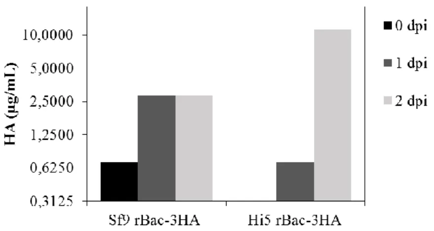
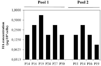
Figure 1 - Replication cycle of Influenza virus (Nayak et al., 2004). ... 2 Figure 2 - Wild-type baculovirus replication cycle (Monteiro et al., 2012). ... 11 Figure 3 - RMCE principle: tagging a locus with a cassette flanked with heterospecific target sites into the genome and then exchanging it for the GOI. Adapted from Turan et al. 2010. ... 17 Figure 4 – Aim of the thesis and strategies adopted. ... 20 Figure 5 - Scheme of the two expression vectors used to transform parental cells. OpIE2 and OpIE1 promoters were selected to drive HA and zeocin resistance gene expression, respectively. ... 34 Figure 6 - HA concentration in culture supernatant. Parental Hi5 and Sf9 cells transfected with construct #1 (Figure 5) were infected with a rBac containing three HA genes at a CCI of 2x10⁶ cell/mL and MOI of 10 total viral particles/cell 48h (for Hi5 cells) and 72h (for Sf9 cells) after transfection. Dpi means days post-infection. ... 34 Figure 7 - Growth profiles of stable Hi5 pools expressing two HA genes, along passages. 35 Figure 8 - Immunofluorescence detection of HA in the membrane of two stable Hi5 pools. Negative control (parental Hi5 cells) was added to evaluate unspecific binding of the antibody used. Secondary antibody was labelled with GFP. Scale bars indicate 100µm... 36 Figure 9 – Concentration of HA in cellular extracts along passages for two stable Hi5 pools. ... 36 Figure 10 – A) Growth profiles for infections at CCI 2x10⁶ cells/mL and CCI 3x10⁶ cells/mL
for both pools. B) HA concentration in culture supernatant during infection for both CCIs and pools. Dpi means days post-infection. ... 37 Figure 11 - A) Growth profiles of supplemented and non-supplemented cultures. Metabolites analysis by 1H-NMR: glucose (Glc), glutamine (Gln), glutamate (Glu), asparagine (Asn), aspartate (Asp) and lactate (Lac) before (B) and after supplementation (C). The arrows indicate the time of the re-feed. ... 39 Figure 12 – Multi-HA VLP production in small-scale shake flask cultures. A) Cell concentration profiles for infections of pool 1 with rBac-3HA and rBac-M1 at CCIs 2x10⁶ cells/mL and 4x10⁶ cells/mL. B) HA concentration in the supernatant for both baculovirus
xi infections at CCI 2x10⁶ cells/mL or 4x10⁶ cells/mL (culture was supplemented 72h after inoculation, i.e 24h before infection). Dpi means days post-infection. ... 40 Figure 13 - Production of pentavalent VLPs in supplemented bioreactor culture by infection of Hi5 pool 1 at a CCI of 4x10⁶ cells/mL. Comparison between shake flask and bioreactor experiments regarding A) cell concentration profiles and viability; B) lactate (lac) concentration during infection and C) HA concentration in supernatant during infection. D) Western blot of culture supernatant for detection of HA and M1 along infection. Dpi stands for days post-infection. E) Electron microscopy analysis of VLPs by negative staining; scale bars represent 100nm. ... 42 Figure 14 - Scheme of tagged populations (A) and target vectors (B) used to address cross-interaction between FwF5 and F13F14. ... 44
Figure 15 - Fluorescence intensity profiles of tagging pools at the day of transfection with target vectors. ... 44 Figure 16 - Evaluation of cross-interaction between target sites Fw/F5 and F13/F14. (A) Flow cytometry and (B) fluorescence microscopy results 48 hours after transfection. Scale bars indicate 100µm. (C) eGFP positive cells (%) in different target populations after two weeks in selection with neomycin. Black bars represent the average of two experiments with cell passage at a high inoculum and grey bars are representative of a third experiment with cell passage at a lower inoculum. ... 46 Figure 17 - A) Construct integrated in clones. B) Target vector encoding M1 and HA genes and C) Resulting population expressing M1 and HA from the same locus after RMCE. .... 47 Figure 18 - Cell line development from tagging to cloning ... 48 Figure 19 - Characterization of Sf9 clones #3 and #4 according to the presence of (A) expression of tagging and target genes by mRNA analysis and (B) flow cytometry. Primers were constructed to anneal with specific regions of each gene in study; L-ladder (Quick-Load 100bp); C) Identification of tagging cassette in clones. Genomic DNA was extracted and primers were located at OpIE2 and OpIE1 promoters amplifying 5,6kbp fragment in a target population and a 2,9kbp in a tagging population. Ladder used: NZY DNA ladder III. ... 49 Figure 20 - Detection of eGFP positive cells in population that was submitted to RMCE to M1-HA by (A) flow cytometry and (B) fluorescence microscopy (scale bars are in 100μm).
xii (C) M1 and HA gene expression analysis. Parental cDNA was added as a negative control in a PCR with primers for M1 and HA amplification and 18S cDNA analysis was added as an endogenous control. Ladder used: Quick-load 100bp. (D) Comparison of fluorescence intensity between a population and clone #3 of Sf9. ... 51
xiii
List of Tables
Table 1 - Insect-cell derived VLPs in preclinical studies (adapted from Krammmer et.al 2010). ... 9 Table 2 - Most used SSR in mammalian cell lines. Adapted from (Wirth et al., 2007). ... 19
xiv
List of Acronyms
AcMNPV Autographa californica multicapsid nucleopolyhedrovirus
Asn Asparagine
BEVS Baculovirus expression vector system
BVs Budded virions
CCI Cell concentration at infection
CRISPR Clustered Regularly Interspaced Short Palindromic Repeats
DSBs Double strand-breaks DSP Downstream processing ECL Enhanced chemiluminescence eGFP Enhanced green fluorescent protein FBS Fetal bovine serum
Flp Flippase
FRT Flippase recognition target site
Glc Glucose
Gln Glutamine
GOI Gene-of-interest
HA Hemagglutinin
hFlpe Humanized Flpe
HR Homologous recombination HRP Horseradish peroxidase
iFlp Insect cells codon-optimized flippase IR Illegitimate recombination
MDCK Madin-Darby Canine Kidney cells
MTT Thiazolyl blue tetrazolium bromide solution
NA Neuraminidase
NEP Nuclear export protein NP Nucleocapsid protein ODVs Occlusion-derived virions
xv PBS Phosphate buffered saline
PDT Population doubling time PTMs Post-translation modifications rBACs Recombinant baculovirus RBC Red blood cells
RdRp RNA-dependent RNA polymerase complex recHA Recombinant hemagglutinin
recNA Recombinant neuraminidase
RMCE Recombinase-mediated cassette exchange SSR Site-specific recombinases
TALENs Transcription activator-like effector nucleases TIVs Trivalent inactivated influenza vaccines TTBS Tween tris buffered saline
VLPs Virus-like particles vRNPs Viral ribonucleoproteins ZFNs Zinc-finger nucleases
1
1 Introduction
1.1 Influenza virus
Influenza viruses are responsible for annual epidemics and, occasionally, pandemics, responsible for acute febrile respiratory tract disease commonly known as “flu”. They belong to Orthomyxoviridae family and are divided into three genera (A, B and C), being genus A the most threatening due to its potential to cause global pandemics (Lowen et al., 2007; Steinhauer and Skehel, 2002). Influenza A viruses are divided into several subtypes depending on their surface glycoproteins hemagglutinin (HA) and neuraminidase (NA). Eighteen different HA subtypes and eleven different NA subtypes have been characterized so far, but only H1, H2, H3, N1 and N2 subtypes have been found to cause human pandemics (CDC, 2014).
Influenza viruses are enveloped containing a segmented, negative single stranded RNA genome and bud from the apical domain of epithelial cells. They have a standard nomenclature which includes the virus type, the species from which it was isolated (if not human), respective location, strain number and year of isolation as well as the hemagglutinin (HA) and neuraminidase (NA) subtype in case of influenza A viruses. Subdivision can also be done into serotypes in terms of antibody responses. The genome contains 8 ssRNA segments, existing as ribonucleoproteins (vRNPs), that can code for 11 proteins, including the M1 matrix protein (bridge between the envelope and the viral core) and the surface glycoproteins HA and NA (virus envelope) (Nayak et al., 2004). The viral core also contains the nucleocapsid protein (NP), the nuclear export protein (NEP) and three different polymerase proteins, PB1, PB2 and PA forming the RNA-dependent RNA polymerase complex (RdRp). The envelope is also composed by M2 ion channel plus host cell’s lipids (Nayak et al., 2004).
The replication cycle of influenza viruses begins with the viral recognition and subsequent binding to the N-acetylneuraminic (sialic) acids of host’s surface, preferentially α-2,3- or α-2,6-carbon linkages (Figure 1). After the binding step, internalization of virus particles occurs via receptor-mediated endocytosis. HA is cleaved by internal proteases and
2 in the acidic environment of the endosome, cleaved HA undergoes conformational changes leading to the fusion of viral and endosomal membranes (Steinhauer, 1999). M2 ion channel opens and allows the release of vRNPs from M1 into the cytoplasm. After this, eight vRNPs that include NP-nuclear transport signals are imported to the nucleus through nuclear pores (Neumann et al., 2000).
After DNA replication and translation, 11 viral proteins are produced of which HA, NA and M2 undergo post-translation modifications (PTMs) in the cis-Golgi apparatus and rough endoplasmic reticulum (Nayak et al., 2004). These glycoproteins are then transported to the budding site together with eight vRNPs and other viral proteins in order to form virions. Budding occurs with the involvement of host and viral components, being HA, NA and M1 key players in this process. M1 is responsible for the encapsidation of the vRNPs into the membrane and for the budding process. On the other hand, NA plays a critical role in the release of the viral particles due to its syalidase activity, cleaving the binding of HA to host sialic acids (Nayak et al., 2009).
3 In human respiratory epithelium, 2,6-carbon linkages are more abundant than α-2,3-carbon linkages. Due to the existence of α-α-2,3-carbon linkages in duck gut epithelium humans can be infected by avian influenza virus. When this transmission occurs, it leads to a more severe infection because α-2,3-linkages are more prevalent in the lower respiratory tract (e.g. lungs) (Couceiro et al., 1993; Matrosovich et al., 2004). Pigs contain both carbon linkages, meaning they can be infected by avian and human strains. In case such double infection occurs, strains may undergo reassortments and a novel strain capable of infecting humans is generated. On the other hand, different strains within the same subtype can also reassort and thus generate a new strain in a phenomenon named antigenic shift. The infections arising from such reassortments are usually severe because people are not immunized against the new strain, as it was the case of 2009 H1N1 pandemics (Steinhauer and Skehel, 2002). In addition, natural mutations can occur during viral genome replication due to errors in the RdRp polymerase enzymes leading to antigenic drift of a given strain (Steinhauer and Skehel, 2002). This is very likely to occur in influenza viruses because their polymerase enzymes do not perform proofreading as they lack a 3’-5’ exonuclease activity that would enable them to repair small errors during DNA replication. Regardless of being minor changes, these mutations can lead to a loss of immunogenicity and thus are held responsible for the renewal of influenza vaccines annually (Steinhauer and Skehel, 2002).
1.1.1 Egg-based influenza vaccines
The market of Influenza vaccines were estimated at $2.9 billion in 2011 and thought to accomplish $3.8 billion by 2018 (Conferences series, 2015).
The most commonly used platform for production of influenza vaccines is hen’s eggs. Production starts by infecting the allantoic fluid of the eggs with influenza viruses. After several rounds of replication, virions are harvested and chemically inactivated (e.g. with formaldehyde) or attenuated (e.g. serial passages at sub-optimal conditions). From this process, a whole virion preparation, a split vaccine or either a subunit vaccine can be achieved (Cox et al., 2008). Due to the fact that HA is the key surface glycoprotein in influenza viruses, triggering an immune response, its presence in a vaccine against influenza infection is
4 essential. Trivalent inactivated influenza vaccines (TIVs), composed by two influenza A viruses (H1N1 and H3N2) and one influenza B virus, are produced with this system. They are standardized in order to contain the same amount of HA of each virus strain, being the most commercialized influenza vaccine (Cox et al., 2008). However, this platform is very laborious, time consuming and costly, requiring large numbers of chicken eggs to produce one shot of vaccine and up to 9 months of production time. Besides this, the presence of eggs’ proteins can trigger allergies in humans which leads to an impairment of biosafety (Zeiger, 2002). In addition, the ability of some strains to replicate to high yields in hen’s eggs is unpredictable and when dealing with a very pathogenic strain the embryos can be killed without producing any virus.
1.1.2 Cell-based influenza vaccines
Mammalian cell lines represent today a robust platform for influenza vaccine production. First results on the effectiveness of continuous cell lines, such as Madin-Darby Canine Kidney cells (MDCK) on influenza virus replication were reported 40 years ago (Meguro et al., 1979; Tobita et al., 1975), providing evidence that mammalian cells could represent a robust platform for influenza vaccine production. A few years later, an inactivated influenza vaccine produced in MDCK cells showed to be more efficient in neutralizing antibody induction in ferrets than egg-grown vaccine (Katz and Webster, 1989). This culminated with the recent FDA approval of Flucelvax (Novartis, 2015), a trivalent inactivated influenza vaccine manufactured using MDCK cell culture technology.
Another continuous cell line used for influenza vaccines production is Vero cells. Vero cells are the most widely accepted continuous cell line by regulatory authorities and have been used for the production of viral vaccines such as for polio and rabies virus (Montagnon, 1989). They enable higher-titer growth of wild-type H5N1 strains (Barrett et al., 2009), which in case of a pandemic is of great importance particularly if a short supply of eggs occur or if the embryos are killed by the highly pathogenic virus strain. Although continuous cell lines like MDCK and Vero have raised some safety questions due to their potential oncogenic properties, regulatory authorities are becoming more receptive given the
5 improved screening technologies to analyse their biosafety. The use of cell-culture grown virus proved to be efficient for influenza vaccine production in a short period of time and with higher antigens yields as well as being capable of inducing neutralizing antibodies (Ehrlich et al., 2008; Kistner et al., 2007).
Despite having several advantages over the egg-based platform, MDCK and Vero cells still have their downsides. They can be transformed over several passages, have oncogenic potential and require a solid matrix to support their growth in bioreactors. The human cell line – PER.C6 (derived from primary cultures of human fetal retinoblast)– has also been showing to be efficient in producing high titers of influenza virus of a variety of subtypes (Pau et al., 2001). The advantage of this cell line in relation to MDCK or Vero is its ability to grow to high cell densities in suspension culture without the need for serum or solid matrix.
Overall, despite the advantages of the cell-based platform for production of influenza vaccines (e.g. higher titers of antigen in a short period of time), isolation of the virus is still required thus leading to the need for biosafety laboratory conditions. Besides this, inactivation or attenuation of the offspring also represents a major shortcoming. Furthermore, adaptation of the virus strains can occur during virus propagation, which can lead to a lower antigenicity of the vaccine.
1.1.3 Subunit vaccines
Recombinant influenza vaccines
Given the downsides of egg-based and cell-based influenza vaccines, efforts have been conducted into the development of safer and more flexible vaccine candidates profiting from recombinant DNA technology.
Recombinant hemagglutinins (recHA) have been shown to be highly immunogenic, inducing the production of broadly reactive neutralizing antibodies representing a potential vaccine candidate against influenza virus infection. One example is FluBlok (Protein Sciences Corporation), which contains three full-length recombinant HA proteins, two from influenza A virus (H1N1 and H3N2) and one from influenza B virus and it was the first recombinant protein based influenza vaccine, approved by FDA in 2013 (Corporation, 2015).
6 The strains included in this vaccine are updated on an annual basis so that it resembles as much as possible the circulating strains thus leading to a more efficient immunization. Also, it contains three times the amount of HA in the TIVs, thus inducing higher antibody titers and has proved to be immunogenic and well tolerated (Cox et al., 2008). Besides this, it is safer because it is a purified antigen free of host or other viral proteins (Cox et al., 2008; Cox and Anderson, 2007). More recently, it was shown that a specific region of hemagglutinin – the stem region – can be recognized by antibodies and is able to stimulate cross-reactive immunization leading to protection against many H1 subtype influenza strains in mice (Yassine et al., 2015).
Neuraminidase, the second most abundant envelope glycoprotein of influenza viruses, naturally forms tetramers and helps in the release of virions from cells. However, after challenge with recombinant neuraminidase (recNA) in mice, immune protection was only shown when coupled with adjuvants, and clinical trials in humans showed no significant vaccination effect with this antigen (Cox, 2008). That being said, a recNA-based vaccine does not represent a good alternative on its own.
Virus-like particles
Virus-like particles (VLPs) are protein structures that self-assemble naturally, mimicking the structure of a native virion lacking the viral genome which is a major advantage in terms of biosafety for implementation as human vaccines. Consequently, several types of VLPs from enveloped and non-enveloped viruses have been explored to become vaccine candidates (Crisci et al., 2012; Kushnir et al., 2012).
Numerous studies have addressed the immunogenicity of VLPs as vaccines reporting their efficacy in mice and in humans (Klausberger et al., 2014; Krammer and Grabherr, 2010). VLPs can be a more effective strategy to induce immunity over inactivated virions (Bright et al., 2007) because during the inactivation process native epitopes lose their folding thus decreasing their ability to stimulate a strong immune response. For example, CervarixTM (GlaxoSmithKline) is a VLP-based vaccine approved by the FDA for vaccination of women
7 against cervical cancer (Monie et al., 2008). It is composed by two viral proteins of human papillomavirus and produced using the insect cells-baculovirus system.
The use of influenza VLPs as vaccine candidates against influenza virus infection has been widely explored. Several reports exist today showing the efficacy of influenza VLPs in generating immune responses in mice after lethal virus challenges (Galarza et al., 2005; Pushko et al., 2005; Quan et al., 2007). Influenza VLPs are traditionally composed by the four major influenza proteins (HA, NA, M1 and M2) (Latham and Galarza, 2001) and their morphology resembles the native influenza virus with spikes on the surface and sizes between 80-120nm (Pushko et al., 2005; Quan et al., 2007). However, it was found that HA and M1 combined are sufficient to generate well assembled and functional VLPs with immunogenic properties (Quan et al., 2008). It was reported that M1 has the ability to colocalize with HA during its exocytic transport to the membrane and in the membrane (Ali et al., 2000; Barman et al., 2001) by association with its cytoplasmic tail and transmembrane domain (Chen et al., 2007). This protein is involved in the budding as it accommodates beneath the lipid bilayer interacting with it, causing its asymmetry and bending, facilitating the initiation of the budding process. However, it was also suggested that it may take a certain amount of M1, like a threshold, for the budding to occur (Bourmakina and García-Sastre, 2005). Depending on the diversity of proteins found in an influenza VLP, it can be monovalent or multivalent. Sometimes a monovalent VLP may not be enough to counteract a disease and there is the need for a multivalent (Pushko et al., 2011). For example, authors showed that a bivalent influenza VLP induced immunity against two viral strains decreasing the viral titers in the lungs (Quan et al., 2008) demonstrating that VLPs are a flexible way of producing candidate vaccines for specific and correlated virus strains.
One major concern when producing influenza VLPs is that protein post-translation modifications (PTMs) such as glycosylation and sialylation resemble as much as possible the in vivo pattern of the native product as they deeply affect biological functionality and antigenicity. It is known that insect cells do not have the same glycosylation pattern as humans cells (Marchal and Jarvis, 2001). Therefore, several studies have been conducted to address the functionality of influenza VLPs produced in insect cells (Bright et al., 2007; Pushko et al., 2005; Quan et al., 2007) and results are clearly positive suggesting that insect
8 cells can perform as good as mammalian cells. Several influenza VLPs produced in insect cells are already being subjected to preclinical trials (Table 1).
The fact that well assembled and functional VLPs can be produced in insect cells, coupled with their efficiency in triggering immune responses and displaying antigens for a number of applications has significantly increased the popularity of these cells in the industrial field.
9 Influenza subtype Influenza proteins Animal
model Results Comments Year
H3N2 HA,NA,M1,M2 First report of Influenza VLPs 2001
H3N2 HA,NA,M1,M2 Mice Protection from challenge
Interleukin-12 tested as an
adjuvant 2005
H9N2 HA,NA,M1 Mice Protection
from challenge 2005
H3N2 HA,NA,M1 Mice and ferrets
High HAI antibody titers
Compared with inactivated
whole virus and rHA 2007
H1N1 HA,M1 Mice Protection from challenge
First report of a cytotoxic
T-cell responde 2007 H1N1 and
H3N2 HA,M1 Mice
Protection from challenge
Bivalent vaccine, comparison
with inactivated whole virus 2008
H5N1 HA,NA,M1 Ferrets Protection
from challenge 2008
H5N1 HA,NA,M1 Mice Protection from challenge
Bivalent vaccine, comparison
with inactivated whole virus 2008
H5N1 HA,NA,M1 Mice Protection
from challenge 2009
H1N1 HA,M1 Mice Protection from challenge
Focus on dose-dependence of protection, bacterial toxins
tested as adjuvants
2009
H1N1 HA,NA,M1 Mice and ferrets
Protection from challenge
VLPs from the 1918 pandemic
strain 2009
H1N1,
H3N2 and B HA,NA,M1 Mice
Protection from challenge
First trivalent approach, compared with split vaccine
Fluarix
2009
H5N1 HA,NA,M1 Mice Protection from challenge
Focus on long-term protective
immune responses 2009 H1N1v HA, M1 Mice HAI titers of
1:2048
Alternative insect cell line, fast
reaction to 2009 pandemic 2010 H1N1v HA, M1 Mice Protection
from challenge
Fast reaction to 2009 pandemic, single-shot strategy 2010
10
1.2 Insect cells
The increasing interest in insect cells led to the generation of a cell line from the ovarian tissues of the cabbage looper - Trichoplusia ni (Hink, 1970). From this cell line, BTI-TN-5B1-4 clones were patented in 1994 (Granados et al., 1994) and Invitrogen then commercialized a more productive clone of this cell line under the name of High-Five™ cells (Hi5). The most important Spodoptera frugiperda insect cell lines - Sf9 and Sf21 - were characterized in 1977 (Vaughn et al., 1977) and were derived from the pupal ovarian tissue of the fall armyworm Spodoptera frugiperda.
Insect cells can be cultivated in static (e.g. T-flasks) and in suspension (e.g erlenmeyer, shake flasks and bioreactors) systems. They grow at 27ºC, in serum free media to high cell densities (Rhiel et al., 1997). They can be sub-cultured for serial passages and do not require CO2 for growth. Besides this, insect cells are typically more resistant to
temperature (Gerbal et al., 2000) and osmolarity (Yang et al., 1996) fluctuations than mammalian cells which constitutes a major advantage for their biotechnological application. It has been shown that insect cells are very efficient at producing recombinant proteins (Cox, 2012) and their scale-up has been successfully implemented and being improved (Bédard et al., 1997; Kioukia et al., 1996; Maranga et al., 2004).
1.2.1 Baculovirus expression vector system (BEVS)
The baculovirus expression vector system (BEVS) was firstly used in 1983 to produce a recombinant protein in insect cells (Smith et al., 1983a). Since then it has proved to be a reasonable platform to express recombinant proteins in insect cells and one of the great advantages of using this platform relies on the good yields of expression that can be achieved with similar eukaryotic PTMs (Harrison and Jarvis, 2007).
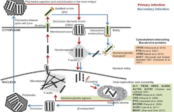
The BEVS relies on the infection of insect cells by recombinant baculoviruses that were genetically modified to carry genes of interest. Baculovirus is a rod-shaped (30-60 nm ×250–300 nm) with double-stranded DNA genome and infects insects and other arthropods (Jehle et al., 2006). The wild type baculovirus replication cycle is biphasic giving rise to two types of virions: occlusion-derived virions (ODVs) and budded virions (BVs), as shown in
11 Figure 2. Virus’s life cycle comprises three phases concerning gene expression: immediate early/early, late and very late (Passarelli and Guarino, 2007). In the very late phase of infection polyhedrin is expressed by a very strong promotor due to its importance in viral ODVs assembly (Volkman, 1997). Given that in biotechnological applications the infection is done with BVs there is no need for polyhedrin protein which gives the opportunity to change this gene for a gene-of-interest (GOI; recombinant protein) (Merrington et al., 1997). Thereby, this allows high productivities that can reach more than 25% of total cell proteins (Caron et al., 1990) although only at the very late stage of infection. Likewise, the p10 protein gene expression is also driven by a very late strong promoter (Smith et al., 1983b) and this gene can also be replaced by a GOI in recombinant baculoviruses not affecting the replication cycle.
12 The most commonly used baculoviruses are Autographa californica multicapsid nucleopolyhedrovirus (AcMNPV) named after its ability to encapsidate multiple nucleocapsids in the occluded particle (polyhedron) and owns a genome of approximately 134kbp (Ayres et al., 1994). It is widely used in lepidopteran derived insect cell lines mentioned above (Sf9, Sf21 and Hi5).
The BEVS has become very popular in the biotechnology field, with numerous commercially available kits. For example, the MultiBacTM system is able to generate multiprotein co-expression which is an evolution over the polycistronic vectors. It was further optimized by eliminating the baculoviral genes v-cath and chiA that encode proteases, abrogating their function (Bieniossek et al., 2012). Subsequently to bacmid transfection, recombinant baculovirus are assembled and released from cells following infection and propagation of the viruses which also leads to recombinant protein production.
To address a potential insect cell PTMs issue, the SweetBac™ system was designed to integrate N-acetylglucosaminyltransferase and β1,4-galactosyltransferase enzymes in the viral genome to generate humanized glycosylation patterns on recombinantly expressed proteins (Palmberger et al., 2012; Palmberger et al., 2015). Another issue is the accumulation of inactive forms in host cells and protein degradation by the ubiquitin proteasome pathway, for example. By co-expressing chaperones (e.g. calnexin and calreticulin chaperones) and folding factors along with the desired protein, authors have managed to enhance the surface expression and protein folding in insect cells (Kato et al., 2005).
In relation to baculovirus vector stability, it may be affected by tandem repetition of promoter sequences in polycistronic constructs (Belyaev and Roy, 1993) and to by-pass this problem identical promoters have been separated into different transcription directions. Moreover, improved stability can also be accomplished by producing a bicistronic mRNA including the GOI coupled to an essential baculoviral gene - gp64 – hence placing a positive selection pressure upon the entire mRNA, thereby ensuring the expression of the protein of interest (Pijlman et al., 2006).
Comparing the two most used cell lines, Hi5 cells have shown to be better recombinant protein producers than Sf9 cells (Davis et al., 1993; Krammer et al., 2010) which are better at producing infectious viral particles (Monteiro et al., 2014). Both cell lines have
13 been proved to undergo oxidative stress during baculovirus infection, resulting in loss of cell viability and consequently cell death (Wang et al., 2001). Despite the popularity of BEVS, many things remain unclear concerning the impact of infection on the cell host, which is a critical subject when the aim is to scale-up the process as efficiently as possible (Maranga et al., 2004; Monteiro et al., 2014).
Although BVs are considered safe because they cannot replicate in mammalian cells, its genome is able to integrate in the human genome (Merrihew et al., 2001) and the consequences of it still remains uncleared. Therefore, BVs and host’s cell DNA contamination are a concern when the goal is to generate a product for human use. Efforts to develop better purification processes have been conducted (Rueda et al., 2000; Vicente et al., 2009) though it is very challenging because in this system virions are co-produced with the desired protein. In order to circumvent this issue, the Geneva Biotech’s ManuBac™ system is being developed which is a virion free protein production platform that uses an induction protocol to turn off virions production at the same time VLP production is turned on (Biotech, 2015). Likewise, to eradicate the presence of baculovirions, a non-replicative baculovirus was engineered by removing the vp80 gene which is implicated in viral protein cleavage, maturation, assembly and release of virions from cells (Marek et al., 2011).
1.2.2 Advantages/disadvantages for production of Influenza vaccines
Considering influenza vaccines, BEVS-based production has proved to be as efficient as more traditional strategies like egg- and cell-based (Bright et al., 2008) with great cultivation benefits compared to mammalian hosts and easily scaled-up. Furthermore, the construction of recombinant baculoviruses (rBacs) is becoming more rapid and versatile, allowing to easily obtain multi-gene expression. Flexibility in rBac construction gives the opportunity of combining genes of different influenza strains, including the most prone to diverge and suffer mutations like HA, allowing the production of broader vaccines. Thus, it offers the great opportunity to renew a vaccine much more rapidly without the need of isolating the circulating influenza virus strain which holds its own biosafety and laborious issues. Hereupon, some shortcomings seen in other vaccines-platforms are overcome such as
14 the virus adaptation to cells, the inability of propagating more pathogenic strains that leads to host death, lack of proteins prone to cause allergies contained in eggs as well as being much more cost effective and faster.
The main bottlenecks of the insect cells-baculovirus system for influenza VLPs production reside in the downstream processing (DSP) (unable to separate rBac from VLPs) and viral stock maintenance (infectious particles titer decreases with time). On top of that, for influenza VLP vaccine candidates that do not contain the M1 protein, a more complex purification process is needed to carefully extract the membrane-anchored HAs without comprising protein integrity.
1.3 Cell line development
The issues associated with BEVS lead to an increasing effort to develop stable systems to produce recombinant proteins in insect cells without the need of using viral infection. Cell line development consists on engineering cells to stably express the GOI and it should be a rapid and standardized process. Stable cell lines are obtained thanks to genetic modifications and one of the main goals is transcriptional efficiency of the GOI where strong promoters, enhancers elements and cis and trans-acting elements play key roles (Nehlsen et al., 2009; Nehlsen et al., 2011). Besides the amount of protein produced, its quality and maintenance of its functionality is very important.
Stable cell lines can be obtained by allowing a GOI to integrate into the genome based on random integration or locus-specific integration. A number of options have been reported for the maintenance of a vector integrated in the genome and most frequently cells are positively selected with a drug (antibiotic for instance) (Fernandes et al., 2012). However, regarding industrial purposes, it is preferable if the process does not need a selection agent because it causes instability in cells and increases the cost of the process (Qiao et al., 2009; Schiedner et al., 2008).
Although offering many advantages, the establishment of stable cell lines still encloses major shortcomings that need to be addressed such as (1) long timeline needed to generate stable, high expressing clones, (2) product yield and quality and (3) flexibility of the cell line.
15
1.3.1 Random integration
Random integration requires a laborious screening process to identify stable and high expressing clones because expression of the GOI greatly depends on the chromosomal elements nearby the integration site, a phenomenon called “position effect”, which makes the integration process irreproducible (Nehlsen et al., 2011; Siegal and Hartl, 1998). Furthermore, random integration can i) lead to the interruption of cellular housekeeping genes, ii) give rise to epigenetic silencing, iii) affect cell’s stability and iv) induce mutagenic effects by inhibiting protective genes or causing gene mutations.
Nevertheless, it offers an advantage over targeted integration as it does not requires knowledge of the genome sequence and chromosomal sites characterization which is not available for some transformed cell lines, being the case of Sf9 and Hi5 cells.
1.3.2 Locus-specific integration
Locus-specific integration is advantageous in the way that if good locus/loci have been previously identified it/they can be tagged and then exchanged for the GOI without the need of screening. When deep knowledge of the working cell line exists, characterized genomic sites can be exploited and reused by homologous recombination (HR) leading to a precise, predictable and reproducible process. Nevertheless, HR is quite inefficient due to dominance of illegitimate recombination (IR) with a ratio of HR/IR of 1:1000 which hampers its broader applicability in transformed cell lines (Turan et al., 2013). Due to this low efficiency, efforts have been made to develop methods capable of achieving higher site-specific integration frequencies.
16 ZFNs, TALENs and CRISPRs
Genome editing methods have expanded and artificial enzymes such as zinc-finger nucleases (ZFNs) and transcription activator-like effector nucleases (TALENs) have been designed to stimulate HR and they rely on the introduction of double strand-breaks (DSBs). Both enzymes have a nuclease activity and a customizable DNA-binding domain which enables to direct them to any target sequence (Mani et al., 2005; Vanamee et al., 2001). In order to broaden their applicability, efforts are being made to design more gene targets in different cell types and to improve the delivery method, targeting specificity and avoiding cytotoxicity. In addition, clustered regularly interspaced short palindromic repeats (CRISPRs) coupled with Cas endonucleases are being used in genome engineering and include programmable RNA-guided DNA endonucleases with ability to modify genomes (Mali et al., 2013; Ronda et al., 2014).
Despite their advantages, concerns are related to potential unspecific cleavage of endogenous genes (Miller et al., 2007). Moreover, such systems rely on genome sequence knowledge of the working cell line.
Recombinase-mediated cassette exchange (RMCE)
RMCE was firstly introduced by Schalke and Bode (1994) and it is a process in which a tagging cassette, flanked by a pair of heterologous recombinase target sites, can be exchanged by a target vector after being integrated into the genome (Oumard et al., 2006). Not only these target sites have to be non-compatible so that the exchange process is accurate but they must also be the same in the tagging and target cassettes.
Typically, the anchored cassette (tagging) encodes a reporter protein and a given selective marker (antibiotic resistance gene, for example), and then it is exchanged for a GOI by means of a site-specific recombinase (SSR), as depicted in Figure 3. The recombinase can be provided in the tagging, the target vector or in a separate vector.
Even though after the tagging step an intensive screening of the best locus is required, the RMCE system enables the reuse of the same locus, decreasing the time spent in further screening process (Gama-Norton et al., 2010; Nehlsen et al., 2009). Moreover, it has been shown that this method offers stable and high levels of gene expression (Coroadinha et al.,
17 2006; Schucht et al., 2006). This system is flexible enough to be used in many applications ranging from the biotechnology field for the establishment of producer cell lines (Coroadinha et al., 2006; Rose et al., 2013), for antibody production (Wiberg et al., 2006) or in the genetic area by allowing a more efficient analysis of gene function in mice (Seibler et al., 1998).
Site-specific recombinases (SSRs)
The most commonly used site-specific recombinases belong to two distinct families according to the structure of their active site: the Tyr-class such as Cre and Flp (Nunes-düby et al., 1998) or the Ser-family like ΦC31 (Smith and Thorpe, 2002) (Table 2).
Concerning ΦC31, it comes from Streptomyces bacteria where its role is to allow the integration of a phage into the bacterial chromosome which occurs via attP/attB sites (Smith and Thorpe, 2002). However, the system cannot be applied to RMCE neither to multiplexing protocols owing to the lack of recombinase efficiency of the enzyme over the integrase efficiency. The Cre enzyme (for “causes recombination”) is a bacteriophage (P1) encoded integrase and was firstly described in bacteria whose function relies on a target site called loxP (locus of crossover in P1). This site is a 34bp sequence that consists of two inverted 13bp repeats separated by an 8bp spacer (Sternberg et al., 1986). Since its discovery, this system has been applied in mammalian cells aiming to be a powerful tool for deeper
Figure 3 - RMCE principle: tagging a locus with a cassette flanked with heterospecific target sites into the genome and then exchanging it for the GOI. Adapted from Turan et al. 2010.
Tagged genomic locus
18 understanding of genomic phenomena in eukaryotes (Sauer and Henderson, 1988) and efforts have been made to improve its efficiency (Koresawa et al., 2000). Despite having some benefits, Cre-induced toxicity as well as impairment of the host’s DNA integrity have been reported (Fernandes et al., 2015; Schmidt et al., 2000).
Flp was identified in the 2µm circle plasmid of Saccharomyces cerevisiae where it is involved in site-specific recombination (Andrew et al., 1985; Mcleod et al., 1986) and its use in mammalian cell lines was firstly reported by O’Gorman (O’Gorman et al., 1991). The Flp enzyme induces a double-reciprocal crossover between two pair of target sites (FRTs) each one consisting on an 8bp asymmetric spacer flanked by a 13pb repeat at one side and two 13bp repeats on the other side, completing a 48bp FRT site. Although the spacer sequence determines the orientation of the site it does not contact directly with the enzyme (Turan et al., 2010). Even though in some cases the Flp/FRT system is less efficient than the Cre/loxP its use has been increasing significantly (Fernandes et al., 2012; Whiteson et al., 2007). Examples are the production of viral vectors for gene therapy (Coroadinha et al., 2006), study of genetic phenomena (Nehlsen et al., 2011; Seibler et al., 1998), recombinant protein production (Kim MS and Lee, 2008; Nehlsen et al., 2009; Wilke et al., 2011) or engineering strains (Cesari et al., 2004). There are various sets of flippase recognition target sites (Flp/FRTs) that were designed by mutagenesis (Schlake and Bode, 1994) and these have different recombination efficiencies and probability of cross-recombination events (Schlake and Bode, 1994; Turan et al., 2010).
Given the need of using Flp enzyme in animal cells, there were several efforts into improving its efficiency. For instance, the wild type Flp (wt Flp) was extremely inefficient at 37ºC because its optimum activity temperature is 30ºC (Buchholz et al., 1996) and efforts were made in order to improve this characteristic. Buchloz was able to construct Flpe successfully which is more termostable at 37ºC (Buchholz et al., 1998). Later on, Flpe enzyme was mouse-codon optimized into Flpo (Raymond and Soriano, 2007) and hFlep (humanized Flpe) also with great success (Kondo et al., 2009). So far toxicity of Flp has not been reported.
Fernandes et al. developed a Sf9 master cell line making use of RMCE and flippase enzyme with the purpose of being a good alternative to BEVS. Firstly, the authors were able
19 to produce the same amount of enhanced green fluorescent protein (eGFP) as in the BEVS system (Fernandes et al., 2012) and then the same principle was successfully applied to the production of more complex proteins such as rotavirus-like particles (Fernandes et al., 2014). This resulted in increased quality and yield of production, which is often compromised in the baculovirus-expression system due to proteolysis in late stages of infection (Monteiro et al., 2012). Stable expression does not compromise the host at such a level as it is seen for BEVS and it can also be adapted to bioreactor strategies for industrial purposes. Thus, it is predicted that this system will be robust enough to outpace the BEVS once some difficulties are overcome like the longer period of time taken to have the product and the lack of chromosomal loci characterization in insect cells.
SSRs RTs employed Cell lines
Cre
LoxP/Lox511
MEL NIH3T3
mES
Fertilised mouse oocytes K562
J558L
LoxP/Lox2272 mES
LoxP/Inverted LoxP mES
Lox66/71 and Lox2272 mES
Lox511/InvertedLox511 MEL
Mouse B hybridoma cells LoxP/LoxP257 HeLa/CHO cells
Primary MEF/mES cells
LoxP/Lox5171 mES Flp FRT/FRT3 BHK mES NIH3T3 FRT/FRT5 NIH3T3 HEK293 mES BHK
ΦC31 attB and attP
Primary epidermal progenitor cells
mES
Cre and Flp LoxP and FRT mES
20 Aim of the thesis
This thesis aims at developing robust insect cells based platforms for production of complex products such as Influenza virus-like particles (VLPs) as vaccine candidates.
In order to achieve such goal two strategies have been designed (Figure 4). The first one consists in combining stable expression of two HAs in Hi5 cells, based on random integration of the GOIs, with baculovirus-mediated expression of M1 and additional HA proteins to produce multivalent VLPs. This approach minimizes the potential risk for instability caused by the addition of many genes in a single baculovirus vector, when developing a production process for multivalent HA VLPs. To by-pass the expression unpredictability of HA random integration, the second strategy consists in generating stable insect cell lines based on our in-house developed flippase-recombinase mediated cassette exchange (Flp-RMCE) platform, to be able to then re-use pre-characterized genomic loci to integrate multiple HA. The feasibility of having two genomic cassettes flanked by different pairs of flippase recognition target sites (FRTs) (double-RMCE platform) will be evaluated.
21
2 Materials and Methods
2.1 Molecular Biology
2.1.1 Plasmid design and construction
Primers sequences are listed in Appendix A.1. Stable HA expression
pIZT/HA1,2 vector: HA1 and HA2 vectors were kindly provided by RedBiotech AG
(Switzerland). Each HA gene was amplified by PCR and cloned into a KpnI or NotI (respectively) excised pIZT/V5-His (Invitrogen, Carlsbad, USA) resulting in pIZT/HA1 and
pIZT/HA2 vectors. OpIE2 promoter and HA2 genes were amplified by PCR from pIZT/HA2
vector and cloned into pIZT/HA1 vector opened by inversePCR.
pIZT/HA2,3 vector: HA3 vector was kindly provided by RedBiotech AG
(Switzerland) and amplified by PCR into a SacI excised pIZT/V5-His (Invitrogen, Carlsbad, USA) resulting in pIZT/HA3 vector.OpIE2 promoter and HA2 genes were amplified by PCR
from pIZT/HA2 vector and cloned into a ClaI excised pIZT/HA3 vector.
Double-RMCE system associated vectors
pTaggF13/F14: The tagging cassette based on the F13 and F14 FRT sites and containing
OpIE2 and OpIE1 promoters (pTagg) was designed by us and synthetized by GenScript (USA). This cassette was then digested with NheI and PsiI. iCherry and hygromycin marker genes were amplified by PCR from an in-house vector and cloned in the previous excised vector.
pTargetF13/F14: To construct the target vector, OpIE2 and OpIE1 promoters were eliminated from pTaggF13/F14. eGFP and neomycin marker genes were amplified by PCR
22 pOpIE2 M1/HA: an in-house vector containing Fw and F5 sites, eGFP and
hygromycin resistance genes was opened by inverted PCR (peGFP/Hygro). OpIE2 promoter and HA genes were amplified by PCR from an in-house construct and cloned in the previous opened peGFP/Hygro vector (pOpIE2 M1/HA+eGFP). eGFP was then eliminated by digestion with BamHI and NotI and M1 (previously amplified by PCR from an in-house vector) was cloned in the excised site.
2.1.2 Techniques supporting plasmid construction
General PCR-protocol
The oligonucleotides used for PCR were custom-made by Sigma Aldrich (St.Louis, USA). A typical PCR-reaction included 4µl of 5x polymerase buffer (Thermo Scientific), 0.4μl of 10mM dNTPs (NZYTech), 0.4μl of 25μM primers (Sigma), 20ng of template DNA and 1 to 5 U of Phusion® High-Fidelity DNa polymerase (Thermo Scientific). RNAse-free water (Sigma) was also added to the final volume of 20μl. The PCR-amplification program started with a 30s denaturation step at 98ºC, followed by 30 cycles of 10sec denaturation at 98ºC, primer annealing for 30s performed up to 5ºC below the melting point of the primer, and extension at 72ºC according to the fragment size. The next step in the cycle was final extension at 72ºC for 10 min.
Agarose gel electrophoresis
Agarose gel electrophoresis was performed to separate DNA-fragments. The concentration of each gel varied according on the size of the fragments in question. Agarose (Lonza) was melted in 1x TAE buffer (Promega) and stained with GelRed or RedSafe (Biotium; iNtRON Biotechnology). Before loading, samples were mixed with loading buffer (NEB; #B7024S) and a standard ladder was used according to the range of fragment sizes expected. For purification of bands, when needed, Illustra GFX kit (GE Healthcare) was used. Gels were photographed using GelDocTM system (Bio-Rad) and DNA quantification was used using Nanodrop ND-2000c (Thermo Scientific).
23 Transformation and vector isolation
Competent E.coli cells were transformed according to the manufacturer’s protocol (NZYTech, ref. MB00401 or Clontech, ref. 636763). Transformed cultures were spread on LB-agar plates containing ampicillin or zeocin and grown overnight at 37 °C. The next day, several isolated colonies were picked and grown separately, in falcon tubes, using 5mL of TB antibiotic supplemented culture medium at 37ºC and 190rpm. After 16-18h, 2mL of cell culture was harvested by centrifugation and DNA was extracted and purified with the miniprep kit (Thermo Scientific) following the manufacturer’s protocol.
To identify whether transformants contained the gene of interest, PCR screening and vector digestion were followed by agarose gel electrophoresis analysis.
Digestion of DNA
DNA-digestion of PCR-fragments or vector-DNA was performed with the appropriate restriction endonucleases according to the manufacturer’s specifications (NEB). When digestion of a vector was desired, further excision and purification from agarose gel was performed with Illustra GFX purification kit (GE Heathcare).
Ligation with In-Fusion
For the ligation of DNA-fragments the In-Fusion® HD Cloning kit was used following the instructions of the manufacturer (Clontech; ref. 638910). The ligated vector-DNA mix was used to transform bacterial cells, as previously described.
24
2.2 Cell line development
2.2.1 Transfection
Foreign DNA was inserted into cells using lipotransfection based on Cellfectin® II reagent (Invitrogen). 8l of Cellfectin and 100ul of Grace’s Insect Medium (Gibco) were used to 1x10⁶ cells (unit of transfection, UT). Transfections were conducted in 125mL shake flasks in 10mL working volume.
For the tagging step in the RMCE strategy, parental Hi5 and Sf9 were transfected at cell concentrations of 0.3x10⁶ cells/mL and 0.5x10⁶ cells/mL, respectively, using 0.3µg/UT
of DNA. Selection was performed with hygromycin (0.2mg/mL; Invivogen) or zeocin (0.1mg/mL; Invivogen) depending on the expression vector resistance marker.
For the establishment of stable Hi5 pools expressing HA genes, parental Hi5 cells were transfected at 0.5x10⁶ cells/mL and selection was performed with zeocin (0.1mg/mL; Invivogen).
2.2.2 Cassette-exchange
To perform RMCE, 0,1µg/UT of target cassette and 0,3µg/UT of iFlp-expressing vector were used and selection was performed with hygromycin (Invivogen) or neomycin (Invivogen) depending on the expression vector resistance marker. Cassette-exchange was performed at a cell density of 1x10⁶ cells/mL for Sf9 cells. When viabilities dropped to 50%, cells were transferred to T-flasks (75cm2). After 24h, the medium was replaced by conditioned medium supplemented with 10% (v/v) fetal bovine serum (FBS) (Gibco) and the respective antibiotic, and then changed every four to five days. Fluorescence intensity and cell colonies’ growth were evaluated by visual inspection (DMI 6000, Leica). When confluent, cells were transferred back to suspension and cultured with the routinely used medium plus antibiotic.
25
2.2.3 Sorting procedures
Cells were sorted in a MoFlo high speed cell sorter (BeckmanCoulter) equipped with a 488 nm laser (200 mWair-cooled Sapphire, Coherent) for scatter measurements and a 561nm laser (50 mW DPSS, CrystaLaser) for iCherry excitation. iCherry was detected using a 630/75 nm bandpass emission filter. As a special requirement for insect cells, cells were resuspended in phosphate buffer saline (PBS) supplemented with Pluronic acid F-68 (PF68; Sigma) (Vidigal et al., 2013). PBS was used as sheath fluid and run at a constant pressure of 207 kPa with a 100µm nozzle and a frequency of drop formation of approximately 30 kHz. Cells were collected into 1 mL of PBS, also supplemented with PF68, and maintained at 4ºC. After sorting, cells were pelleted (200g for 10 min) and seeded in 6-well plates. They were kept for one week in culture medium with antibiotics–antimycotics (Invitrogen).
2.2.4 Cloning
Cloning by limiting dilution is a procedure to separate cells through serially dilutions of the culture suspension until the amount of 1 cell in 100 μl of final solution is reached. The medium is composed by 50% conditioned (the supernatant of exponentially growing parental cells) and 50% fresh medium and G418 (an analogous of neomycin). Then, 100 μl of this mixture was transferred into a separate 96-wells plate so each well receives one cell. When confluency was achieved, each clone was transferred to a 48-well, 24-well, 12-well and then to a 6-well and finally to 10mL suspension culture. From one cell per well to suspension cultures it took about 2-3 months.
2.3 Cell culture
Sf9 cells were purchased from Invitrogen and Hi5 cells were kindly provided by RedBiotech. For suspension cultures, cells were routinely cultured in 125mL or 500 shake flasks (working volume of 10-20mL or 30-50mL, respectively) at 27ºC in orbital shakers at 100rpm. Sf900 II serum-free medium (Gibco) and Insect X-press (Lonza) were used for Sf9 and Hi5 cultures, respectively. Cells were sub-cultured every 3-4 days when cell density reached 2-3x10⁶ cells/mL. Hi5 cells expressing HA genes were supplemented with lipids
26 concentration and viability were calculated by haemocytometer counting (Brand, Wertheim, Germany) using trypan blue exclusion dye (Merck, Darmstadt, Germany). For adherent cultures, cell cultivation was maintained in T-flasks (75cm2) with conditioned medium supplemented with 10% (v/v) of FBS (Gibco) and sub-cultured when confluency was reached.
2.3.1 Freezing and thawing cells
Cells at exponential growth phase (2-3x10⁶ cells/mL) were centrifuged at 200g, 4ºC for 10min, and cell pellets were ressuspended in cryopreservation media (CryoStor®, Sigma) to obtain a concentration of 1-2x10⁷ cells/mL. Aliquots were frozen using a freezing container (Mr. Frosty) (Thermo Fisher Scientific) and stored at -80ºC until further use. Thawing was performed by centrifuging cells with 12mL of fresh medium at 200g for 10 min to eliminate cryo preservation medium. After this, the cell pellet was re-suspended in medium, according the volume to the cell density desired. Suspension culture was then performed in standard conditions.
2.4 Baculovirus
2.4.1 Virus amplification
Recombinant baculoviruses were kindly provided by RedBiotech AG (Switzerland) and virus titters determined using the Virocyt virus counter (Virocyt, USA). Whenever needed, virus amplification was performed by infecting Sf9 cells at a CCI of 1x10⁶ cells/mL using a virus (V0 stock) dilution of 1:500, in 2L shake flasks with 300mL of working volume. After reaching a viability of 70-80%, cultures were harvested by centrifuging at 200g and 4ºC for 10 min and supernatant was collected and stored at 4ºC in the dark until further use.
2.4.2 Virus titration
Virus titration was performed using the MTT method (Mena et al., 2003; Roldão et al., 2009). Briefly, 100 µL of 0.5x10⁶ cells/mL of Sf9 cells were seeded into 96-well plates
(Nunc, Roskilde, Denmark) and allowed to attach to the plate for at least one hour at 27°C in the dark. Then, culture supernatant was removed and cells were infected with serial dilutions
27 of baculovirus. Both positive (non diluted baculovirus stock) and negative (virus free culture media) controls were added to the assay. Plates were incubated for 6 days at 27ºC in the dark. After this period, 10µl of 0.5 mg/mL MTT solution was added to each well and plates were incubated for 4h at 27ºC. After removing the supernatant, the formazan crystals were solubilized with dimethyl sulfoxide (Sigma–Aldrich) (150µl/well) and incubated for additional 20min under constant shaking. The absorbance (570/690nm wavelength) was measured using a plate reader (Tecan, Switzerland). The collected data was analyzed using GraphPad Prism 4 (Graph-Pad Software, Inc., La Jolla, CA).
2.4.3 Infection of insect cells with baculovirus
Hi5 cells were inoculated at 0.3x10⁶ cells/mL and were allowed to grow until the time of the infection. Infections were performed at different CCIs of 2, 3 and 4x10⁶ cells/mL though always with the same MOI of 10 total viral particles/cell.
2.5 Production of influenza VLPs in 2L bioreactors
Bioreactor culture was performed in BIOSTAT® B-DCU 2L vessels (Sartorius, Goettingen, DE). The pH, temperature and dissolved oxygen were monitored on-line. The dissolved oxygen was set to 30% of air saturation and was maintained by automated stirring and air/oxygen supply on demand. The gas flow rate was set to 0.02 L/min. The inoculum was 0.5x10⁶ cells/mL and 40h later a supplement mixture was added, as described above. Infection was performed when cells reached a concentration of 4x10⁶ cells/mL with a MOI of 10 total viral particles/cell. Samples were taken daily to analyze cell viability and density as well as the HA titer. Medium additions and sampling proceedings were performed aseptically in a moveable flow chamber (Cruma 670 FL, Spain).
2.5.1 Downstream processing of influenza VLPs
After cells reached viabilities around 50-60%, the bioreactor bulk was centrifuged at 200g, for 10min at 4ºC. Supernatant was collected and then supplemented with 50 U/mL of benzonase (Merck Millipore, Germany) for 15min at room temperature to digest any host and/or viral DNA in solution. Supernatant was then filtered using a Sartopore 2 membrane