Assessment of enoxacin effect on cancer growth and microrna expression in prostate cell lines
88
0
0
Texto
(2)
(3) ELSA JOANA FERREIRA DE SOUSA. ASSESSMENT OF ENOXACIN EFFECT ON CANCER GROWTH AND MICRORNA EXPRESSION IN PROSTATE CELL LINES. Dissertation for applying to a Master’s degree in Oncology – Specialization in Molecular Oncology submitted to the Institute of Biomedical Sciences Abel Salazar, University of Porto. Supervisor Carmen de Lurdes Fonseca Jerónimo, PhD Guest Associate Professor with Aggregation Department of Pathology and Molecular Immunology Institute of Biomedical Sciences Abel Salazar – University of Porto Assistant Investigator and Coordinator of the Cancer Epigenetics Group Department of Genetics and Research Centre Portuguese Oncology Institute – Porto Co-Supervisor Rui Manuel Ferreira Henrique, MD, PhD Guest Assistant Professor with Aggregation Department of Pathology and Molecular Immunology Institute of Biomedical Sciences Abel Salazar – University of Porto Director of the Department of Pathology Senior researcher of the Cancer Epigenetics Group Portuguese Oncology Institute – Porto.
(4)
(5) ACKNOWLEDGMENTS A realização do trabalho aqui apresentado só foi possível devido à contribuição de várias pessoas a quem quero expressar a minha sincera gratidão. À minha orientadora, a Professora Doutora Carmen Jerónimo, um agradecimento muito especial por me ter recebido no Grupo de Epigenética do Cancro e pelo apoio científico, motivação e entusiasmo com que me orientou ao longo do desenvolvimento deste projeto. Muito obrigada pela confiança que depositou no meu trabalho e por me ter incentivado a fazer sempre mais e melhor. Ao meu co-orientador, o Professor Doutor Rui Henrique, pelo rigor científico, disponibilidade e sugestões fundamentais na conceção deste trabalho. Obrigada também pela sua importante contribuição como Patologista, sem a qual este trabalho não teria sido possível. Ao Professor Doutor Manuel Teixeira, Diretor do Serviço de Genética e do Centro de Investigação do Instituto Português de Oncologia do Porto, por ter permitido o desenvolvimento deste trabalho no Centro que dirige. Ao Professor Doutor Carlos Palmeira, pela oportunidade de realizar a análise do ciclo celular por citometria de fluxo e pela ajuda e disponibilidade demonstrada. À Inês, pela partilha de ideias e experiências que me permitiu aprender tanto e que foi fundamental ao longo deste percurso. Este trabalho não teria sido possível sem o teu encorajamento, otimismo, espírito crítico e paciência. Muito obrigada pela amizade, pelas longas conversas, pela ajuda e companhia nas inúmeras horas passadas na câmara de fluxo, pela cumplicidade e por nunca perderes a tua boa disposição contagiante... Foi um enorme prazer ter trabalhado contigo! À Filipa, pela importante ajuda na fase final deste trabalho, pelo incentivo, pela boa disposição constante e por todos os momentos que partilhamos ao longo desta etapa da minha vida. Acima de tudo, obrigada pela amizade sincera e pelo companheirismo. Ao Pedro, pela amizade, troca de ideias e disponibilidade constante. Muito obrigada por me proporcionares tantas gargalhadas e momentos divertidos! À Joana, por ser uma amiga preocupada e sempre disponível, pelas conversas e pelos momentos de diversão. Ao Tiago, pela prontidão em ajudar, pelo espírito crítico constante e pela boa disposição.. III.
(6) ACKNOWLEDGEMENTS À Mafalda, pela ajuda na fase inicial deste trabalho e, sobretudo, pela amizade. À Susana, minha companheira nesta aventura, pela amizade e apoio constante. A todos os membros do Grupo de Epigenética do Cancro, por me terem proporcionado um ambiente de trabalho fantástico, pela ajuda prestada e pelos momentos de descontração. Obrigada pela alegria que trouxeram a cada um dos dias que passei na vossa companhia! Agradeço também a todos os que trabalham no Serviço de Genética pela forma simpática como me receberam e pela disponibilidade demonstrada. À Catarina, minha amiga de todos os momentos e minha companheira ao longo de todo o meu percurso académico, agradeço o apoio constante, os desabafos, as palavras de incentivo e a amizade sincera. É bom saber que existes e que posso contar contigo! Ao Diogo, por me fazer acreditar que todos os meus sonhos se podem tornar realidade… Obrigada pela tua inesgotável paciência, pelo apoio incondicional, pela constante motivação, pelo amor e, acima de tudo, por me fazeres tão feliz. Sem dúvida, tudo se torna mais fácil por caminharmos lado a lado! Às minhas avós, pelo carinho, preocupação e orgulho que sentem por mim. Espero nunca vos desapontar! À minha querida Inês, por me fazer esquecer qualquer preocupação ou cansaço com o seu sorriso maravilhoso, pelos momentos de ternura, pela cumplicidade e pelas maluqueiras de irmãs! Obrigada por preencheres o meu coração de amor e de felicidade e por seres a irmã que sempre quis ter! Por fim, um agradecimento muito especial aos meus Pais, a quem devo tudo o que sou. Obrigada pela força e determinação que me transmitem, pelo encorajamento, pela compreensão, pela infinita paciência, pelos sábios conselhos e, acima de tudo, pelo amor incondicional. Muito obrigada por apoiarem cada um dos meus passos e por me ajudarem a encontrar o caminho para a felicidade!. Agradeço ainda à Liga Portuguesa Contra o Cancro – Núcleo Regional do Norte pela bolsa concedida durante o ano de realização do trabalho experimental apresentado nesta dissertação de Mestrado.. Este estudo foi financiado pela Liga Portuguesa Contra o Cancro – Núcleo Regional do Norte, pelo “European Community’s Seventh Framework Programme – Grant number FP7-HEALTH-F5-2009-241783” e pelo Centro de Investigação do Instituto Português de Oncologia do Porto (CI-IPOP 4-2008).. IV.
(7) RELEVANT ABBREVIATIONS A. Adenine. AMO. Anti-miRNA Oligonucleotide. AS. Active Surveillance. BCA. Bicinchoninic Acid. BPH. Benign Prostatic Hyperplasia. BSA. Bovine Serum Albumin. C. Cytosine. CASP3. Caspase 3. cDNA. Complementary DNA. CpG. Cytosine-phosphate-Guanine. CRPC. Castration-Resistant Prostate Cancer. DAB. 3,3-Diaminobenzidine. DGCR8. Di George Syndrome Critical Region 8. DMSO. Dimethyl Sulfoxide. DNA. Deoxyribonucleic Acid. DNMT. DNA Methyltransferase. DRE. Digital Rectal Examination. dsRNA. Double-stranded RNA. EMT. Epithelial-to-Mesenchymal Transition. FBS. Fetal Bovine Serum. FI. Fluorescence Index. G. Guanine. GS. Gleason Score. GUSB. Beta-Glucuronidase. HAT. Histone Acetyltransferase. HDAC. Histone Deacetylase. HDAC1. Histone Deacetylase 1. HDM. Histone Demethylase. HGPIN. High-Grade Prostatic Intraepithelial Neoplasia. HMT. Histone Methyltransferase. LNA. Locked Nucleic Acid. miRNA. MicroRNA. mRNA. Messenger RNA. V.
(8) RELEVANT ABBREVIATIONS MTT. 3-(4,5-dimethylthiazol-2-yl)-2,5-diphenyltetrazolium-bromide. ncRNA. Non-coding RNA. OD. Optical Density. PBS. Phosphate-Buffered Saline. PCa. Prostate Cancer. PCR. Polymerase Chain Reaction. PIA. Proliferative Inflammatory Atrophy. PIN. Prostatic Intraepithelial Neoplasia. Pre-miRNA. Precursor-miRNA. Pri-miRNA. Primary-miRNA. PSA. Prostate-Specific Antigen. RIPA. Radio Immuno Precipitation Assay. RISC. RNA-induced Silencing Complex. RNA. Ribonucleic Acid. RNAi. RNA interference. RP. Radical Prostatectomy. SDS. Sodium Dodecyl Sulfate. SNP. Single Nucleotide Polymorphism. T. Timine. TARBP2. Trans-Activation-Responsive RNA-Binding Protein 2. TBS. Tris-Buffered Saline. TRUS. Transrectal Ultrasound. UTR. Untranslated Region. XPO5. Exportin-5. VI.
(9) SUMMARY Background: Prostate cancer (PCa) is one of the most incident malignancies worldwide and represents a leading cause of cancer-related morbidity and mortality. Although efficient therapy is available for early-stage PCa, treatment of advanced disease is mainly ineffective and remains a clinical challenge. Thus, new therapeutic strategies, based on the biology of PCa, are urgently needed. MicroRNA (miRNA) dysregulation is associated with PCa development and progression. In fact, several studies have reported a widespread downregulation of miRNAs in this disease, which highlights the importance of studying compounds with the ability to restore the global miRNA expression. Aims: The main aim of this study was to define the usefulness of enoxacin as an anti-tumoral agent in PCa, due to its ability to induce miRNA biogenesis in a Transactivator RNA-binding protein (TRBP)-mediated manner. Material and Methods: A panel of five PCa cell lines was screened for TARBP2 mutations by direct sequencing and the protein levels of TRBP were evaluated by Western Blot. Immunohistochemistry was performed to assess the protein levels of TRBP in primary prostate carcinomas. After exposure of PCa cell lines to enoxacin, cell viability, apoptosis, cell cycle, and cell invasion assays were carried out to evaluate the effects of the drug. A miRCURY LNA™ array was used to determine the impact of enoxacin on the expression profile of miRNAs. Then, the protein levels of histone deacetylase 1 (HDAC1), a direct target of one of the overexpressed miRNAs, were assessed by Western Blot. Results and Discussion: All PCa cell lines analyzed were TARBP2 wild-type and expressed TRBP protein. Furthermore, primary prostate carcinomas displayed normal levels of TRBP protein, rendering them sensitive to restoration of normal miRNA biogenesis by enoxacin. Remarkably, enoxacin was able to decrease cell viability, induce apoptosis, lead to cell cycle arrest, and inhibit the invasive potential of PCa cell lines. Enoxacin was also effective in restoring the global expression of miRNAs, enhancing the expression of tumor-suppressor miRNAs in PCa. Moreover, the overexpression of miR449a, one of the tumor-suppressor miRNAs implicated in PCa, was associated with the downregulation of its direct target oncoprotein, HDAC1. Conclusions: These results demonstrated that PCa cells are highly responsive to the anti-tumoral effects of enoxacin. Therefore, enoxacin constitutes a promising therapeutic agent and in vivo studies should be performed to further support the potential of enoxacin for PCa treatment.. VII.
(10)
(11) RESUMO Introdução: O cancro da próstata (CaP) é uma das neoplasias malignas mais incidentes no mundo, representando uma causa importante de morbilidade e mortalidade. Embora estejam já disponíveis terapias eficazes para a fase inicial da doença, o tratamento da doença avançada apresenta limitações importantes, constituindo um desafio clínico. Assim, torna-se imperativo o desenvolvimento de novas estratégias terapêuticas, baseadas nas características biológicas do CaP. A desregulação dos microRNAs (miRNAs) está associada ao desenvolvimento e progressão do CaP. De facto, diversos estudos têm descrito uma sub-expressão global dos miRNAs nesta neoplasia, destacando a importância de investigar compostos com a capacidade de restabelecer a expressão global dos miRNAs. Objetivos: O principal objetivo deste estudo foi investigar a utilidade da enoxacina como agente anti-tumoral no CaP, tendo como base a sua capacidade de promover a biogénese dos miRNAs de forma dependente da TRBP. Material e Métodos: Foram pesquisadas mutações no gene TARBP2 por sequenciação direta e avaliados os níveis proteicos da TRBP por Western Blot em cinco linhas celulares de CaP. A expressão proteica da TRBP em adenocarcinomas primários da próstata foi determinada por imunohistoquímica. Após exposição das linhas celulares à enoxacina, realizaram-se ensaios para avaliar os efeitos do fármaco na viabilidade celular, apoptose, ciclo celular e invasão. O impacto da enoxacina no perfil de expressão dos miRNAs foi determinado através da análise de arrays miRCURY LNA™. De seguida, os níveis proteicos da HDAC1, alvo direto de um dos miRNAs sobre-expressos, foram avaliados por Western Blot. Resultados e Discussão: Não foram encontradas mutações no gene TARBP2 em nenhuma das linhas celulares e todas expressaram a proteína TRBP. Adicionalmente, os tumores prostáticos primários exibiram níveis proteicos normais de TRBP, o que os torna sensíveis ao restabelecimento da biogénese dos miRNAs pela ação da enoxacina. Este fármaco foi capaz de reduzir a viabilidade celular, induzir apoptose, levar a uma paragem do ciclo celular e inibir o potencial invasor em linhas celulares de CaP. A enoxacina foi igualmente eficaz na restituição da expressão global dos miRNAs, promovendo a expressão dos miRNAs supressores tumorais no CaP. Adicionalmente, a sobre-expressão do miR-449a, um dos miRNAs supressores tumorais implicados no CaP, induziu a sub-expressão da sua oncoproteína alvo, a HDAC1.. IX.
(12) RESUMO Conclusão: Estes resultados demonstraram que as linhas celulares de CaP respondem significativamente aos efeitos anti-tumorais da enoxacina. Assim, a enoxacina constitui um agente terapêutico promissor para o tratamento do adenocarcinoma da próstata, sendo ainda necessária a realização de estudos in vivo para confirmar o seu potencial clínico.. X.
(13) TABLE OF CONTENTS ACKNOWLEDGMENTS ............................................................................................................ III RELEVANT ABBREVIATIONS ................................................................................................... V SUMMARY .......................................................................................................................... VII RESUMO ............................................................................................................................. IX TABLE OF CONTENTS........................................................................................................... XI FIGURES INDEX ................................................................................................................. XIII TABLES INDEX .................................................................................................................... XV INTRODUCTION ...................................................................................................................... 1 1. Prostate Cancer ........................................................................................................... 1 1.1. Anatomy and Histology of the Prostate ................................................................. 1 1.2. Pathologic Conditions of the Prostate Gland ......................................................... 2 1.3. Epidemiology ......................................................................................................... 3 1.3.1. Risk Factors .................................................................................................... 5 1.4. Diagnosis ............................................................................................................... 6 1.5. The Gleason Grading System ............................................................................... 6 1.6. Clinical and Pathologic Staging ............................................................................. 8 1.7. Therapy ............................................................................................................... 10 1.7.1. Treatment of Clinically Localized Disease .................................................... 10 1.7.2. Treatment of Advanced and Castration-Resistant PCa ................................ 11 2. Epigenetics................................................................................................................. 12 2.1. MicroRNA Biogenesis and Mode of Action.......................................................... 15 2.2. MicroRNAs and Cancer ....................................................................................... 17 2.3. MicroRNA-based Therapies ................................................................................ 18 2.3.1. Enoxacin ....................................................................................................... 19 3. MicroRNA Dysregulation in PCa ................................................................................ 20 AIMS OF THE STUDY ............................................................................................................ 23 MATERIAL AND METHODS .................................................................................................... 25 1. Cell Culture ................................................................................................................ 25 2. TARBP2 Analysis ....................................................................................................... 26 2.1. Mutational Status Evaluation of PCa Cell Lines .................................................. 26 2.1.1. DNA Extraction ............................................................................................. 26 2.1.2. Direct Sequencing ......................................................................................... 27 2.2. Assessment of Protein Levels of PCa Cell Lines ................................................ 28 2.2.1. Protein Extraction.......................................................................................... 28 . XI.
(14) TABLE OF CONTENTS 2.2.2. Western Blot ................................................................................................. 29 2.3. Assessment of Protein Levels of PCa Tumor Samples ...................................... 30 2.3.1. Patients and Sample Collection .................................................................... 30 2.3.2. Immunohistochemistry .................................................................................. 30 3. Evaluation of the Effect of Enoxacin on PCa Cell Lines ............................................ 31 3.1. Enoxacin Exposure ............................................................................................. 31 3.2. Cell Viability Assay .............................................................................................. 32 3.3. Apoptosis Assay.................................................................................................. 33 3.4. Assessment of CASP3 mRNA Expression ......................................................... 34 3.4.1. RNA Extraction ............................................................................................. 34 3.4.2. cDNA Synthesis ............................................................................................ 35 3.4.3. Quantitative Reverse Transcription PCR ...................................................... 35 3.5. Cell Cycle Analysis.............................................................................................. 37 3.6. Cell Invasion Assay ............................................................................................. 37 3.7. Assessment of miRNA Expression by Microarray............................................... 38 3.7.1. RNA Extraction and cDNA Synthesis ........................................................... 38 3.7.2. Quantitative Reverse Transcription PCR ...................................................... 38 3.7.3. Microarray Analysis ...................................................................................... 39 3.8. Protein Expression Evaluation of HDAC1 ........................................................... 40 3.8.1. Protein Extraction ......................................................................................... 40 3.8.2. Western Blot ................................................................................................. 40 4. Statistical analysis ..................................................................................................... 40 RESULTS ............................................................................................................................ 41 1. Mutational and Expression Status of TRBP in PCa Cell Lines .................................. 41 2. TRBP Expression in PCa Tumor Samples ................................................................ 42 3. Anti-tumoral Effect of Enoxacin on PCa Cell Lines.................................................... 42 3.1. Cell Viability......................................................................................................... 42 3.2. Apoptosis ............................................................................................................ 44 3.3. Cell Cycle ............................................................................................................ 47 3.4. Cell Invasion........................................................................................................ 49 3.5. MicroRNA Expression ......................................................................................... 49 3.5.1. Protein Expression Status of HDAC1 – a miR-449a target .......................... 51 DISCUSSION........................................................................................................................ 53 CONCLUSIONS AND FUTURE PERSPECTIVES ........................................................................ 57 REFERENCES ...................................................................................................................... 59 . XII.
(15) FIGURES INDEX Figure 1. Zonal anatomy of the normal prostate ................................................................. 1 Figure 2. Incidence of different types of cancer in Europe and Portugal, in males ............. 3 Figure 3. Estimated age-standardized incidence rate per 100,000 worldwide ................... 4 Figure 4. Updated Gleason score for histological grading of prostate tumors .................... 7 Figure 5. miRNA biogenesis and mechanism of action .................................................... 16 Figure 6. Mechanisms of miRNA dysregulation ................................................................ 18 Figure 7. Chemical structure of enoxacin ......................................................................... 19 Figure 8. Reduction of MTT: the chemical basis of the cell viability assay ....................... 32 Figure 9. Apoptotic cells staining using APOPercentage Assay Kit .................................. 33 Figure 10. TaqMan® technology chemistry. ...................................................................... 36 Figure 11. Schematic outline of the miRCURY LNA™ Universal RT microRNA PCR System. .............................................................................................................................. 39 Figure 13. TRBP protein expression analysis by Western Blot in PCa cell lines .............. 41 Figure 14. Immunohistochemical stain for TRBP protein expression in glands of prostate adenocarcinoma. ............................................................................................................... 42 Figure 15. Cell viability evaluations by MTT assay in LNCaP (A), 22Rv1 (B), VCaP (C), DU145 (D), and PC-3 (E) cell lines .................................................................................... 43 Figure 16. Effect of enoxacin on PCa cell viability measured by MTT assay after 5 days of exposure ............................................................................................................................ 44 Figure 17. Effect of enoxacin on apoptosis by APOPercentage assay at days 2 and 5 in LNCaP (A), 22Rv1 (B), VCaP (C), DU145 (D), and PC-3 (E) cell lines ............................. 45 Figure 18. Digital images of PCa cells exposed to enoxacin or vehicle for 5 days (Magnification x10) ............................................................................................................ 46 Figure 19. CASP3 mRNA expression by qRT-PCR at day 5 ............................................ 46 Figure 20. Cell cycle distribution by flow cytometry on LNCaP (A), 22Rv1 (B), VCaP (C), DU145 (D) and PC-3 (E) cell lines ..................................................................................... 48 Figure 21. Effect of enoxacin on the invasive potential of PCa cells ................................ 49 Figure 22. Graphic representation of global alterations induced by enoxacin in miRNA expression ......................................................................................................................... 50 Figure 23. HDAC1 protein expression analysis by Western Blot in PCa cell lines exposed to enoxacin (E) versus vehicle (V) ..................................................................................... 51 . XIII.
(16)
(17) TABLES INDEX Table 1. The 2010 American Joint Committee on Cancer/International Union Against Cancer TNM Staging Classification for prostate cancer ...................................................... 9 Table 2. Characteristics of cultured cell lines. ................................................................... 26 Table 3. Sequences of the primers used in sequencing analysis. .................................... 27 Table 4. Impact of enoxacin on the percentage of cells in SubG1 phase. ........................ 47 Table 5. Effect of enoxacin on the expression of several miRNAs already implicated in PCa. ................................................................................................................................... 50 . XV.
(18)
(19) INTRODUCTION 1. Prostate Cancer 1.1. Anatomy and Histology of the Prostate The prostate is a walnut-shaped gland located deep in the pelvis between the bladder neck and the urogenital diaphragm and is part of the male reproductive system [1, 2]. A normal adult prostate measures approximately 25 cm3 and weighs about 20 g [1, 3]. This gland is responsible for producing a secretion that makes up part of the seminal fluid [1]. As described by McNeal, the prostate presents a specific architecture being composed by four distinct zones: peripheral, transition, and central zones, and anterior fibromuscular stroma (AFMS) (Figure 1) [2, 3]. The peripheral zone represents the bulk of the normal gland and comprises all the prostatic glandular tissue at the apex as well as all of the tissue located posteriorly near the capsule. The transition zone, which normally encompasses only about 5% of the prostatic glandular tissue, is located centrally and surrounds the urethra. The central zone is a conical structure surrounding the ejaculatory ducts that arises from the confluence of the seminal vesicles with the vas deferens on each side. The AFMS accounts for the convexity of the anterior external surface. The apical portion of this area is mainly composed of striated muscle, which blends into the gland and the pelvic diaphragm, whereas smooth muscle cells are predominant at the base, blending into the fibers of the bladder neck. The gland is surrounded by a layer of fibrous tissue usually referred in the literature as a capsule [1, 3].. Figure 1. Zonal anatomy of the normal prostate. Adapted from [3].. 1.
(20) INTRODUCTION At histological level, the human prostate is composed of several branched ductal glands. Each gland is delimited by two cell layers, an inner luminal secretory columnar cell layer and an outer basal cell layer. Neuroendocrine cells are also present in normal prostatic epithelium being interspersed between the luminal and basal cells and representing a rare population [3].. 1.2. Pathologic Conditions of the Prostate Gland Benign prostatic hyperplasia (BPH) represents the most common urologic disease among elderly males, affecting about 75% of men over age 50 years [4, 5]. BPH usually develops from the transition zone of the gland and is defined by a hyperplastic growth of both epithelial and stromal components of the prostate [5, 6]. BPH is considered a chronic disease characterized by prostate enlargement accompanied by lower urinary tract symptoms [4, 5]. Despite the high impact of BPH on public health, the pathogenesis of this disease is still largely understood [4, 5]. Prostatic intraepithelial neoplasia (PIN) is characterized by the presence of cytologically atypical epithelial cells and architectural derangement without compromise of the basement membrane [1, 7]. It is subdivided into low-grade and high-grade and the main differences between the two grades are that high-grade prostatic intraepithelial neoplasia (HGPIN) presents cells with large nuclei of relatively uniform size, an increased chromatin content, and prominent nucleoli [8]. Currently, it is widely accepted that only HGPIN represents a precursor lesion to prostatic carcinoma [9]. In fact, the occurrence of areas of HGPIN carries a 30% to 50% risk of finding neoplastic tissue on a subsequent biopsy. Furthermore, it has been shown that these lesions can harbor many molecular abnormalities seen in prostatic carcinoma [1]. Another lesion that has been proposed as a prostatic carcinoma precursor is proliferative inflammatory atrophy (PIA), an atrophic but highly proliferative condition associated with chronic inflammation [1]. It has been postulated that the atrophic epithelial cells in PIA lesions give rise to carcinoma directly or indirectly through the development of HGPIN [10]. Indeed, there is some evidence in the literature supporting that a significant fraction of PIN and/or prostatic adenocarcinoma may originate in these atrophic lesions [11]. Finally, prostate adenocarcinoma, which accounts for over 95% of prostatic neoplasms [2], has a variable natural history, ranging from indolent, with a long preclinical phase, to strikingly aggressive [12]. Most tumors arise in the peripheral zone of the prostate, but a significant minority of prostate adenocarcinoma foci arise in the transition. 2.
(21) INTRODUCTION and central zones [3]. A feature common to almost all prostate adenocarcinomas is the presence of only a single cell type without a basal cell layer [13].. 1.3. Epidemiology Prostate cancer (PCa) is the second most incident cancer among men worldwide, only behind lung cancer, and ranks fifth overall.. An estimated 914,000 new cases. occurred in 2008, accounting for 14% of the total cancer cases [14]. In Europe, PCa was reported as the most common non-skin cancer neoplasm in men, with an estimated 370,733 cases occurring in 2008 (Figure 2). In Portugal, the estimated number of newly diagnosed cases in 2008 was 5,140 being also the leading cancer among male population (Figure 2) [14].. Incidence Europe. Portugal. Figure 2. Incidence of different types of cancer in Europe and Portugal, in males. Adapted from [14].. The trends in PCa incidence are quite heterogeneous across countries being estimated a 25-fold variation in occurrence worldwide. Highest incidence rates are estimated in Oceania, Western and Northern Europe, and Northern America, mainly because of the wide utilization of prostate-specific antigen (PSA) testing in those regions,. 3.
(22) INTRODUCTION while the lowest are found in South-Central Asia [14]. Indeed, about three-quarters of the registered cases occur in more developed countries (Figure 3) [14]. Over the past decades, incidence of PCa has noticeably changed due to several reasons depending on the country [15]. Interestingly, temporal trends in incident rates in countries with higher PSA testing, such as the United States, Australia, Canada and the Nordic countries, followed similar patterns [16, 17]. There was a rapid rise in incidence in the early 1990s, soon after the introduction of PSA testing, followed by a sharp reduction in rates. In contrast, in countries with a lower prevalence of PSA screening, such as the United Kingdom (England and Wales) and Japan, incidence rates are slightly increasing, and a peak has not been observed yet [17].. Figure 3. Estimated age-standardized incidence rate per 100,000 worldwide. Adapted from [14].. Regarding mortality, about 258,400 deaths from PCa were estimated to have occurred in 2008 worldwide (6.1% of the total), being the sixth leading cause of death from cancer in men. In Europe as well as in Portugal, PCa is the third most frequent cause of cancer death [14]. Because PSA testing has a stronger effect on incidence than on mortality, there is a less variation in mortality rates worldwide (10-fold) than is observed for incidence. Hence, the difference in the number of deaths between developed and developing regions is less accentuated (136,000 and 122,000, respectively) [14].. 4.
(23) INTRODUCTION. 1.3.1. Risk Factors There are only three well-established risk factors for PCa: age, ethnicity, and a positive family history of PCa [18, 19]. In addition to these non-modifiable risk factors, numerous modifiable or behavioral factors, such as high intake of meat or obesity, have been associated with this malignancy [19]. Age The association between increasing age and PCa risk is very strong [12]. PCa in men under 50 is rare corresponding to less than 0.1% of all patients [20]. After that age, the incidence rate increases sharply, being the mean age of diagnosed patients with this malignancy between 72 and 74 years [19, 20]. Ethnic Origin Incidence of PCa varies widely between ethnic populations [20]. African-American men in the United States have one of the highest incidences of PCa worldwide, presenting more cases diagnosed at a younger age, more aggressive forms of PCa, and lower survival rates than European-American men [1, 17]. In addition, several studies in African and Caribbean men have suggested that Sub-Saharan African ancestry may be more relevant than location [1]. The lowest rates are observed in Asian men both living in Asia and in the United States [1, 17]. Although the exact reasons for these racial differences remain unclear, they could include genetic susceptibility, exposure to unknown external risk factors, or artifactual reasons, such as differences in cancer registration and in health care across countries [17, 20]. Family History A family history of PCa is an important risk factor for disease development [12]. Familial clustering of PCa can be explained by genetic susceptibility, exposure to common environmental factors, or chance alone due to the high prevalence of this malignancy [20]. Men with first-degree relatives with PCa are more susceptible to develop this disease. Moreover, with an increase in the number of affected individuals in the family and/or a decrease in the age at diagnosis, the risk of PCa occurrence is even higher [1, 20].. 5.
(24) INTRODUCTION. 1.4. Diagnosis Currently, diagnosis of PCa is based on histological examination of prostatic tissue and the standard method to obtain material from the prostatic gland is transrectal ultrasound (TRUS)-guided systemic biopsy [21, 22]. The most common indications for a prostate biopsy are an elevated PSA level and/or a suspicious digital rectal examination (DRE). Furthermore, the patient’s age, potential comorbidities, and therapeutic consequences should also be considered [22]. The DRE was the primary diagnostic tool employed by physicians for PCa detection [23]. Nevertheless, its central role in PCa diagnosis was superseded by the widespread application of serum PSA [21]. PSA is a kallikrein-related serine protease that is produced in normal prostate secretions being released into the blood due to the disruption of the normal prostatic membrane structures [24, 25]. Hence, PSA is specific to the prostate but not to PCa. In fact, most men with increased serum PSA do not have PCa since benign prostatic diseases are also responsible for an increased PSA [24]. The availability of a highly accessible blood test for PSA has revolutionized the diagnosis of PCa over the past three decades. Although widely practiced, PSA screening for PCa is still controversial since it has led to an overdetection and therefore overtreatment of indolent tumors [24, 25]. Moreover, it has become increasingly clear that there is no cutoff point below which there is no risk of having a biopsy positive for PCa [1]. Usually, men with PSA levels of 4.0 ng/mL or greater and/or those with abnormal DRE result are candidates to perform a prostatic biopsy [13, 26]. According to the 2011 Guidelines on Prostate Cancer from the European Association of Urology, a minimum of 10 systemic laterally directed cores are recommended and, in patients with prostate volumes greater than 40 mL, more cores should be sampled [22]. One set of repeat biopsies may be necessary in cases with persistent suspicion of PCa [22].. 1.5. The Gleason Grading System The Gleason grading system was first described by Donald F. Gleason in 1966 and it is currently the most commonly used pathologic grading system for PCa. Although many aspects of PCa have changed since the introduction of the Gleason grading system, it has remained timely because of gradual adaptations to accommodate the changing practice of medicine [27]. This system is exclusively based on glandular architecture of the tumor and defines five different histological grades with decreasing differentiation (Figure 4) [27]. As PCa has a marked morphological heterogeneity, usually presenting more than one of the. 6.
(25) INTRODUCTION five patterns defined by Gleason in the same tumor, the Gleason Score (GS) was developed, resulting from the sum of the primary (most prevalent) and the secondary (second most prevalent) grades. Consequently, the GS possibilities range from 2 (1+1) for tumors in which the only histological pattern is 1 to 10 (5+5) for tumors in which the only histological pattern is 5 [3, 13].. Figure 4. Updated Gleason score for histological grading of prostate tumors. Pattern 1 – Closely-packed, uniform, rounded to oval glands; Pattern 2 – More loosely arranged glands with smooth ends that may minimally invade non-neoplastic tissue; Pattern 3 – Irregular size and shape glands with more infiltrative margins; Pattern 4 – Fused, cribriform or ill-defined glands; Pattern 5 – Essentially no glandular differentiation. Adapted from [27].. Although the Gleason system is now widely accepted, a number of controversial issues have concerning it as a grading system. Most notably, Gleason grading is observer dependent and may vary with the level of experience. Another limitation is that most men diagnosed today fall into the Gleason 6-7 category, an intermediate prognostic range limiting the potential usefulness of a 10-point scale [3]. Nevertheless, GS is a very important prognostic indicator enabling the prediction of the natural history of PCa and the assessment of the risk of recurrence after total prostatectomy or radiotherapy [13].. 7.
(26) INTRODUCTION. 1.6. Clinical and Pathologic Staging The most widely used staging system for PCa is the tumor node metastasis (TNM) system (Table 1) which is based on the size and extent of the primary tumor (T), involvement of regional lymph nodes (N), and the presence or absence of distant metastases (M) [3, 28]. Stage may be defined at different points in the care of the cancer patient, including pretreatment or clinical stage, and postsurgical or pathologic stage [28]. Clinical stage refers to the extent of disease defined by diagnostic study before information is available from surgical resection [28]. It is mainly established through the evaluation of the patient by DRE and, less commonly, by transrectal ultrasonography, and magnetic resonance imaging (MRI). Further information can be provided by biopsy histopathological evaluation and serum PSA levels [1, 15, 21]. On the other hand, pathologic stage is determined after surgical removal of the prostate through adequate analysis of the prostatectomy specimen, and enables the prediction of disease recurrence much more accurately [1]. Indeed, prognosis can be estimated with more precision though the combination of several factors in nomograms thus leading to an improvement of biological characterization of a given tumor and helping in clinical decision concerning treatment options. These prognostic factors include extraprostatic tumor invasion, seminal vesicles involvement, lymph node metastases, and distant metastases, as well as the preoperative serum PSA levels, GS in the prostatectomy specimen, and surgical margins [1]. The gold-standard method to assess N-staging is pelvic lymphadenectomy, whereas for the classification of M-staging, bone scan remains the most sensitive method, but it is only recommended in high-risk patients (i. e., symptomatic or asymptomatic patients with a PSA level > 20 ng/mL and a GS of 8 or higher) [15, 21].. 8.
(27) INTRODUCTION Table 1. The 2010 American Joint Committee on Cancer/International Union Against Cancer TNM Staging Classification for prostate cancer. Adapted from [28].. Primary Tumor Clinical TX Primary tumor cannot be assessed T0 No evidence of primary tumor T1 Clinically inapparent tumor neither palpable nor visible by imaging T1a Tumor incidental histologic finding in 5% or less of tissue resected T1b Tumor incidental histologic finding in more than 5% of tissue resected T1c Tumor identified by needle biopsy T2 Tumor confined within prostate T2a Tumor involves one-half of one lobe or less T2b Tumor involves more than one-half of one lobe but not both lobes T2c Tumor involves both lobes T3 Tumor extends through the prostate capsule T3a Extracapsular extension (unilateral or bilateral) T3b Tumor invades seminal vesicle(s) T4 Tumor is fixed or invades adjacent structures other than seminal vesicles such as external sphincter, rectum, bladder, levator muscles, and/or pelvic wall Pathologic pT2 Organ confined pT2a Unilateral, one-half of one side or less pT2b Unilateral, involving more than one-half of side but not both sides pT2c Bilateral disease pT3 Extraprostatic extension pT3a Extraprostatic extension or microscopic invasion of bladder neck pT3b Seminal vesicle invasion pT4 Invasion of rectum, levator muscles, and/or pelvic wall Regional Lymph Nodes Clinical NX Regional lymph nodes were not assessed N0 No regional lymph node metastasis N1 Metastasis in regional lymph node(s) Pathologic* pNX Regional nodes not sampled pN0 No positive regional nodes pN1 Metastasis in regional node(s) Distant Metastasis M0 No distant metastasis M1 Distant metastasis M1a Non-regional lymph node(s) M1b Bone(s) M1c Other site(s) with or without bone disease * There is no pathologic T1 classification.. 9.
(28) INTRODUCTION. 1.7. Therapy Therapeutic management of PCa must be based on a multidisciplinary approach, taking into account the TNM classification, GS, preoperative serum PSA level, patient’s age, comorbidity, life expectancy, and quality of life [21].. 1.7.1. Treatment of Clinically Localized Disease The main therapeutic options for early-stage PCa are watchful waiting/active surveillance, surgery, and radiotherapy [22]. Traditionally, watchful waiting has meant that no active treatment is administered until a patient develops evidence of symptomatic disease progression, at which time androgen-deprivation therapy is initiated. This approach do not attempt to administer potentially curative treatment, but aims to limit morbidity from the disease and therapy [1]. A more recent concept, termed active surveillance (AS), assumes that delayed treatment will be as curative as immediate treatment and attempts not only to avoid overtreatment in the majority of patients, but also to administer curative therapy to selected cases [1]. Therefore, men with low-risk PCa (i.e., stage T1 to T2, GS of 6 or less and serum PSA lower than 10 ng/mL) and more than 10 years of life expectancy are good candidates for AS [22]. Patients who are offered AS must be followed-up carefully with serial PSA measurements and periodic prostate rebiopsies [21]. Radical prostatectomy (RP), which consists of removing the whole prostate gland and the seminal vesicles, is the only treatment for localized PCa that has shown a cancerspecific survival benefit when compared to AS [21]. This procedure has been refined, resulting in high cure rates with decreased morbidities, such as urinary incontinence or erectile dysfunction, which are frequent in patients submitted to RP [29]. Alternatively to RP, external beam-radiotherapy and brachytherapy have achieved disease-free survival rates comparable to those of the surgical procedure in the treatment of early-stage PCa, but with a different spectrum of side effects [29]. Because it is noninvasive and has no anesthesia risk, external beam-radiotherapy may be offered to a wide range of patients with PCa [29]. Patients with local failure after prostatectomy may also be submitted to external-beam radiotherapy [15]. Brachytherapy, which involves placement of the radiation source directly into the region of interest, is offered to patients with clinically localized, low-volume and low-grade disease [15]. More recently, hormonal therapy has been evaluated as adjuvant therapy in combination with RP or radiotherapy in the treatment of early-stage PCa. However, only in combination with radiotherapy it was shown an improvement in survival [29].. 10.
(29) INTRODUCTION Besides these conventional procedures, cryosurgical ablation of the prostate and high-intensity focused ultrasound have emerged as alternative therapeutic options in patients with clinically localized PCa [21].. 1.7.2. Treatment of Advanced and Castration-Resistant PCa Androgen-deprivation therapy has long been the mainstay for management of advanced PCa [15], and is usually performed by administration of gonadotropin-releasing hormone analogs and/or surgical castration (orchiectomy), often in combination with antiandrogens such as flutamide or bicatulamide [2]. Although this therapeutic strategy reduces symptoms in about 70-80% of patients [15], most tumors progress to castrationresistant disease after a median duration of response of 18 to 24 months [30]. Castration-resistant PCa (CRPC) has been essentially untreatable, with the most effective standard chemotherapeutic regimens (i.e., docetaxel in combination with either prednisone or estramustine) demonstrating a limited survival benefit of approximately 2 months [2, 30]. Moreover, many patients develop painful metastases, usually osseous, and are not amenable to chemotherapy. Metastatic PCa is incurable and all therapy available is merely for palliative purposes [31, 32]. Given this scenario, it is clear that new therapeutic strategies are urgently needed.. 11.
(30) INTRODUCTION. 2. Epigenetics The word “Epigenetics” was coined by Conrad Waddington in the early 1940s [33]. Derived from the word “epigenesis”, the term “Epigenetics” was first introduced to describe “the causal interactions between genes and their products, which bring the phenotype into being” [34]. This definition was originally used in the context of embryonic development but it is now superseded since epigenetics have been implicated in a wide variety of biological processes [35]. Therefore, in 1996 Arthur Riggs and colleagues defined epigenetics as “the study of mitotically and/or meiotically heritable changes in gene function that cannot be explained by changes in DNA sequence” [36]. As used today the term “Epigenetics” encompasses both heritable and transient changes in gene expression that do not involve a change in primary DNA sequence [36, 37]. The mammalian genome is organized and packaged into chromatin, a highly compact and structured complex consisting of DNA and histone proteins [38]. Epigenetic mechanisms that modify chromatin structure can be divided into three main categories: DNA methylation, post-translational histone modifications, and expression of non-coding RNAs [39]. The continuous interplay of these mechanisms creates what Waddington called the “epigenetic landscape” and today is designated by the “epigenome” – the epigenetic status that determines the way the mammalian genome manifests itself in different cell types, developmental stages and disease [33, 35]. DNA Methylation DNA methylation is perhaps the most studied epigenetic mechanism [35]. Although this modification is found in the genome of almost all living organisms, the patterns and levels are variable across species [40, 41]. In mammals, DNA methylation is mostly confined to cytosines that precede a guanine, in the so-called CpG dinucleotides. This process is catalyzed by DNA methyltransferases (DNMTs), which add a methyl group to the fifth carbon position of a cytosine residue ring, resulting in the formation of a new DNA base – 5-methylcytosine (m5C). The DNMTs use S-adenosyl-L-methionine (SAM) as a donor of methyl groups [42, 43]. The DNMTs fit into two main categories based on their preferred DNA substrate. The de novo methyltransferases DNMT3a and DNMT3b are able to methylate previously unmethylated CpG sequences, whereas the maintenance methyltransferase DNMT1 has preferential activity for hemi-methylated DNA, copying the pre-existing methylation marks onto the new DNA strand during replication [41, 44, 45]. The DNA methyltransferases family includes other two members: DNMT2 and DNMT3L. DNMT2 is the smallest. 12.
(31) INTRODUCTION mammalian DNMT and has shown weak DNA methyltransferase activity. DNMT3L is a DNMT-related protein that does not contain intrinsic DNA methyltransferase activity, but interacts with de novo DNMTs and modulates their catalytic activity [44]. DNA methylation is associated with a repressed chromatin state and inhibition of gene expression. In fact, there are two general mechanisms by which DNA methylation inhibits gene expression. Although it can inhibit the association of some DNA-binding factors with their cognate DNA recognition sequences, repression seems to occur largely indirectly, via recruitment of methyl-CpG binding proteins (MBPs) that induce chromatin changes. MBPs use transcriptional co-repressor molecules to silence transcription and to modify surrounding chromatin, providing a link between DNA methylation and chromatin remodeling and modification [44, 46]. In human somatic cells, m5C accounts for nearly 1% of total DNA bases and affects 60% to 90% of all CpG dinucleotides in the mammalian genome. The exceptions are CpG islands, CG-rich sequences that frequently coincide with gene promoter regions and are normally unmethylated [40, 45]. Nonetheless, a small but significant proportion of all CpG islands become methylated during development, which results in stable silencing of the associated promoter. Developmentally programmed CpG island methylation of this kind is involved in genomic imprinting and X-chromosome inactivation [40, 43]. Moreover, alterations in DNA methylation are linked to many human diseases, including cancer [46]. Histone Modifications Histones are small basic proteins around which DNA is packaged within the chromatin, and consist of a globular C-terminal domain and a flexible unstructured Nterminal tail [47]. The N-terminal tail of histones can undergo a variety of post-translational covalent modifications, among which the most studied are acetylation and methylation [48]. These modifications are known to play a critical role in chromatin packaging and key cellular processes, such as replication, transcription, and DNA repair [49]. In general, acetylation of lysine residues decreases the affinity of histones for DNA creating an “open” chromatin conformation that enables gene transcription. This modification is catalyzed by histone acetyltransferases (HATs) and can be reversed by the enzymatic action of histone deacetylases (HDACs) [50]. Thus, the interplay between HATs and HDACs activities regulates cellular histone acetylation levels through a dynamic equilibrium [51]. Contrarily to acetylation, histone methylation can lead to either transcriptional activation or repression depending on the altered residue and its position. For instance, methylation of lysines 4, 36, and 79 of histone 3 (H3K4me3, H3K36me, and H3K79me) is associated with transcriptionally active regions, whereas methylation of lysines 9 and 27. 13.
(32) INTRODUCTION of histone 3 (H3K9 and H3K27) and of lysine 20 of histone 4 (H4K20) is generally linked to heterochromatin formation and the presence of inactive promoters [52]. Histone methylation-modifying enzymes include histone methyltransferases (HMTs) and histone demethylases (HDMs), which present high substrate specificity [49]. Remarkably, histone modifications are also implicated in the previously mentioned epigenetic mechanisms present in normal cells, namely genomic imprinting and Xchromosome inactivation, as well as in embryonic stem cell development, and differentiation [49, 52]. In malignant cells, genome-wide histone modifications are also altered in concert with changes in DNA methylation [35]. Non-coding RNA Non-coding RNAs (ncRNAs) are a growing class of transcripts that does not encode for proteins, but is biologically functional [53, 54]. Recently, multiple ncRNAs have been demonstrated to play a role in transcriptional regulation through their interaction with several transcription factors. This class of transcripts also interacts with chromatinmodifying enzymes demonstrating its relevance in maintaining a proper chromatin conformation [55, 56]. Indeed, these molecules have been recognized as an important component in the epigenetic modification mechanisms, as shown by silencing of transposable elements, X-chromosome inactivation, and DNA imprinting [57]. Therefore, it has been suggested that ncRNA transcripts are crucial players in eukaryotic molecular biology and that their dysregulation might be involved in the development of many different human diseases [54, 57]. NcRNAs comprise several different classes, such as microRNAs (miRNAs), transcribed ultraconserved regions (T-UCRs), small nucleolar RNAs (snoRNAs), PIWIinteracting RNAs (piRNAs), large intergenic non-coding RNAs (lincRNAs), and the heterogeneous group of long non-coding RNAs (lncRNAs) [54]. The functional relevance of the non-coding genome for normal development and physiology and for disease have been particularly evident for miRNAs [54].. 14.
(33) INTRODUCTION. 2.1. MicroRNA Biogenesis and Mode of Action MiRNAs have been the most widely investigated class of ncRNAs characterized by only having approximately 18 to 25 nucleotides in length [54, 58]. The first miRNA to be identified was lin-4, in 1993, from a study of developmental timing in the nematode Caenorhabditis elegans [59, 60]. Since then, hundreds of miRNAs have been identified and currently, 2,042 human miRNAs are registered in the miRBase database (release 19, August 2012). The canonical miRNA biogenesis pathway begins in the nucleus with the synthesis by RNA polymerase II of a long capped and polyadenylated RNA with hairpin structures known as primary transcript (pri-miRNA) (Figure 5) [58, 59]. Following transcription, a primiRNA forms a stem-loop structure with a double-stranded RNA (dsRNA) of approximately 33 nucleotides [61]. This dsRNA intermediate is then recognized and excised by a complex known as Microprocessor which is composed of RNase III Drosha and its dsRNA-binding partner Di George syndrome critical region 8 (DGCR8). The cleavage produces a hairpin RNA of about 65 nucleotides that is termed a precursormiRNA (pre-miRNA) [58, 61]. Alternatively, some miRNAs (termed mirtrons), which are located in short introns of host genes, bypass the Drosha processing step and get processed using the splicing machinery. After splicing, the excised intron is debranched and trimmed by lariat-debranching enzyme to generate the pre-miRNA [54, 58, 60]. At this point, the mirtron pathway merges with the canonical miRNA pathway [58], and premiRNAs are actively transported to the cytoplasm by the nuclear export factors Exportin-5 (XPO5) and RanGTPase [61, 62]. In the cytoplasm, the loop of miRNA is cleaved off by another RNase III named Dicer, which acts in complex with the dsRNA-binding protein Trans-activator RNA-binding protein 2, TARBP2 (TRBP) [18, 60, 62]. The cleavage results in the production of a short RNA duplex, which consists of a functional miRNA (called the guide strand) and its partially complementary passenger strand (denoted as miRNA*) [62]. The strand with the 5’ segment more loosely paired with the opposite strand is chosen as the functional one and is loaded into an Argonaute protein to form the RNA-induced silencing complex (RISC), whereas the passenger strand is subjected to degradation [59, 62].. 15.
(34) INTRODUCTION. Figure 5. miRNA biogenesis and mechanism of action. Adapted from [60].. RISC directs the regulation of gene expression at post-transcriptional level, binding mostly through partial complementarity to a sequence in the target mRNA generally located at the 3’-untranslated region (3’UTR) [54, 60]. This regulation is performed by two main ways depending on level of complementarity between miRNA and its target mRNA sequence: if perfect or near-perfect complementarity is established, the RISC induces mRNA degradation; if they exhibit an imperfect pairing, mRNA translation into a protein is blocked (Figure 5) [60, 63]. More recent studies have reported that miRNAs do not only bind to 3’UTR but also to the open reading frame or to the 5’UTR of the target mRNAs [64, 65]. Additionally, they can bind to ribonucleoproteins in a seed sequence in a RISCindependent manner interfering with their RNA binding functions [66]. Furthermore, there are three reports that describe miRNAs as being able to regulate gene expression at transcriptional level by direct binding to the DNA [67-69]. Surprisingly, it was recently shown that miRNAs can activate, rather than inhibit gene expression [70]. Given the complexity of mechanisms regulating the interaction between a miRNA and its target mRNA, it is accepted that each miRNA has the potential to target several different mRNAs. Conversely, a single mRNA can be targeted by multiple miRNAs [59, 60].. 16.
(35) INTRODUCTION In mammals, miRNAs are predicted to regulate the translation of more than 60% of protein-coding genes and play crucial roles in the regulation of multiple pathways and processes, including proliferation, differentiation, apoptosis, development, and metabolism [54, 71]. Due to their involvement in almost every biological processes, the abnormal expression or alteration of miRNAs contributes to a range of human diseases, including cancer [54, 58, 72].. 2.2. MicroRNAs and Cancer The first evidence of the association between aberrant miRNA expression and human cancer derived from studies on B-cell chronic lymphocytic leukemia. In 2002, Calin and colleagues showed that miR-15-a and miR-16-1 were downregulated or deleted in a majority of patients with B-cell chronic lymphocytic leukemia due to chromosomal deletion at the 13q14 locus [73]. Since then, several studies have identified miRNA expression signatures that distinguish between normal and cancer tissues [74, 75]. Additionally, miRNA profiles might also discriminate different subtypes of a particular cancer [76-78], or even specific oncogenic abnormalities [79]. Alterations of miRNA expression have been described in most cancers and can arise from either genetic or epigenetic means (Figure 6) [80]. Chromosomal abnormalities represent an important mechanism of alteration, as suggested by the evidence that miRNAs are frequently located within fragile chromosomal sites which are often deleted or rearranged in cancer [73, 81, 82]. Mutations and single nucleotide polymorphisms (SNPs) within miRNAs have also been described in different types of cancer [83-85]. Moreover, miRNA dysregulation may result from increased or decreased transcription activity of a transcription factor at the promoter [86]. Concerning epigenetic regulation, between 20% and 40% of miRNA genes are located close to CpG islands, which suggests that they represent candidate targets of the DNA methylation machinery being susceptible to epigenetic silencing [87]. Conversely, methylation is not the only epigenetic mechanism that might affect miRNA expression, since it has been shown that HDAC inhibitors have the potential to promote downregulation of some miRNAs [71]. In addition to genetic and epigenetic alterations targeting miRNA genes, miRNA expression can also be modulated by defects in the machinery implicated in miRNA biogenesis [60]. Indeed, changes in miRNA levels might. be a consequence of genetic alterations in different molecules. involved in miRNA processing, such as Drosha [88], XPO5 [89], Dicer [90], and TARBP2 [91]. These alterations affect the production of the pri-miRNA, their processing to mature miRNA form and/or interaction with mRNA targets, and have been already implicated in neoplastic transformation [54]. 17.
(36) INTRODUCTION. Figure 6. Mechanisms of miRNA dysregulation. Adapted from [80].. As same as for protein-coding genes, miRNAs have been proposed to function as oncogenes or tumor-suppressor genes, depending on the target genes and the tissue context in which they are expressed [92]. Although it has been known that both overexpression and depletion of specific miRNAs play pathogenic roles in tumor progression, most human tumors are characterized by a general defect in miRNA production that results in global downregulation of miRNA expression [74, 93]. Taken together, these findings have suggested the promising potential of miRNAs as cancer biomarkers, for prognosis and/or prediction of response to specific therapies [94-97]. The translational applications for miRNAs also include their use as novel therapeutic targets [98].. 2.3. MicroRNA-based Therapies The association of aberrant miRNA expression with cellular transformation and maintenance of the malignant state placed miRNAs as attractive targets for cancer treatment [98]. The identification of their dual role in carcinogenesis has led to the development of two different therapeutic approaches: inhibition of oncogenic miRNAs and restoration of tumor-suppressor miRNAs expression [99]. MiRNAs with oncogenic functions might be inhibited by several RNA interferencetype strategies, namely anti-miRNA oligonucleotides (AMOs), miRNA sponges and miRNA masking [100]. AMOs competitively block the interaction between miRNAs and target mRNAs and have been chemically modified in a variety of ways to improve their stability [99, 100]. One example of these chemically altered AMOs is locked nucleic acids (LNAs) which have many advantages over traditional AMOs, since they do not require a. 18.
(37) INTRODUCTION vector and present higher thermal stability and lower toxicity [99, 100]. Alternatively, it is possible to use a sponge vector containing multiple artificial miRNA binding sites that are placed under the control of strong promoters to produce large quantities of transcript. They act as sponges for cognate miRNAs, preventing their association with natural targets [54]. Finally, a miR-mask, which is synthesized as a single-stranded 2′-O-methylmodified oligoribonucleotide, has perfect complementarity to an endogenous miRNA binding site in the 3′UTR of a protein-coding gene [100]. Regarding tumor-suppressor miRNAs that are downregulated in cancer, the fundamental principle of miRNA-based therapy relies on restoring their expression to normal levels [99, 100]. This can be achieved through miRNA mimics or viral vector-based miRNA replacement [100, 101]. MiRNA mimics are small chemically modified dsRNA molecules that imitate endogenous miRNAs, whereas viral vectors encoding miRNAs function as miRNA deliver vehicles [100]. However, because most human tumors show a general downregulation of miRNA expression, a miRNAome-based approach may be much more effective for therapy than strategies that aim to regulate a single miRNA. Therefore, compounds with the ability to restore the global miRNA expression might be an excellent therapeutic option for cancer.. 2.3.1. Enoxacin Enoxacin is a member of a family of synthetic antibacterial compounds based on a fluoroquinolone skeleton (Figure 7). It has been used to treat a wide variety of bacterial infections such as gonorrhea and urinary tract infections [102, 103]. Clinically, side effects have been minimal in adults, with an incidence of adverse events ranging from 0% to 24% [103].. Figure 7. Chemical structure of enoxacin. Adapted from [104].. In 2008, the small molecule enoxacin was found to promote RNA interference (RNAi), the mechanism that uses small ncRNAs to post-transcriptionally silence gene expression. This finding was achieved through a chemical screen of a collection of 2,000 US Food and Drug Administration (FDA)-approved compounds and natural products to. 19.
(38) INTRODUCTION identify small molecules that enhance RNAi [105]. Additionally, of 10 fluoroquinolones analyzed, enoxacin was the only one capable of enhancing RNAi, suggesting that this ability of enoxacin does not depend on general fluoroquinolone activity, but rather on the unique chemical structure of the molecule [105]. Recently, enoxacin was shown to be effective in the inhibition of tumor cell growth in vitro and in vivo by enhancing the production of miRNAs with tumor-suppressor functions [106]. Remarkably, the drug did not affect normal cells and was not associated with toxicity in mice models [106]. Moreover, both studies reported that enoxacin promotes RNAi and miRNA biogenesis by facilitating the interaction between TRBP and miRNA precursors [105, 106]. Thus, it has been demonstrated that enoxacin acts in a TRBP-dependent manner [105, 106] and, consequently, TARBP2-mutant cells are less responsive to enoxacin [91, 106].. 3. MicroRNA Dysregulation in PCa Currently, there are more than 100 published studies investigating miRNA expression in PCa and a vast number of miRNAs have been reported to be abnormally expressed in this malignancy [107, 108]. Although still controversial, several studies have reported a widespread downregulation of miRNAs in human PCa [93, 109]. Nonetheless, only a few miRNAs have been experimentally proven to contribute to prostate carcinogenesis and the knowledge about their respective target genes is still limited [107]. Despite the vast number of differentially expressed miRNAs in PCa, they mostly converge on key cellular pathways which are often dysregulated in cancer, specifically in epigenetic reprogramming, apoptosis, cell cycle, migration, invasion, and androgen signaling [110]. The relationship between miRNAs and androgen signaling is indicative of their role in PCa progression, with miRNAs modulating the androgen pathway or being regulated by androgens [111]. Indeed, miR-146a expression has been found to be lower in androgenindependent PCa cell lines than in androgen-dependent PCa cell lines [112]. The contribution of miR-146a to PCa progression has been supported by its capacity to repress Rho-activated protein kinase 1 (ROCK1) [112], an enzyme involved in the activation of hyaluronan-mediated hormone-refractory PCa transition [113]. On the other hand, miR-141, which presents higher levels in PCa, is an androgen-responsive miRNA and it has been recently demonstrated that its expression was affected by the integrity of the androgen pathway [114]. Moreover, it has been reported that overexpression of miR141 in LNCaP cells increased cell proliferation, suggesting a role in the progression of PCa [114]. Recently, different studies have also shown the value of miR-141 expression as a noninvasive biomarker for tumor progression and therapeutic response [115, 116]. 20.
(39) INTRODUCTION The two closely related miR-221 and miR-222 were also described as massively overexpressed in CRPC cells [117]. Moreover, the induced overexpression of miR-221 and miR-222 in androgen-dependent LNCaP cells altered cells’ responsiveness to dihydrotestosterone, assessed by PSA expression, and increased androgen-independent growth. Hence, these findings suggest the importance of specific miRNAs in maintaining the castration-resistant phenotype [117]. Interestingly, miRNAs also regulate androgen signaling through the cross-talk between androgen receptor (AR) and v-erb-b2 erythroblastic leukemia viral oncogene homolog 2, neuro/glioblastoma derived oncogene homolog (avian) (HER2) signaling pathways. Sun et al. reported that reduced levels of miR-331-3p in PCa promoted high HER2 expression, which increased AR signaling [118]. Moreover, transfection of PCa cell lines with miR-331-3p reduced HER2 expression, as well as, downstream PI3K/AKT signaling, and blocked AR signaling [118]. In addition to their role in androgen signaling, miRNAs are also involved in apoptosis avoidance during carcinogenesis [107]. For instance, the miR-34 family has been implicated in the p53 tumor-suppressor network which is an important mediator of apoptosis [119, 120]. MiR-34a expression was found to be lost in androgen-independent cell lines and its reconstitution in PC-3 cells resulted in cell-cycle arrest and apoptosis [121]. Furthermore, it was demonstrated that miR-34a inhibits silent information regulator 1 (SIRT1) [122], a gene that suppresses p53-dependent apoptosis [123]. Likewise, miR34c, which negatively regulates the oncogenes E2F transcription factor 3 (E2F3) and Bcell CLL/Lymphoma 2 (BCL2), was found to be downregulated in PCa, further contributing to apoptosis evasion by neoplastic cells [124]. Another illustrative example is provided by miR-449a which presents decreased expression levels in PCa compared to normal prostate tissues. In fact, its reintroduction in PC-3 cells resulted in cell-cycle arrest, apoptosis, and a senescent-like phenotype [125]. Additionally, it has been established that miR-449a modulates the expression of histone deacetylase 1 (HDAC1) [125], which is overexpressed in about 70% of PCa [126]. Therefore, it has been proposed that miR-449a regulates cell growth and viability in part by repressing the expression of HDAC1 in PCa cells [125]. Remarkably, miRNAs might function either as metastatic inducers or suppressors by regulating multiple critical steps in the metastatic cascade. Several miRNAs have also been implicated in the regulation of the multistep metastatic cascade by targeting various proteins that play major roles in this process [127]. So far, only a few studies have investigated miRNAs associated with metastatic PCa. A recent study, using next generation sequencing, has demonstrated that the miRNA profile of a transplantable metastasis is rather different from a non-metastatic PCa xenograft [128]. Moreover, miR205, which plays a crucial role in epithelial organization maintenance in human prostatic. 21.
(40) INTRODUCTION tissue mainly through the suppression of protein kinase Cɛ, has been shown to be downregulated in PCa [129]. Interestingly, miR-205 restoration in aggressive cancer cells induced marked morphological changes consistent with a reverse transition from a mesenchymal to an epithelial state [129]. Finally, miR-29b which acts as an antimetastatic miRNA by modulating the expression of multiple proteins involved in metastasis formation, including metalloproteinase-2 (MMP2), E-cadherin, N-cadherin, Snail and Twist, has been also found to be decreased in PCa [130, 131]. Remarkably, its ability to diminish migration and invasiveness has been confirmed by both in vitro and in vivo functional assays [131]. Therefore, the involvement of miRNAs dysregulation in PCa not only strongly supports its importance in PCa carcinogenesis, but it is also indicative of its putative value as tumor biomarkers for diagnosis and prognosis assessment. Ultimately, it may also open new avenues for innovative PCa treatment approaches.. 22.
(41) AIMS OF THE STUDY PCa is a complex disease and when it reaches advanced stages there are no effective treatment options available. Therefore, new therapeutic strategies, based on the knowledge of its biology, are urgently needed. Since it is well known that epigenetic changes, namely miRNA dysregulation, are implicated in PCa development and progression, miRNAs might be a promising therapeutic target for this disease. Therefore, the main objective of this Thesis was to define the usefulness of enoxacin, an antibacterial compound which promotes miRNA biogenesis in a TRBP-mediated manner, as an antitumoral agent, especially in the inhibition of the cell growth and in the restoration of normal miRNA levels in PCa cells. Specifically, the aims of this study were: Analyze the mutational status of TARBP2 and the protein levels of TRBP in PCa cell lines. Assess the expression levels of TRBP in primary tumor tissues to evaluate the clinical usefulness of enoxacin in PCa treatment. Evaluate the phenotypic effects of enoxacin on PCa cell lines. Test the effect of enoxacin on the expression of miRNAs involved in prostate carcinogenesis.. 23.
(42)
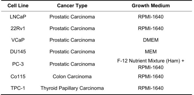
(43) MATERIAL AND METHODS 1. Cell Culture In this study, five cell lines derived from human metastatic PCa were selected: LNCaP, 22Rv1, VCaP, DU145, and PC-3. These PCa cell lines are representative of different relevant features of prostatic adenocarcinoma: LNCaP, 22Rv1 and VCaP are hormone-sensitive cell lines, whilst DU145 and PC-3 are castration-resistant cell lines. DU145 was obtained from the American Type Culture Collection (ATCC, Lockville, MD, USA), whereas LNCaP, VCaP and PC-3 were kindly provided by Prof. Ragnhild A. Lothe from the Department of Cancer Prevention at The Institute for Cancer Research, Oslo, Norway, and 22Rv1 by Dr. David Sidransky from the Johns Hopkins University School of Medicine, Baltimore, MD, USA. For control purposes, we used the human colon carcinoma-derived cell line Co115 and the human thyroid papillary carcinoma-derived cell line TPC-1. Co115 was kindly provided by Prof. Fátima Baltazar from the Life and Health Sciences Research Institute at the University of Minho, Braga, Portugal and TPC-1 by Prof. Paula Soares from the Institute of Molecular Pathology and Immunology of the University of Porto, Porto, Portugal. All cell lines were cultured in the recommended medium (Table 2), supplemented with 10% Fetal Bovine Serum (FBS) (GIBCO, Invitrogen, Carlsbad, CA, USA) and 1% Penicillin-Streptomycin (GIBCO). Cells were maintained at 37°C in a humidified atmosphere containing 5% CO2. Cell lines were subcultured, using the dissociation reagent TrypLE™ Express (GIBCO) to harvest them as many times as necessary in order to obtain the desired number of 75 cm3 cell culture flasks. All PCa cell lines were routinely tested for Mycoplasma spp. contamination.. 25.
Imagem
![Figure 1. Zonal anatomy of the normal prostate. Adapted from [3].](https://thumb-eu.123doks.com/thumbv2/123dok_br/19238375.970473/19.892.254.628.840.1122/figure-zonal-anatomy-normal-prostate-adapted.webp)
![Figure 2. Incidence of different types of cancer in Europe and Portugal, in males. Adapted from [14]](https://thumb-eu.123doks.com/thumbv2/123dok_br/19238375.970473/21.892.222.697.484.934/figure-incidence-different-types-cancer-europe-portugal-adapted.webp)
![Figure 3. Estimated age-standardized incidence rate per 100,000 worldwide. Adapted from [14]](https://thumb-eu.123doks.com/thumbv2/123dok_br/19238375.970473/22.892.122.752.417.781/figure-estimated-age-standardized-incidence-rate-worldwide-adapted.webp)
![Figure 7. Chemical structure of enoxacin. Adapted from [104].](https://thumb-eu.123doks.com/thumbv2/123dok_br/19238375.970473/37.892.334.581.840.970/figure-chemical-structure-enoxacin-adapted.webp)
+7


Documentos relacionados
High PD-L1 expressing basal breast cancer cell lines (N = 12) demonstrate higher levels of STAT1 expression and lower levels of IRF2BP2 compared to low PD-L1 expressing cell lines (N
Forced expression of miR-7 in aggressive breast cancer cell lines suppressed tumor cell monolayer proliferation, anchorage independent growth, three-dimensional growth in
miR-644a Downregulates GAPDH and b -actin Expression While studying the effect of a panel of miRNAs on target mRNA expression in prostate cancer cell lines, we found that the use
Tnao38, high five and Sf9--evaluation of host-virus interactions in three different insect cell lines: baculovirus production and recombinant protein
High expression levels of TP have also been observed in lung, bladder and prostate cancer cell lines, leading to increased cell proliferation, migration and invasion
To understand the expression of Oct4 in OSCC cell lines (OSCCs), the endogenous protein level of Oct4 in nine established OSCC cell lines and one normal oral epithelial cell line SG
Expression of the Carboxy-Terminal Portion of MUC16/CA125 Induces Transformation and Tumor Invasion.
While the expression of MUC16 protein in 3T3 cells was clearly linked to hallmarks of trans- formation, some fully transformed ovarian cancer cell lines lack MUC16 expression
Interestingly, we found that co-expression of RASSF1C and IGFBP-5 reduced PIWIL1 mRNA and protein levels in lung cancer cell lines, while co-expression of RASSF1A- RASSF1C did not
