U
minho | 20
1
3
Outubro de 2013
Maria Alexandra Barroso Azevedo
Development of nanostructures for
encapsulation of vitamins
Maria Ale xandra Barr oso Aze vedo De velopment of nanos tructures for encapsulation of vit aminsDissertação de Mestrado
Mestrado em Bioengenharia
Trabalho efetuado sob a orientação do
Professor Doutor António Augusto Martins
de Oliveira Soares Vicente
Outubro de 2013
Maria Alexandra Barroso Azevedo
Development of nanostructures for
encapsulation of vitamins
e do
II
Autor: Maria Alexandra Barroso Azevedo e-mail: [email protected] Título
Desenvolvimento de nanoestruturas para o encapsulamento de vitaminas. Development of nanostructures for encapsulation of vitamins.
Orientador
Professor Doutor António Augusto Martins de Oliveira Soares Vicente Co-orientador
Doutor Miguel Ângelo Parente Ribeiro Cerqueira Ano de conclusão: 2013
Mestrado em Bioengenharia
É AUTORIZADA A REPRODUÇÃO INTEGRAL DESTA TESE/TRABALHO APENAS PARA EFEITOS DE INVESTIGAÇÃO, MEDIANTE DECLARAÇÃO ESCRITA DO INTERESSADO, QUE A TAL SE COMPROMETE
Universidade do Minho, ___ /___ /___
III Com a conclusão desta tese/trabalho mais uma etapa se finda nesta minha curta, mas espero que longa, caminhada pelo “mundo científico”. Durante um ano, esta exigiu muito trabalho e dedicação, mas agora no final sinto que ganhei muito mais do que aquilo que dei. Cresci, não só ao nível do conhecimento científico como também a nível pessoal, e isso muito se deve a todas as pessoas que me acompanharam ao longo desta tese e que fizeram com que as dificuldades encontradas fossem mais fáceis de ultrapassar. Deste modo, não poderia terminar sem lhes dedicar o meu sincero agradecimento.
Em primeiro lugar, quero agradecer ao meu orientador, Professor António Vicente, por mais uma vez ter aceite orientar o meu trabalho e por me ter dado a oportunidade de voltar a fazer parte de uma grande equipa. Muito obrigada, professor, pela partilha de saberes, pela paciência, ajuda e amizade ao longo desta etapa.
Também, de uma forma especial, quero agradecer ao meu co-orientador Doutor Miguel Cerqueira por toda ajuda prestada, pelo incentivo e apoio. Muito obrigada pela paciência e amizade e por toda a disponibilidade sempre demonstrada para me ajudar e orientar.
Quero também agradecer a todos os meus colegas do Laboratório de Indústria e Processo (LIP), em especial aos “lipinhos” Ana Isabel, Philippe, Ricardo, Joana, Melissa, Ana Cristina e Ariana, pela ajuda, pelos pertinentes comentários, incentivos e sugestões, e pelas oportunas manisfestações de companheirismo e encorajamento.
Aos meus amigos que já fazem parte da minha vida há uns aninhos, Mário, Elisabete, Elísia, e Vanessa, um sincero, profundo e sentido obrigada por mais uma vez estarem presentes numa etapa muito importante para mim. Aos que recentemente conheci, Débora, Diana e Rui, e que se tornaram numa agradável surpresa, um obrigada por tudo e por agora fazerem parte do meu pequeno, mas valioso, grupo de amigos. Aos que não referi, mas que também foram muito importantes na concretização desta etapa, um muito obrigada.
À Enga Magda Graça do Departamento de Biologia da Universidade do Minho e ao Doutor Rui
Fernandes do Instituto de Biologia Molecular e Celular (IBMC) um obrigada pela disponibilidade em colaborar sempre que solicitava a sua ajuda.
IV
E, finalmente, um agradecimento muito especial às pessoas mais importantes – à minha familia: Um muito obrigada aos meus pais por todo amor, apoio, ânimo, compreensão e incentivo, pois sem eles nada disto seria possível. Quero agradecer também à minha irmã, ao meu irmão e ao mais recente membro da familia, Paulo Teixeira, pelo apoio, incentivo, força, carinho e amizade.
A todos aqueles que contibuiram para a elaboração e conclusão desta tese, o meu profundo e sentido, muito obrigada!
V and humidity conditions. For the food industry it is important to reduce some of these limitations and being with nanosystems arise a promising solution.
This work aims at the development of nanosystems for the encapsulation of riboflavin (water-soluble) and α-tocopherol (liposoluble) using biopolymer and their further characterization. For encapsulation of riboflavin an ionotropic polyelectrolyte pre-gelation was used as production method being chitosan and alginate used as main materials. ( )-α-tocopherol was encapsulated through the self-assembly of zein, one of the major proteins of corn with an amphiphilic character.
Initially a preliminary study was done to determine the optimal concentrations of biopolymers used and encapsulated vitamin that allow the highest efficiency and the production of nano-sized structures. For the alginate and chitosan system the optimal concentrations were 0.63 of alginate, 0.4 of chitosan and 0.065 mg/ml of riboflavin. For the zein system 2 and 1 mg/ml of zein and ( )-α-tocopherol, respectively, were used. Nanosystems were characterized in terms of average size, polydispersity index and zeta potential (through Dynamic Light Scattering) and vitamin entrapment efficiency. The average size (by number) for alginate/chitosan nanoparticles with riboflavin was 110.17 (± 47.71) nm. The nanosystems present values of polydispersity index and the zeta potential of 0.520 (± 0.041) and -29.64 (± 0.97) mV, respectively. Zein nanoparticles with ( )-α-tocopherol present an average size of 93.60 (± 1.09) nm and a polydispersity index of 0.222 (±0.020) with a zeta potential of +26.08 (± 2.51) mV. Both nanosystems have successful with encapsulation of vitamins, being the values of encapsulation efficiency (EE) and loading capacity (LC) obtained for (-)-riboflavin 55.91 ( 5.56) % and 2.18 ( 0.63) %, respectively, and for ( )-α-tocopherol was 94.95( 4.17) % of EE and 8.53( 1.90) % of LC. In order to complete the characterization of the nanoparticles was also done release profiles was also done for both vitamins and the diffusion coefficient (D) was calculated based on the fitting of model: for (-)-riboflavin at 37 °C D was 2.28 - m s at pH 7 and 4.95 0.74 - m s at pH 2, and at 25 °C D was 1.02 - m s and 1.88 - m s for pH 7 and 2, respectively; for ( )-α-tocopherol encapsulated into zein nanoparticles at 37°C pH 7, the ( )-α-tocopherol is not released and at pH 2 D was 2.8 - m s. Lastly, the stability of both nanoparticles was assessed being evaluated the effect of temperature and storage time in particles sizes and PDI.
Alginate, chitosan and zein are biodegradable, biocompatible, food-grade with good physicochemical properties and can be used in the development of biopolymer-based nanosystems. This work shows that these biopolymer-based nanosystems can be used for the encapsulation of water-soluble and liposoluble vitamins with a great potential for application in food products.
VII luz e humidade. Para a indústria alimentar é importante reduzir algumas destas limitações e os nanosistemas aparecem como uma solução promissora.
Este trabalho tem como principal objectivo o desenvolvimento e caracterização de nanosistemas para a encapsulação de (-)-riboflavina (vitamina hidrosolúvel) e ( )-α-tocoferol (vitamina liposolúvel) usando biopolímeros. Para a encapsulação da (-)-riboflavina foi usado o método pré-gelificação ionotrópica, sendo o alginato e o quitosano os principais materiais. O ( )-α-tocoferol foi encapsulado através de automontagem da zeína, uma proteína do milho com carácter amfifílico.
Inicialmente foi feito um estudo preliminar com o objectivo de determinar os parâmetros e concentrações óptimas dos biopolímeros e das vitaminas. Para o sistema do alginato e quitosano as concentrações óptimas foram 0,63 de alginato, 0,4 de quitosano e 0,065 mg/ml de (-)-riboflavina. Para o sistama da zeína foram usados 2 e 1 mg/ml de zeína e ( )-α-tocoferol, respectivamente. Os nanosistemas foram caracterizados em termos de médias de tamanhos, índice de polidispersividade e potencial zeta (através de espalhamento dinâmico de luz) e eficiência de encapsulação da vitamina. A média dos tamanhos (por números) para as nanopartículas de alginato/quitosano com (-)-riboflavina foi de 110,17 (± 47,71) nm. O nanosistema apresenta valores de índice de polidispersividade e potencial zeta de 0,520 (± 0,041) e -29,64 (± 0,97) mV, respectivamente. As nanopartículas de zeína com ( )-α-tocoferol apresentam uma média de tamanhos de 93,60 (± 1,09) nm e têm um índice de polidispersividade de 0,222 (± 0,020) com um potencial zeta de +26,08 (± 2,51) mV. Ambos os sistemas foram bem sucedidos na encapsulação das respectivas vitaminas, para a (-)-riboflavina os valores de eficiência de encapsulação (EE) e de capacidade de incorporação (LC) foram 55,91 ( 5,56) % e 2,18 ( 0,63) %, respectivamente, e 94,95 ( 4,17) % de EE e 8,53 ( 1,90) % de LC para o ( )-α-tocoferol. De forma a completar a caracterização de ambas as nanopartículas, também foram avaliados os perfis de libertação de ambas as vitaminas e foram calculados os coeficientes de difusão (D): para a (-)-riboflavina a 37 °C o D foi de 2,28
- m s para pH 7 e 4,95 0,74 - m
s para pH 2 e a 25 °C o D foi de 1,02 - m s e 1,88 - m s para pH 7 e 2, respectivamente; para o ( )-α-tocoferol encapsulado em nanopartículas de zeína a 37 °C pH 7 não houve libertação da vitamina e a pH 2 o D foi de 2,8 - m s. Por fim, a estabilidade de ambos os sistemas foi medida, sendo avaliados os efeitos da temperatura e do tempo de armazenamento nos tamanhos e PDI das nanopartículas.
O alginato, quitosano e zeína são biodegradáveis, biocompatíveis, edíveis e com boas propriedades físico-químicas e estes podem ser usados no desenvolvimento de nanosistemas biopoliméricos. Este trabalho mostra que esses sistemas biopoliméricos podem ser usados para a encapsulação de vitaminas hidro- e liposolúveis com um grande potencial para aplicação em produtos na indústria alimentar.
IX Maria A. Azevedo, Miguel A. Cerqueira and António A. Vicente, Development of an edible, bio-based nanostructure for encapsulation of water soluble vitamins. IFT’ 3 - Annual Meeting & Food Expo, Chicago, 13-16 July 2013 (poster presentation).
Maria A. Azevedo, Miguel A. Cerqueira and António A. Vicente, Development of biopolymer based-nanosystems for vitamins delivery. ESBP 2013 – European Symposium on Biopolymers, Lisbon, Portugal, 7-9 October 2013 (poster presentation).
Miguel A. Cerqueira, Ana C. Pinheiro, Hélder D. Silva, Philippe E. Ramos, Maria A. Azevedo, Maria L. F. López, Melissa C. Rivera, Ana I. Bourbon, Óscar L. Ramos, António Vicente (2013). Design of bio-nanosystems for functional compounds delivery, Food Engineering Reviews, DOI 10.1007/s12393-013-9074-3.
Maria A. Azevedo, Ana I. Bourbon, Miguel A. Cerqueira and António A. Vicente (2013). Development and characterization of alginate/chitosan nanoparticles for controlled release of vitamin B2. To be submitted to International Journal of Biological Macromolecules.
Maria A. Azevedo, Ana I. Bourbon, Miguel A. Cerqueira and António A. Vicente (2013). Development α-tocopherol loaded nanoparticles through zein self-assembly. To be submitted to Journal of Agricultural and Food Chemistry.
XI AGRADECIMENTOS III ABSTRACT V RESUMO VII LIST OF PUBLICATIONS IX TABLE OF CONTENTS XI LIST OF IMAGES XV
LIST OF TABLES XIX
LIST OF GENERAL NOMENCLATURE XXI
CHAPTER I. MOTIVATION, OBJECTIVE AND OUTLINE 1
1.1. THESIS MOTIVATION 3
1.2. RESEARCH AIMS 4
1.3. THESIS OUTLINE 4
CHAPTER II. INTRODUCTION 5
2.1 NANOTECHNOLOGY 7
2.2 EDIBLE AND BIO-BASED MATERIALS 9
2.2.1 POLYSACCHARIDES 10
2.2.1.1 Alginate 11
2.2.1.2 Chitosan 13
2.2.2 PROTEINS 14
2.2.2.1 Zein 16
2.3 NANOSYSTEMS IN FOOD SECTOR: PREPARATION AND CHARACTERIZATION 18
2.3.1NANOCAPSULES 19
2.3.1.1 Preparation Methods 19
XII
Self-assembly 20
2.3.1.2 Characterization Methods 21
Dynamic Light Scattering (DLS) 21
Nanoparticle Tracking Analysis (NTA) 22
Transmission Electron Microscopy (TEM) 23
Release Profile 23
2.4 BIOACTIVE FOOD COMPOUNDS 24
2.4.1 VITAMINS 25
2.4.1.1 Riboflavin (Vitamin B2) 26
2.4.1.2 Tocopherol (Vitamin E) 27
CHAPTER III. MATERIALS AND METHODS 29
3.1. DEVELOPMENT OF NANOPARTICLES 31
3.1.1 ALGINATE/CHITOSAN NANOPARTICLES PREPARATION 31
3.1.1.1 Materials 31
3.1.1.2 Methodology 31
3.1.2 ZEIN NANOPARTICLES PREPARATION 32
3.1.2.1 Materials 32
3.1.2.2 Methodology 32
3.2 CHARACTERIZATION 32
3.2.1 NANOPARTICLE SIZE, POLYDISPERSITY INDEX AND ZETA POTENTIAL 32
3.2.2 ENCAPSULATION EFFICIENCY AND LOADING CAPACITY 33
3.2.3 MORPHOLOGY 35
3.2.4 RELEASE PROFILE 35
3.2.4.1 Materials 35
3.2.4.2 Dialysis membrane method 35
3.2.4.3 Kinetics of Release 36
3.2.5 STABILITY MEASUREMENT 36
3.2.5.1 Temperature 36
3.2.5.2 Storage Time 37
XIII
4.1.1 OPTIMIZATION OF THE FORMULATION 42
4.1.2 CHARACTERIZATION 43
4.1.2.1 Size, Polidispersity index (PDI) and Zeta Potential 43
4.1.2.2 Morphological observation 45
4.1.2.3 Encapsulation Efficiency and Loading Capacity 46
4.1.2.4 Release profile of (-)-riboflavin from alginate/chitosan nanoparticles 47
4.1.2.5 Stability measurement 49
Temperature 50
Storage time 51
4.2 ZEIN NANOPARTICLES 53
4.2.1 OPTIMIZATION OF THE FORMULATION 54
4.2.2 CHARACTERIZATION 54
4.2.2.1 Size, Polydispersity index (PDI) and Potential Zeta 54
4.2.2.2 Morphological observation 56
4.2.2.3 Encapsulation Efficiency and Loading Capacity 57
4.2.2.4 Release Profile of TOC from zein nanoparticles 57
4.2.2.5 Stability 59 Temperature 59 Storage Time 60 CHAPTER V. CONCLUSION 61 5.1 CONCLUSIONS 63 5.2 RECOMMENDATIONS 64 REFERENCES 65 ANNEXES 79 ANNEX A 81
XIV
ANNEX B 85
MAXIMUM ABSORBANCE PEAK 85
ANNEX C 87
XV
CHAPTER II
Figure 2.1 - Applications of nanotechnology in food sector. Image adapted from Blasco and Picó
(2011) and Weiss, Takhistov and McClements (2006). 9
Figure 2.2 – Some basic structure of polymers. Image adapted from U. S. Congress (1993). 10 Figure 2.3 – Chemical structure of alginate. The monomeric units of alginate, 1,4-linked- -D-mannuronic acid (M) and α-L-guluronic acid (G), can be linked in varying sequences, as blocks of alternating gulutonic and mannuronic residues, blocks of guluronic acids and of mannuronic acids (Sundar et al., 2010). Image from Vrignaud, Benoit, and Saulnier (2011). 12 Figure 2.4 – (a) Binding of cation Ca2+ and guluronic acid residue of alginate; (b) “Egg box”
model. Image adapted from Chavanpatil et al. (2007) and Myrvold and Onsoyen (2004). 13 Figure 2.5 – Chemical structure of chitosan. Image adapted from Kumar (2000) and Mourya and
Inamdar (2008). 14
Figure 2.6 – Classification of proteins and their characteristics. 15
Figure 2.7 – Structure models of zein: (a) Argo’s model; (b) Matsushima’s model; (c) Momany model. Image (a) and (b) adapted from Y. Wang and Padua (2012) and (c) adapted from
Momany et al. (2006). 17
Figure 2.8 – Top down and bottom up approaches. Image adapted from Reverchon and Adami
(2006). 18
Figure 2.9 – (a) Schematic of Ionic pre-gelation/coacervation method; (b) Structure of nanoparticle with bioactive component loaded: Bioactive component; Chitosan; Alginate. Images adapted from (a) Nagavarma et al. (2012) and (b) Y. Zhang et al. (2011) and
Mora-Huertas, Fessi, and Elaissari (2010). 20
Figure 2.10 – Schematic presentation of set-up for release profile by dialysis membrane method. Image adapted from Prata et al. (2008) and S. S. D. Souza and Deluca (2005). 24
XVI
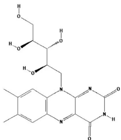
Figure 2.11 – The Chemical structure of riboflavin. 26
Figure 2.12 – Chemical structure of α-tocopherol. 28
CHAPTER III
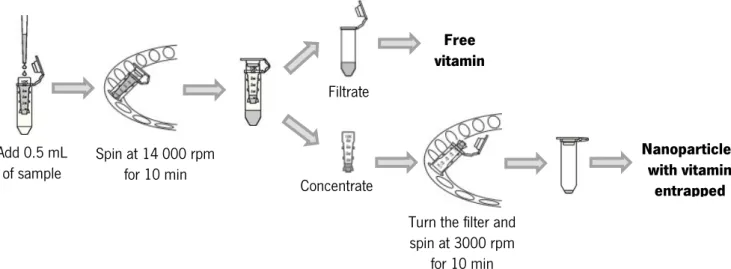
Figure 3.1 – Membrane separation method with an Amicon® ultra-0.5 centrifugal filter device.
Image adapted from User Guide Amicon® ultra-0.5 centrifugal filter device. 34 CHAPTER IV
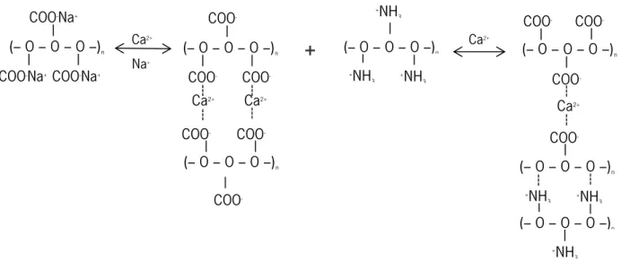
Figure 4.1 – Electrostactic interactions between Ca2+ ions, -COO- groups of alginate and amine groups of chitosan. Image adapted from Bajpai and Tankhiwale (2006). 42 Figure 4.2 – Transmission electron microscopy (TEM) images of alginate/chitosan nanoparticles without (-)-riboflavin at a scale bar of 2 µm, 7000x (a); 1 µm, 12000x (b); 0.5 µm, 20000x (c).
46 Figure 4.3 – Fitting of Eq. 3.3 to (-)-riboflavin controlled release experimental data from alginate chitosan nanoparticles ((•) experimental results and ( o ) model-generated values): (a) at 37°C, pH 7; (b) at 37°C pH 2; (c) at 25°C, pH 7; (d) at 25°C, pH 2. 48 Figure 4.4 – Effect of temperature in particle size and PDI of alginate/chitosan without
(-)-riboflavin. 50
Figure 4.5 – Stability of alginate/chitosan nanoparticles: (a) without riboflavin; (b) with
(-)-riboflavin. 52
Figure 4.6 – Schematic illustration of formation of zein nanoparticles with TOC. Image adapted
from Luo et al. 2011. 53
Figure 4.7 - Transmission electron microscopy (TEM) images of zein nanoparticles: (a) Scale bar: 0.2 µm, 85000x and (b) Scale bar: 0.5 µm, 30000x without TOC and (c) Scale bar: 0.5 µm,
30000x; and (d) Scale bar: 0.5 µm, 20000x with TOC. 56
Figure 4.8 – Fitting of Eq. 3.3 to TOC controlled release experimental data from zein nanoparticles at 37°C, pH ((•) experimental results and ( o ) model-generated values). 58
XVII
ANNEX
Figure A.1 – UV–Vis spectrum of (-)-riboflavin. Spectra were overlaid after scanning from 200 to
600 nm. 85
Figure A.2 – UV–Vis spectrum of TOC. Spectra were overlaid after scanning from 200 to 400 nm. 86 Figure A.3 – Calibration curve of (-)-riboflavin. Absorbance versus concentration for (-)-riboflavin. 87 Figure A.4 – Calibration curve of (±)-α-tocopherol. Absorbance versus concentration for
XIX
CHAPTER IV
Table 4.1 – Particle size (by z-average and number), Polydispersity index (PDI) and zeta potential for alginate/chitosan nanoparticles with and without (-)-riboflavin, by DLS instrument (Zetasizer
Nano ZS). 44
Table 4.2 – Particle size (mean and mode) for alginate/chitosan nanoparticles with and without
(-)-riboflavin, by NTA (NanoSight). 45
Table 4.3 – Particle size (by z-average and number), polydispersity (PDI) and zeta potential for
zein nanoparticles with and without ( )-α-tocopherol (TOC). 54
Table 4.4 – Particle size (mean and mode) for zein nanoparticles with and without (
)-α-tocopherol (TOC), by NTA (NanoSight). 55
ANNEX
Table A.1 – Different formulations tested for development of alginate/chitosan nanoparticles. 81 Table A.2 – Particle size and PDI values of different formulations for development of
alginate/chitosan nanoparticles. 82
Table A.3 – Different formulations testes for development of zein nanoparticles. 83 Table A.4 – Particle size and PDI values of different formulations for development of zein
nanoparticles. 84
Table A.5 – Dilutions and their absorbance values for calibration curve of (-)-riboflavin. 87 Table A. 6 – Dilutions and their absorbance values for calibration curve of TOC. 88
XXI Abs – Absorbance
° C – Celsius
CaCl2 – Calcium Chloride Corp. – Corporation cP – Centipoise
D – Translational diffusion coefficient d (H) - Hydrodynamic diameter Da – Dalton
DLS – Dynamic Light Scattering
DSC – Differential Scanning Calorimetry EE – Encapsulation Efficiency
e.g. – For example
FTIR – Fourier transform infrared spectroscopy g – Unit of measure of acceleration (m/s2)
H – Hours LC – Loading Capacity K – Boltzmann’s constant M – Molar µL – Microliters µm – Micrometers
m s – Square meter per second mL – milliliters min – minute mm – millimeters mM – millimolar Mw – Molecular weight nm - Nanometers
NTA – Nanoparticle Tracking Analysis PCS – Photon Correlation Spectroscopy PDI – Polydispersity
XXII
PBS – Phosphate Buffer Saline QLS – Quasi-Elastic Light Scattering RF- – Anionic form of riboflavin RFH – Neutral form of riboflavin RFH2+ – Cationic form of riboflavin rpm – Rotations per minute (r/min) SD – Standard Deviation
T – Absolute temperature
TEM – Transmission Electron Microscopy TGA – Thermogravimetric analysis TOC – ( )-α-Tocopherol
Tris-HCl – Tris-Hydrochloride UK – United Kingdom
USA – United States of America – Viscosity
UV – Ultraviolet λ – Wavelength ζ – Zeta Potential
DEVELOPMENT OF NANOSTRUCTURES FOR ENCAPSULATION OF VITAMINS | 1
CHAPTER I
CHAPTER I
MOTIVATION, OBJECTIVE AND OUTLINE
DEVELOPMENT OF NANOSTRUCTURES FOR ENCAPSULATION OF VITAMINS | 3
1.1. THESIS MOTIVATION
The food industry has as main challenges: to improve the production efficiency, food safety and food characteristics. This way, the maintenance and increase of chemical, physical, microbiologic and nutritional stability of food and compounds used in food is an essential aim to achieve. Vitamins have a special attention from food industry, because they have bioactive properties that are essential for normal maintenance, growth and development of human organism and their absence causes a specific deficiency syndrome. However, humans do not have the capacity to synthesize vitamins, except vitamin D and B3, meaning that they must obtain vitamins through ingestion of food (Ball, 2008; Combs Jr, 2012). The enrichment of food and/or beverages with vitamins is a good idea to complete a feed with absence of vitamins, but they can also be used to mask the flavor of vitamins and mineral or to improve taste of the food products. Nonetheless, vitamins are sensitive and unstable when exposed to inadequate temperature, oxygen, light and moisture. This way, the nanoencapsulation can be a solution to keep stability of vitamins (De Britto et al., 2012; Luo et al., 2012).
Nanotechnology is an emergent and multidisciplinary field that is stimulating much interest across many areas of research and industries. This field involves the application, production and processing of materials with sizes at nano-scale and due to sizes of materials, the nanostructures when applied in food industry allow e.g. great improvements in bio-adhesive properties, appearance, texture, stability, or flavor of the product and release of bioactive compounds, higher bioavailability and can solve some problems such as compatibility and loss of activity of some functional compounds, affected by light, oxygen and temperature when dispersed in the food (Acosta, 2009; Kuan et al., 2012). In order to improve the efficiency and stability of nanostructures it is essential to find adequate materials; in the particular case of the food sector it is also important the replacement of non-food-grade materials by bio-based and biodegradable food-grade materials. Biopolymers, such as polysaccharides, proteins and lipids, open the door to new functionalities and applications due distinct advantages of biodegradability, edibility and lack of toxicity (Chassenieux et al., 2013).
In doing so, the encapsulation of vitamins can solve the problems associated with used and incorporated of vitamins in food and/or beverages and can lead to an increase of stability and to
CHAPTER I
MOTIVATION, OBJECTIVE AND OUTLINE
4 | DEVELOPMENT OF NANOSTRUCTURES FOR ENCAPSULATION OF VITAMINS
promote their bioactivity. It is possible that encapsulation of vitamins also improves the controlled release of vitamins in human digestive system.
1.2. RESEARCH AIMS
The main objective of this thesis was the development of nanosystems for the encapsulation of (-)-riboflavin (water-soluble) and ( )-α-tocopherol (liposoluble) using biopolymer and their further characterization. The main focus areas were:
i) The development and characterization of nanosystems;
ii) Assessment of the intake of vitamins in nanosystems developed.
1.3. THESIS OUTLINE
The thesis is organized in five chapters, being this chapter the first and is described the motivation, research aims and outline of the thesis. The chapters 2 to 5 are distributed as follows:
CHAPTER II - “INTRODUCTION”: Presents a review of the relevant literature about the theme of the thesis, being provide to the reader the basic information to understand the main issues of this work.
CHAPTER III – “MATERIALS AND METHODS”: In this chapter is approached the materials and methodologies used for nanoparticles preparation and their characterization.
CHAPTER IV – “RESULTS AND DISCUSSION”: Shows all results and their discussion. In this work was development two nanosystems with different characteristics, so this chapter has two main titles: “ALGINATE CHITOSAN NANOPARTICLES” and “ZEIN NANOPARTICLES”.
CHAPTER V – “GENERAL CONCLUSIONS”: Presents the overall conclusions and recommendations and suggestions for future work.
DEVELOPMENT OF NANOSTRUCTURES FOR ENCAPSULATION OF VITAMINS | 5
CHAPTER II
CHAPTER II INTRODUCTION
DEVELOPMENT OF NANOSTRUCTURES FOR ENCAPSULATION OF VITAMINS | 7
2.1 NANOTECHNOLOGY
On 29 December 1959 the physicist Richard Feynman (Nobel Prize for Physics 1965) gave a speech entitled “There’s Plenty of Room at the Bottom” in annual meeting of the American Physical Society at the California Institute of Technology and proposed the exploration of materials at scale of atoms and molecules resulting in something that human wouldn’t see (Asiyanbola and Soboyejo, 2008; Miyazaki and Islam, 2007). This way began the history of nanotechnology. However, just in 1974 the nanotechnology term was first used and defined by Norio Taniguchi (researcher of University of Sciences in Tokyo) to refer the processing, separation, consolidation and deformation of materials at scale of an atom or a molecule (Asiyanbola and Soboyejo, 2008; Kuan et al., 2012; Miyazaki and Islam, 2007). Currently, the nanotechnology is defined as a science that involves the design, synthesis, characterization and application of materials at the nano-scale, being one nanometer (nm) one-billionth of a meter (Calster, 2006; Commission, 2004; Kuan et al., 2012). Generally, all particles with a size less 1 micrometer (µm) are classified as nanoparticles (Asiyanbola and Soboyejo, 2008; Mohanraj and Chen, 2006), but different interpretations of the nanoparticle dimensions have been proposed and the more stringent classifications for nanoparticle just include particles with a size in the range of 1-100 nm (Calster, 2006; Cushen et al., 2012; Reverchon and Adami, 2006). These stringent classifications are justified by the fact that some physical properties appear when the materials reach the values 1-100 nm, but the legitimate definition classifies the sub-micron scale as nano and extends the limit to 1 µm (Buzea et al., 2007; Naahidi et al., 2013).
The materials at nano-scale change their physics, chemistry and biologic properties and these changes are due to increase of relative surface and quantum effects that begins to control the material at this scale. This way, the utilization of nanomaterials, when compared with normal utilization (micro or macro), is an added advantaged because they can have better properties such as: increased physical strength, chemical reactivity, electrical conductivity, magnetism and optical effects (Buzea et al., 2007; Chau et al., 2007; Siegrist et al., 2008). As result, nowadays, exist ever more technological sectors, e.g. medical, information technologies, energy production and storage, materials science, manufacturing, instrumentation, food, water and environmental and security, that betting on nanotechnology a way to achieve better results for various problems and to create new technologies. In doing so, the investments in nanomaterials research have
CHAPTER II INTRODUCTION
8 | DEVELOPMENT OF NANOSTRUCTURES FOR ENCAPSULATION OF VITAMINS
increased, making the nanotechnology an expanding technology with many applications (Commission, 2004; Miyazaki and Islam, 2007; Pitkethly, 2004).
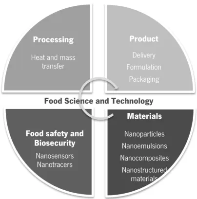
Nanotechnology can help the food sector achieving some of its main goals, i.e. improve production efficiency, increase food safety and enhance food characteristics; to do that applications in food industry are focused on the development of nano-size delivery systems for functional ingredients and additives, and innovative packaging. Actually, food sector is one of the sectors with fewer applications at nano-scale, but it is expected that in the near future the nanomaterials in food industry increase (Cushen et al., 2012; Weiss et al., 2006). This increase can be explained by advantages related with the size reduction of materials e. g. improvement of bio-adhesive properties, that includes an increase of adhesive force and prolonged gastrointestinal transit time, possible leading to a higher bioavailability (Acosta, 2009). Based on these unique characteristics nanosytems can solve some problems occurring when using systems at macro- and microscale for delivery of functional compounds; such problems are: compatibility (e.g. aggregation and phase separation) with the food matrix (influencing e.g. appearance, texture, stability, or flavor of the product); release, that should be controlled and only activated once inside the human gut (i.e. some compounds start to be released when mixed with the food product and functional compounds lose their activity); and loss of activity of some functional compounds, affected by light, oxygen and temperature when dispersed in the food matrix. This versatility and limitless potential of nanotechnology allow its application in different systems that are part of food sector (Figure 2.1).
DEVELOPMENT OF NANOSTRUCTURES FOR ENCAPSULATION OF VITAMINS | 9
2.2 EDIBLE AND BIO-BASED MATERIALS
The technological sectors that use nanotechnology have a greatest challenge: to find materials that ensure the efficiency and stability of nanosystems; and in the food sector is also important replacement of non-food-grade materials by bio-based and biodegradable food-grade materials. In food sector, the physic-chemical properties, such as solubility, molecular weight, glass/melting transition, crytallinity, diffusivity, film forming and emulsifying properties, or costs of material are important factors in materials selection (Sagalowicz and Leser, 2010). Besides that, it is also necessary to take account the future characteristics of nanosystems, e.g. size, morphology, biodegradability, biocompatibility, cytotoxicity, drug loading, release profile and preparation method (Matalanis et al., 2011; Sundar et al., 2010). Initially, the synthetic polymers were the basis of nanosystems, because they have great properties of durability and strength. However, the use of synthetic polymers triggered a lot of environment and health problems, because they are produced from fossil fuels, are not biodegradable and the synthesis of some polymeric materials involves the use of toxic compounds or generation of toxic products. So, it is important to use renewable source-based biopolymers materials of natural origin that not involve the use of
Product Delivery Formulation Packaging Materials Nanoparticles Nanoemulsions Nanocomposites Nanostructured materials
Food safety and Biosecurity
Nanosensors Nanotracers
Processing
Heat and mass transfer
Food Science and Technology
Figure 2.1 - Applications of nanotechnology in food sector. Image adapted from Blasco and Picó (2011) and Weiss et al. (2006).
CHAPTER II INTRODUCTION
10 | DEVELOPMENT OF NANOSTRUCTURES FOR ENCAPSULATION OF VITAMINS
toxic or harmful components in their development, use or degradation (U.S. Congress 1993; Wang et al., 2005).
Biopolymers are polymers produced by biological systems (e.g. microorganisms, plants and animal) or using chemical processes derived from biological starting materials such as amino acids, sugars, natural fats or oils. They are mainly composed by repeating units of saccharides, amino acids, lipids, peptides and nucleic acids, Figure 2.2, and have the characteristic of being degraded by biological activities (Chassenieux et al., 2013; Kuan et al., 2012; U.S. Congress, 1993). Polysaccharides (e.g. alginate, pectin, dextran and chitosan), proteins (e.g. zein, whey protein isolate) and lipids (e.g. medium chain triglycerides, tristearin and corn oil) are some examples of biopolymers. The utilization of biopolymers open the door to new functionalities and applications due distinct advantages of biodegradability, edibility and lack of toxicity (Chassenieux et al., 2013).
´
2.2.1 POLYSACCHARIDES
Polysaccharides are natural polymers composed by simple sugars (monosaccharide residues) that are linked by different glucosidic bonds (U.S. Congress, 1993; Yang and Zhang, 2009). The Figure 2.2 – Some basic structure of polymers. Image adapted from U. S. Congress (1993).
Monomer
Repeat Unit (Formed when different units are linked together)
Linear homopolymer
Linear copolymer Branched homopolymer
DEVELOPMENT OF NANOSTRUCTURES FOR ENCAPSULATION OF VITAMINS | 11 (environmental conditions and history) and their monomer sequence can be responsible for the molecular structure of polysaccharides (Matalanis et al., 2011; Yang and Zhang, 2009). Each polysaccharide has different chemical structures (e.g. type, number, sequence and bonding of the monosaccharide within the polymer chain), which are responsible by molecular weight, degree of branching, structure, flexibility, electrical charge and interaction between polysaccharides. Besides, depending of the ionic groups presented in the polysaccharide chain, some polysaccharides can be neutral (e.g. starch and cellulose), anionic (e.g. alginate, carrageenan, xanthan and gum Arabic), or cationic (e.g. chitosan). This differences allow to polysaccharides present different functional properties such as: solubility, thickening, gelation, water holding capacity, surface activity, emulsification and digestibility (Yang and Zhang, 2009). Based on the existing great variety of polysaccharides it is necessary when choosing one or more polysaccharides for bio-based nanosystems preparation to considerer their physicochemical properties and the desired characteristics of the bio-based nanosystems (e.g. morphology, density, refractive index, size, charge and stability) (U.S. Congress, 1993; Yang and Zhang, 2009).
For the presented work two polysaccharides (alginate and chitosan) were used as material for the development of bio-based nanosystem. Below, are presented the most important characteristics of these two polysaccharides.
2.2.1.1 Alginate
Alginate is an hydrophilic polysaccharide extracted from marine brown algae of the Phaeophyta family. It is a linear biopolymer composed by two uronic acids, 1,4-linked- -D-mannuronic acid (M) and α-L-guluronic acid (G), being carboxylic groups from uronic acids responsible by their negative charge (Figure 2.3). Alginate is considered a non-toxic, biocompatible, biodegradable and mucoadhesive polysaccharide (Silva et al., 2006; Sundar et al., 2010; Vrignaud et al., 2011) and it approved for application in pharmaceutical and food industry.
CHAPTER II INTRODUCTION
12 | DEVELOPMENT OF NANOSTRUCTURES FOR ENCAPSULATION OF VITAMINS
Figure 2.3 – Chemical structure of alginate. The monomeric units of alginate, 1,4-linked- -D-mannuronic acid (M) and α-L-guluronic acid (G), can be linked in varying sequences, as blocks of alternating gulutonic and mannuronic residues, blocks of guluronic acids and of mannuronic acids (Sundar et al., 2010). Image from Vrignaud et al. (2011).
The solubility of alginate in water depends on the associated cations, for example sodium alginate is soluble in water but when a solution with multivalent cations (e.g. calcium - Ca2+) is
used the biopolymer can form a reversible gel (Sarmento et al., 2007; Silva et al., 2006; Sundar et al., 2010). The gelling property of alginate is promoted by the high selectivity of guluronic acid residues that in the presence Ca2+ organized in side-by-side blocks formatting the so called “egg box” model. The arrangement is due to an electrostatic interaction between Ca2+ and the
negatively charged oxygen atoms (Figure 2.4). The gelling property of alginate depends of following factors: sequential order and composition of mannuronic and guluronic acid residues, molecular weight of biopolymer and concentration of counter ions in solution (Sarmento et al., 2007; Silva et al., 2006). However, the addition of a polyelectrolyte complex such as chitosan or poly-L-lysine induces a polyelectrolyte complex formation and stabilization of the alginate pre-gel nucleus into individual sponge-like particles (Sarmento et al., 2007). The hydrophilic and gelling properties, make of alginate a biopolymer with a considerable potential for encapsulation of hydrophilic components (Vrignaud et al., 2011).
DEVELOPMENT OF NANOSTRUCTURES FOR ENCAPSULATION OF VITAMINS | 13 Figure 2.4 – (a) Binding of cation Ca2+ and guluronic acid residue of alginate; (b) “Egg box” model. Image
adapted from Chavanpatil et al. (2007) and Myrvold and Onsoyen (2004).
2.2.1.2 Chitosan
Chitosan is a cationic polysaccharide derived from the N-deacetylation of chitin, the second most abundant natural biopolymer. The chitin is obtained from exoskeletons of marine arthropods (e.g. shells of crabs, shrimps and krill), walls of fungi (e.g. fungal mycelia) and cuticle of insects. It is a linear cationic copolymer composed by Nacetylglucosamine and glucosamine residues with -1,4-linkage (Mourya and Inamdar, 2008; Shahidi et al., 1999; Sundar et al., 2010). Chitin has unique properties such as ability to polyoxysalt formation, chelate metal ions and optical structural characteristics. However, it is insoluble in water and in most organic solvents, being its biodegradation very slow. So, chitosan, chitin deacetylated form, appears as a great solution for these solve that problems (Kumar, 2000; Shahidi et al., 1999).
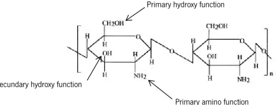
Chitosan is a linear copolymer composed by repeating units of 2-amino-2-deoxy- -D-glucan with glycosidic linkages, where the amine groups confer of chitosan special properties (e.g. high charge density, readily available for chemical reactions and salt formation with salts) (Agnihotri et al., 2004; Sundar et al., 2010). Chitosan present three types of reactive functional groups: an amino group as well as both primary and secondary hydroxyl groups at the C-2, C-3 and C-6 position, respectively (Shahidi et al., 1999). The position of free amino and N-acetyl groups is responsible by solubility of chitosan; nevertheless, the chitosan solubility is improved with organic solvents as aqueous acids (e.g. formic acid and acetic acid) (Agnihotri et al., 2004; Kumar, 2000; Mourya and Inamdar, 2008). The increase of solubility is due to protonation of amino groups by acids that along the chitosan chain increase the polarity and the degree of electrostatic repulsion (Mourya and Inamdar, 2008). N-deacetylation of chitin during chitosan formation is not complete, and chitosan can have a degree of deacetylation between 40-98% (Mourya and
CHAPTER II INTRODUCTION
14 | DEVELOPMENT OF NANOSTRUCTURES FOR ENCAPSULATION OF VITAMINS
Inamdar, 2008) and molecular weight ranged between 3.8 and 2000 KDa; these two characteristics can influence chitosan properties being important factors for its use in several applications (Mourya and Inamdar, 2008; Sundar et al., 2010).
This biopolymer has great properties such as biocompatibility, biodegradability, non-toxicity, and several studies indicated chitosan with a great adsorption and muchoadhesive properties, antifungal activity, ability to promote metabolic changes, an excellent film-forming ability and micro/nanoparticles developing. Due to these properties, the chitosan have a great potential for food, environmental, pharmaceutical, medical and agriculture sectors (Agnihotri et al., 2004; Kumar, 2000; Peniche and Acosta, 2003; Sarmento et al., 2007; Sundar et al., 2010; Weber, 2000).
Figure 2.5 – Chemical structure of chitosan. Image adapted from Kumar (2000) and Mourya and Inamdar (2008).
2.2.2 PROTEINS
Proteins are biopolymers that are present in plants (e.g. zein, gluten and soy) or animals (e.g. casein, whey, collagen and keratin) composed by a linear sequence of amino acids linked by peptide bonds; exist 20 amino acids and many different combinations and sequences, so the proteins can have distinct structures and properties (Weber, 2000; Nelson and Cox, 2000). Amino acids are composed by a carboxyl group (COOH) and an amino group (NH3) linked to a
carbon atom. Besides that, they have a side chain, R group, which varies in structure, size and electrical charge for each amino acid being responsible for their solubility and polarity. Taking into account properties of R group, the amino acids can be classified as aromatic, nonpolar or polar and uncharged or positively or negatively charged (Nelson and Cox, 2000). The amino
Primary amino function Secundary hydroxy function
DEVELOPMENT OF NANOSTRUCTURES FOR ENCAPSULATION OF VITAMINS | 15 carboxyl group of another by removal of the elements of water, these linkages are called of peptide bonds. Polypeptide is the product of linkages between two or more amino acids and have a low molecular weight, when the linkages contain more than 50 amino acids presenting a higher molecular weight (ranged between 6/10 and 40 000 KDa), they are called of protein (McKee and Mckee, 2013; Wade, 2012).
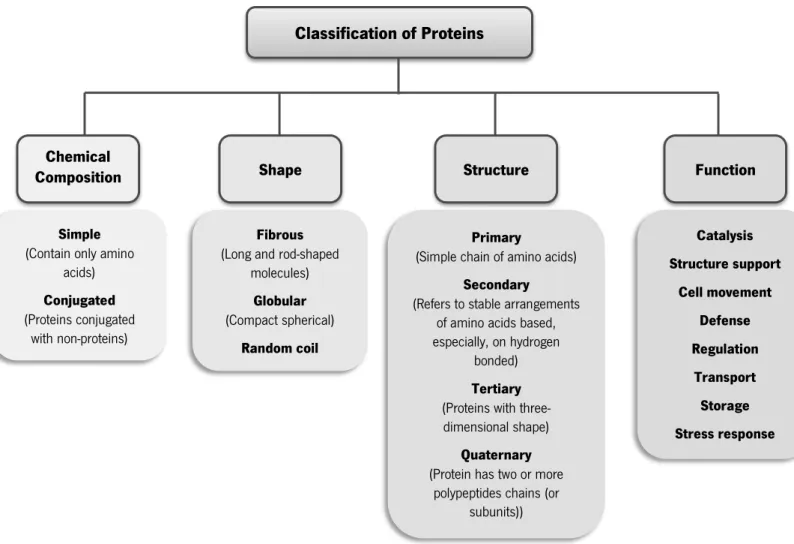
Proteins have a great range of structural and catalytic properties, being their amino acids sequence, environmental conditions and the physico-chemical treatment (e.g. exposure to different temperatures, pressures, solvents, pH values and ionic composition) responsible for it. As a result of their diversity/complexity, they can be classified according to chemical composition, shape, structure or function as shown in Figure 2.6 (McKee and Mckee, 2013; Wade 2012; Nelson and Cox, 2000).
Figure 2.6 – Classification of proteins and their characteristics.
Classification of Proteins
Chemical
Composition Shape Structure Function
Simple
(Contain only amino acids)
Conjugated
(Proteins conjugated with non-proteins)
Fibrous
(Long and rod-shaped molecules)
Globular
(Compact spherical)
Random coil
Primary
(Simple chain of amino acids)
Secondary
(Refers to stable arrangements of amino acids based, especially, on hydrogen
bonded)
Tertiary
(Proteins with three-dimensional shape)
Quaternary
(Protein has two or more polypeptides chains (or
subunits)) Catalysis Structure support Cell movement Defense Regulation Transport Storage Stress response
CHAPTER II INTRODUCTION
16 | DEVELOPMENT OF NANOSTRUCTURES FOR ENCAPSULATION OF VITAMINS
The proteins not only have a great biochemical interest, but also, due to their functional properties, such as self-assembly, emulsification, gelation, foaming, water binding capacity and gas barrier, have a big potential as technological biomaterial (Chassenieux et al., 2013; Chen et al., 2006; Weber, 2000). For example, in food sector, they can be used to produce biopolymer-based delivery systems or film forming solutions for packaging materials being these systems relative easy to prepare, biodegradable, immunogenic, nontoxic and present a greater stability in vivo and during storage (Sundar et al., 2010). However, different factors must be considered when proteins are chosen as biomaterial and is important to: a) establish appropriate physicochemical conditions for protein associate with other proteins or non-proteins; b) establish the electrical characteristics between the involved molecules when electrostatic interactions are used to structure formation; c) know the properties of biopolymer particles formed after protein association (e.g. morphology, physical properties, size, charge and stability) (Matalanis et al., 2011).
Due to the potential of proteins as bio-based material zein was used for nanostructure preparation. Below are presented the most important characteristics and the reason for this choice.
2.2.2.1 Zein
Corn or maize (Zea mays) is one of the most important cereals on food industry and their main components are starch, proteins and water. The proteins represent one of the greatest components and about 75 % are present in endosperm tissue, being zein the major storage protein of corn (Lawton, 2002; Shukla and Cheryan, 2001).
Zein was first isolated in 1821 by John Gorham and was described as a soft, ductile, tenacious and elastic protein, but only in 1939 began the commercial production due their deficiency in essential amino acids (e. g. lysine and tryptophan) and insolubility in water. However, since its isolation in 1821, zein has scientific interest as a polymeric material and their properties such as ability to form tough, glossy, hydrophobic grease-proof coatings and their resistance to microbial attack has been explored (Lawton, 2002; Shukla and Cheryan, 2001). Zein is also biodegradable and renewable and is one of the few cereal proteins that are extracted in relatively pure form. So, zein is considered as safe and food grade ingredient with a great potential for: film, coatings and plastics applications, encapsulation and controlled release of fat-soluble compounds and for that
DEVELOPMENT OF NANOSTRUCTURES FOR ENCAPSULATION OF VITAMINS | 17 Cheryan, 2001; Weber, 2000).
Zein is considered a prolamin and is amphiphilic due their structure containing three quarter of lipophilic and one quarter of hydrophilic amino acids residues (Luo et al., 2011). The composition of amino acids sequence is responsible for its solubility: zein is insoluble in water except in the present of alcohol such as ethanol and isopropanol, or in the presence of high concentrations of urea or alkali and anionic detergents (Lawton, 2002; Shukla and Cheryan, 2001). It has a surface charged that varies according to the pH of environment (Deo et al., 2003). During zein extraction from corn is used a suitable solvents and as a result is obtained a native form of protein in heterogeneous mixture of disulfide-linked aggregates with a molecular weight of 44 000 Da (Shukla and Cheryan, 2001). This protein is composed by a mixture of four distinct types of peptides that varying in molecular size, solubility and charge and can be separated using their different solubility. The four peptides are denominated as α-, -, - and δ-zein, being α- and -zein the most abundant types (Lawton, 2002; Shukla and Cheryan, 2001). Exist three models for structure of zein (Shukla and Cheryan, 2001; Wang and Padua, 2012a): a) helical wheel model for zein where antiparallel helices are clustered within a distorted cylinder stabilized by hydrogen bonds (Argos et al. 1982); b) model where helices are linked by glutamine-rich bridges that are aligned in an antiparallel fashion (Matsushima et al. 1998); c) a three-dimensional structure, being a coiled-coil super helix with lutein at the core (Momany et al. 2006) (Figure 2.7).
Figure 2.7 – Structure models of zein: (a) Argo’s model; (b) Matsushima’s model; (c) Momany model. Image (a) and (b) adapted from Y. Wang and Padua (2012) and (c) adapted from Momany et al. (2006).
(a) (b)
CHAPTER II INTRODUCTION
18 | DEVELOPMENT OF NANOSTRUCTURES FOR ENCAPSULATION OF VITAMINS
Top down Bottom up
2.3 NANOSYSTEMS IN FOOD SECTOR: PREPARATION AND CHARACTERIZATION
In general, the selection of materials to prepare nanosystems is very important and a great challenge, but the nanosystem and their characteristics are one of the most important factors to ensure the efficiency and stability of nanosystem selected.
Nanosystems can be classified based on ( Morris 2010; Silva et al. 2011): - the major material used in their fabrication;
- the production method (e.g. bottom-up or top-down);
- the predominant forces in the system (e.g. electrostatic, hydrogen bonding); - the main properties of the system (e.g. mechanical and optical properties) and - the system’s overall free energy (thermodynamic or kinetic stable systems).
One of the major trends in the development of nanosystems is to combine different approaches such as: mixtures of the materials used, combination of bottom-up strategies (nanosystems are obtained by interaction of small components such as layer-by-layer and self-assembly) and top down strategies (formation of nanosystems by physical-chemical processes that involve reduction of particle size to nano-size, e.g. homogenization), and intervention of different types of forces during the production process (Choi et al., 2011; Hu et al., 2007; Lertsutthiwong et al., 2008; Yu et al., 2006) in order to achieve a desired functionality. In the last years a large number of different delivery nanosystems have been developed, often using a trial-and-error approach, which leads to a great number of developed and well characterized nanosystems however without a final and conclusive application.
In the food sector, the utilization of delivery systems, in particularly, at the nano scale showed to be promising as active vectors. Some of the reasons are their capacity to release bioactive compounds and their high intracellular uptake. Their capacity to improve the stability of active substance and their high encapsulation efficiency (Mora-Huertas et al., 2010).
DEVELOPMENT OF NANOSTRUCTURES FOR ENCAPSULATION OF VITAMINS | 19 Nanocapsules, also called nanoparticles, are constituted by an external cavity consisting of a polymeric membrane and an internal part composed by a liquid or polymeric matrix that contains the active compound (Fang and Bhandari, 2010; Mora-Huertas et al., 2010). The selection of an appropriate method for nanocapsules production is an important step in order to have nanostructures with properties allowing the desired performance and functionality. The method will depends on the physicochemical character of the polymer, the bioactive compound, the final application and the desired properties for the nanocapsules (e.g. particle size, particle size distribution, surface area, shape, solubility, encapsulation efficiency and release mechanism) (Ezhilarasi et al., 2013; Pal et al., 2011; Rao and Geckeler, 2011). The methods for preparation of nanocapsules are divided in three main techniques: polymerization (preparation of nanocapsules through polymerization of monomers using classical polymerization or polyreactions), dispersion of preformed polymers (nanocapsules are obtained directly preformed from synthetic, semi-synthetic or natural polymers) and ionotropic pre-gelation/coacervation (Pinto Reis et al., 2006; Rao and Geckeler, 2011). During and after nanocapsules formation is important their characterization and in order to evaluate main characteristics. For that is commonly used two types of methods: characterization methods (e.g. measurement of size, morphology, charge and stability) and chemical composition methods (Blasco and Picó, 2011).
2.3.1.1 Preparation Methods Ionotropic pre-gelation/coacervation
Ionotropic pre-gelation/coacervation methods are based on the ability of polyelectrolites to cross-link in the presence of a counter-ion (Patil et al., 2010) being commonly used biodegradable hydrophilic polymers (e.g. chitosan, sodium alginate and gelatin) to form nanocapsules (Mohanraj and Chen, 2006). Ionotropic pre-gelation/coacervation involves the blend of a polymer, with positive or negative charge, and a cationic counter-ion (e.g. calcium chloride) or polyanionic (e.g. sodium tripolyphosphate). Then a second polymer is added that allows polyelectrolyte complexation and nanocapsules formation. The bioactive compound is entrapped in the core of the first polymer and stabilized after the addition of the second polymer. The polyelectrolyte solutions and bioactive compound are added in a counter-ion solution or vice-versa drop wise with a needle under magnetic stirring (Calvo et al., 1997; Sarmento et al., 2007). This
CHAPTER II INTRODUCTION
20 | DEVELOPMENT OF NANOSTRUCTURES FOR ENCAPSULATION OF VITAMINS
methodology is based in physical-chemical mechanisms; therefore it is affected by several parameters such as stirring, flow rate of solutions, polymers characteristics (e.g. molar mass, flexibility and charge), pH, ionic strength, concentration and polymers ratio (Ezhilarasi et al., 2013). The nanocapsules produced by this method can be used, for example, to encapsulation of riboflavin, curcumin, tea catechins and capsaicin (Das et al., 2010; Hu et al., 2007; Wang et al., 2008).
Self-assembly
The self-assembly process uses polymers with capacity to form spontaneously compact and stable nanocapsules. One of the main driving forces for self-assembly is amphiphilicity and some weak interactions such as van der waals, capillary, π-π and hydrogen bonds. Besides that, it is also important to exist a long-range repulsion, for example, thermodynamic incompatibility, phase separation, excluded volume and columbic repulsion, between the polymers and the medium. Thus, this method consists in a polymer structure organization without help or guidance from external agents (Reches and Gazit, 2006; Sanguansri and Augustin, 2006; Wang and Padua, 2012a). The nanocapsules formed by self-assembly are dependent on the size and shape of polymer, composition of solution and environmental stresses. Materials such as zein, casein, Figure 2.9 – (a) Schematic of Ionic pre-gelation/coacervation method; (b) Structure of nanoparticle with bioactive component loaded: Bioactive component; Chitosan; Alginate. Images adapted from (a) Nagavarma et al. (2012) and (b) Y. Zhang et al. (2011) and Mora-Huertas et al. (2010).
Addition of a counter ion solution (e.g. CaCl2)
Solution of biopolymer with positive or negative
charge (e.g. Alginate) with bioactive component
(e.g. Riboflavin) Addition of a second biopolymer (e.g. Chitosan) Solution with bioactive component loaded nanoparticles (a) (b)
DEVELOPMENT OF NANOSTRUCTURES FOR ENCAPSULATION OF VITAMINS | 21 this method. Typically, nanocapsules have an average size of 50-100 nm (Sanguansri and Augustin, 2006). Some works show that this process is able to produce nanocapsules that can be utilized in food industry e.g. in the encapsulation of vitamins (Li et al., 2011).
2.3.1.2 Characterization Methods Dynamic Light Scattering (DLS)
Dynamic Light Scattering (DLS) or also known as Photon Correlation Spectroscopy (PCS) or Quasi-Elastic Light Scattering (QLS) is a not invasive, versatile and useful set of techniques to measure the particle size, polydispersity index (PDI), zeta potential and (in some cases) the shapes of particles with a size down to 1µm in solution. Normally, this technique is used in the characterization of emulsions, micelles, polymers, proteins, nanoparticles or colloids (Malvern Instruments, 2004; Pecora, 2000).
The particle size is the diameter of nanoparticles and the polydispersity index (PDI) is parameter that gave us the distribution of nanoparticles size. The PDI is dimensionless and values greater than 0.2 indicate that the sample is not monodisperse, in other words, has a very broad size distribution. The measurements of these parameters are based in a simple principle: illuminating of sample with a laser and analyzing the scattered light, where the detector position can be at either 173° or 90° depending on the model of Zetasizer Nano model. Due to different sizes of particles, the light are scattered in different directions and with different intensities, being monitored the scattered light intensity (particle size) and Brownian motion (random movement of particles carrying on the time - the large particles have a slow moving and small particles have a quick). The relationship between these parameters (movement and size of particles) allow to determine the diameter of particles using the Stokes Einstein equation, Eq. 2.1 (Dahneke, 1983; Goldburg, 1999; Malvern Instruments, 2004).
d (H) T 3 π D
CHAPTER II INTRODUCTION
22 | DEVELOPMENT OF NANOSTRUCTURES FOR ENCAPSULATION OF VITAMINS
Hydrodynamic diameter, d (H); Boltzmann’s constant ( ); Absolute temperature (T); Viscosity ( );
Translational diffusion coefficient (D).
The average size (z-average diameter) estimated by DLS is obtained through an intensity distribution, but these results can be converted in a volume distribution that can also be converted to a number distribution (Malvern Instruments, 2004).
The Zeta Potential (ζ) is other parameter that can be measurement by DLS and consist, essentially, in measurement of electrostatic/charge at the surface of the nanoparticle through a laser that passes the sample cell. The scattering is detected at an angle of 90°. This measurement can give information related with dispersion, aggregation or flocculation of dispersions, emulsions and suspensions (Kirby and Hasselbrink, 2004; Malvern Instruments, 2004).
Nanoparticle Tracking Analysis (NTA)
The NTA is a recent and innovative technique that combines a laser light scattering microscopy with a charge-coupled device camera. So, is possible the visualization and recording of particles in a solution. NTA determines particle size and measures the concentration of particles with size ranged from 30 to 1000 nm. These measurements are possible due the capacity equipment to identify and track the particles through Brownian motion than then are used in a expression derived of Stokes-Einstein equation for particle size calculation. For the measurement of particles concentration is calculated the scattering volume through dimensions of field of view and the depth of the laser beam, then scattering volume calculated is extrapolated and is determined the average of concentration (number of particles per milliliter of sample). Furthermore, the scattered light is captured by a charge-coupled device camera, being obtained an image and recorded a video of the particles of sample (Filipe et al., 2010; Gillespie et al., 2011).
DEVELOPMENT OF NANOSTRUCTURES FOR ENCAPSULATION OF VITAMINS | 23 TEM is a conventional microscopy technique based in transmission of electron and is considered the most powerful of microscopes. The aim of this technique is to provide information about morphologic, compositional and crystallographic of samples. In general, the TEM has an electron source, thermionic gun, electron beam, electromagnetic lenses, vacuum chamber, condensers, sample stage, phosphor or fluorescence and a computer (Reimer, 1984; Wang, 2000).
TEM images with present high-resolution and are two dimensional, being obtained through an electron interaction whit sample. In generally, an electron beam is emitted and will be propagate along different directions, when this electron beam interacts with sample that are present in TEM grids can happen three different interactions: unscattered electrons – transmitted beam, elastically scattered electrons – diffracted beam and inelastically scattered electrons. The contrast of image depend of the amount of sample that electron beam pass and the sample material, being a good image when a contrast of sample is greater relative to the background. Due to their advantages (e.g. high-quality and detailed images, can provide information about surface, shape, size, compounds and structure of particles), this technique is used in different fields, such as nanotechnology, medical, biological, life sciences and industrial fields (Reimer, 1984; Voutou and Stefanaki, 2008).
Release Profile
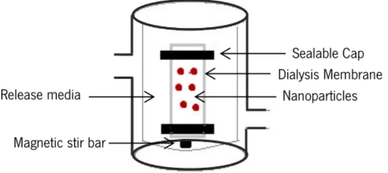
The release profile of a bioactive compound is equally important as the bioactive polymer formulation because to understand the release profile can be crucial for product development and potential applications (Kumari et al., 2010; Pinheiro et al., 2012a, 2012b). Commonly, the release profile is also determined as an alternative to characterize nanoparticles, along with others parameters (e.g. particle shape, size, zeta-potential and encapsulation efficiency) (Zambito et al., 2012).
A method used to evaluate the release profile is dialysis membrane and this method consists, essentially, in to separate the nanoparticles with bioactive compound loaded from the bulk media by a dialyzing membrane, Figure 2.10. Over the time, occurs the passage of compound loaded in nanoparticles through the membrane into the release media and a sample is withdrawn at intermittent intervals to evaluate the compound release (Souza and Deluca, 2005).
CHAPTER II INTRODUCTION
24 | DEVELOPMENT OF NANOSTRUCTURES FOR ENCAPSULATION OF VITAMINS
There are four mechanisms that can be responsible by release profile of bioactive compound from nanoparticles: Mechanism of Fic ’s diffusion, polymer matrix swelling, polymer erosion and degradation; and the different mechanisms depending on the system and environmental conditions. However, to understand the release mechanism involved is necessary to use mathematical modeling (Pinheiro et al., 2012b). The release profile depends of various parameters, such as value of diffusion coefficient, size and shape of bioactive compound and polarity of the matrix (Romero-cano and Vincent, 2002).
2.4 BIOACTIVE FOOD COMPOUNDS
The normal development and growth of human depend of different constituents, being one of them bioactive compounds. These components are substances/compounds present in animal and plants that can have different physiological functions, such as health promoting or disease preventing effect (De Vos et al., 2010). Each bioactive component has their own molecular characteristics (e.g. molecular weights, conformations, polarity and charge) that are responsible by physicochemical differences between bioactive components (e.g. solubility, partitioning, physical state, interactions, optical characteristics and chemical stability). They can be divided into bioactive molecules such as vitamins, bioactive peptides, antioxidants, fatty acids and minerals; and bioactive living cell such as probiotics (McClements et al., 2009; de Vos et al., 2010), which can be used in food industry to improve taste, aroma, stability, nutritional value and appearance of fodd products (Augustin and Hemar, 2009; Raybaudi-Massilia and Mosqueda-Melgar, 2012). Some of these bioactive compounds are sensitive to heat, moisture and/or pH and can be slowly degraded and lose their activity during utilization. This way, the food industry has a great challenge: protect these components during production, storage and consumption.
Sealable Cap Dialysis Membrane
Nanoparticles Release media
Magnetic stir bar
Figure 2.10 – Schematic presentation of set-up for release profile by dialysis membrane method. Image adapted from Prata et al. (2008) and Souza and Deluca (2005).
DEVELOPMENT OF NANOSTRUCTURES FOR ENCAPSULATION OF VITAMINS | 25 problems, in order to protect the bioactive compound against adverse conditions and simultaneous keep the functionality of the bioactive compound. Due to their different properties, is needed to develop micro- or nanostructurs with specific physicochemical characteristics to promote compatibility and stability between bioactives compounds and the structure used. Besides that, it is also essential to select a system easily incorporated into the food and that does not interfere with the texture and taste of the food (De Vos et al., 2010).
As already mentioned the vitamins are an example of bioactive component that plants and microorganisms have the capability to produce for the normal organism functioning. The human haven’t this ability, but it is recognized that vitamins are essential for maintain health and well-being of human (Spitzer and Schweigert, 2007). Below are presented the most important characteristics of them and their importance for us.
2.4.1 VITAMINS
In nineteenth century, the physiologists thought that food was a source of only four types of nutrients (protein, fat, carbohydrate and ash) and water, being vitamin only associated with the preventing of vitamin disease and their biochemical functions. Today, this opinion is different and the term vitamin is a common word that reveals an interrelationship between diet/food and health (Combs Jr, 2012).
Vitamins are defined as organic molecules with small dimension and low molecular weight being presented in food in small amounts. They are considered essential for normal maintenance, growth and development of human organism and their absence cause a specific deficiency syndrome. The incapacity of human to synthetize some vitamins (exception of vitamin D and B3) lead to their necessity to get through food products (Ball, 2008; Combs Jr, 2012). In general, the pathway of vitamins biosynthesis is complex and is biologically more efficient for human to ingest the vitamins that synthetize through simples’ molecules. This fact has a disadvantage that is the dependency of the other organisms to get essentials components to life (Berg et al., 2004). In general, all vitamins have similar characteristics, but their chemical properties are very different (e.g. the vitamins A, K and C are enzymatic co-factors, E and C act as antioxidants and vitamins A and D are hormones). Besides that, vitamins can be divided into liposoluble and water