Faculdade de Medicina de Lisboa
Deciphering the mechanisms underlying
the loss of BDNF neuroprotection in an
Alzheimer’s Disease model
Sara Luísa Ramalho Tanqueiro
Orientador: Professora Doutora Maria José de Oliveira Diógenes Nogueira Co-orientador: Doutora Rita Cruz Coelho de Mira Ramalho
Dissertação especialmente elaborada para obtenção do grau de
Mestre em Neurociências
Universidade de Lisboa
Faculdade de Medicina de Lisboa
Deciphering the mechanisms underlying
the loss of BDNF neuroprotection in an
Alzheimer’s Disease model
Sara Luísa Ramalho Tanqueiro
Orientador: Professora Doutora Maria José de Oliveira Diógenes Nogueira Co-orientador: Doutora Rita Cruz Coelho de Mira Ramalho
Dissertação especialmente elaborada para obtenção do grau de
Mestre em Neurociências
Todas as afirmações efetuadas no presente documento são da exclusiva responsabilidade do seu autor, não cabendo qualquer responsabilidade à Faculdade de Medicina de Lisboa pelos conteúdos nele apresentados.
“A impressão desta dissertação foi aprovada pelo Conselho Científico da
Faculdade de Medicina de Lisboa em reunião de 19 de Janeiro.”
v
|
PUBLICATIONS
Papers where the author of this thesis participate during her master:
- Jerónimo-Santos A, Fonseca-Gomes J, Guimarães DA, Tanqueiro SR, Ramalho RM, Ribeiro JA, Sebastião AM, Diógenes MJ (2015). Brain-Derived Neurotrophic Factor mediates neuroprotection against Aβ-induced toxicity through a mechanism independent on adenosine 2A receptor activation. Growth Factors, 33: 298-308. - Ribeiro FF, Xapelli S, Miranda-Lourenço C, Tanqueiro SR, Fonseca-Gomes J, Diógenes MJ, Ribeiro JA, Sebastião AM (2015). The role of purines in neuroregeneration and neuroprotection. Neuropharmacology, S0028-3908(15)30170-2.
vii
|
RESUMO
O fator neurotrófico derivado do cérebro (Brain-derived neurotrophic factor - BDNF) desempenha importantes funções no sistema nervoso central, nomeadamente diferenciação e sobrevivência neuronais e regulação da transmissão e plasticidade sinápticas. Em algumas doenças neurodegenerativas, como na doença de Alzheimer (Alzheimer’s disease - AD) que se caracteriza por declínio cognitivo e perda de memória, sabe-se que a sinalização mediada pelo BDNF se encontra diminuída. De facto, tanto em doentes como em modelos animais de AD, existem evidências de que os níveis proteicos de BDNF e da isoforma completa do seu recetor, TrkB-FL (full length – FL), se encontram diminuídos. O BDNF tem também a capacidade de se ligar a recetores TrkB truncados (truncated TrkB – TrkB-TC), porém estes recetores são moduladores negativos de TrkB-FL, uma vez que são incapazes de iniciar as vias de sinalização mediadas pelo BDNF. Estudos recentes revelaram que o recetor TrkB-FL é clivado por um grupo de proteases, designadas por calpaínas, resultando na formação de um novo recetor TrkB truncado (TrkB-T’) e de um fragmento intracelular (intracellular domain - ICD) que é translocado para o núcleo. Não se conhecem ainda em profundidade as ações destes novos fragmentos mas sabe-se que a função do BDNF fica severamente comprometida. As calpaínas são proteases dependentes de cálcio, sendo por isso ativadas por um aumento dos níveis intracelulares deste catião. Os recetores N-metil-D-aspartato (NMDARs), importantes mediadores da plasticidade sináptica, são permeáveis a cálcio e podem ser encontrados tanto na região sináptica como na extrassináptica. Enquanto a ativação dos recetores NMDAR sinápticos resulta em alterações que promovem a neuroprotecção, a ativação dos NMDARs extrassinápticos induz, preferencialmente, fenómenos de morte neuronal. Curiosamente, sabe-se que os eNMDARs se encontram sobreactivados em diversas condições patológicas, inclusivamente em modelos de AD, e que podem ter um importante papel na desregulação dos níveis de cálcio intracelular. Assim, o trabalho desenvolvido nesta tese teve como objetivo investigar se a ativação dos eNMDARs contribui para a ativação das calpaínas e consequente clivagem dos recetores TrkB-FL, assim como a perda da sinalização do BDNF.
viii
Em primeiro lugar, investigou-se se a prevenção da ativação dos eNMDARs, em neurónios expostos ao péptido β amilóide (amyloid β – Aβ), inibia a clivagem dos recetores TrkB-FL pelas calpaínas. Para testar esta hipótese, culturas primárias de neurónios de rato Sprague-Dawley com 14 dias in vitro (DIV14), foram incubadas durante 24 h com Aβ25-35 (25 µM), o mais pequeno fragmento tóxico do péptido Aβ, e memantina (1 µM), fármaco que bloqueia preferencialmente eNMDARs sobreactivados. Os resultados obtidos indicaram, como esperado, que Aβ25-35 induz um aumento dos níveis dos produtos específicos da clivagem da αII-espectrina mediada pelas calpaínas (specific spectrin breakdown products – SBDP150) sugerindo que existe uma forte ativação destas proteases. Esta alteração traduziu-se na diminuição significativa dos níveis proteicos de TrkB-FL e num aumento nos níveis de TrkB-ICD. Por outro lado, os resultados mostraram, pela primeira vez, que os efeitos de Aβ25-35 são prevenidos pela co-incubação com a memantina: i) a formação de SBDP150 diminuiu, ii) os níveis dos recetores TrkB-FL aumentaram e iii) os níveis de TrkB-ICD diminuíram. Assim, os resultados indicam que a ativação dos eNMDAR parece estar envolvida na ativação das calpaínas e, consequentemente, na clivagem dos recetores TrkB-FL.
Uma vez que a formação de novas sinapses são processos que estão na base da formação de memória, pensa-se que as alterações que decorrem no número de espinhas dendríticas num neurónio de um doente de AD tem um papel preponderante nos défices cognitivos que se desenvolvem possivelmente adjacentes à perda neuronal. Sabe-se que o BDNF aumenta o número de espinhas dendríticas, protusões sinápticas através das quais a maioria das sinapses excitatórias ocorre e que correspondem à força de atividade sináptica de um neurónio. Assim, propusemo-nos avaliar, através de imunocitoquímica, se a ativação dos eNMDARs está relacionada com a perda de espinhas dendríticas num neurónio. Os resultados foram obtidos a partir de neurónios provenientes da cultura primária de rato a DIV14. O número de protusões (espinhas dendríticas e filopodia, protusões mais finas) foi quantificado em frações de 10 µm da dentrite-mãe a uma distância de 25 µm do corpo celular do neurónio.
Os resultados indicam, como esperado, que Aβ25-35 diminui significativamente o número de protusões e que o BDNF aumenta o número de protusões per se. Na presença de Aβ25-35, os resultados sugerem que o BDNF perde a sua ação no aumento
ix
do número de protusões. Essa função é recuperada aquando bloqueio dos eNMDARs com memantina, assim como bloqueio da atividade das calpaínas com MDL28170 (20 µM). Estes resultados propõem que os mecanismos através dos quais Aβ interfere com as espinhas dendríticas envolvem não só a ativação das calpaínas, como visto anteriormente, como também a ativação dos eNMDARs.
Por outro lado, foi também nosso propósito avaliar se o bloqueio dos eNMDARs restaurava o efeito do BDNF na potenciação de longa duração (long-term potentiation – LTP) na área CA1 do hipocampo, o mecanismo fisiológico da aprendizagem e memória, cuja magnitude se encontra diminuída na presença de Aβ.
Para tal, foram preparadas fatias de hipocampo de rato Wistar com 8-12 semanas de vida e após incubação durante 3h com Aβ25-35 (25 μM) e/ou memantina (1 μM) procedemos ao registo extracelular dos potenciais pós-sinápticos excitatórios de campo (field excitatory postsynaptic potentials – fEPSP) e à indução de LTP na ausência ou presença de BDNF (20 ng/mL). Os dados sugerem, como esperado que, em fatias incubadas exclusivamente com líquido cefalorraquidiano artificial (artificial
cerebrospinal fluid - aCSF), o BDNF induz um aumento significativamente da magnitude
da LTP e que, na presença de Aβ, o BDNF perde a sua ação na LTP. Curiosamente, os nossos resultados indicam, pela primeira vez, que a co-incubação de memantina e Aβ25–35 restaura a capacidade do BDNF em facilitar a LTP. Estes resultados indicam que a sinalização mediada pelo BDNF na LTP se encontra diminuída na presença de Aβ e que esta diminuição pode ser mediada pela ativação dos eNMDARs.
Em conclusão, os resultados sugerem que, na presença de Aβ, a sinalização mediada pelo BDNF se encontra severamente diminuída, afetando as suas ações sinápticas, através de um mecanismo que possivelmente é mediado pela ativação dos eNMDARs. Estas evidências realçam a consequência funcional da clivagem dos recetores TrkB-FL induzida pelo Aβ e propõem a modulação dos eNMDARs de modo a prevenir a desregulação dos níveis intracelulares de cálcio e, consequentemente, a perda dos mecanismos neuroprotetores mediados pelo BDNF.
Palavras-Chave: BDNF, péptido β-amilóide, recetor TrkB, calpaínas, recetores
xi
|
ABSTRACT
The brain-derived neurotrophic factor (BDNF) plays important functions in the central nervous system, such as cell survival, neuronal outgrowth, differentiation and plasticity. In contrast, BDNF signaling is known to be impaired in some neurodegenerative diseases, including Alzheimer’s disease (AD), which is characterized by cognitive decline and loss of memory. In fact, in AD patients and in several AD models a decrease in BDNF and its main receptor, TrkB-full length (TrkB-FL), has been reported.
BDNF can also bind to truncated TrkB (TrkB-TC), however these receptors act as dominant negative inhibitor of TrkB-FL since they cannot initiate BDNF signaling. Recent evidences revealed that TrkB-FL is processed by calpains, which results in the formation of a new truncated TrkB (TrkB-T’) and in the formation of an intracellular domain (ICD) fragment. Thus, this cleavage culminates in the receptor loss of function. Calpains are Ca2+-dependent proteases that are activated by increased intracellular levels of this cation. N-methyl-d-aspartate receptors (NMDARs), which are known to be permeable to Ca2+, are essential mediators of brain synaptic plasticity and can be found at synaptic and extrasynaptic sites. Synaptic NMDARs are neuroprotective, whereas extrasynaptic NMDARs (eNMDARs) preferentially initiate cell death pathways. Importantly, eNMDARs are known to be over activated in AD. Furthermore, NMDARs have been proposed as one of the molecules that might be involved in intracellular Ca2+ deregulation. Thus, we purposed to investigate if, by preventing eNMDAR activation in primary rat neurons or hippocampal slices exposed to the active fragment of amyloid β (Aβ25-35) (25 µM), one of the main neurotoxic species that contribute to AD progression, we could inhibit the truncation of TrkB-FL by calpains, restoring the functions of BDNF. Our results have shown that the inhibition of eNMDAR by memantine (1 µM), which preferentially blocks extrasynaptic receptors over synaptic receptors, reduces significantly the activation of calpains. These findings are related with an increase in TrkB-FL levels and a decrease in ICD levels. Moreover, it is known that BDNF increases the number of spines in one neuron, which are synaptic protrusions where the majority of excitatory post-synaptic domains are localized and that can highly predict the strength of synaptic activity. Our results indicate that, in the
xii
presence of Aβ, BDNF loses its function upon spine density, which is prevented when calpains activity is inhibited with MDL28170 (20 µM) or while eNMDAR blockade. Finally, data suggest that the inhibition of eNMDAR restores the capacity of BDNF to enhance long-term potentiation in hippocampal slices, the physiological basis for learning and memory that is known to be impaired in the presence of Aβ. Finally, the focus of our work was to clarify the mechanism by which BDNF loss its function upon synapses. In conclusion, data suggest that, in primary neuronal cultures and hippocampal slices, Aβ severely impairs BDNF/TrkB-FL signaling affecting the synaptic actions of BDNF by a mechanism that is, at least in part, mediated by eNMDARs activation. These findings highlight the functional consequence of the Aβ-induced cleavage of TrkB receptors and propose eNMDAR modulation to prevent the disruption of Ca2+ homeostasis and, consequently, the loss of physiological mechanisms that depend on BDNF.
Keywords: BDNF, β-amyloid peptide, TrkB receptor, calpains, extrasynaptic NMDA
xiii
|
FIGURE INDEX
Figure 1 - Schematic representation of (A) healthy neurons and (B) the abnormal deposits described by Dr. Alzheimer.
Figure 2 - Generation of Aβ through proteolytic processing of APP. Figure 3 – Formation of amyloid plaques.
Figure 4 - Expression of NMDARs subunits in the mouse brain at postnatal day 0 (P0), which is the day of birth, 2 weeks following birth (P14) and at the adult stage.
Figure 5 – Schematic representation of a hippocampus slice. Figure 6 – Schematic representation of dendritic spines. Figure 7 – Molecular mechanisms underlying LTP. Figure 8 - Structural changes associated with LTP. Figure 9 - The NMDA paradox.
Figure 10 - Mediators of synaptic and extrasynaptic NMDAR activity effects.
Figure 11 – Schematic representation of the mechanisms whereby Aβ leads to synaptic dysfunction and neurodegeneration.
Figure 12 - Neurotrophin receptors and their specificity for the neurotrophins. Figure 13 –TrkB isoforms.
Figure 14 - BDNF/TrkB-FL signaling pathway.
Figure 15 – BDNF facilitation upon LTP in a glutamatergic synapse. Figure 16 - TrkB receptor dimer combinations.
Figure 17 – TrkB-FL cleavage.
Figure 18 – Rat hippocampal slices preparation.
Figure 19 - Schematic representation of extracellular recordings in hippocampal slices. Figure 20 – The inhibition of eNMDAR reduces the cleavage of TrkB-FL by modulating calpains activation.
Figure 21 – BDNF restores its capacity to increase dendritic spines number after inhibition of Aβ-induced eNMDAR activation.
Figure 22 – The alterations in the number of spines are not associated with changes in different NMDAR subunits expression.
Figure 23 - The inhibition of eNMDAR activation by Aβ restores the facilitatory effect of BDNF upon θ-burst-induced LTP.
|
INDEX
Resumo …….……… vii
Abstract ……...……… xi
Figure Index ……….. xiii
Abbreviations List ………..… 3 1|INTRODUCTION 1.1|Alzheimer’s Disease 1.1.1| Overview ……….. 5 1.1.2| Pathophysiology ..………..….. 6 1.1.3| Aβ peptide ....……….... 7
1.1.4|Neurotoxicity and synaptic failure mediated by Aβ peptide .…………... 9
1.2|The NMDA receptor 1.2.1|NMDAR composition ………. 10
1.2.2|NMDAR and synaptic plasticity ………. 11
1.2.3|Synaptic and extrasynaptic NMDARs ……….. 16
1.2.4. Extrasynaptic NMDARs in AD ………. 18
1.3|Neurotrophins 1.3.1| BDNF signaling and its function ………. 22
1.3.2|TrkB-FL cleavage and loss of BDNF signaling in AD ……….…………. 26
2|AIM ……….. 29
3|METHODS 3.1|Primary Neuronal Cultures and Drug Treatments ………..….. 31
3.2|Western Blotting ...……….………...…. 32
3.3|Immunocytochemistry ..……….….. 33
3.4|Freshly Prepared Hippocampal Slices ..………..….………... 34
3.5|Ex-vivo Electrophysiology Recordings: LTP induction ..……….…..…. 35
3.6|Data analysis ...……….………. 36
4|RESULTS
4.1|The blockade of eNMDAR can limit TrkB-FL truncation induced by Aβ and restore the ability of BDNF to increase spine density on primary neuronal cultures ....……… 39
4.2| The inhibition of eNMDAR can partially rescue the facilitatory effect of BDNF upon
LTP on hippocampal slices ………..……… 45
5|DISCUSSION
5.1|The inhibition of eNMDAR limits Aβ-induced TrkB-FL truncation and restores the ability of BDNF to increase spine density on primary neuronal cultures ……… 51
5.2| The inhibition of eNMDAR can partially restore BDNF synaptic function in the
presence of Aβ ………. 53
5.3| eNMDAR activation in AD: The initial trigger of calcium dyshomeostasis? ……. 56
6|CONCLUSIONSANDFUTUREDIRECTIONS……….………... 59
7|REFERENCES……….. 63
3
|ABBREVIATIONS LIST
Aβ Amyloid-β
aCSF Artificial cerebrospinal fluid AD Alzheimer’s disease
AMPA Alpha-amino-3-hydroxy-5-methyl-4-isoxazolepropionic acid
AMPAR AMPA receptor APOE Apolipoprotein E
APP Amyloid precursor protein BAD Bcl-2-associated death promoter BDNF Brain-derived neurotrophic factor BSA Bovine serum albumin
CA1 Cornu ammonis area 1 CA3 Cornu ammonis area 3
CaMK Ca2+-calmodulin-regulated Kinase CDK5 Cyclin-dependent kinase 5 CNS Central nervous system CREB CRE binding protein CTR Control
DAG Diacylglycerol DIV Days in vitro DTT 1,4-dithiothreitol
EDTA Ethylenediaminetetraacetic acid E-LTP Early LTP
ER Endoplasmic reticulum
ERK Extracellular signal-regulated kinase eNMDAR Extrasynaptic NMDA receptor fEPSP Field excitatory postsynaptic potential
FL Full-length
GAPDH Glyceraldehyde 3-phosphate dehydrogenase
HBSS Hanks' balanced salt solution HFS High frequency stimulation ICD Intracellular domain IkB Inhibitor of kappa B IP3 Inositol 1,4,5-triphosphate IP3R IP3 receptor
L-LTP Late phase of LTP LTP Long-term potentiation
MAP Microtubule-associated protein MAPK Mitogen-activated protein kinase
MDL28170
N-[N- [(phenylmethoxy)carbonyl]-L-valyl]-phenylalaninal
MEK Mitogen-activated protein kinase kinase
NFkB Nuclear Factor Kappa-light-chain-enhancer of activated B cells
NFT Neurofibrillary tangles NGF Nerve growth factor
NMDAR N-Methyl-D-aspartate receptor NT Neurotrophin
p75NTR p75 neurotrophin receptor PBS Phosphate buffered saline PI3K Phosphatidylinositol 3-kinase PIP3 Phosphatidylinositol-3,4,5-trisphosphate PKC Protein kinase C PLC Phospholipase C PSD Postsynaptic density PVDF Polyvinylidene difluoride RIPA Radioimmunoprecipitation assay buffer
ROS Reactive oxygen species RT Room temperature
SBDP Spectrin breakdown product SDS-PAGE Sodium dodecyl sulfate-polyacrylamide gel electrophoresis SEM Standard error of the mean sNMDAR Synaptic NMDA receptor TC Truncated TBS-T Tris-buffered saline-Tween 20 Trk Tropomyosin-related kinase TrkB-FL TrkB full-length TrkB-T’ TrkB truncated (calpain-generated) TrkB-T1 TrkB truncated isoform 1 TrkB-T2 TrkB truncated isoform 2 TrkB-TC TrkB truncated (total pool) TrkB-ICD TrkB intracellular domain TRPC Canonical transient receptor potential
5
1
|
INTRODUCTION
1.1|
Alzheimer's Disease
1.1.1| Overview
Alzheimer’s disease (AD) is the most common form of dementia and the most prevalent neurodegenerative disease in the elderly population, affecting almost 40 million people worldwide (Brookmeyer et al., 2007, Alzheimer's, 2015). AD progression has been associated with a gradual damage in function and structure in the hippocampus and neocortex, brain areas involved in memory and cognition (Braak et al., 1993). As a consequence, the most common symptom is the gradually worsening ability to remember new information. AD is ultimately fatal due to brain changes that severely impair basic physiological functions (Alzheimer's, 2014).
The precise pathophysiological changes that trigger the development of AD remain largely unknown. Only approximatly 1% of AD cases are caused by three known genetic mutations, involving the gene for the amyloid precursor protein (APP) and the genes for the presenilin 1 and presenilin 2 proteins (Rogaeva, 2002). The majority of cases, however, are sporadic, with the disease onset starting usually after 65 years old. Thus, as a multifactorial disease, increased age is considered the main risk factor for developing AD (Guerreiro et al., 2012). In addition, individuals with the ε4 form of the gene apolipoprotein E (APOE ε4) for brain cholesterol transport are also at increased risk (Kim et al., 2009). Moreover, a diagnosis of mild cognitive impairment, cardiovascular disease, high blood cholesterol, diabetes, obesity and traumatic brain injury are also associated with a higher risk of developing AD (Boyle et al., 2006, Stampfer, 2006, Barbagallo and Dominguez, 2014, Naderali et al., 2009).
Although first described in 1906 by the neuropathologist Alois Alzheimer, the research into AD symptoms, causes and risk factors has only gained impetus in the last 25 years, while efficient treatment is still lacking (Goedert and Spillantini, 2006).
6
1.1.2| Pathophysiology
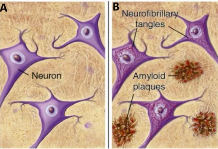
More than 100 years ago, Dr. Alois Alzheimer identified neurofibrillary tangles (NFT) and neuritic plaques in a 51-years-old brain’s patient that presented a severe cognitive decline by the time of death (Hippius and Neundorfer, 2003). Later, the identification of tau protein as the major NFT component and amyloid β (Aβ) peptide as the major plaque component (Figure 1) led to a new era of research on AD (Kidd, 1963, Terry et al., 1964).
Figure 1 - Schematic representation of (A) healthy neurons and (B) the abnormal deposits described by
Dr. Alzheimer. Amyloid plaques are localized extracellularly whereas NFT are localized inside neurons
(Mitra and Dey, 2013).
Under physiological conditions, phosphorylation of tau is important to maintain the structure of cytoskeleton. The balance of phosphorylated and unphosphorylated tau regulates the stability of microtubules, which defines the normal morphology of neurons and maintain the axoplasmic flow (Kosik, 1993). In AD, however, hyperphosphorylated tau protein accumulates inside the cell, dimerizing to paired helical filaments, which aggregate to form the typical NFTs within neurons (Goedert et al., 2006). An imbalance between the activation of phosphorylating protein kinases and dephosphorylating protein phosphatases is thought to occur in AD, leading to excessive tau phosphorylation, microtubule instability, axonal transport impairment and, consequently, cell death (Mandelkow et al., 1995).
On the other hand, extracellular amyloid plaques, also called senile or neuritic plaques, are mainly composed by Aβ peptide. These amyloid plaques are preferential
B
A
7
localized in the cortex and hippocampus and correlated with the primary cognitive and memory disturbances (Killiany et al., 2002).
Due to its neurotoxic effects and accumulation in AD, Aβ is believed to play a central role in the pathogenesis of the disease (Hardy and Higgins, 1992). Moreover, since it is known that Aβ aggregates into toxic plaques up to several years before the first clinical symptoms appear, their detection and monitoring became of primary interest, both for diagnostic purposes and for fundamental research (Goedert and Spillantini, 2006).
1.1.3| Aβ peptide
In 1991, Hardy and Allsop proposed, for the first time, the amyloid hypothesis for AD pathogenesis and this postulate still continues to be the hypothesis best scientifically supported nowadays (Carrillo-Mora et al., 2014). It assumes that Aβ, in multiple forms, triggers several cascades that can led to synapse loss and neurodegeneration (Morris et al., 2014). In fact, it was demonstrated that Aβ production are increased in familial forms of AD (Vetrivel and Thinakaran, 2006), and several toxic effects of this peptide have been described both in vitro and in vivo studies (Atwood et al., 2003). A study using two mouse AD lines (AβPPPS1-21 and Tau22 mice) described that AβPPPS1-21 transgenic mice were impaired in spatial, fear, aversion and extinction learning deficits, whereas Tau22 animals were impaired in appetitive responding. The deficits in AβPPPS1-21 mice suggested that amyloid-related pathology might be more pervasive and/or widespread than tau pathology (Lo et al. 2013). Furthermore, research on the pathological changes in AD indicates that accumulated Aβ in vivo may initiate the hyperphosphorylation of tau (Huang and Jiang 2009). Thus, an excessive production of Aβ or an impairment in its adequate clearance have been suggested as key events in the origin and progression of the neuronal damage (Mawuenyega et al., 2010).
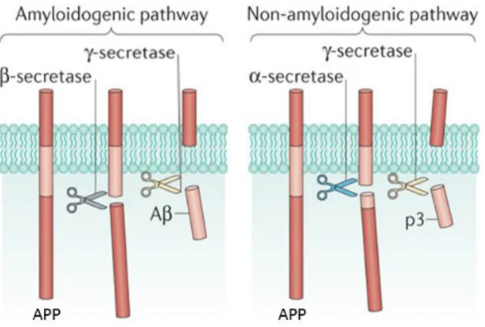
Aβ is a peptide of 39 to 42 aminoacids produced in all neurons through proteolitic processing of the transmembrane amyloid precursor protein (APP) by β- and γ-secretases. The cleavage site used by γ-secretase in the amyloidogenic pathway determines whether the predominant Aβ1-40 or the more aggregation-prone and neurotoxic Aβ1-42 species of the peptide is generated. Within the nonamyloidogenic
8
pathway, APP is cleaved by α-secretase, preventing Aβ formation (Figure 2) (Hardy and Selkoe, 2002).
Figure 2 - Generation of Aβ through proteolytic processing of APP. Cleavage by β-secretase generates the N-terminal and intramembranous cleavage by γ-secretase gives rise to the C-terminal of Aβ. Cleavage by α-secretase precludes Aβ formation, producing another fragment (figure adapted from Heppner et al., 2015).
Interestingly, Aβ has been shown to have physiological functions within the brain at pM concentrations (Puzzo et al., 2008). In fact, Aβ can enhance synaptic plasticity, increase synaptic vesicles release probability (Abramov et al., 2009) and even protect against excitotoxic insults (Giuffrida et al., 2009). In contrast, at supraphysiological concentrations, Aβ self-aggregates into higher order structures. Initially, soluble Aβ acquires higher β-sheet content, leading to self-dimerization. This is followed by oligomerization into soluble high molecular aggregates, protofibril formation and finally aggregation into insoluble fibrillary structures characteristic of amyloid plaques (Figure 3) (Serpell, 2000).
Figure 3 – Formation of amyloid plaques. Aβ monomers aggregate to form oligomers, fibrils and/or plaques, depending on mutations in the Aβ coding region of APP and/or post-translational modifications (figure adapted from Heppner et al., 2015).
9
In AD brain, insoluble extracellular amyloid plaques aggregate around neurons and glia and their main component is Aβ1-42 (Hardy and Higgins, 1992). Aβ1–42, with its two additional hydrophobic amino acids, has a higher tendency to aggregate than Aβ1–40 and has been ascribed to be the main pathogenic form (Citron, 2010). Furthermore, since evidences suggest that Aβ toxicity implicate oligomers as the primary toxic species (Haass and Selkoe, 2007, Kayed and Lasagna-Reeves, 2013, Ferreira et al., 2007), a shift had occurred in amyloid hypothesis focus from plaque to soluble forms of Aβ.
1.1.4|Neurotoxicity and synaptic failure mediated by Aβ peptide
The accumulation of intracellular Aβ occurs early in the neuropathological phenotype of AD, even before the formation of NTFs and plaque deposition (Gouras et al., 2000). Although extracellular Aβ can bind to several receptors, producing Aβ-receptor complexes that can also be internalized into early endosomes, the amyloidogenic cleavage of APP also occurs in Golgi and endoplasmic reticulum membranes (LaFerla et al., 2007). Therefore, Aβ can accumulate in mitochondria, culminating in reactive oxygen species (ROS) production, leading to oxidative stress (Manczak et al., 2006, Caspersen et al., 2005). Furthermore, mitochondrial Aβ also can initiate a cascade of events that activates caspase-3, triggering the intrinsic apoptotic pathway (D'Amelio et al., 2011).On the other hand, extracellular Aβ oligomers can also lead to the dysregulation of different signaling pathways that culminate in multiple mechanisms of synaptic failure (Kayed and Lasagna-Reeves, 2013). Most of the mechanisms whereby both intracellular and extracellular Aβ leads to synaptic dysfunction and neurodegeneration will be schematized later when all of them were described.
Interestingly, the notion that dementia is a consequence of synaptic degeneration was raised when Santiago Ramon y Cajal suggested that "dementia could result when synapses between neurons are weakened as a result of a more or less pathological condition, that is, when processes atrophy and no longer form contacts, when cortical mnemonic or association areas suffer partial disorganization" (Shankar and Walsh, 2009). Accordingly, several studies have shown that neuronal death in AD patients is closely associated with extensive synapse loss in the neocortex (Terry et al., 1991) and decreased synapse density is the strongest neuropathological correlate of the
10
degree of dementia in AD (Arendt, 2009, Masliah et al., 2006). Importantly, Aβ exerts neurotoxic effects by disrupting the integrity of both plasma and intracellular membranes (Demuro et al., 2005) and by accumulating at excitatory synapses, impairing synapse function (De Felice et al., 2007, Deshpande et al., 2009). Furthermore, it has been reported that Aβcan disrupt the postsynaptic density, which organizes synaptic proteins to mediate the functional and structural plasticity of the excitatory synapse and to maintain synaptic homeostasis (Gong and Lippa, 2010, Lacor et al., 2004, Reddy et al., 2005, Snyder et al., 2005, Hsieh et al., 2006).
1.2|
The NMDA receptor
Glutamate is the major fast excitatory neurotransmitter and it is involved in almost all central nervous system (CNS) functions, particularly in cortical and hippocampal regions (Parsons et al., 1998), including synaptic transmission, neuronal growth, cell differentiation, synaptic plasticity, learning and memory (Butterfield and Pocernich, 2003, Francis, 2003).
The ionotropic glutamate receptors subtype N-methyl D-aspartate (NMDARs), which are known to be concentrated on postsynaptic spines of neuronal dendrites (Mattson et al., 1998), are cationic channels gated by glutamate, permeable to Na+, K+ and Ca2+ (Danysz and Parsons, 2012). At the resting membrane potential, the Ca2+ channel of the NMDA receptor is blocked by Mg2+ ions, which is associated with a low background level of postsynaptic intracellular Ca2+. Mg2+ ion removal from the pore, which will allow the flow of ions, requires a membrane depolarization of sufficient amplitude and duration (Duguid IC, 2009). Importantly, NMDARs can differ in their subunit composition, a characteristic that also varies across CNS regions during development and in disease states, subcellular localization, pharmacological properties and their interacting proteins.
1.2.1|
NMDAR composition
NMDAR subunits are encoded by three families of genes coding for GluN1, GluN2 and GluN3 subunits (Cull-Candy et al., 2001). Functional NMDARs are heterotetramers composed by two glycine or D-serine-binding GluN1 subunits and two glutamate binding GluN2 (GluN2A-D) subunits or, in some cases, glycine binding GluN3 (GluN3A/B)
11
subunits. The most widely expressed NMDARs contain the obligatory subunit GluN1 plus either GluN2B or GluN2A or a mixture of the two (Kohr, 2006). GluN3A and GluN3B are mostly expressed in oligodendrocytes and astrocytes (Cull-Candy et al. 2001).
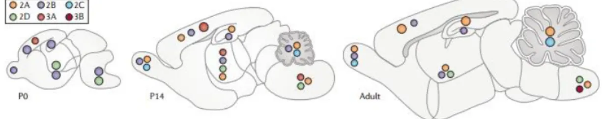
Interestingly, during development and even in some adult synapses, the composition of NMDARs changes in response to neuronal activity. In early postnatal development, NMDARs switch their subunit composition from primarily containing GluN2B subunits to predominantly containing GluN2A subunits. Nevertheless, GluN2B subunits still populate many regions of the adult forebrain (Figure 4) (Paoletti et al., 2013).
Figure 4 - Expression of NMDARs subunits in the mouse brain at postnatal day 0 (P0), which is the day of
birth, 2 weeks following birth (P14) and at the adult stage. In adults, GluN2A is ubiquitously expressed
in the brain, GluN2B is mostly restricted to the forebrain, GluN2C is limited to the cerebellum, and GluN2D is expressed in small numbers of cells in selected brain regions (Paoletti et al., 2013).
1.2.2|
NMDAR and synaptic plasticity
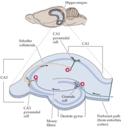
Long term potentiation (LTP), which is a long-lasting increase in synaptic strength, is widely accepted as the neurophysiological basis for learning and memory (Bliss and Collingridge, 1993). The best-characterized form of LTP occurs between pyramidal neurons of Cornu Ammonis 3 (CA3) and Cornu Ammonis 1 (CA1) in the hippocampus (Figure 5) (Malenka, 1994). “CA” refers to cornu Ammon, the latin for Ammon’s horn, since the ram’s horn resembles the shape of the hippocampus (Purves D, 2001).
12
Figure 5 – Schematic representation of a hippocampus slice. The arrangement of neurons allows the hippocampus to be sectioned in slices, maintaining the relevant circuitry intact. The cell bodies of the pyramidal neurons lie in a single densely packed layer. This layer is divided into CA1 and CA3, where the dendrites of pyramidal cells in CA1 (stratum radiatum) receive synapses from the Schaffer collaterals, which are axons of pyramidal cells in the CA3 region (Purves D, 2001).
Electrical stimulation of Schaffer collaterals generates excitatory postsynaptic potentials (EPSPs) in the postsynaptic CA1 cells. A brief, high-frequency stimulation (HFS) to the same axons causes LTP, which is evident as a long-lasting increase in EPSP amplitude (Purves D, 2001). In addition, the mechanisms underlying electrically induced LTP, which can performed in laboratory with appropriate protocols, remains as a model of synaptic and cellular events that may underlie synaptic changes in brain during learning and memory formation. Supporting the emerging interest in LTP as a potential mechanism of memory, LTP exhibits numerous properties expected of a synaptic associative memory mechanism, such as rapid induction, synapse specificity, associative interactions, persistence, and dependence on correlated synaptic activity (Escobar and Derrick, 2007). Although LTP has become one of the most extensively studied topics in neuroscience, the molecular mechanisms underlying LTP are still not fully understood (Lømo, 2003). It is known that LTP is divided into early LTP (E-LTP) and late LTP (L-LTP). E-LTP requires modifications in existing proteins, whereas L-LTP is only induced by strong stimulation and requires synaptic growth, gene transcription and new protein synthesis (Pang and Lu, 2004).
It is clear that some types of LTP do not involve NMDAR (Johnston et al., 1992), however NMDAR-dependent LTP, which has received more attention, is known to occur in the CA1 area of the hippocampus (Lüscher et al., 2012). In NMDAR-dependent LTP,
13
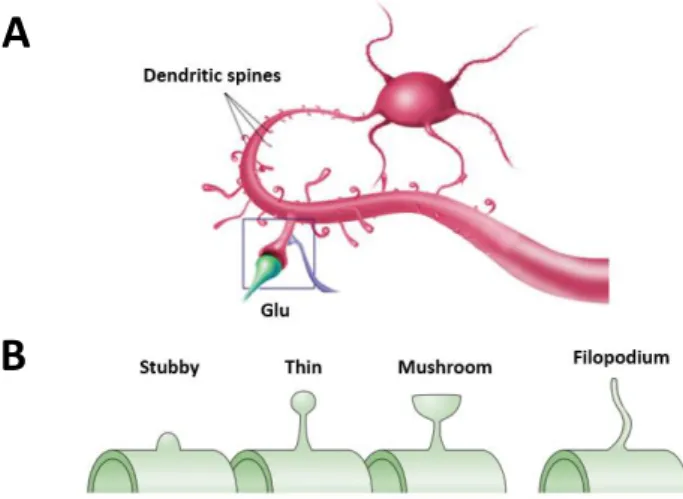
during HFS, glutamate binds to Alpha-Amino-3-Hydroxy-5-Methyl-4-Isoxazolepropionic Acid receptors (AMPARs), also ionotropic glutamate receptors, leading to cell depolarization, which allows the removal of Mg2+ from NMDAR channel. Thus, NMDARs are activated and, due to the Ca2+ influx through these channels, a rapid rise of intracellular Ca2+ occurs within the dendritic spines of the postsynaptic cell (Purves D, 2001). Dendritic spines are postsynaptic protrusions along dendrites, which can assume different morphologies (Figure 6B), where excitatory glutamatergic synapses occur (Figure 6A). They are highly motile, can undergo remodeling even in the adult nervous system and their number highly predict the strength of synaptic activity in one neuron (Knobloch and Mansuy, 2008).
Figure 6 – Schematic representation of dendritic spines. (A) Glutamatergic synapse occurring through spines of two different neurons. (B) Examples of different spine morphologies (adapted from Yuste and Bonhoeffer, 2004).
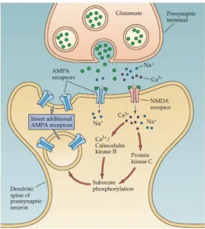
Upon Ca2+ influx within the dendritic spine through activated NMDAR, Ca2+ -dependent proteins, such as calcium/calmodulin--dependent protein kinase II (CaMKII) and protein kinase C (PKC), are activated, inducing different intracellular cascades necessary for LTP induction (Figure 7) (Miyamoto, 2006). AMPARs, which can be phosphorylated in this process (Derkach et al., 1999), are further recruited to post-synaptic density (PSD), resulting in a greater post-post-synaptic response to glutamate (Malenka and Bear, 2004).
A
14
Figure 7 – Molecular mechanisms underlying LTP. During HFS, glutamate binds to AMPARs and, if the postsynaptic cell is sufficiently depolarized, NMDARs are activated. The Ca2+ ions that enter the cell
through these channels activate postsynaptic protein kinases. These kinases may act postsynaptically to insert new AMPARs into the postsynaptic spine, thereby increasing the sensitivity to glutamate (Purves D, 2001).
AMPAR are tightly anchored in the PSD by numerous scaffolding proteins linking them to cytoskeletal elements, including actin. The insertion of additional receptors therefore is likely to affect synapse structure, and in fact, spines associated with synapses that underwent LTP become enlarged (Matsuzaki et al., 2004, Harvey and Svoboda, 2007, Holtmaat and Svoboda, 2009, Kasai et al., 2010).
L-LTP involves interactions with transcription factors, both local dendritic and nuclear transcription, and somatic translation, where the synthesis of required proteins for the maintenance of functional and structural plasticity after LTP triggering occurs (Nguyen et al., 1994). Moreover, LTP is correlated with the formation of new spines within minutes of induction, which leads to an increase in spine density (Figure 8) (Toni et al., 1999).
15
Figure 8 - Structural changes associated with LTP. Synaptic strength correlates with spine volume and the area of PSD. LTP can also lead to the formation of new spines (Luscher and Malenka, 2012).
Importantly, there are evidences that Aβ peptides could impair LTP (Lambert et al., 1998). In fact, perfusion of rat hippocampal slices with low concentrations (200 nM or 1 mM) of Aβ1-42, Aβ1-40 or their active fragment Aβ25-35 has known to significantly impair LTP (Chen et al., 2000, Lee et al., 2000). On the contrary, in previous studies performed in our laboratory any significant change in LTP magnitude induced by a very-weak θ-burst in hippocampal slices exposed to Aβ was detected (Jeronimo-Santos et al., 2015). The stimulation protocol, the Aβ preparation, the developmental age or genetic background of the animals used (Smith et al., 2009) could explain this absence of Aβ effect upon LTP. Soluble oligomeric Aβ1–42 significantly blocked hippocampal LTP when induced by HFS but not by θ-burst (Smith et al., 2009), the type of stimulation used.
Remarkably, synaptic dysfunction and loss caused by age-dependent accumulation of Aβ1-42 in AD brains has been proposed to underlie cognitive decline (McLean et al., 1999, Lue et al., 1999, Wang et al., 1999, Izzo et al., 2014).Curiously, the loss of dendritic spines has also been described in AD (Spires et al., 2005, Spires-Jones et al., 2007). Since spine remodeling and the formation of new synapses are activity-dependent processes that provide a basis for memory formation, alterations in spine density are thought to be responsible for cognitive deficits long before or even in the absence of neuronal loss (Knobloch and Mansuy, 2008). Furthermore, studies demonstrated that the blockade of NMDARs prevent the decrease in synaptic density observed with AD animal models. Thus, it was suggested that NMDAR activation is required for Aβ to exert its effects on spines (Shankar et al., 2007, Shankar et al., 2008, Wei et al., 2010). Another reasonable causal hypothesis for the loss of synapses that leads to functionally disconnection between regions of the neocortex involved in cognition might involve deficiency or failure of delivery of trophic factors (Selkoe, 2002) which will also be discussed later on.
16
1.2.3|
Synaptic and extrasynaptic NMDARs
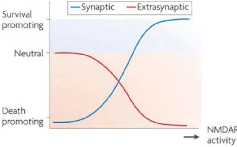
NMDARs play an important role in several cell processes, including LTP, as described above. However, they can also lead to neurodegeneration. An important work has revealed that the localization of NMDARs influences whether they are coupled to pro-death or pro-survival signals (Hardingham and Bading, 2010).
NMDARs can be synaptic (sNMDAR), localized in synapses and gated by the co-agonist D-serine released by astrocytes (Kang et al., 2013), or extrasynaptic (eNMDAR), localized outside synapses and gated by the co-agonist glycine released by both astrocytes and neurons (Holopainen and Kontro, 1989). sNMDARs are neuroprotective, whereas eNMDARs preferentially initiate cell death pathways (Hardingham and Bading, 2010). In fact, low levels of eNMDARs activation have no effects on neuronal survival but high levels of eNMDAR activity enhances cell death pathways and exacerbates certain neurodegenerative processes, reducing, consequently, neuronal survival (Figure 9) (Hardingham and Bading, 2010).
Figure 9 - The NMDA paradox. The ascending curve represents increased neuroprotection due to increased synaptic NMDAR activity, which is superimposed on a descending curve that illustrates the progressive decrease in neuroprotection due to increasing extrasynaptic NMDAR activity (Hardingham and Bading, 2010).
The activation of sNMDAR promotes neuroprotection, resulting from changes in gene expression that have multiple effects within the cell. Activation of sNMDARs initiates a chain of reactions that enhances antioxidant defenses, which contributes to neuroprotection against oxidative insults (Papadia et al., 2008). sNMDARs activity can also have anti-apoptotic effects, including the inactivation of pro-death transcription factors, such as forkhead box protein O (FOXO) and p53, and the transcription of
pro-17
survival factors, including CREB (cyclic-AMP response element binding protein) (Hardingham and Bading, 2010), that results in Brain-Derived Neurotrophic factor (BDNF) transcription (Tao et al., 1998).
In contrast, increased eNMDAR activity preferentially induces pro-death effects, such as the activation of CREB shut-off pathway, which block BDNF expression (Hardingham et al., 2002), ERK1/2 inactivation, which is necessary for BDNF function on spines (Hardingham et al., 2002), FOXO activation, the Ca2+-dependent cysteine proteases calpains activation and subsequent striatal enriched tyrosine phosphatase (STEP) cleavage that prevents STEP from inhibiting p38 MAP kinase, further contributing to neuronal death (Figure 10).
Concerning the calpains, they are involved in multiple cell functions, including proliferation, differentiation, growth cone motility and guidance, and apoptosis. They are also known to play an important role in learning and memory in physiological conditions (Goll et al., 2003, Ono and Sorimachi, 2012). However, when overactivated, calpains cleave and change the function of several proteins, such as synaptic, structural or signaling proteins (Lee et al., 2000). For example, calpains activate cyclin-dependent kinase 5 (CDK5) by cleaving the CDK5-modulator p35 into p25. Increased CDK5 activity leads to tau hyperphosphorylation and to AD progression (Lee et al., 2000). Furthermore, there are two types of calpains: μ-calpain, which is activated in the presence of μM concentrations of Ca2+ and m-calpain that requires mM concentrations for its activation. These local Ca2+ concentrations can trigger calpain activation in distinct subcellular domains and calpains will regulate different substrates to produce opposite effects on neuronal fate (Wang et al., 2013). Synaptic NMDAR activation stimulates μ-calpain, resulting in the activation of pro-survival pathways, and extrasynaptic NMDAR activation induces m-calpain activation, resulting in cell death (Figure 10) (Xu et al., 2009, Ferreira, 2012).
18
Figure 10 - Mediators of synaptic and extrasynaptic NMDAR activity effects. The activation of sNMDARs and eNMDARs triggers different pathways, such as the increase or decrease in the activation, expression or function of a particular intracellular signal that culminates in neuroprotection or neurodegeneration, respectively.
Although the proportion of sNMDARs increases with development, a significant population of NMDARs remains extrasynaptic in adulthood (Petralia et al., 2010). Physiological studies indicate that about 75% of NMDARs are extrasynaptic at 7 days in
vitro (DIV), decreasing the levels to 20–50% by DIV14 (Gladding and Raymond, 2011).
Additionally, a hippocampal slice study indicates that about 36% of NMDARs are extrasynaptic at DIV14-21 (Petralia et al., 2012). However, it is known that pathological conditions can increase the expression or activation of eNMDARs and thus favor these pro-death pathways (Danysz and Parsons, 2012).
1.2.4|
Extrasynaptic NMDARs in AD
Studies have been indicating that activation of NMDARs by Aβ accumulation may occur at early stages of AD (Parameshwaran et al., 2008). Accordingly, it was shown that Aβ induces a sustained Ca2+ influx by interacting directly with NMDAR(Alberdi et al., 2010, Texido et al., 2011, Ferreira et al., 2012), modulating its properties (Hu et al., 2009). Interestingly, it was proposed that Ca2+ entry through NMDARs is particularly effective at killing neurons compared to entry through other channels (Tymianski et al., 1993). Early neuronal dysfunction induced by Aβ is known to be mediated by an activation of GluN2B subunits, the most abundant subunit of eNMDAR, in primary
19
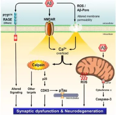
neuronal cultures and hippocampal slices from rat and mouse (Rönicke et al., 2011). In fact, other reports also showed that Aβ oligomeric species evoke an immediately rise in intracellular Ca2+ in cultured cortical neurons through activation of GluN2B-containing NMDARs, suggesting that the activation of this subunit is involved in Aβ-induced Ca2+ homeostasis deregulation (Ferreira et al., 2012a). Importantly, by increasing the Ca2+ influx, Aβ, among other things, leads to an overactivation of calpains (Figure 11).
Figure 11 – Schematic representation of the mechanisms whereby Aβ leads to synaptic dysfunction and
neurodegeneration. Intracellular Aβ accumulates in mitochondria, leading to ROS production and the
activation of pro-apoptotic pathways. Extracellular Aβ oligomers interact with several receptors, such as p75NTR and RAGE, leading to the dysregulation of different signaling pathways that culminate in synaptic dysfunction and neurodegeneration. By interacting directly with NMDARs or by the formation of cation conducting pores, Aβ causes a sustained Ca2+ influx that culminates in calpains activation. Overactivated
calpains change the function of several important proteins, leading to tau hyperphosphorylation and to AD progression (Jerónimo-Santos, 2014).
Interestingly, inhibition of calpain activity prevents excitotoxic neuronal death in
vitro (Caba et al., 2002) and restores normal synaptic function and spatial memory in AD
animal models (Trinchese et al., 2008, Granic et al., 2010, Medeiros et al., 2012). Since calpastatin, the endogenous inhibitor of calpains, is known to be depleted in AD brains (Rao et al., 2008), it would be important to find an alternative way that could modulate calpain activation. General pharmacological calpain inhibitors would not be the best solution, given the important physiological functions of these proteases. Thereby,
20
factors that selectively affect NMDA receptor-mediated Ca2+ influx could be candidates to efficiently modulate calpain activation and its deleterious effects in neurons (Bullock, 2006).
1.3|
Neurotrophins
The discovery of neurotrophins family began with Levi-Montalcini, Cohen and Hamburger, who discovered the nerve growth factor (NGF), a factor required for axonal growth (Levi-Montalcini, 1987). Only about 30 years later, BDNF, neurotrophin-3 (NT-3), neurotrophin-4 (NT-4), NT-6 and NT-7 were identified (Nilsson et al., 1998, Gotz et al., 1994).
Neurotrophins are dimers, which are secreted in an unprocessed form called pro-neurotrophins, synthesized in the hippocampus and cortex and retrogradely transported to the basal forebrain (Holsinger et al., 2000). They mediate their effects through binding to two different receptors – tropomyosin-related kinase (Trk) receptors and the p75 neurotrophin receptor (p75NTR) (Arevalo and Wu, 2006). p75NTR is known to bind the unprocessed pro-neurotrophins (Roux and Barker, 2002) and several reports support a dual role of p75NTR in cell death, as well as in survival, depending on the physiological or developmental stage of the cells (Mamidipudi and Wooten, 2002). Moreover, when expressed within the same cell, p75NTR can interact with Trk receptor activating a set of signaling pathways that are synergistic to those activated by Trk receptors (Huang and Reihardt, 2003).
Focusing on Trk receptors, they are subdivided in three types of receptors, each of which is a single transmembrane protein with a cytoplasmic tyrosine kinase domain that dimerizes and become active upon neurotrophin ligand binding. TrkA is primarily a receptor for NGF, TrkB a receptor for BDNF and NT-4/5, and TrkC a receptor for NT-3 (Figure 12). Neurotrophins bind with equal affinity to p75NTR and preferentially to specific Trk receptors (McAllister et al., 1999).
21
Figure 12 - Neurotrophin receptors and their specificity for the neurotrophins. Neurotrophins are important regulators of neuronal survival, development, function, and plasticity. NGF binds to TrkA, BDNF and NT-4 bind to TrkB and NT-3 binds to TrkC. All of these neurotrophins binds to p75NTR, however pro-neurotrophins bind to this receptor with more affinity (McAllister et al., 1999).
Regarding to TrkB, it is important to clarify that, in the human brain, multiple isoforms of this receptor are expressed. There are three major isoforms of the TrkB receptor characterized: the full-length (TrkB-FL) and two C-terminal truncated TrkB receptors (TrkB-TC). Both C-terminal truncated isoforms are generated by alternative splicing of the TrkB pre-mRNA but differ in that each contain unique amino acid sequences at their C-terminal. TrkB-Shc isoform was also indentified and includes the sarc homology containing (Shc) binding domain that is absent in TrkB-TC (Figure 13) (Stoilov et al., 2002).
Figure 13 – TrkB isoforms. The full-length (TrkB-FL) isoform and three C-terminal truncated TrkB receptors (TrkB-TC): TrkB-T1 with an 11 amino acid tail, TrkB-T2 with a unique 9 amino acid tail and TrkB-Shc, the sarc homology containing TrkB.
22
1.3.1|
BDNF signaling and its function
BDNF is widely expressed in the CNS (Durany et al., 2000) and it has been demonstrated, in several cell culture and animal models, that its signaling is critical for neuronal differentiation, survival, plasticity and cognition (Bartkowska et al., 2010, Lowenstein and Arsenault, 1996).
Upon BDNF binding, monomeric TrkB-FL dimerizes and tyrosine kinase domains activate, leading to a cross-phosphorylation of the tyrosine residues on each domain. Phosphorilated tyrosine residues can act as a “docking” site for signaling molecules, initiating different signaling pathways that promote neuronal survival (via phosphatidylinositol-3-kinase (PI3K)/Akt pathway), growth and differentiation (via Ras/MAPK pathway) and activity-dependent plasticity (via PLCγ) (Segal and Greenberg, 1996, Blum and Konnerth, 2005, Arevalo and Wu, 2006).
There are two tyrosine phosphorylation residues outside the kinase activation domain of TrkB. TrkB phosphorylation site on Tyr515 recruits Shc to TrkB and phosphorylates it, thus allowing the activation of PI3K pathway (Reichardt, 2006). Activation of PI3K changes the composition of inositol phospholipids in the inner leaflet of the plasma membrane. This results in the translocation of Akt/protein kinase B to the plasma membrane. Akt then phosphorylates and controls the biological functions of several proteins important cell survival modulation (Yuan and Yankner, 2000), such as BAD, a Bcl-2 family member that promotes apoptosis, or IκB (Datta et al., 1999), whose phosphorylation leads to degradation and activation of NFκB, resulting in transcription to promote neuronal survival (Middleton et al., 2000).
Recruitment of Shc to the Trk receptors also allows the activation of Ras that activates the downstream kinase B-raf, MEK and ERK family of MAP kinase (Grewal et al., 1999, Huang and Reichardt, 2003). MEK-MAPK/ERK signaling influences transcription events (Xing et al., 1998), such as the activation of the CREB transcription factor (Shaywitz and Greenberg, 1999). CREB regulates genes whose products are essential for prolonged neurotrophin-dependent survival of neurons (Riccio et al., 1999). MAPK signaling pathway is involved in the structural remodeling of excitatory spine synapses triggered by neurotrophins (Alonso et al., 2004), as well as in dendritic growth (Kumar et al., 2005).
23
TrkB phosphorylation site on Tyr816 recruits and activates PLCγ, which hydrolyses phosphatidyl inositides to generate inositol trisphosphate (IP3) and diacylglycerol (DAG) (Vetter et al., 1991). IP3 induces the release of Ca2+ from intracellular stores, increasing levels of intracellular Ca2+. This results in activation of various enzymes regulated by Ca2+, including protein kinases and phosphatases regulated by Ca2+-calmodulin. The formation of DAG stimulates the activity of protein kinase C (PKC) isoforms (Arevalo and Wu, 2006), that have an important role in LTP, as described above (Figure 14).
Figure 14 - BDNF/TrkB-FL signaling pathway. BDNF binds to TrkB-FL inducing receptors dimerization and after tyrosine kinase domains activation, different signaling pathways are initiated. Activation of PI3 kinase through Ras promotes survival and growth of neurons and other cells. Activation of Ras results in activation of the MAP kinase-signaling cascade, which promotes neuronal differentiation, including neurite outgrowth. Activation of PLCγ results in activation of PKC-regulated pathways that promote synaptic plasticity.
In fact, several studies have reinforced that, in the adult brain, BDNF facilitate spine formation (Tartaglia et al., 2001). Indeed, chronic treatment of hippocampal slice cultures with BDNF increases synapse number and spine density in apical dendrites of CA1 pyramidal neurons. Consistently, BDNF can also enhance synaptic transmission and synaptic plasticity (Lu et al., 2008, Korte et al., 1998),which is particularly favored when weak θ-burst stimuli is applied at the synapses from CA3 pyramidal neurons onto CA1
24
pyramidal neurons (Diogenes et al., 2011). Accordingly, impaired LTP in BDNF knockout mice can be recovered by direct application of BDNF (Pozzo-Miller et al., 1999, Patterson et al., 1996). Furthermore, it has been provided evidences about how BDNF facilitates LTP at glutamatergic hippocampal synapses. It is know that endogenous BDNF is released from glutamatergic synapses, in a Ca2+-dependent way, in response to stimulus used to induce LTP (Balkowiec and Katz, 2002, Aicardi et al., 2004). Released BDNF can facilitate LTP at excitatory CA1 synapses, by increasing presynaptic release of glutamate, and by amplifying the postsynaptic response to this neurotransmitter (Carvalho et al., 2008). Through its postsynaptic TrkB-FL receptor, BDNF stimulates tyrosine kinase Fyn, which in turn phosphorylates the NMDAR and increases its activity (Levine et al., 1998, Mizuno et al., 2003). Moreover, BDNF/TrkB-FL signaling can induce cation influx through canonical transient receptor potential channels (TRPC). In this case, activation of TrkB-FL and PLCγ leads to IP3-dependent Ca2+ store depletion, which activates the influx of Ca2+ and Na+ through TRPC3, altering membrane potentials that might, in turn, facilitate synaptic Ca2+ entry through voltage-gated channels or NMDARs (Li et al., 1999, Amaral and Pozzo-Miller, 2007). Ca2+ influx through NMDARs activates CaMKII, which, in turn, contributes to the induction and expression of LTP. Finally, BDNF/TrkB-FL signaling modulates AMPAR expression and trafficking to postsynaptic terminal, a mechanism also dependent on IP3 receptor and TRPC calcium signaling (Figure 15) (Caldeira et al., 2007, Nakata and Nakamura, 2007).
25
Figure 15 – BDNF facilitation upon LTP in a glutamatergic synapse. Presynaptic TrkB activation by BDNF increases glutamate release. Postsynaptic activation of TrkB: increases the open probability of NMDAR by activating the protein tyrosine kinase Fyn; promotes the influx of cations, through TRPC, depolarizing the postsynaptic terminal and facilitating the Ca2+ entry through voltage-gated channels or NMDARs; and
modulates AMPAR expression and trafficking (Minichiello, 2009).
Ample studies indicate that BDNF is also involved in L-LTP. Acute application of BDNF to hippocampal slices induces synaptic potentiation in the hippocampal CA1 region (Kang and Schuman, 1996). Furthermore, evidences suggest that pairing BDNF perfusion and weak θ-burst stimulation produces a reliable L-LTP in CA1 area of hippocampus by regulating local dendritic protein translation and concomitantly increasing synthesis of LTP-associated proteins, such as CaMKII. BDNF can also regulate actin cytoskeletal dynamics, which are required for structural changes of synapses and L-LTP formation (Pang et al., 2004). Briefly, BDNF signaling, in the presence of synaptic activity, can facilitate spine morphogenesis, particularly because similar changes in spine morphology have been associated with long-term synaptic potentiation, where considerable evidence for the requirement of TrkB-FL signaling exists. Importantly, increase in glutamatergic synapse activity stimulates the release of BDNF (Hartmann et al., 2001). BDNF, in turn, facilitates the growth of immature spines into mature spines (Yoshii and Constantine-Paton, 2010).
26
The regulation of BDNF upon synaptic plasticity and synaptic growth suggests that it has a crucial role in cognitive functions. Accordingly, a genetical or pharmacological reduction of hippocampal BDNF levels leads not only to impaired LTP and reduced number of synapses, but also causes deficits in the formation and consolidation of hippocampus-dependent memory (Mu et al., 1999, Bekinschtein et al., 2008, Bekinschtein et al., 2007).
1.3.2|
TrkB-FL cleavage and loss of BDNF signaling in AD
Importantly, BDNF/FL signaling is modulated by alterations in TrkB-FL/TrkB-TC ratio (Eide et al., 1996). Since they lack the tyrosine kinase domain, truncated receptors cannot initiate BDNF signaling and act as a dominant-negative inhibitor of TrkB-FL, by sequestering BDNF so that it cannot bind to TrkB-FL, or by the formation of non-funtional heterodimers with TrkB-FL (Figure 16) (Wong, 2013). Thus, TrkB-FL isoform is the principal mediator of the neurotrophic effects of BDNF (Biffo et al., 1995, Eide et al., 1996).
Figure 16 - TrkB receptor dimer combinations. All dimer combinations of TrkB receptors can bind to BDNF. However, only a homodimer of TrkB-FL can initiate TrkB signaling pathways.
Particularly, on primary neuronal cultures, Aβ is known to induce a decrease in TrkB-FL receptors and an increase in truncated TrkB receptors, which is independent of
27
the presence of glial cells (Kemppainen et al., 2012). These effects of Aβ exposure on TrkB receptors are time and concentration-dependent, thus longer incubation times with Aβ or higher concentrations of Aβ produce a more robust change on TrkB isoforms levels (Rodrigues et al., 2000). Importantly, corroborating this data, TrkB-FL isoform was also found to be decreased (Connor and Dragunow, 1998), whereas TrkB-TC isoforms were found to be increased in postmortem brain samples of AD patients (Connor et al., 1996, Ferrer et al., 1999).
It is well-known from previous studies that the influence of Aβ upon TrkB-FL occurs at the post-translational level, since Aβ strongly reduces TrkB-FL protein levels. In fact, Aβ selectively increases mRNA levels for the truncated TrkB isoforms without affecting TrkB-FL mRNA levels (Jeronimo-Santos et al., 2015). Regarding the unbalance in the ratio of TrkB isoforms, it was proposed a calpain processing of TrkB-FL as a possible mechanism of TrkB regulation (Vidaurre et al., 2012). Indeed, Aβ induces a calpain-mediated cleavage of TrkB-FL receptors, originating a new truncated TrkB receptor (TrkB-T′) and an intracellular fragment (TrkB-ICD) (Figure 17). TrkB-T′ is heavier than the natural truncated TrkB isoforms T1 and TrkB-ICD is a fragment of approximately 32 kDa. Notably, this intracellular fragment is also detected in
postmortem human brain samples, showing that human endogenous calpains can also
cleave human TrkB-FL receptor (Jeronimo-Santos et al., 2015). Interestingly, it is suggested that ICD fragments, which result from the proteolytic cleavage of some members of the receptor tyrosine kinase family, can have a biological function, since they can bind to transcription factors in the nucleus (Ancot et al., 2009). Recent unpublished data of our lab has been indicating that TrkB-ICD is translocated to the nucleus, but its function is still not clear.
28
Figure 17 – TrkB-FL cleavage. The cleavage of TrkB-FL is mediated by calpains, resulting in the formation of a new truncated TrkB receptor (TrkB-T’), which is incapable of initiating TrkB signaling, thus impairing BDNF functions, and an intracellular fragment (TrkB-ICD) whose function is not known yet.
Moreover, it was also demonstrated that, upon calpain-dependent TrkB truncation, BDNF becomes unable to modulate neurotransmitter release from hippocampal nerve terminals, as well as LTP in hippocampal slices (Jeronimo-Santos et al., 2015). This suggests that calpains overactivation induced by Aβ also affects BDNF synaptic actions.
Taken together, the observations that BDNF and TrkB-FL receptor are required for synaptic plasticity and neuronal survival on CNS (Alcantara et al., 1997) and that increased TrkB-FL and BDNF signaling ameliorate the neurodegeneration and cognitive impairment in multiple AD models (Lu et al., 2013)lead to the hypothesis that the loss of BDNF signaling might be involved in AD pathology (Arancio and Chao, 2007, Schindowski et al., 2008).
29
2
|
AIM
Aβ peptide induces an increase in intracellular Ca2+ levels, which results in calpains activation. These proteases lead to TrkB-FL truncation and, consequently, to an impairment of BDNF signaling. However, the causes for this intracellular Ca2+ deregulation remains to be clarified.
Extrasynaptic NMDARs, which are known to be overactivated in AD and associated to harmful effects in neurons, are permeable to Ca2+,allowing the influx of this ion to the cell.Therefore,in the present work,the hypothesis that these receptors could have a central role, contributing to the calpains activation, was considered. Accordingly, the aim of this thesis was to clarify whether the role of BDNF could be recovered by inhibiting the activation of eNMDARs, in neuronal cultures or hippocampal slices exposed to Aβ.
Taking into account the main aim of this work, two specific objectives were proposed: i) to explore if the inhibition of eNMDAR could limit the activation of calpains and, ii) to study whether eNMDAR inhibition could prevent the truncation of TrkB-FL induced by Aβ and therefore facilitate the BDNF mediated actions.
31
3
|
MATERIALS
AND
METHODS
3.1|Primary Neuronal Cultures and drug treatments
Primary neuronal cultures were obtained from fetuses of 18/19-day pregnant Sprague-Dawley females. Animals were purchased from Charles River (Barcelona, Spain) and were handled according to European Community guidelines and Portuguese law concerning animal care (86/609/EEC). Unless stated otherwise, culture reagents and supplements were from Gibco (Paisley, UK). The fetuses were collected in Hanks’ balanced salt solution (HBSS). After brain dissection, the cerebral cortex was isolated and meninges were removed. The tissue was mechanically fragmented and its digestion was performed with 0,025% of trypsin solution in HBSS for 15 min at 37°C. After tissue digestion, cells were precipitated by centrifugation at 1200 rpm. The supernatant was removed and 20% of Fetal Bovine Serum (FBS) was added to HBSS. Cells were again precipitated by centrifugation, the supernatant removed and 2 mL of HBSS were added to the solution. Cells resuspension by pipete aspiration was required between centrifugations in order to dissociate cells. This washing process was repeated four more times to neutralize trypsin. After washed, cells were resuspended in Neurobasal medium supplemented with 0.5 mM L-glutamine, 25 mM glutamic acid, 2% B-27, and 25 U/mL penicillin/streptomycin. To obtain single cells and avoid cellular clusters or tissue fragments, the suspension was filtrated with a nylon filter (BD Falcon™ Cell Strainer 70 µM, Thermo Fisher Scientific, Waltham, MA, USA). Cells were plated at 6 x 10⁴ cells/cm2 and 5 x 10⁴ cells/cm2 on coverslips to perform western blotting and immunocytochemistry, respectively, and maintained at 37°C in a humidified atmosphere of 5% CO2. This coverslips were previously sterilized under UV light and coated with 10 μg/mL of poly-D-lysine (Sigma-Aldrich, St. Louis, MO, USA), which is a synthetic amino acid that enhance cell attachment and adhesion to both plasticware and glass surfaces, overnight and washed with sterile H2O.
Primary neuronal cultures were incubated with 25 µM of Aβ25-35 (Bachem (Bubendorf, Switzerland) at DIV13 for 24 hours at 37°C, as previously described (Kemppainen et al., 2012). In these experiments, cells were also co-incubated with Aβ peptide and 1 μM of Memantine, a NMDAR antagonist, which was a gift by Merz (Frankfurt, Germany). Finally, for immunocytochemistry, cells were co-incubated with