UNIVERSIDADE DE LISBOA FACULDADE DE CIÊNCIAS
DEPARTAMENTO DE BIOLOGIA ANIMAL
Directed evolution of activation-induced
cytidine deaminase (AID) in its natural
environment
DANIEL ANTÓNIO BURGOS ESPADINHA
MESTRADO EM BIOLOGIA HUMANA E AMBIENTE 2009
UNIVERSIDADE DE LISBOA FACULDADE DE CIÊNCIAS
DEPARTAMENTO DE BIOLOGIA ANIMAL
Directed evolution of activation-induced
cytidine deaminase (AID) in its natural
environment
DANIEL ANTÓNIO BURGOS ESPADINHA Tese orientada pelos Prof.es Doutores
Vasco Barreto
GRUPO EPIGENÉTICA E SOMA INSTITUTO GULBENKIAN DE CIÊNCIA
e
Gabriela Rodigues
DEPARTAMENTO DE BIOLOGIA ANIMAL
FACULDADE DE CIÊNCIAS DA UNIVERSIDADE DE LISBOA
MESTRADO EM BIOLOGIA HUMANA E AMBIENTE 2009
i
Abstract
Activation-induced cytidine deaminase (AID) is responsible for the induction of three reactions of DNA somatic modification employed by jawed vertebrates in the context of adaptive immunity: Somatic Hypermutation (SHM), Class Switch Recombination (CSR) and Immunoglobulin Gene Conversion (Ig GC). However, in conditions of deregulation, AID has also been implicated in both lymphoid and non-lymphoid neoplasias. One of most puzzling questions is how the activity of this enzyme is controlled and, in normal conditions, restricted to act only on the Ig loci. Many structural and functional aspects of AID have been discovered by analyzing mutations in AID-impaired patients that have Hyper-IgM syndrome type II. Nevertheless, this approach seems to be exhausted and over the last few years it has not produced new relevant findings. Here, I employ the exact opposite strategy. Since there are no patients with mutated genes encoding hypermorphic AID forms, probably because these would have severe genotoxic and oncogenic effects, I am designing a directed evolution experiment of AID to isolate mutants with enhanced activity. Thus, I plan to unleash the mutagenic potential of this deaminase by mimicking the process of natural evolution in culture, at the protein level, through the reiteration of two main events: Diversification & Selection. I anticipate that the high-activity mutants will result from a number of different possibilities, including the bypass of regulatory mechanisms acting at the protein level. If so, by studying in detail the isolated mutations it should be possible to infer interactions with AID regulatory proteins. The mutants will also be valid tools to create novel cancer murine models, more heuristic and refined than the previous ones. Finally, the mutants will be relevant for several biotechnological applications.
ii
Summary (Portuguese)
O nosso sistema imunitário adaptativo, juntamente com o de todos os outros vertebrados mandibulados, depende da geração de uma infinita variedade de receptores que, no seu conjunto, são capazes de reconhecer toda a diversidade de eventuais patogéneos. Uma vez que a existência de um gene para cada receptor é inviável, devido ao extraordinário número em questão (mais de 1012), mecanismos de diversificação somática do ADN evoluíram de modo a reorganizar e expandir a diversidade já codificada na linha germinal. O primeiro destes mecanismos a ser descoberto foi o da recombinação V(D)J, mediado pelas enzimas RAG1/RAG2, através do qual os receptores das células T e o repertório primário de receptores das células B são formados pela combinação aleatória de diferentes segmentos presentes nos loci das Imunoglobulinas (Igs). No entanto, as células B possuem três formas adicionais de diversificação dos seus receptores. Estas reacções - hipermutação somática (SHM), mudança de classe por recombinação (CSR) e conversão génica das imunoglobulinas (Ig GC) - são todas reguladas pela mesma enzima, a Desaminase de Citidinas Induzida por Activação (AID). É pois esta a enzima que induz o aumento de afinidade das Igs para os respectivos antigéneos (SHM), a modificação da especificidade funcional das Igs (CSR) e, em alguns animais (ex: galinha), a diversificação adicional da rearranjada região variável pela cópia de segmentos localizados a montante (Ig GC). A AID pertence à família de desaminases de citidina AID/ABOPEC, da qual fazem parte enzimas capazes de mutar o ADN, o ARN e ainda o ADN complementar, e que desempenham papéis diversos, do metabolismo de lípidos à imunidade inata, nomeadamente na defesa contra vírus. A AID exerce a sua função através da desaminação de desoxi-citidinas (dCs) que se transforman em desoxi-uracilos (dUs), daí resultando emparelhamentos incorrectos do tipo dU:dG. Ao serem processados por mecanismos de reparação propensos a erros, originam-se mutações pontuais na região variável dos loci das Igs, que depois são seleccionadas no contexto do processo de maturação de afinidade, ou então quebras na dupla cadeia de ADN nas regiões “switch” (S), que desencadearão os eventos de recombinação característicos da CSR. Embora a AID tenha sido descoberta há já uma década, vários aspectos respeitantes à sua regulação permanecem ainda por desvendar. Como se pode imaginar, devido à sua capacidade de modificar somaticamente o DNA, esta enzima possui um perigoso potencial mutagénico. De facto, a AID já foi implicada na geração de linfomas de células B, nomeadamente na formação de translocações cromossómicas, assim como na inserção de mutações em proto-oncogenes. Inesperadamente, parece estar envolvida também em neoplasias não-linfóides, sobretudo como resultado de activação ectópica.
Uma das questões mais enigmáticas prende-se com a forma como em condições normais a AID é direccionada de modo a agir apenas nos loci das Igs. Entender este mecanismo poderá elucidar a forma como a AID está envolvida na geração de tumores. Muitos dos aspectos estruturais e funcionais da AID, como os domínios específicos da SHM e CSR, bem como uma sequência sinal de exportação nuclear, foram descobertos por meio da análise de mutações no gene da AID em pacientes com hiperglobulémia de IgM autossómica recessiva do tipo II. No entanto, esta abordagem parece agora ter chegado ao seu ponto de saturação, e ao longo dos últimos anos nada de novo foi descoberto pelo seu uso. Aqui, proponho a estratégia diametralmente oposta, que consiste em orientar a evolução da AID para o isolamento de mutantes com actividade acrescida. A inexistência
iii de pacientes com formas hiperactivas de AID é explicável pelo efeito genotóxico e oncogénico, sem necessidade de postular constrangimentos estruturais que possam limitar a evolução desta molécula para formas mais activas. De facto, ao longo da evolução a AID pode ter vindo a ser moldada por duas pressões opostas: capacidade de introduzir mutações e modificar os loci das Igs versus inibição de uma actividade demasiado alta que possa comprometer a integridade do restante genoma. No entanto, ao remover este segundo constrangimento, em condições laboratoriais controladas, deverá ser possível libertar em pleno o potencial mutagénico da AID. Para tal, irei imitar em cultura o processo de evolução, recorrendo à reiteração de dois eventos principais: Diversificação e Selecção. Uma biblioteca de mutantes de AID foi originada por PCR propensa à introdução de erros, tendo sido introduzidas, em média, 1 a 6 mutações pontuais por sequência. De seguida, a biblioteca foi transfectada na linha celular de empacotamento viral BOSC23, de modo a produzir retrovírus que carreguem individualmente as formas mutantes de AID. Para avaliar e seleccionar os mutantes, transduziram-se células capazes de efectuar CSR com os retrovírus portadores da biblioteca, de modo a que a actividade acrescida surja num contexto fisiológico - utiliza-se uma linha celular (CH12F3-2) que quando estimulada é capaz de efectuar mudança de classe de IgM para IgA. Ao seleccionar as células que conseguem efectuar a CSR durante um período de tempo estringente, estão a ser seleccionados mutantes com actividade acrescida. No entanto, este passo não é repetido isoladamente de maneira consecutiva, como se faria num típico “screen”. Após o isolamento das células por citometria de fluxo e amplificação das respectivas formas de AID por PCR, procede-se à mistura aleatória de fragmentos provenientes de diferentes moléculas (“DNA shuffling”), de modo a promover a combinação de mutações isoladas e a explorar o seu eventual efeito sinergístico. Para tal, decidi utilizar a técnica StEP (PCR-based staggered extension process) que consiste na repetição de ciclos compostos essencialmente por dois passos: um de hibridação e outro de extensão muito curta. A nova biblioteca é seguidamente reclonada no vector retroviral e transformada por electroporação em Escherichia coli, gerando-se as colónias a partir das quais se purifica o ADN que será usado num novo ciclo de selecção. Uma vez que a linha celular de células B CH12F3-2 possui AID, antes de iniciar a evolução dirigida propriamente dita concebi um “knock-down” específico para a expressão de AID endógena. Para tal, desenhei “short-hairpin RNAs” (shRNAs) que possuem como alvo a região 3’ não traduzida, a qual não existe nas formas mutantes introduzidas pelo retrovírus. Executei também uma bateria de optimizações dos diversos passos envolvidos em cada ciclo de evolução dirigida. Optimizei diversos parâmetros da transfecção da linha celular de empacotamento, como sejam a quantidade de ADN, o rácio plasmídeo vector retroviral / plasmídeo ”helper”, entre outros. Foram também afinados os parâmetros da transdução da linha celular de células B e da sua estimulação para a CSR, bem como a amplificação das formas mutantes e a recombinação de mutações por StEP. Efectuei ainda testes piloto para demonstrar que apenas amplifico AID proveniente das células seleccionadas e não de plasmídeos contaminantes que eventualmente ainda permanecessem em suspensão. Por fim, demonstrei que o StEP induz de facto a recombinação de mutações provenientes de diferentes moléculas de AID. Em suma, ao reiterar as duas grandes etapas de Selecção e Diversificação, o objectivo é adquirir uma colecção de mutantes com elevada actividade que, entre outras, inclua formas da enzima capazes de escapar a mecanismos reguladores. Ao estudar em pormenor as mutações
iv responsáveis pela hiperactividade, poderá ser possível deduzir interacções entre a AID e proteínas reguladoras. Os mutantes serão também importante per se, nomeadamente na criação de novos modelos de ratinho para estudar cancro, mais ecléticos, heurísticos e refinados que os anteriores. Por fim, os mutantes seleccionados podem também vir a ser úteis para diversas aplicações biotecnológicas, como a produção de soros policlonais e anticorpos monoclonais, entre outras.
v
List of Abbreviations
HBS HEPES - Buffered Saline
A Adenine
Ab-MLV Abelson Murine Leukemia Virus AID Activation-Induced Cytidine Deaminase
AMP Ampicillin
AP Apyrimidinic Endonuclease apoB Apolipoprotein B
APOBEC-1 Apolipoprotein B RNA Editing Catalytic Component 1 APOBEC-3 Apolipoprotein B RNA Editing Catalytic Component 3 ATM Ataxia Telangiectasia Mutated
BCL-6 B-Cell Lymphoma 6
C Cytosine
CSR Class Switch Recombination D region Diversity Region
dA Deoxy-Adenine
dC Deoxy-Cytidine
DNase I Deoxyribonuclease I
dG Deoxy-Guanine
DLBCL Difuse Large B-Cell Lymphomas DMEM Dulbecco’s Modified Eagle Medium
DNA Deoxyribonucleic Acid
DNA-PKcs
DSB DNA-Dependent Protein Kinase Catalytic Subunit Double-strand break
dT Deoxy-Thymine
dU Deoxy-Uracil
E. coli Escherichia Coli
E-box Enhancer Box sequences EBV Epstein-Barr Virus
EDTA Ethylenediamine Tetra-Acetic Acid
FBS Fetal Bovine Serum
G Guanine
GFP Green Fluorescent Protein GSP Gene Specific Primer HCV Hepatitis C Virus
HEPES 4-(2-Hydroxyethyl)-1-Piperazineethanesulfonic Acid HIV Human Immunodeficiency Virus
ID2 Inhibitor of DNA Binding 2 ID3 Inhibitor of DNA Binding 3
Ig Immunoglobulin
Ig GC Immunoglobulin Gene Conversion IgH Immunoglobulin Heavy Chain
IL-4 Interleukin-4
vi IRES Internal Ribosome Entry Site
J region Join Region
LB Luria Broth
LPS Lipopolysaccharide
MDM2 Murine Double Minute 2 Oncogene
miRNAS MicroRNAs
MMR Mismatch-Repair
mRNA Messenger Ribonucleic Acid
MSH2 MutS Homolog 2
MSH6 MutS Homolog 6
NF-kB Nuclear Factor kappa of Activated B-Cells NHEJ Non-Homologous End Joining Mechanism NK cells Natural Killer Cells
p19 Tumor Protein 19
p53 Tumor Protein 53
PAX5 Paired Box Gene 5
PBS Phosphate Buffered Saline
PIM1 Proto-Oncogene Serine/Threonine-Protein Kinase 1
PKA Protein Kinase A
RB Retinoblastoma
RH Random Hexamer
RNA Ribonucleic Acid
RPA Replication Protein A S region Switch Region
SHM Somatic Hypermutation
shRNA short hairpin Ribonucleic Acid
ssDNA single stranded Deoxyribonucleic Acid
STAT6 Signal Transducer And Activator Of Transcription 6
T Thymine
TCR T-cell receptor
TGF-β Transforming Growth Factor beta TNF-α Tumor Necrosis Factor alpha
U Uracil
UNG Uracil-N-DNA Glycosylase V region Variable Region
vii
Index
Abstract ...i Keywords ... i Summary (Portuguese)... ii Palavras-chave ...iv List of Abbreviations ...v Index ...vii 1. Introduction ...11.1 DNA somatic modification reactions in the context of adaptive immunity ...1
1.2 Activation-induced cytidine deaminase and beyond ...1
1.2.1 RNA editing versus DNA deamination hypothesis ...1
1.2.2 Error-prone repair recruitment ...3
1.2.3 AID in SHM and CSR ...4
1.2.4 Quaternary structure ...4
1.2.6 Post-transcriptional restraint ...5
1.2.7 Nucleo-cytoplasmic trafficking ...5
1.2.8 Post-translational control ...5
1.2.9 Transcription-driven AID targeting ...5
1.2.10 Epigenetic regulation ...6
1.2.11 Role in innate immunity ...6
1.2.12 Pathological scenarios – AID’s dark side ...7
1.2.13 Potential role in developmental processes and evolution ...8
1.3 Directed evolution ...8 1.3.1 Mimicking nature ...8 1.3.2 Potential applications ...9 1.3.3 General principles ...9 1.3.4 Diversification techniques ...9 1.3.5 Selection strategy ... 11 1.4 Objectives ... 12
2. Materials and Methods ... 13
2.1 Molecular Biology techniques ... 13
2.1.1 epPCR and generation of the original library ... 13
2.1.2 Bacteria Transformation ... 13
2.1.2.1 Escherichia coli (DH5α) transformation by Heat shock ... 13
2.1.2.2 E. coli (DH10B) transformation by Electroporation ... 13
viii
2.1.4 PCR products purification ... 14
2.1.5 Gel Electrophoresis ... 14
2.1.6 DNA extraction from agarose gel ... 14
2.1.7 RNA extraction ... 14
2.1.8 DNA depletion of RNA samples ... 14
2.1.9 cDNA formation ... 15
2.1.10 AID amplification ... 15
2.1.11 PCR-based staggered extension process (StEP) ... 16
2.1.12 DNA Digestion ... 16
2.1.13 Ligase Reaction ... 16
2.1.14 shRNA design ... 17
2.1.15 Sequencing ... 17
2.2 Cell Culture and Manipulation ... 18
2.2.1 Cell lines maintenance ... 18
2.2.2 Transfection by Calcium Phosphate Technique ... 18
2.2.2.1 Retroviral production purposes ... 18
2.2.2.1.2 Six-well plate (optimized recipe) ... 18
2.2.2.1.2 Petri dish (directed evolution experiment) ... 19
2.2.2.2 Lentiviral production purposes ... 19
2.2.3 Transduction ... 19
2.2.3.1 Retrovirus ... 19
2.2.3.1.1 Six-well plate (optimized recipe) ... 19
2.2.3.1.2 Petri dish (directed evolution experiment) ... 19
2.2.3.2 Lentivirus ... 20
2.2.4 IgA Class-switch Recombination Stimulation (optimized) ... 20
2.2.4.1 Six-well plate ... 20
2.2.4.2 Petri dish (directed evolution experiment) ... 20
2.3 Cell Immunostaining, flow cytometry, FACS and MACS ... 20
2.3.1 Cell Immunostaining ... 20
2.3.1.1 Cells stimulated in 24-well plates (optimization purposes) ... 20
2.3.1.2 Cells stimulated in petri dishes (directed evolution experiment) ... 20
2.3.2 Flow Cytometry ... 21
2.3.3 Fluorescence-activated cell sorting (FACS) ... 21
2.3.4 Magnetic-activated cell sorting (MACS) ... 21
3. Results ... 22
3.1 epPCR library ... 22
3.2 Transfection ... 24
3.2.1 DNA quantity and total volume reaction ... 25
ix 3.2.3 Chloroquine ... 27 3.3 Transduction ... 28 3.3.1 Pilot transduction ... 28 3.3.2 Centrifugation ... 29 3.3.3 Polybrene concentration ... 29
3.3.4 Time post-transfection to use retroviral supernatant ... 29
3.4 CSR stimulation ... 30
3.4.1 Total cell number and requirement of all 3 stimulants ... 30
3.4.2 Stimulation of AID transduced cells ... 31
3.4.3 Titration of stimulants concentration ... 31
3.5 Endogenous AID knock-down ... 33
3.5.1 Puromycin titration ... 33
3.5.2 shRNA transduction into CH12F3-2 cells ... 34
3.5.3 Knock-down efficiency ... 35
3.5.4 Transfection of BOSC23 with AID ... 35
3.5.5 Transduction of CH12F3-2 cells with AID ... 37
3.5.6 CSR activity of cells under control of shRNA transduced with AID ... 38
3.5.7 Repetition of shRNA introduction into CH12F3-2 cells followed by MACS ... 39
3.5.8 CSR stimulation repetition ... 40
3.5.9 CSR stimulation of AID transduced cells followed through several time-points . 40 3.6 AID amplification ... 42
3.6.1 DNAse usage to deplete DNA from RNA samples ... 42
3.6.2 DreamTaq versus Pfu ... 43
3.6.3 PCR optimization ... 44
3.6.4 Primers comparison ... 44
3.6.5 Semi-quantitative PCR ... 45
3.7 StEP proof-of-principle ... 46
3.7.1 Recombination of proximal point mutations ... 46
3.7.2 Recombination of the ends of the molecule ... 47
3.8 Directed Evolution 1st cycle ... 48
4. Discussion ... 49
1
1. Introduction
1.1 DNA somatic modification reactions in the context of adaptive immunity
The adaptive immunity of vertebrates relies on the creation of a myriad of receptors capable of recognizing, as a whole, any potential antigen from pathogens. In order to expand the diversity already encoded in the germline genome, which is far from sufficient for the creation of the tremendous diversity of receptors that is needed, distinct and complex mechanisms of DNA modification in somatic tissues evolved. The immune system of all jawed vertebrates depends on the V(D)J recombination reaction [1], in which the multiple segments of the T and B cell receptor genes are randomly and imprecisely assembled into a functional gene. Several species of vertebrates rely on additional reactions that further diversify and increase the versatility of the immunoglobulin (Ig) repertoires, namely, somatic hypermutation (SHM), class switch recombination (CSR) and immunoglobulin gene conversion (Ig GC) [2]. SHM consists of the introduction of mutations, usually single-nucleotide substitutions, into the variable (V) region of the light and heavy chains of the Ig locus in germinal center activated B cells. This phenomenon is the driving force for antibody affinity maturation by generating the diversity required for the process of cellular selection to occur. On the other hand, CSR involves the excision of DNA between two switch (S) regions in order to attach to the variable region of the heavy chain a different constant region that renders new effector functions to the immunoglobulin. Finally, Ig GC is employed by some animals (e.g. chicken) to further diversify the primary immunoglobulin repertoires through the introduction in the already recombined V region, of sequence patches from V pseudo-genes located upstream of the Ig loci. Although the mechanism of V(D)J recombination was discovered in the 1970’s and the genes encoding the responsible recombinase were cloned in the late 1980’s [3, 4] the molecular basis of SHM and CSR remained unclear for a much longer period and even today it has not been fully elucidated.
1.2 Activation-induced cytidine deaminase and beyond
Nine years ago, the discovery that the gene for the activation-induced cytidine deaminase (AID) is selectively expressed in germinal center B cells upon activation to undergo CSR was the prelude to a breakthrough [5]. Shortly after its identification, AID was shown to be necessary and sufficient for the induction of SHM and CSR [6] and for the induction of Ig GC as well [7]. This changed the understanding of the peripheral mechanisms of immunoglobulin gene diversity. It was an unexpected and surprising discovery that one enzyme was responsible for the induction of such seemingly different molecular events, initially considered to be regulated by different mechanisms. Mutations on the AID gene were found to be responsible for a severe immune deficiency in humans called hyper IgM syndrome type II. This disease causes the absence or low levels of Ig isotypes other than IgM as well as the absence of hypermutation, thereby resulting in a severe susceptibility to bacterial infection [8].
1.2.1 RNA editing versus DNA deamination hypothesis
The exact molecular mechanism through which AID performs its function has been actively debated, and two hypotheses have been proposed: RNA editing [9] and DNA
2 deamination [10]. The RNA editing hypothesis predicts that AID and an associated cofactor recognize a putative mRNA precursor and convert it to a mRNA encoding an endonuclease, or a molecule that guides a preexisting nuclease to its target sites. The endonuclease would then cleave DNA in the V region for SHM or in the S region for CSR. On the other hand, the DNA deamination hypothesis predicts that AID itself modifies DNA bases, by deamination of cytidines in S regions for CSR or in variable regions for SHM. The resulting U:G mismatch can then be resolved through uracil excision by uracil-N-DNA glycosylase (UNG) or by alternative repair mechanisms, as base-excision repair or mismatch repair (MMR).
AID has strong homology (34% amino acid identity) to the apolipoprotein B RNA editing catalytic component 1 (APOBEC-1), the catalytic subunit of the apolipoprotein B (apoB) RNA editing enzyme. It was mostly due to this structural similarity that the first hypothesis was initially proposed. However, the alleged mRNA precursor has never been identified. On the other hand, in favor of the DNA deamination hypothesis it was demonstrated that purified AID in vitro deaminates cytidines on single-strand DNA, most notably within the same motif preference observed in vivo [11-14]. Additionally, it was demonstrated that SHM, CSR and IgGC are impaired in the absence of UNG activity [15-17] and that mutations at G:C pairs are biased toward T:A transitions, as one would predict from the direct outcome of AID action [18]. It has also been shown that AID interacts with DNA in the S regions during CSR [19]. Furthermore, it has been reported that the MMR protein MutS Homolog 2 (MSH2) is involved in both SHM and CSR, and the combined absence of UNG and MSH2 totally disrupts CSR [20]. Therefore, based on all the experimental data available to date, the majority of the scientific community has adopted the DNA deamination model.
In the current DNA deamination model for AID function, subsequently to the deamination of dC (deoxy-cytidine) and consequent formation of dU:dG mismatch, three possible pathways can be followed (Fig. 1). If the mismatch is not recognized and processed before replication takes place, a dC to dT transition occurs. On the contrary, if the mismatch is recognized by UNG, the U is removed and an abasic site is created which upon replication can lead to the introduction of any type of base. However, if an apyrimidinic endonuclease (AP) recognizes the abasic site, it originates a nick on the DNA strand. Then, the conventional base-excision repair mechanism can intervene, repairing the nick and restoring the initial dC:dG pair; alternatively, the non-homologous end joining (NHEJ) mechanism can process the nick and lead, for instance, to CSR, which implies the formation of intermediate double-strand breaks (DSBs). Yet another possibility is that the abasic site is recognized by MMR molecules in combination with error prone polymerases – in fact, it is through this pathway that the majority of the mutations in A-T pairs around the mutational hotspots are originated.
3 Figure 1. DNA deamination model of AID [21].
This model per se does not answer a number of questions. How is AID directed to perform either SHM or CSR in activated B cells? Are there negative regulators acting on AID? How come error-prone polymerases are recruited to act on the AID induced lesions while in the rest of the genome error-free polymerases are the ones operating? How is AID targeted to act on the Ig locus and not in other loci? What are the cofactors that regulate AID? Does AID have additional functions in the innate immune system? What pathological situations can arise upon impairment of the regulatory mechanisms of AID?
1.2.2 Error-prone repair recruitment
It is still not entirely understood why U:G mismatches and abasic sites in the Ig loci lead to error-prone resolution, whereas in the rest of the genome these mutations are repaired in an error-free manner [22]. It has been suggested that this could be due to the establishment of an interaction between AID and specific downstream repair pathways [23]. Alternatively, it has been proposed that high levels of dC deamination induced by AID could saturate the normal error-free repair mechanisms or that error-prone repair pathways could be specifically induced in response to stimuli that promote SHM [23]. A recent study demonstrated that AID deaminates many more genes than it was previously assumed, with the majority of the lesions being corrected by gene-specific high-fidelity DNA repair [24]. The conclusion was achieved by sequencing several transcribed non-Ig genes from germinal center B cells derived from Ung/Msh2 double knockout mice, which are thought to reflect all deamination events, catalyzed by AID. However, AID preferred the Ig loci to all non-Ig loci examined by at least an order of magnitude and there were many transcribed non-Ig loci that did not accumulate mutations. Therefore, the existence of a substantial level of targeting of AID was corroborated.
4
1.2.3 AID in SHM and CSR
The way AID performs SHM or CSR is also not entirely clear. AID mutants with deletion or replacement of the C-terminus lack CSR activity both in vitro and in vivo [25, 26]. In spite of losing the CSR induction ability, these mutants still retain SHM activity as well as the ability to catalyze gene conversion [25]. These results imply that the C-terminal domain of AID is specifically crucial for CSR, independently of the cytidine deaminase capability (Fig. 2). It may be important for recruiting putative CSR-specific cofactor(s) that aid in recognition of target S regions or for connecting AID to the CSR-specific NHEJ machinery [27-29]. With respect to the N-terminal region, it was reported that mouse AID mutants bearing point mutations located in it, show almost normal CSR activity but absent SHM activity [30]. Therefore the N-terminal domain might be involved in the interaction with SHM-specific cofactor(s) that regulate target specificity (Fig. 2). At present, no definitive AID-specific cofactor has been reported. Although the C-terminus of AID interacts with Murine Double Minute 2 Oncogene (MDM2) [31], an ubiquitin ligase that targets cytoplasmic p53 for degradation, the functional relevance of this interaction in CSR is unclear. Replication protein A (RPA), protein kinase A (PKA), DNA-dependent protein kinase catalytic subunit (DNA-PKcs), and RNA polymerase II have been implicated as AID-interacting molecules [19, 29, 32, 33]. For example, the classical NHEJ factors DNA-PKcs and Artemis were reported to be necessary for joining a subset of AID-dependent DSBs [34].
1.2.4 Quaternary structure
It was shown that AID functions as a dimer, just like its structural and phylogenetic relative APOBEC-1, although with different dimerization motifs [26] (Fig. 2). All mutations impairing dimerization disrupt both CSR [35] and SHM (Barreto and Mcbride, unpublished). Nevertheless, dimer formation is autonomous and independent of modifications and cofactors, including nucleic acids [35]. Therefore, dimerization of AID is necessary but not sufficient for its activity, as the interaction with putative substrate-specific cofactors for either CSR or SHM, at the respective domains, is required.
1.2.5 Transcriptional control
AID has a mutagenic potential that implies a tight regulation in order to keep the enzyme’s activity restricted to the appropriate cell type (mature B cells), loci (immunoglobulin genes), time and place (the germinal center reaction). However, the precise mechanism by which AID is regulated is still an incomplete puzzle. It is known that IL-4, anti-CD40 and lipopolysaccharide (LPS) induce AID expression, possibly through the action of the transcription factors signal transducer and activator of transcription 6 (STAT6) and nuclear factor-kB (NF-kB) on the proximal promoter [36, 37]. It was also reported that a highly conserved intronic Enhancer Box sequences (E-box) exists in the AID gene, on which E47/E12 and ID3 might regulate AID expression in positive and negative ways, respectively [38]. A similar counter-balanced regulation was reported to exist in the minimal promoter region through the action of paired box gene 5 (PAX5) positive and inhibitor of DNA binding 2 (ID2) negative actions [39]. Additionally, it was reported that GC boxes and GT motifs, on which the ubiquitous transcription factor Sp acts, exist on the AID minimal promoter [40]. Hence, it appears that a complex combination of transcription factors act in concert to regulate AID
5 expression: nevertheless, these regulators still await experimental assessment in vivo.
1.2.6 Post-transcriptional restraint
Two different microRNAs (miRNAS), namely miR155 [41, 42] and miR-181b [43], were recently reported to act over AID mRNA. They appear to regulate AID in different stages, with the first narrowing AID function after B cells have been activated and the later preventing premature AID activity while allowing proper AID transcriptional activation at early time points.
1.2.7 Nucleo-cytoplasmic trafficking
AID has a potential nuclear localization signal (NLS) and a nuclear export signal (NES) at its N- and C-termini, respectively (Fig. 2) [44-46]. Thus, the molecule moves between the nucleus and the cytoplasm, where it accumulates, which may contribute to protect the DNA from excessive amounts of the AID protein.
Figure 2. AID primary structure (Adapted from [50])
1.2.8 Post-translational control
It has been reported that upon phosphorylation of serine 38, presumably by PKA, AID´s activity is increased, implying that post-translational regulatory mechanisms also aid in the regulation of AID function [32, 33, 47, 48]. This AID phosphorylation by PKA seems to preferentially happen in B cells. Furthermore, it seems that the ability of AID to mediate both SHM and CSR is increased, due to a facilitated access to ssDNA in V and S regions, respectively.
In another experiment [49] it was shown that in murine B cells, where AID is over-expressed, both CSR and SHM are impaired, suggesting that there are additionally regulatory mechanisms, particularly those that repress AID activity, still to be unveiled.
1.2.9 Transcription-driven AID targeting
Although the amount of AID in the nucleus is limited through the regulation of its expression and active export to the cytoplasm, thus limiting AID activity, that does not explain how is the molecule targeted to the Ig loci. The simplest explanation for targeting would be the primary DNA sequence itself. In fact, AID does not act randomly in the cytidines of V regions when performing SHM but instead it acts on WRC (A/T-A/G-C) mutational hotspots both in vivo as well as in vitro, indicating that the preference is due to its intrinsic specificity [2, 14]. The association between AID and RPA has been reported to increase WRC targeting [33]. Nevertheless, when AID is ectopically expressed outside B cells or even in heterologous systems such as Escherichia coli, it still has the same WRC motif preference and thus the importance of the interaction with RPA might rather be due to an increase in the accessibility to transcribed substrates or to unknown regulatory mechanisms. It has also been shown that the WRC targeting of AID can be
6 modulated in vivo by mutS homolog 6 (MSH6), indicating that MMR proteins might be involved in the selection of the target sequence [51]. In any case, despite the proved sequence bias shown by AID, the WRC motifs have a degenerated nature and cannot explain the Ig locus specificity. Furthermore, even when the V and S regions are replaced by heterologous sequences, SHM and CSR still takes place [52, 53]. In addition, the hotspot requirement is not absolute; AID can also mutate sequences without hotspots. It has been shown that for SHM and CSR to take place, the occurrence of transcription of V and S regions, respectively, is necessary [52, 53]. There has been also evidence of correlation between those levels of transcription and the rates of SHM and CSR [52, 53]. It is interesting to notice that, in general, mutations start at 100 bp downstream of the promoter, have a maximum pick at 200 bp and slowly decrease to vestigial levels at around 2 Kb away from the promoter [54]. Moreover, whereas AID is capable of deaminating ssDNA in vitro but not dsDNA, transcription is sufficient for AID to become capable of deaminating dsDNA both in vitro and in Escherichia coli [12, 55, 56]. Hence, one can easily deduce that the need for transcription of V and S regions is especially due to a mechanistic role in allowing the access of AID to ssDNA.
1.2.10 Epigenetic regulation
Several processes such as transcription, replication, recombination and repair are known to be promoted and modulated by DNA methylation and histone modification. In regard to AID regulation, it was shown that hyperacetylation of histone 3 (H3) at S regions is related to CSR and germline transcription, possibly by preceding and marking the IgH locus for AID to act on [19, 57, 58]. In addition, it was reported that hyperacetylation of H4 at switch regions correlates with CSR in an AID-dependent manner [58], indicating that this modification might not influence AID directly but instead aid in the recruitment of DSBs repair factors.
1.2.11 Role in innate immunity
It has been suggested that AID might also have functions beyond those previously discussed. The family of AID/APOBEC cytidine deaminases has been implicated in the innate host response to viral infection, which suggest an intriguing evolutionary connection of innate and adaptive immune mechanisms [59]. One of the family members, APOBEC3G, was the first to be recognized as having an antiviral activity [60], namely as a potent inhibitor of Human Immunodeficiency Virus (HIV) through extensive dC-to-dU mutations of the minus-strand viral DNA formed during reverse transcription [61, 62]. It is now known that APOBEC3 proteins have targets beyond HIV, including a wide variety of viruses as well as host-encoded retrotransposable genetic elements [63, 64]. AID also possesses antiviral properties, although it appears to function through a different mechanism [65]. While APOBEC3 proteins target the virus itself, AID targets the host cell genome leading to the inhibition of proliferation and targeting of the infected cells by NK cells. Thus, AID protects against transformation by the oncogenic Abelson murine leukemia virus (Ab-MLV) [65] independently of its antibody diversification functions. Additionally it has also been implicated in the host response to other pathogenic viruses like Epstein-Barr Virus (EBV) [66, 67] and Hepatitis C Virus (HCV) [68, 69] . More recently, it was shown that AID is also able to inhibit the replication of retroelements like L1 or MusD, further demonstrating the importance of this enzyme in both adaptive and
7 innate immunity [70].
1.2.12 Pathological scenarios – AID’s dark side
A question that might arise is whether AID can actually mutate genes other than the Ig loci and once that happens what kind of pathologies can it induce. It is interesting to notice that 95% of the lymphomas are derived from B cells, especially from germinal center (GC) or post-GC B cells, and are characterized for having chromosome translocations between Ig and proto-oncogene genes that cluster either at V or at S regions [71]. As a result of the chromosome fusion, the oncogene comes under the control of the Ig cis sequences leading to its deregulation and constitutive expression [72]. One of such translocations occurs between the c-myc proto-oncogene and the IgH locus, which is typical of Burkitt’s lymphoma in human, and of spontaneously developed plasmacytomas in IL-6 transgenic mice. However, when AID is knocked-out in these mice, the onset of the disease is delayed and the otherwise typical c-myc/IgH translocations disappear [73], showing the requirement of AID in the formation of these translocations through a mechanism common to CSR.
When AID is expressed at normal levels, the occurrence of c-myc/IgH translocations is rare both in vivo and in vitro, suggesting that in physiological conditions the cellular surveillance mechanisms prevent the formation of translocations and the growth of translocation harboring cells [74, 75]. Indeed, it has been reported that B cells with knock-out of different genes that code for DNA-damage response proteins are more prone to accumulate aberrations and chromosomal breaks at IgH locus as well as c-myc/IgH translocations, and in some cases it was demonstrated that these events were dependent on the presence of AID [74-76]. It has also been reported that impairment of the p53 signalling leads to AID-dependent c-myc/IgH translocations [75].
Mutations on tumor suppressor genes, such as p53, retinoblastoma (RB), p19 and ataxia telangiectasia mutated (ATM) are also known to contribute to the lymphoma pathogenesis [71]. It has been shown that when AID is overexpressed in a B cell line, it can induce SHM on a GFP transgene [77]. Even when it is overexpressed in an entirely different cell type, such as fibroblasts, AID is capable of inducing CSR and SHM on highly transcribed substrates [78, 79]. As a matter of fact, in transgenic mice where AID is ubiquitously and constitutively expressed, T-cell lymphomas are formed with extensive mutations in the TCR and Myc genes [80]. In the context of physiological conditions, it has been reported that human B cells of the peripheral blood also can exhibit SHM in genes other than Ig locus, including proto-oncogenes, such as BCL-6 (B-cell lymphoma 6) [81] and FAS [82, 83] as well as non-oncogenes [84]. For instance BCL-6, which is translocated in 35% of difuse large B-cell lymphomas (DLBCL), is mutated in 73% of DLBCL and 47% of follicular lymphomas [85]. In addition, other proto-oncogenes like PIM1, MYC, RHOH and PAX5 are also shown to be mutated in DLBCL [86]. It was reported that besides the occurrence of WRC mutational hotspots, the mutations accumulate downstream of active promoters, further supporting the idea that these mutations are dependent on AID.
The carcinogenic potential of AID is clearly not restricted to the germinal center activated B cells and neither to lymphoma tissues. Further analyses of AID-transgenic mice showed they also develop epithelial tumors in the lung, liver and stomach [87, 88]. Under physiologic conditions, AID transcription seems to be mostly restricted to activated B cells and thus almost no AID expression is observed in most human tissues.
8 However this scenario can be critically changed in contexts of inflammation and viral infection. For instance, Helicobacter pylori infection and the resultant inflammatory stimulation have been shown to trigger aberrant AID expression in gastric epithelial cells [88]. Additionally, Epstein-Barr virus is capable of inducing AID expression in human peripheral blood B cells [67]. Hepatitis C virus is also implicated in the induction of a mutator phenotype [68]. Furthermore, AID ectopic expression was reported in hepatocytes from patients with chronic liver disease [89] and in hepatoma cell lines. Treatment with the cytokines TGFβ and TNFα was shown to induce AID expression in human hepatocytes [87, 89]. It has also been detected AID expression in epithelial breast cancer cell lines [90] and very recently it was shown that estrogen activates AID transcription in breast and ovarian tissues, and in B cells [91]. Overall, the discovery that AID can cause several types of tumors when aberrantly expressed is beginning to open new fields in tumor biology [50].
1.2.13 Potential role in developmental processes and evolution
Interestingly, AID mRNA levels as high as those encountered in lymph nodes were detected in mouse oocytes and human spermatocytes [92, 93]. Additionally, embryonic germ cells, primordial germ cells and embryonic stem cells also exhibit low levels of AID mRNA. If the AID protein is indeed confirmed to exist and to be functional in those tissues, the ability of AID to target non-Ig genes could have physiological roles in developmental processes and in evolution. For instance, AID could generate germline mutations and thereby contribute to genetic diversity or it may play a role in meotic recombination. A role for AID in epigenetic reprogramming and development was also proposed, since the molecule was implicated in programmed DNA demethylation of the zebrafish genome during embryonic development [94].
1.3 Directed evolution 1.3.1 Mimicking nature
All living organisms are believed to have evolved primarily by consecutive rounds of genetic diversification and selection through the differential contribution of each individual to the novel generation. This is based on the principle that the fittest organisms under a given set of conditions survive and outcompete others in a population. Evolution requires a diversification of genotype, selection pressure favouring a new phenotype, and a linkage of genotype and phenotype. These features have long been broadly applied to create useful plants, animals and microorganisms, and explore gene function [95]. One common example is the whole range of canine breeds that are the result of iterative cycles of selection imposed by man. The advances of technology in the field of molecular biology have allowed to mimic this process at the molecular level, making possible the development of artificial enzymes with catalytic properties that conform to selection constraints imposed by the experimenter [96]. While nature-based evolution of polypeptides has a head start of millions of years, protein design by artificial evolutionary mimicry is progressing at a far rapid pace. The mutation, selection, and amplification steps of the evolutionary cycle may be imitated in the laboratory using existing proteins, or even molecules created de novo from random sequences, as starting templates [97].
9
1.3.2 Potential applications
In little more than a decade, since the first reported applications of evolutionary design to enzyme engineering, directed evolution has matured to the point where it now represents the centerpiece of industrial biocatalyst’s development and it is being practiced by thousands of scientists in companies and universities around the world [98]. Directed molecular evolution has become a powerful strategy to forge countless protein properties, including thermal and solvent stability, enzyme selectivity, specific activity, protease susceptibility, allosteric control of protein function, ligand binding, transcriptional activation, solubility, or even for generating proteins with novel enzymatic properties [98, 99]. Moreover, the range of applications has expanded to the engineering of more complex functions such as those performed by multiple proteins acting in concert (in biosynthetic pathways) or as part of macromolecular complexes and biological networks [98].
The alteration of protein properties is of great interest, as these proteins might be useful for medical applications [100], industrial use [101], and the study of biophysical properties of proteins [102]. Others have used these methods to study Darwinian evolution itself, taking advantage of the controlled laboratory setting, which provides access to all genotypic and phenotypic parameters [103]. It is easy to understand why directed evolution is so appealing: since it is conceptually straightforward, it can be practiced without any special instrumentation, and, most importantly, it frequently yields useful solutions. Moreover, a significant advantage of directed evolution over rational designed methods is that new functional properties can be evolved without requiring knowledge of enzyme structures or catalytic mechanism [104].
1.3.3 General principles
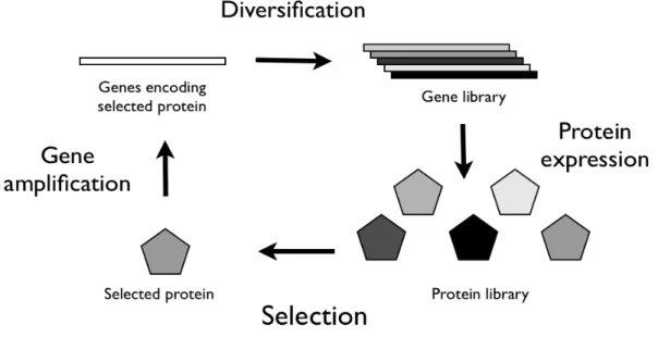
In a directed evolution experiment it is necessary to create a library of mutant genes of the protein we are interested in altering the properties, so that there is diversity on which selection can work. Then, after the collection of mutant sequences is inserted into a suitable expression system, those proteins that perform the desired function according to the specified criteria are selected and the genes encoding them are amplified. Thus, the inferior enzymes and the corresponding genes are discarded and the genes encoding the improved enzymes are used as “parents” for the next round of evolution, repeating the whole process as often as necessary. After applying these two steps in an iterative way for a sufficient number of rounds, the experiment is essentially done (Fig. 3) [105]. The first step of diversification of molecules is carried out by introducing mutations, such as point mutations, deletions or elongations, into the genes encoding the protein we want to modify. It is even possible to recombine parts of different genes in order to create a novel protein.
1.3.4 Diversification techniques
The quality of the diversity method is crucial and the performance of the chosen protocol has a direct impact on the success rate of obtaining improved variants as well as on the time and cost effectiveness of the ensuing screening or selection process [106, 107]. There are several ways to create molecular diversity, but the first method to achieve this was error prone Polymerase Chain Reaction (epPCR) [108, 109], which takes advantage
10 of the lack of proofreading activity of thermostable polymerases. For example, Taq polymerase incorporates wrong nucleotides at a frequency of 0.1×10⁻⁴ to 2×10⁻⁴ per nucleotide per cycle of polymerisation, which can infinitely be varied by increasing the concentration of MgCl2 [110], addition of MnCl2 [111], using unbalanced concentrations
of nucleotides [112], using a mixture of triphosphate nucleoside analogs [113], or a combination of all these to achieve higher rates of mutations. Still the epPCR technique has been accused of some limitations, including the bias for A→T or T→A substitutions and the rarely occurrence of mutations next to each other, resulting in limited mutation frequencies and low product yields [108]. Furthermore epPCR introduces random point mutations that may be too gradual to allow block changes that are required for continued sequence evolution.
When DNA shuffling was introduced by Stemmer in 1994 [114, 115] it became the most popular method to recombine genes and one that could compensate for the various epPCR drawbacks. Through this technique parental genes are randomly fragmented using DNaseI. The resulting fragments are recombined using a primer-free PCR with a high temperature denaturation, followed by annealing to other fragments, and extension by DNA polymerase. After cycles of assembly, PCR amplification with primers is used to selectively amplify full-length sequences. Yet, DNA shuffling also carries problems of its own: large amounts of template DNA are needed; it is time consuming; crossover points are biased towards regions of high sequence identity [116]; the yield of chimeras can be quite low, particularly when short genes are being shuffled; parental background ranging from around 20% [117, 118] to almost 100% [119, 120] has been reported. Meanwhile, an alternative shuffling technique which does not involve DNA cleavage was introduced:
Staggered Extension Protocol (StEP) [121]. In this protocol the template sequences
undergo repeated cycles of a denaturation step and an extremely short annealing/extension step. In each cycle, the growing strands anneal to different templates based on sequence complementarity and extend further. This is repeated until full-length sequences are formed. Such fragmentation-free PCR-based protocol has advantages regarding the original Stemmer´s technique, because it is less time-consuming, and less technically-demanding, as it bypasses the DNaseI fragmentation step by generating chimeric genes through simple template switching. Even so, some biases were still pointed out, mainly the occurrence of preferential crossing-overs in regions of high sequence identity in the alignment [122].
Overall, no matter by which method random libraries are generated, there are certain limitations and the libraries are clearly not entirely random. Each method gives a different bias of base substitutions that result in a different range of amino acid replacements.
Beyond the random incorporation of mutations, it is also possible to direct the randomization to a particular number of residues that belong to the active site. This strategy might be particularly fruitful when a small number of active-site residues that trigger the required change in substrate specificity can be identified [123]. Usually the short gene segments that are subjected to mutagenesis are either hotspots identified upon screening of epPCR libraries or are important domains of enzyme analyzed through structural data [124]. These procedures ignore the fact that many mutations altering the function might occur at unexpected positions away from the active site [124, 125]. Although in some cases, the targeted mutagenesis yielded better results [123, 126], in
11 others the random approach was quite comparable [127] and in the end both are able to improve activity.
Beyond the “in vitro” recombination techniques referred so far, there are “in situ” strategies. In this case, the genetic diversity is generated within cells as well as the following steps of protein expression and screening. Mostly, yeast [128], or Escherichia coli, cells (e.g. carrying a mutator polymerase [129]) have been used and one of the main advantages of this technique is the capability of affording very large library sizes independent of transformation efficiency.
1.3.5 Selection strategy
Independently of the library generation strategy of choice, it is critical to have in place a robust and high-throughput method for either screening or selection of the desired function [130]. In many ways, this step can be the most challenging. No matter how cleverly designed or diverse the starting library is, without an effective screening strategy the ability to isolate useful clones is severely diminished. The ideal screen should be high throughput, in order to increase the likelihood of identifying useful clones, it should have a good signal to noise ratio, to allow the isolation from lower activity clones early in evolution and be sufficiently reproducible, and it should be robust, so that mutants displaying subtle improvements will not be lost. Obviously, it must be properly designed in such a way that it would be specifically sensitive to the desired function [105].
12
1.4 Objectives
The main objectives of my master thesis were to plan, optimize and execute all steps involved in the directed evolution of activation-induced cytidine deaminase (AID) towards more efficient variants, within a physiological environment. Briefly, this would be achieved by putting a retroviral library of AID mutants through rounds of selection and diversification in the B cell line CH12F3-2, which is able to drive the AID-dependent CSR. The isolation of molecules with enhanced activity, which have not been found, would open previously unexplored territory in the mutational landscape. By studying in detail the mutations responsible for the increased activity, a disruption of an unknown regulatory mechanism could be discovered. Thus a significant contribution to a better understanding of the way AID is regulated at post-translational level, which is still poorly understood, could be made. Besides gaining information from the study of the causes of the enhanced activity, the activity of the mutants itself could be explored in a number of different biotechnological settings. Additionally, the hyperactive mutants could be used to generate novel AID knock-in mice models.
During the course of my thesis I was able to test and optimize all the steps involved in each cycle of directed evolution. Furthermore, I confirmed that the PCR-based staggered extension process (StEP) technique is indeed capable of recombining mutations from different molecules encoding AID. Finally, I performed the first cycle of the directed evolution of AID and I am planning the subsequent ones, in order to complete this project.
13
2. Materials and Methods
2.1 Molecular Biology techniques
2.1.1 epPCR and generation of the original library
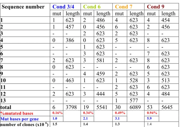
Error-prone PCR (epPCR) was perfomed with oligos aid5 and aid3 (see table 1) on PMX-AID, a retroviral vector that expressed the murine PMX-AID, using the Diversify PCR random mutagenesis kit (Clontech) and according to the instructions of the manufacturers. Four different conditions were used, which generated four types of amplicons that differ in the average number of mutations per clone (see figure 5). Amplicons were digested with BamHI and EcoRI, and cloned back in the PMX vector. The ligation was used to transform bacteria by electroporation. More than 1 million total transformants were produced. Around 20 individual transformants were sequenced to confirm the introduction of the mutations (see figure 5). The library was replated only once prior to the onset of the experiments here described. It is assumed that the starting complexity of the library is 1 milion transformants.
2.1.2 Bacteria Transformation
2.1.2.1 Escherichia coli (DH5α) transformation by Heat shock
After cells were thawed on ice, 50 to 1000 ng of plasmid (in a 10 µL maximum volume) were carefully mixed with 50 µL of cells in a 1.5 ml polypropylene tube. The tube was kept on ice for 20 minutes and it was submitted to a heat shock of 90 seconds at 42 ºC. The cells were then chilled on ice for 2 minutes, after which 1 ml of SOC medium was added and the mix was incubated for 1 hour at 37ºC with shaking (300 rpm). Following the incubation step, the tubes were centrifuged for 5 minutes at 5000 rpm on a bench microcentrifuge, after which 900ml of supernatant was discarded. The cell pellet was resuspended and plated on a Luria Broth/ampicillin (Sigma) Petri dish, which was incubated overnight at 37ºC.
2.1.2.2 E. coli (DH10B) transformation by Electroporation
Cells were thawed on ice and, for each sample to be electroporated, a 1.5 ml polypropylene microfuge tube and a 0.1 cm electroporation cuvette were placed on ice. In the cold tube, 20 µl of the cell suspension were mixed with 1 µl of DNA and were incubated on ice for 1 minute. The mixture of cells and DNA was transferred to the chilled electroporation cuvette and the suspension was tapped to the bottom. The MicroPulser was set to 1.8 kV, which corresponds to Ec1 program in Bio-Rad apparatus, and was pulsed once. The cuvette was removed from the chamber and 1 ml of SOC medium was immediately added, followed by a quick and gently resuspension of the cells with a Pasteur pipette. The cell suspension was transferred to 1.5 ml polypropylene microfuge tube and it was incubated at 37 °C for 1 hour, shaking at 225 rpm, after which the cells were plated on LB/ampicillin medium.
14
2.1.3 Plasmid Purification
For plasmid DNA low scale purification the QIAprep Spin Miniprep kit (Quiagen) was used. In case of high scale purification the QIAprep Spin Maxiprep kit was used. The instructions of the manuals were followed.
2.1.4 PCR products purification
In order to purify amplicons, either the QIAquick PCR Purification Kit (Quiagen) was used or the PCR reaction was ran in an agarose gel, followed by gel band extraction (see 2.1.6).
2.1.5 Gel Electrophoresis
DNA samples were loaded onto 1% agarose gels, and ran in Tris-acetate-EDTA (TAE): 40 mM Tris-acetate, 1 mM EDTA buffer. DNA band size was estimated by using the 1kb plus DNA ladder (Fermentas).
2.1.6 DNA extraction from agarose gel
DNA bands were excised from 1% agarose gels and DNA was purified using QIAquick gel extraction (Qiagen) kit, according to the manual instructions.
2.1.7 RNA extraction
The RNA from the CH12F3-2 cells was extracted using the TriPure Isolation Reagent (Roche). After sorting cells, they were centrifuged in a polypropylene centrifuge tube and the supernatant was removed. 1 mL of TriPure Isolation Reagent was added directly to the tube containing the cell pellet (1–10 x 106 cells) at 25°C. The cells were lysed by
repetitive pipetting and then transferred to a polypropylene centrifuge tube. The homogenized sample was incubated for 5 minutes at 25°C to ensure the complete dissociation of nucleoprotein complexes. 200 µl of chloroform (Sigma) were added, the tube securely capped and shaken vigorously for 15 seconds. The tube was then incubated at 25°C for 15 minutes. To separate the solution into three phases, the tube was centrifuged at 12,000 x g for 15 minutes, at 4°C. The colorless upper aqueous phase was transferred to a new polypropylene centrifuge tube. 500 µL of isopropanol were added to the aqueous phase. The tube was capped and inverted several times to mix it thoroughly. The sample was incubated for 10 minutes at 25°C to allow the RNA precipitate to form, followed by centrifugation at 12,000 x g for 10 minutes at 4ºC. The supernatant was discarded and 1 mL of 75% ethanol was added. The RNA pellet was washed in the ethanol by vortexing. The sample was centrifuged at 7500 x g for 5 minutes at 4°C. The supernatant was discarded and the excess ethanol was removed from the RNA pellet by air-drying. The RNA pellet was resuspended in 10 µL of diethylpyrocarbonate (DEPC)-treated RNase-free water and dissolved by passing the solution through a pipette tip several times and then by a incubation for 15 minutes at 57°C.
2.1.8 DNA depletion of RNA samples
In order to remove DNA contamination from RNA samples, for each µg of RNA, 1µL of 10X reaction buffer with MgCl2,0.5 µL of Ribonuclease inhibitor, DEPC-treated water to a volume of 9µl and 1 µl (1 unit) of Deoxyribonuclease I (DNase I), RNase-free (Fermentas) were added. The reaction mixture was incubated at 37°C for 30 minutes.
15 Finally, 1µl of 25mM EDTA was added and the mixture was incubated at 65°C for 10 minutes. The RNA was then used as a template for reverse transcriptase.
2.1.9 cDNA formation
In order to generate cDNA from the extracted RNA, the enzyme SuperScriptTM II Reverse Transcriptase (Invitrogen) was used. In the first pilot experiments both a gene specific primer (pmx3.1) and a random hexamer primer were tested. Since the amplification was successful with both in generating considerable amounts of cDNA, the random hexamer primer was the one chosen to do all the remaining experiments, because it allowed to monitor for a control mRNA, like the cytoskeletal beta and gamma actins. To each 5 µg of mRNA, in a nuclease-free microcentrifuge tube, the following reagents were added: either 250 ng of random hexamer primer or 2 pmole of gene-specific primer (GSP), 1 µl of dNTP Mix (10 mM each), and sterile, distilled water to a total volume of 12 µl. The mixture was heated to 65 ºC for 5 minutes and immediately chilled on ice. The contents of the tube were collected by brief centrifugation and then the following reagents were added: 4 µl of 5X First-Strand Buffer, 2 µl of 0.1 M DTT, and 1 µl of RNaseOUT™ (40 units/µl). The contents of the tube were gently mixed. The tube was incubated at 42 °C for 2 minutes, in the case of using the GSP, or incubated at 25 ºC for 2 minutes in the case of using the random hexamer. 1 µl (200 units) of SuperScript™ II RT was added and mixed by pipetting gently up and down. In the case of using the random hexamer primers, the tube was incubated at 25 ºC for 10 minutes. The reaction mixture was incubated at 42 ºC for 50 minutes, followed by a heating step at 70 ºC for 15 minutes in order to inactivate the enzyme.
2.1.10 AID amplification
Half of the library was amplified by one set of primers (pmx5.2/3.1), which binds to the PMX vector, and the other half was amplified by a set of primers (aid5/3) that directly binds to AID. The optimized PCR consisted in an initial denaturation at 95 ºC for 2 minutes followed by 30 cycles with pmx5.2/3.1 primers or 22 cycles in the case of aid5/3 primers. Each cycle was composed of a denaturation at 95 ºC for 30 seconds, an annealing step at 59 ºC for 30 seconds and an extension step at 72 ºC for 1 minute. After the cycling phase, the reactions were incubated at 72 ºC for 7 minutes. The enzyme was DreamTaq DNA polymerase (Fermentas) incubated in the 10X DreamTaq buffer containing 20 mM MgCl2.
Table 1 List of primers used.
Name Primer Sequence (5' to 3')
pmx5.2 CCCCACCGCCCTCAAAGTAGACGGC pmx3.1 GGGGGGGCGGAATTTACGTAGCGGC aid5 CGGGATCCACCATGGACAGCCTTCTGATGAAGCAAAAGAAGTTTC aid3 CCGGAATTCCTCAAAATCCCAACATACGAAATGCATCTCGCAAG actin5 GCTCCGGCATGTGCAA actin3 AGGATCTTCATGAGGTAGT
16
2.1.11 PCR-based staggered extension process (StEP)
The combination of mutations from the selected AID forms was performed as described in [131] (Fig. 4). In a sterile PCR tube the following reagents were combined:5 µL of 10 X Pfu buffer with MgSO4; 5 µL of 10 X dNTP mix; 0.15 pmol of total template DNA; 30
pmol of each primer; 1.25 units of Pfu DNA polymerase; and sterile nuclease-free water to 50 µL total volume reaction.Then, a denaturation step at 94 ºC for 30 seconds and an annealing/extension at 55 ºC for 5 seconds were reiterated 79 more times. Finally, the reaction mixture was run on a 1% agarose gel and the AID corresponding band was extracted and purified.
Figure 4. Principle of the Step method [131].
2.1.12 DNA Digestion
To digest DNA, 1-2 µg were added to a 1.5 mL polypropylene centrifuge tube, 5 µl of the adequate 10X Buffer (Fermentas), enzyme (the last to be added) in a maximum quantity of 2.5 µl (≤5% of total volume) and nuclease-free water to a final volume of 50 µl. The reaction mixture was gently mixed, spun down for a few seconds and finally incubated at 37°C for 1-2 hours. This digestion reaction was also scaled either up or down. In the case of vector digestion, calf intestinal alkaline phosphatase (NEB) was used to prevent vector re-ligation without insert.
To digest the pMX vector as well as the AID molecules selected from each cycle of the directed evolution (to be inserted in pMX), the 2X tango buffer was used, which means 10 µl of 10X tango buffer in a 50 µl total reaction. 12 units of BamHI and 6 units of EcoRI were used per digestion.
2.1.13 Ligase Reaction
In order to ligate an insert to the desirable vector, 50-200 ng of the digested vector were mixed with 3-fold more moles of insert in a microcentrifuge tube, then 1 µl of 10X T4 ligase buffer, 1 µl of T4 DNA Ligase (Fermentas) (the last to be added) and water nuclease-free to 10 µl were added. The reaction was mixed by up and down, spun down and incubated at room temperature for 1 hour.
17
2.1.14 shRNA design
In order to knockdown the endogenous AID expression of the CH12F3-2 cell line, I designed 3 alternative shRNAs that would target the 3-untranslated region of murine´s AID mRNA. The first candidate (shRNA1) was selected based on Clontech´s shRNA sequence designer which is based on the rules of Elbashir et al. [132]. On the other hand, both the second (shRNA2) and the third (shRNA3) candidates were selected based on GeneLink´s shRNA Explorer™. A BLAST search was done to all candidates to ensure that the target sequence was not homologous to other murine gene sequences. Although for shRNA 1 and 2 the loop sequence was TTCAAGAGA, for shRNA3 an alternative sequence was selected: CTCGAG (as suggested by The RNAi consortium). Both AgeI and EcoRI overhangs were added to all shRNAs for pLKO.1 insertion.
Table 2. Target sequences of the shRNAs
Table 3. Complete shRNA sequences
2.1.15 Sequencing
DNA samples were sent to Stab Vida Company where they were sequenced with the universal primers T7 and SP6.
Name Target Sequence (5' to 3')
shRNA1 GAGGCAGGAGGATTGTAAA
shRNA2 AACTGAGCTTGCTGTGCAA
shRNA3 AACAACGATCTTTGCTAATGA
Name Complete shRNA Sequence (5' to 3')
shRNA1 ccggGAGGCAGGAGGATTGTAAATTCAAGAGATTTACAATCCTCCTGCCTCTTTTTTg Sense Strand Antisense Strand aattcAAAAAAGAGGCAGGAGGATTGTAAATCTCTTGAATTTACAATCCTCCTGCCTC shRNA2 Sense Strand ccggAACTGAGCTTGCTGTGCAATTCAAGAGATTGCACAGCAAGCTCAGTTTTTTTTg Antisense Strand aattcAAAAAAAACTGAGCTTGCTGTGCAATCTCTTGAATTGCACAGCAAGCTCAGTT
shRNA3 ccggAACAACGATCTTTGCTAATGACTCGAGTCATTAGCAAAGATCGTTGTTTTTTTg Sense Strand Antisense Strand
18
2.2 Cell Culture and Manipulation
2.2.1 Cell lines maintenance
BOSC23 and 293T LENTI cells were cultured at 37ºC, 5% CO2, in Dulbecco’s Modified
Eagle Medium (DMEM; GIBCO) supplemented with 10% heat-inactivated Fetal Bovine Serum (GIBCO), 1 mM sodium pyruvate (GIBCO) and penicillin/streptomycin (GIBCO) at 100 u/mL and 100 µg/mL, respectively (DMEM complete medium).
CH12F3-2 cells were cultured at 37°C, 5% CO2, in RPMI 1640 glutamax medium
(GIBCO) supplemented with 10% heat-inactivated Fetal Bovine Serum (FBS; GIBCO), 50 µM 2-mercaptoethanol, 1 mM sodium pyruvate (GIBCO) and penicillin/streptomycin (GIBCO) at 100 u/mL and 100 µg/mL respectively (RPMI complete medium). In the case of CH12F3-2 cells transduced with a shRNA,the medium also contained 0.4 µg/ mL of puromycin (Calbiochem) (in order to select for the transduced cells).
Cells were cultured either in 75 cm2 or 150 cm2 T flasks, with 15 or 30 mL of the adequate complete medium, respectively. In the case of the adherent cell lines, namely BOSC23 and 293T LENTI, for cell passages the medium was aspired, 7.5/15 mL of pre-warmed trypsin (GIBCO) at 37ºC were added and the flask was gently tapped until cells were completely dissociated from the bottom. Then 7.5/15 mL of pre-warmed complete medium at 37ºC were added, the medium was pipetted up and down to dissociate cells clumps, and cells were diluted in the range of 1:10 to 1:30 in pre-warmed complete medium at 37ºC. In the case of CH12F3-2 cells, which are not adherent cells, the cultures were regularly diluted 1/10-1/30 in complete medium pre-warmed at 37ºC.
Cells were passed every 2-3 days.
2.2.2 Transfection by Calcium Phosphate Technique 2.2.2.1 Retroviral production purposes
2.2.2.1.2 Six-well plate (optimized recipe)
Each well was plated with 6x10⁵ BOSC23 in 2 mL of DMEM complete medium and kept overnight at 37 °C. The following day, when cells were between 40-60% confluent, for each well, two 1.5 ml polypropylene tubes were prepared: one with 75 µL of 2XHBS and another containing 8 µg of DNA (1:8 pCL-Ampho/PMX-AID), 7.5 µL of 2.5 M CaCl2
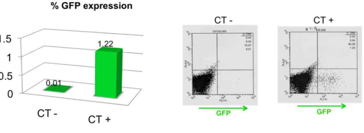
and water in a 75 µL total volume reaction. After bubbling the 2XHBS with a yellow tip, the DNA/Ca solution was added dropwise to the 2XHBS with concomitant shaking. The mixture was bubbled with a yellow tip and then added dropwise to the petri dish, which was being carefully moved in circles. About 16-18 hours later, the medium of each well was aspired and 2 mL of DMEM complete medium pre-warmed at 37 ºC were added. Finally, after 36-48 hours post-transfection, the retroviral supernatant was ready for transduction and the cells were analyzed by flow cytometry for GFP expression in order to quantify the transfection percentage.
![Figure 1. DNA deamination model of AID [21].](https://thumb-eu.123doks.com/thumbv2/123dok_br/19189878.949416/15.918.242.675.111.463/figure-dna-deamination-model-aid.webp)
![Figure 4. Principle of the Step method [131].](https://thumb-eu.123doks.com/thumbv2/123dok_br/19189878.949416/28.918.261.655.306.615/figure-principle-step-method.webp)