Faculdade de Ciências
Departamento de Biologia Vegetal
Dynamics of β-lactamases in
Gram-negative bacteria
Vera Mónica Martins Gonçalves Manageiro
DOUTORAMENTO EM BIOLOGIA
(Microbiologia)
Lisboa
2011
Faculdade de Ciências
Departamento de Biologia Vegetal
Dynamics of β-lactamases in
Gram-negative bacteria
Vera Mónica Martins Gonçalves Manageiro
Thesis supervised by: Prof. Doctor Manuela Caniça
Instituto Nacional de Saúde Dr. Ricardo Jorge co-supervised by: Prof. Doctor Maria Filomena Caeiro
Faculdade de Ciências, Universidade de Lisboa
DOUTORAMENTO EM BIOLOGIA
(Microbiologia)
Lisboa
2011
Departamento de Doenças Infecciosas
Laboratório Nacional de Referência da Resistência
aos Antimicrobianos
Dynamics of β-lactamases in
Gram-negative bacteria
Vera Mónica Martins Gonçalves Manageiro
DOUTORAMENTO EM BIOLOGIA
(Microbiologia)
Lisboa
2011
A
CKNOWLEDGEMENTS/A
GRADECIMENTOSI would like to express my gratitude to a group of people who participated and contributed in some way to the achievement of the challenging project that was my PhD thesis:
À Professora Doutora Manuela Caniça, o apoio incondicional, incentivo e confiança, especialmente nos momentos menos bons. Agradeço também por me ter permitido crescer pessoal e profissionalmente, por todos os estágios que realizei e lugares que conheci, os quais me proporcionaram um vasto conhecimento e experiência. É para mim um grande privilégio trabalhar sob sua orientação e fazer parte do Laboratório Nacional de Referência da Resistência aos Antimicrobianos, do Instituto Nacional de Saúde Dr. Ricardo Jorge.
À Professora Doutora Filomena Caeiro, por ter aceite a orientação interna da minha tese de doutoramento na Faculdade de Ciências da Universidade de Lisboa. Obrigada pelo acompanhamento, disponibilidade e ajuda durante estes quatro anos.
To Professor Richard Bonnet, for having accepted me in the Laboratoire de Bactériologie, Faculté de Médecine, Université d'Auvergne and in the Laboratoire de Bactériologie Clinique, Centre de Biologie, CHU Clermont-Ferrand, where I performed part of the experimental work presented in this thesis. Thanks also for the continuous and fruitful collaboration on kinetic studies, which I hope will continue for the next years.
À Dani, ―my private English interpreter‖, toda a sua amizade e apoio, assim como toda a ajuda, esforço e dedicação que despendeu em inúmeros ensaios, análises e discussões durante a escrita da minha tese.
À Doutora Eugénia Ferreira, a sua grande paciência no decorrer do meu doutoramento e também por todo o interesse e incentivo que sempre dispensou à realização deste trabalho. Obrigada pela sua amizade e por partilhar a sua apaixonante história de vida em terras africanas.
À D. Deolinda, a sua completa disponibilidade para ajudar em tudo, quer fosse uma simples solução de antibiótico ou semear um isolado porque ―eu tinha coisas mais importantes para fazer‖; pela sua simpatia e amizade que tornou a realização desde trabalho possível. E desde já agradeço pelos inúmeros telefonemas que iremos ter de fazer quando se reformar…
Aos ex-URRAnianos, David Félix, Germana Domingues, Joana Leitão, Nuno Mendonça, Patrícia Francisco, Doutora Paula Lavado e Ricardo Dias, agradeço a companhia, amizade e a ajuda prestada. Ao David agradeço ainda ter aceite fazer
―Choose a job you love, and you’ll never have to work a day in your life‖. Confucius
ACKNOWLEDGEMENTS
To all co-authors of the manuscripts included in this work for their valuable contributions, suggestions and improvements to the manuscripts.
To Marlene Jan and Roland Perroux for technical assistance and support during my stay in Clermont-Ferrand.
Ao Instituto Nacional de Saúde Dr. Ricardo Jorge, pela disponibilização dos meios necessários para a realização da minha tese de doutoramento e pela oportunidade de integrar um Laboratório Nacional de Referência.
À Fundação para a Ciência e a Tecnologia pela bolsa de doutoramento que possibilitou a execução desta tese.
To the Federation of European Microbiological Societies (FEMS), for awarding me a fellowship entitled ―Biochemical study of a new β-lactamase inhibitor enzyme produced by a clinical Klebsiella pneumoniae strain‖, that enabled the development of a part of this PhD thesis at Richard Bonnet’s lab.
To the organizing committee of the European Congress of Clinical Microbiology and Infectious Diseases (ECCMID), whose financial support allowed me to attend international scientific meeting that was essential for the progress of my work. Aos laboratórios participantes no ARSIP (―Antibiotic Resistance Surveillance Program in Portugal‖) pelo envio de isolados clínicos de Enterobacteriaceae e de Acinetobacter baumannii, ao Laboratório Nacional de Referência da Resistência aos Antimicrobianos, do Instituto Nacional de Saúde Dr. Ricardo Jorge.
A todos os meus amigos, por todo o apoio, momentos inesquecíveis e conversas inspiradoras durante todo este período. Obrigado pela vossa amizade.
Aos meus pais e aos meus avós Manuel e Lucinda, sem os quais nunca teria chegado onde cheguei. Obrigada por terem sempre acreditado em mim e nas minhas capacidades; não existem palavras suficientes para exprimir a minha gratidão. Obrigada por terem tomado conta do Miguel quando o cansaço não mo permitia.
Finalmente, porque os últimos são os primeiros, obrigada aos meus amores, a quem dedico esta tese. Luís: ―Amo como ama o amor. Não conheço nenhuma outra razão para amar senão amar. Que queres que te diga, além de que te amo, se o que quero dizer-te é que te amo?‖, obrigada pelo teu amor e pela tua dedicação ao nosso Miguinhas… Filho, obrigada por existires, os teus miminhos são a minha motivação para não desistir!
The work presented in this thesis was performed in the National Reference Laboratory of Antimicrobial Resistances, Department of Infectious Diseases, National Institute of Health Dr. Ricardo Jorge. Vera Manageiro was financial supported by a PhD fellowship from Fundação para a Ciência e a Tecnologia (SFRH/BD/32578/2006).
In accordance with Paragraph 1 of ―Artigo 41, Capítulo V, do Regulamento de Estudos Pós-Graduados da Universidade de Lisboa, publicado no Diário da República – II Série No. 209, de 30 de Outubro de 2006‖, it is clarified that full scientific articles already published (4), and under revision (1) or submitted (3) for publication in peer-reviewed scientific journals, were used in the elaboration of this dissertation. Hence, the candidate states that was involved in the study design, execution of experimental work, in the analysis and interpretation of results, and in their preparation for publication, with the exception for the two publications ―The Lys234Arg substitution in the enzyme SHV-72 is a determinant for resistance to clavulanic acid inhibition‖ and ―Biochemical characterization of SHV-55, an extended-spectrum class A beta-lactamase from Klebsiella pneumoniae‖, in which the candidate was not the leading author.
P
REFACEThe introduction of a large collection of β-lactam antibiotics into clinical practice, namely the third-generation cephalosporins, in response to the increased prevalence of pathogenic organisms-producing β-lactamases, lead to the emergence of an even larger variety of β-lactamases conferring resistance to those agents, both in clinical and community settings. Hence, the overall aim of this PhD thesis was to contribute to the knowledge of molecular epidemiology of β-lactamases, as the most important antibiotic resistance mechanism among Gram-negative isolates, and to the understanding of their diversity in a structural and functional level. To accomplish this aim, several studies with different approaches were performed.
After a general overview about antimicrobial resistance (Section I: Introduction), the results presented in this PhD thesis are branched into two chapters (presented in Section II, Results), including, overall, eight papers:
Chapter 1 (Paper I to III), entitled ―β-lactamases: impact of antibiotic resistance, dissemination and co-resistance‖ sheds light on the emergence of Enterobacteriaceae strains producing extended-spectrum β-lactamases and AmpC plasmid-mediated β-lactamases. Furthermore, the dissemination of multidrug-resistant Acinetobacter baumannii strains due to the expression of carbapenem-hydrolyzing class D β-lactamases was analyzed. Chapter 2 (Paper IV to VIII) entitled ―Class A β-lactamases: function meets
structure‖, reports the correlation of function and structure in novel clinical important β-lactamases identified throughout this PhD thesis, highlighting protein evolution and diversification as a mechanism of rapid adaptation of bacterial populations.
Each Paper I to VIII (Section II: Results) contains a specific background and a detailed discussion about the respective results. At the end, Section III (Concluding remarks), focus on an overall discussion regarding the main results of the different chapters.
The presentation of each paper in the present PhD dissertation does not necessarily reflect a chronological order, since some of the studies described
PREFACE
below were done simultaneously and the results obtained during one particular work would influence the progress of the other and vice-versa.
Publications and Manuscripts included in the thesis: Chapter 1:
Paper I. Manageiro V, Ferreira E, Jones-Dias D, Louro D, Pinto M, Diogo J, Caniça M (2011) Emergence of β-lactamase-mediated resistance to oxyimino-β-lactams in Enterobacteriaceae isolates in various services in a single centre: risk factors and contribution of the newly detected CTX-M-3 variant in Portugal. Submitted to Int J Antimicrob Agents.
Paper II. Manageiro V, Jones-Dias D, Ferreira E, Antimicrobial Resistance Surveillance Program in Portugal (ARSIP), Caniça M (2011) Diversity of plasmid-encoded AmpC β-lactamases among clinical isolates of Enterobacteriaceae lacking inducible chromosomal ampC gene from Portuguese Hospitals. Submitted to Antimicrob Agents Chemother.
Paper III. Manageiro V, Jones-Dias D, Ferreira E, Louro D, Antimicrobial Resistance Surveillance Program in Portugal (ARSIP), Caniça M (2011) Carbapenem-hydrolyzing class D β-lactamase-producing Acinetobacter baumannii isolated in community and health care facilities in Portugal. Submitted to J Antimicrob Chemother.
Chapter 2:
Paper IV. Mendonça N, Manageiro V, Robin F, Salgado MJ, Ferreira E, Caniça M, Bonnet R (2008) The Lys234Arg substitution in the enzyme SHV-72 is a determinant for resistance to clavulanic acid inhibition. Antimicrob Agents Chemother 52: 1806-1811. doi:10.1128/AAC.01381-07.
Paper V. Manageiro V, Ferreira E, Albuquerque L, Bonnet R, Canica M (2010) Biochemical study of a new inhibitor-resistant beta-lactamase, SHV-84, produced by a clinical Escherichia coli strain. Antimicrob Agents Chemother
54: 2271-2272. doi:10.1128/AAC.01442-09.
Paper VI. Manageiro V, Ferreira E, Albuquerque L, M. C, Bonnet R (2011) Characterization of the inhibitor-resistant SHV β-lactamase (SHV-107) in a
clinical Klebsiella pneumoniae strain co-producing GES-7 enzyme. Under review at Antimicrob Agents Chemother.
Paper VII. Mendonça N, Manageiro V, Bonnet R, Caniça M (2008) Biochemical characterization of SHV-55, an extended-spectrum class A beta-lactamase from Klebsiella pneumoniae. Antimicrob Agents Chemother 52: 1897-1898. doi:10.1128/AAC.01525-07.
Paper VIII.Ramdani-Bouguessa N*, Manageiro V*, Jones-Dias D, Ferreira E, Tazir M, Canica M (2011) Role of SHV-beta-lactamase variants in resistance of clinical Klebsiella pneumoniae strains to beta-lactams in an Algerian hospital. J Med Microbiol 60:983-987. doi:10.1099/jmm.1090.030577-030570.
T
ABLE OFC
ONTENTSAcknowledgements/Agradecimentos _________________________________ i Preface ________________________________________________________ v Table of Contents ________________________________________________ ix List of Figures __________________________________________________ xii List of Tables __________________________________________________ xiii List of Abbreviations ____________________________________________ xiv Resumo da Tese _______________________________________________ xvii Abstract ______________________________________________________ xxiii S
SEECCTTIIOONNII.. Introduction __________________________________________ 1
Chapter 1. An overview on antibiotic resistance ___________________________ 3
1.1.Importance of antibiotic-resistant Gram-negative bacteria in public health ___________ 3 1.2.Monitoring of Resistance _________________________________________________ 3
Chapter 2. Antibiotic Agents ___________________________________________ 5
2.1.History of antibiotics _____________________________________________________ 5 2.2.Antibiotic Targets _______________________________________________________ 6 2.2.1.Inhibition of Cell Wall Synthesis ________________________________________ 7 2.2.2.Inhibition of Protein Synthesis __________________________________________ 7 2.2.3.Alteration of Cell Membranes __________________________________________ 8 2.2.4.Inhibition of Nucleic Acid Synthesis _____________________________________ 8 2.2.5.Competitive Inhibitors ________________________________________________ 8 2.3.The β-lactam antibiotics __________________________________________________ 9 2.3.1.Structure __________________________________________________________ 9 2.3.2.Mechanism of action ________________________________________________ 11
TABLE OF CONTENTS
3.1.Mechanisms of antibiotic resistance ________________________________________ 16 3.1.1.Altered target sites __________________________________________________ 16 3.1.2.Decreased uptake __________________________________________________ 17 3.1.3.Active efflux system _________________________________________________ 17 3.1.4.Enzymatic inactivation or modification ___________________________________ 17 3.2.Mechanisms of dissemination of resistance genes _____________________________ 18
Chapter 4. β-lactamases: the most important antibiotic resistance mechanism in Gram-negative bacteria _______________________________________________ 21
4.1.Overview and classification schemes _______________________________________ 21 4.2.β-lactamase active site and hydrolytic mechanisms ____________________________ 24 4.2.1.Serine β-lactamases _________________________________________________ 24 4.2.1.1. Mechanism of hydrolysis of Ser-β-lactamases _______________________ 25 4.2.1.2. β-Lactamase inhibitors and pathway of inhibition by clavulanic acid ______ 26 4.2.1.3. Class A β-lactamases: the catalytically important residues _____________ 29 4.2.2.Metallo-β-lactamases ________________________________________________ 32 4.3.β-lactamase mediated-resistance __________________________________________ 33 4.3.1.Extended-spectrum β-lactamases ______________________________________ 34 4.3.2.Inhibitor-resistant class A β-lactamases __________________________________ 37 4.3.3.Plasmid-mediated AmpC β-lactamases __________________________________ 38 4.3.4.The challenge of carbapenem resistance ________________________________ 39
S
SEECCTTIIOONNIIII.. Results _____________________________________________ 43
Chapter 1. β-lactamases: impact in the antibiotic resistance, dissemination and co-resistance _______________________________________________________ 45
Paper I. Emergence of β-lactamase-mediated resistance to oxyimino-β-lactams in
Enterobacteriaceae isolates in various services in a single centre: risk factors and
contribution of the newly detected CTX-M-3 variant in Portugal ___________________ 45 Paper II. Diversity of plasmid-encoded AmpC β-lactamases among 2570 clinical isolates of
Enterobacteriaceae lacking inducible chromosomal ampC gene from Portuguese
Paper III. Carbapenem-hydrolyzing class D β-lactamase-producing Acinetobacter baumannii isolated in community and health care facilities in Portugal _____________________ 101
Chapter 2. Class A β-lactamases: function meets structure _______________ 113
Paper IV. The Lys234Arg substitution in the enzyme SHV-72 is a determinant for resistance to clavulanic acid inhibition ______________________________________________ 113 Paper V. Biochemical study of a new inhibitor-resistant β-lactamase, SHV-84, produced by a
clinical Escherichia coli strain ____________________________________________ 121 Paper VI. Characterization of the inhibitor-resistant SHV β-lactamase (SHV-107) in a clinical
Klebsiella pneumoniae strain co-producing GES-7 enzyme ____________________ 125
Paper VII.Biochemical characterization of SHV-55, an extended-spectrum class A
β-lactamase from Klebsiella pneumoniae ____________________________________ 143 Paper VIII.Role of SHV-β-lactamase variants in resistance of clinical Klebsiella pneumoniae
strains to β-lactams in an Algerian hospital _________________________________ 147
S
SEECCTTIIOONNIIIIII..Concluding Remarks ________________________________ 155
S
LIST OF FIGURES
L
IST OFF
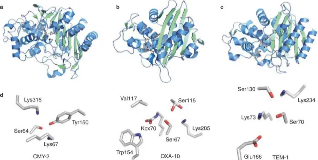
IGURESFigure 1 - History of antibiotic discovery and concomitant development of antibiotic resistance.__6 Figure 2 - Antibiotic targets and mechanisms of resistance.______________________________7 Figure 3 – Graphical illustration of Gram-positive and Gram-negative cell wall structures.______12 Figure 4 - N-acetylglucosamine (NAG) and N-acetylmuramic acid (NAM) chains forming the structure of peptidoglycan._______________________________________________________12 Figure 5 – Schematic representation of the biosynthesis of the cell wall.____________________13 Figure 6 – β-lactams acts as a substrate analog, binding to the substrate-anchoring site normally occupied by D-alanyl-D-alanine.___________________________________________________13 Figure 7 - The modular and hierarchical composition of MGEs.___________________________20 Figure 8 – Ser-β-lactamases structures: Class C (CMY-2); Class D (OXA-10) and class A (TEM-1).24 Figure 9 – Action of Ser-β-lactamase._______________________________________________26 Figure 10 – Mechanism of action of clavulanate and amoxicillin.__________________________27 Figure 11 – Representation of the general mechanism of action of irreversible inhibitors._______28 Figure 12 - The SHV β-lactamase active site pocket.___________________________________29 Figure 13 – Structures of B1 (BcII), B2 (CphA) and B3 (FEZ-1) MBLs subclasses.____________33 Figure 14 - Global distribution of CTX-M β-lactamases._________________________________36 Figure 15 – Proportion of E. coli invasive isolates resistant to third-generation cephalosporins in 2006 and in 2009, in Europe. ____________________________________________________158 Figure 16 - Proportion of K. pneumoniae invasive isolates resistant to third-generation
L
IST OFT
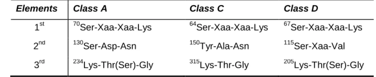
ABLESTable 1 - Classification scheme for β-lactam antibiotics, based on chemical structure. ________ 10 Table 2 – Examples of intrinsic resistance phenotypes. ________________________________ 15 Table 3 – Characteristics of the most important MGEs._________________________________ 19 Table 4 - Evolution of the molecular and functional classification of β-lactamases. ___________ 22 Table 5 - Classification schemes for the major families of β-lactamases of clinical importance in Gram-negative bacteria. _________________________________________________________ 23 Table 6 – Conserved motifs (elements) in the active site of Ser-β-lactamases. ______________ 25 Table 7 - Chronology and homology of plasmid-mediated AmpC β-lactamases. _____________ 39
LST OF ABBREVIATIONS
L
IST OFA
BBREVIATIONSIn this thesis acronyms are expanded on first usage and whenever deemed necessary to improve clarity.
Aac(6’)-Ib-cr PMQR determinant
Ab Acinetobacter baumannii
ARSIP Antibiotic Resistance Surveillance Program in Portugal
bla β-Lactamase coding gene
BSAC British Society for Antimicrobial Chemotherapy
CA-SFM Antibiogram Committee of the SFM CHDL carbapenem-hydrolysing class D
β-lactamase
CI 95% Confidence Interval
CLSI Clinical and Laboratory Standards Institute CMS Complex Mutant SHV
CMT Complex Mutant TEM DAP diaminopimelic acid
DDST Disk diffusion susceptibility test
EARS-Net European Antimicrobial Resistance Surveillance Network
EARSS European Antimicrobial Resistance Surveillance System
ECDC European Centre for Disease Prevention and Control
EDTA Ethylenediaminetetraacetic acid EMBL European Molecular Biology Laboratory ESAC Extended-spectrum AmpC
cephalosporinase
ESBL Extended-Spectrum β-Lactamase EUCAST European Committee on Antimicrobial
HGT Horizontal Gene Transfer
IC50 Fifty Percent Inhibitory Concentration IEF Isoelectric Focusing
IPTG isopropyl β-D-thiogalactoside IR Inverted Repeat Sequences IRS Inhibitor Resistant SHV IRT Inhibitor Resistant TEM IS Insertion Sequence
ISCR Insertion Sequence Common Region
kcat Catalytic Activity Constant
Ki Inhibitor constant
kinact Inactivation rate constant
Km Michaelis Constant MBL Metallo-β-lactamase MDR Multidrug-resistance
MDR-Ab Multidrug-resistant Acinetobacter
baumannii
MDS Molecular Dynamic Simulation MGE Mobile Genetic Element MIC Minimal Inhibitory Concentration MLST Multilocus Sequence Typing NAG N-acetylglucosamine NAM N-acetylmuramic acid NIH National Institute of Health
NRL-AR National Reference Laboratory of Antimicrobial Resistances
OMP Outer Membrane Protein
OR Odds Ratio
ORF Open Reading Frame PBP Penicillin-Binding Protein PCR Polymerase Chain Reaction PDR Pandrug-resistant
PFGE Pulsed-Field Gel Electrophoresis PMAβ Plasmid-mediated AmpC β-lactamase PMQR Plasmid-mediated quinolone resistance Qnr PMQR determinant
QRDR Quinolone-resistance determining region
RIVM National Institute for Public Health and the Environment
RMSD Root Mean Squared Deviation
Ser-β-lactamase β-lactamase with serine active site
SFM Société Française de Microbiologie UPGMA Unweighted Pair Group Method Vmax Rates of hydrolys
WHO World Health Organization
β-Lactamase Abbreviations:
ACC Ambler class C ACT AmpC type BES Brazilian ESBLs
CFE derived from Citrobacter freundii
CMY Active on Cephamycins (cephamycinase) CTX-M Active on Cefotaxime, First Isolated at
Munich
DHA Discovered at Dhahran Hospital in Saudi Arabia
DIM Dutch imipenemase FOX active on Cefoxitin GES Guyana ESBLs GIM German Imipenemase
IBC Integron-Borne Cephalosporinase IMI Imipenem-hydrolyzing β-lactamase IMP Active on Imipenem
KPC K. pneumoniae carbapenemase
LAT Active on Latamoxef
MIR Discovered at Miriam Hospital in Providence MOX Active on Moxalactam
NDM New Delhi MBL
NMC-A Enterobacter cloacae carbapenemase OXA Active on Oxacillin
PER Pseudomonas Extended Resistant PSE Pseudomonas-Specific Enzyme SFO Serratia fonticola
SHV Sulphydryl Reagent Variable SIMSeoul imipenemase
SME Serratia marcescens carbapenemase SPM São Paulo MBL
TEM Named after patient Temoniera TLA Tlahuicas (Indian tribe)
VEB Vietnam ESBLs
LST OF ABBREVIATIONS
Amino Acid Abbreviations:
Ala, A Alanine Arg, R Arginine Asn, N Asparagine Asp, D Aspartic Acid Cys, C Cysteine Gln, Q Glutamine Glu, E Glutamic Acid
Gly, G Glycine His, H Histidine Ile, I Isoleucine Leu, L Leucine Lys, K Lysine Met, M Methionine Phe, F Phenylalanine Ser, S Serine Thr, Threonine Tyr, Y Tyrosine Val, V Valine
Xaa Variable amino acid
Latin abbreviations:
i.e. id est, that is to say
e.g. exempli gratia, for example
R
ESUMO DAT
ESEA introdução de antibióticos β-lactâmicos na prática clínica, nomeadamente de oximino-β-lactâmicos e carbapenemos, em resposta ao aumento da prevalência de bactérias patogénicas produtoras de β-lactamases, tais como E. coli e K. pneumoniae, levou ao aparecimento de uma maior diversidade dessas enzimas, com uma actividade catalítica alargada. Actualmente existem mais de 860
β-lactamases descritas em bactérias clinicamente importantes
(http://www.lahey.org/Studies/).
O trabalho apresentado nesta tese de doutoramento pretendeu responder a várias questões na área da resistência aos antibióticos, nomeadamente:
Quais os actuais mecanismos de resistência aos oximino-β-lactâmicos e aos carbapenemos em isolados de Enterobacteriaceae e Acinetobacter baumannii e se, porventura emergentes, quais os mecanismos implicados na sua disseminação?
Se propagados à comunidade, quais os mecanismos subjacentes? Quais os factores de risco associados?
Que substituições aminoacídicas são responsáveis por alterações estruturais nas β-lactamases e qual a consequência a nível da actividade hidrolítica? As respostas a estas questões são apresentadas em dois capítulos distintos desta dissertação, correspondendo aos resultados obtidos durante a realização da mesma (Secção II). O Capítulo 1, intitulado "β-lactamases: o impacto na resistência aos antibióticos, disseminação e co-resistência", permitiu avaliar a emergência de isolados produtores de β-lactamases de espectro alargado (ESBL; extended-spectrum β-lactamases) e de -lactamases AmpC plasmídicas (PMAβ, plasmid-mediated AmpC -lactamases) (Artigo I e II, respectivamente). A disseminação de isolados de A. baumannii multirresistentes foi também identificada e correlacionada com a expressão de β-lactamases de Class D capazes de hidrolisar carbapenemos (CHDL, carbapenem-hydrolyzing class D β-lactamase) (Artigo III).
O Capítulo 2 avalia a estrutura e função de cinco novas β-lactamases com importância clínica: três SHV resistentes aos inibidores (IRS, inhibitor-resistant SHV) (SHV-72, SHV-84 e SHV- 107), uma ESBL (SHV-55) e uma SHV parental
RESUMO DA TESE
(SHV-99). Glovalmente, os resultados aqui apresentados contribuíram para o conhecimento da epidemiologia molecular das β-lactamases, assim como para a compreensão da actividade associada à sua estrutura secundária e terciária.
O primeiro estudo realizado (Artigo I) teve como principal objectivo avaliar a disseminação e a evolução dos mecanismos de resistência aos antibióticos oximino-β-lactâmicos, de entre os quais se destacam as cefalosporinas de terceira geração, em isolados de Enterobacteriaceae. Para tal, foram estudados 220 isolados, apresentando multirresistência a diferentes desses antibióticos, provenientes de um hospital da área de Lisboa (1999 e 2004-2008). Este estudo documenta a emergência e disseminação de genes blaESBL em Portugal, desde, pelo menos, o ano de 2004. No primeiro período do estudo, todas as β-lactamases detectadas eram não-ESBL, incluindo TEM-1, SHV-1 e enzimas da família CMY-2. Durante o segundo período, houve um aumento significativo ESBLs encontradas (8 em 2004, 58 em 2008), de entre as quais 94,8% eram da família CTX-M, sendo a variante mais prevalente a β-lactamase CTX-M-15 (80,5%). De notar, a presença de um novo clone epidémico local, transitório, de K. pneumoniae, produtor de CTX-M-3, associado a um aumento da frequência de ESBL da família CTX-M. Esta foi a primeira descrição de estirpes produtoras de CTX-M-3 em Portugal, a qual foi inicialmente reportada em 1995, na Polónia, em isolados de Citrobacter freundii e Escherichia coli. Para este hospital, foram identificados como factores de risco associados à resistência ao antibióticos oximino-β-lactâmicos, a idade superior a 65 anos, infecções nosocomiais e produção de β-lactamases da família CTX-M.
A resistência aos antibióticos oximino-β-lactâmicos no Artigo I, não foi, no entanto, atribuída apenas à produção de ESBLs. No decorrer do estudo foram detectadas não só β-lactamases não-ESBLs da família TEM e SHV, mas também PMAβs, nomeadamente CMY-2.
Em Portugal, à data de início da presente tese, pouco se conhecia acerca da existência e disseminação de PMAβ, quer em meio hospital, quer na comunidade. Assim, no Artigo II, colmatando a necessidade de conhecimento nesta matéria, foi pesquisada a eventual produção de PMAβ em isolados de Enterobacteriaceae, nos quais não existe expressão de ampC cromossómica, nomeadamente, E. coli,
Klebsiella spp. e Proteus mirabilis. Neste estudo, identificou-se uma prevalência de 2,8% de isolados produtores de PMAβ num total de 2570 isolados clínicos, obtidos em 28 hospitais portugueses, em diferentes períodos (1999 e 2004-2009). Entre os isolados produtores de PMAβ, 9.9% foram identificados em 1999 e 90,1% no segundo período. Globalmente, foi encontrada uma importante diversidade de β-lactamases, destacando-se não só PMAβs (DHA-1, CMY-2, CMY-39, MIR-1, MIR-3, FOX-5 e as novas CMY-46 e CMY-50), como também ESBLs da família CTX-M, e enzimas da família SHV e TEM. Foi também pesquisada a ocorrência de determinantes plasmídicos de resistência às quinolonas (PMQR, plasmid-mediated quinolone resistance), assim como a sua associação com ESBLs e PMAβs, o que era desconhecido até então. Os resultados obtidos demonstraram uma co-expressão de PMAβs com ESBLs (50,7%) e com PMQRs (78,9%), respectivamente. No decorrer deste estudo foi detectada a presença de QnrC em dois isolados, os quais representam a segunda descrição deste determinante PMQR, para além da primeira publicada na China, em 2009.
A elevada disseminação de enzimas PMAβ poderá estar associada à transferência horizontal dos respectivos genes codificantes, através de plasmídeos e/ou de elementos móveis, como ISEcp1, IS26 e IS903, encontrados com uma frequência de 23,5%, 17,6% e 46,0%, respectivamente. Acresce o facto de 91.5% dos isolados produtores de PMAβ do estudo possuírem o gene intl1, o que indica a presença de integrões de classe 1, e realça o papel importante que os elementos genéticos móveis têm na disseminação da resistência.
O estudo da clonalidade, realizada por electroforese em campo pulsado (PFGE), evidenciou heterogeneidade genética dos clones produtores de PMAβs, embora tenham sido identificados clones epidémicos locais, particularmente produtores de β-lactamases da família DHA. Em relação aos clones produtores de CMY, 66,7% não eram geneticamente relacionados, apesar de distribuídos por diferentes serviços hospitalares e pela comunidade.
O uso excessivo de antibióticos no tratamento de infecções bacterianas, nomeadamente de carbapenemos, tem contribuído para a emergência de β-lactamases capazes de hidrolisar esses antibióticos. A expressão de CHDLs em A. baumannii quer de origem cromossómica, através de uma sobre-expressão do
RESUMO DA TESE
gene blaOXA-51-tipo, quer através da aquisição de plasmídeos que codificam, por exemplo, genes da família blaOXA-23 e/ou blaOXA-24/40, é preocupante. Em Portugal, a resistência aos carbapenemos em isolados de A. baumannii tem sido associada à produção de OXA-24 (ou OXA-40), e atribuída à disseminação de um clone endémico, multirresistente. Para conhecimento da situação actual, em Portugal, foram avaliados 172 isolados provenientes de nove hospitais portugueses, de Abril de 2009 a Abril de 2010 (Artigo III). Os resultados obtidos demonstram uma alteração na epidemiologia molecular dos isolados de A. baumannii no país. Assim, os clones ST98, produtores de 24/40, e ST92, produtores de OXA-23, os quais coexistiam numa situação endémica antes de 2009, verificou-se terem sido substituídos pelo clone ST118, produtor de OXA-23, identificado em todo o mundo como responsável por epidemias de isolados clínicos de A. baumannii multirresistentes.
Em suma, os resultados obtidos no capítulo 1 contribuíram para o conhecimento da epidemiologia molecular de diferentes β-lactamases, realçando clones específicos de Enterobacteriaceae, produtores de ESBL e/ou PMAβ, e de A. baumannii, produtores de CHDLs, com capacidade de persistir e disseminar em ambiente hospitalar e na comunidade, dando origem a uma situação endémica complexa.
No Capítulo 2, intitulado "Classe A β-lactamases: função vs estrutura", é descrita a relação estrutura-função de β-lactamases com importância clinica, levando a um maior conhecimento da proteína, enquanto mecanismo de resistência.
No decorrer da caracterização genotípica, foram detectados genes que codificam novas enzimas, algumas das quais envolvidas na resistência a cefalosporinas de terceira geração (ESBL) ou a inibidores de β-lactamases (IRS, Inhibitor Resistant SHV). A caracterização bioquímica de cinco novas β-lactamases permitiu compreender a sua actividade catalítica sobre os diferentes substratos, os antibióticos β-lactâmicos.
Numa primeira fase, foi efectuada a caracterização bioquímica de três novas β-lactamases (SHV-72, SHV-84 e SHV-107) expressando um aumento da resistência à combinação amoxicilina/ácido clavulânico (Paper IV ao VI). No total, com a identificação destas três novas enzimas, o número de β-lactamases da
(http//www.lahey.org/studies/webt.asp). Na β-lactamase SHV-72 identificaram-se as substituições aminoacídicas Ile8Phe, Ala146Val e Lys234Arg. As constantes cinéticas mostraram um aumento na concentração de ácido clavulânico para a inibição de 50% da actividade enzimática (em relação à parental SHV-1). De referir que as mutações observadas na β-lactamase SHV-72 não alteraram significativamente os valores de afinidade (Km) e não conferiram uma diminuição
da actividade catalítica (Kcat) para as penicilinas, como reportado para as outras
IRS descritas, nomeadamente SHV-84 (Paper V) e SHV-59 (nas quais a substituição Lys234Arg é única ou associada a Leu35Gln, respectivamente). Assim, este estudo sugere que outras substituições aminoacídicas na β-lactamase SHV-72, que não Lys234Arg, conferem um aumento da afinidade da enzima para as penicilinas, assim como uma maior actividade catalítica. Simulações, efectuadas por dinâmica molecular, sugeriram que a substituição aminoacídica Lys234Arg, presente em SHV-72, era responsável pela estabilização da conformação da cadeia lateral de Ser130, impedindo a sua ligação com Ser70, o que poderá diminuir a susceptibilidade ao ácido clavulânico.
As propriedades catalíticas da enzima SHV-107, uma IRS apresentando as substituições aminoacídicas Leu35Gln e Thr235Ala, demonstraram que esta mutação não confere alterações significativas na resistência às penicilinas, apesar de a actividade catalítica ser inferior em SHV-1. Simulações, efectuadas por dinâmica molecular, para a substituição aminoacídica Thr235Ala, sugeriram que esta modifica a acomodação do ácido clavulânico no sítio activo da enzima e, consequentemente, altera a sua função de inibição. Em conclusão, os resultados obtidos no estudo bioquímico do mecanismo de resistência ao ácido clavulânico, permite-nos inferir que o motivo Lys234-Ser/Thr235-Gly236, conservado em β-lactamases de classe A, é um ponto essencial para a inibição das β-β-lactamases, o que significa que novos inibidores poderão ser desenhados, tendo em consideração as características estruturais e funcionais da β-lactamase nesse motivo.
As propriedades enzimáticas da nova β-lactamase SHV-55 (com as mutações Tyr73Phe, Gly238Ser e Glu240Lys) evidenciaram a sua maior afinidade para as cefalosporinas de espectro alargado, característica das ESBLs, em contraste com a enzima parental SHV-1. Por outro lado, a β-lactamase SHV-99, que apresentava
RESUMO DA TESE
para o aztreonam, sugerindo que este resíduo é importante para a ligação e reconhecimento da enzima ao substrato. Assim, mesmo não conferindo um fenótipo ESBL, este estudo mostra a importância da caracterização de novas β-lactamases, uma vez que, sob pressão de selecção, a evolução das ESBL e IRS terá o seu início nas β-lactamases parentais.
Em conclusão, o trabalho apresentado nesta dissertação, permite a elucidação da dinâmica das β-lactamases produzidas por bactérias de Gram negativo, em Portugal. A ocorrência de Enterobacteriaceae produtoras de ESBL, assim como a disseminação de novos clones epidémicos, nomeadamente de A. baumannii multi- ou pan-resistentes, é de grande preocupação. De facto, os resultados apresentados reforçam a necessidade de desenvolver estratégias de intervenção, no sentido, quer de prevenir o desenvolvimento de bactérias resistentes aos antibióticos, quer de reduzir a sua disseminação nos hospitais e destes para a comunidade.
Palavras chave: Bactérias, Gram negativo, Resistência antimicrobiana,
A
BSTRACTβ-Lactamase production is the most important resistance mechanism among Gram-negative bacteria. The overall aim of this PhD thesis was to contribute to the knowledge of molecular epidemiology of β-lactamases and to the understanding of their diversity in a structural-functional level. To accomplish this aim, several studies with different approaches were performed.
The emergence of β-lactamase-producing isolates, as well as the appearance of new epidemic clones, is of great concern. The studies presented in the first chapter of results, have clearly shown that specific extended-spectrum β-lactamase (ESBL)-, plasmid-mediated AmpC β-lactamase (PMAβ)- and carbapenem-hydrolyzing class D β-lactamase (CHDL)-producing clones are able to persist in clinical settings for long periods, resulting in a complex β-lactamase endemic situation. A high diversity of β-lactamases was encountered, specifically: CTX-M family which is the most prevalent ESBL, and PMAβ (e.g., DHA-1, CMY-2, CMY-39, MIR-1, MIR-3, FOX-5 and the novel CMY-46 and CMY-50), both in Enterobacteriaceae, as well as CHDLs OXA-23 and OXA-24/40 in Acinetobacter baumannii. The results obtained in this thesis also highlight different strategies for bacterial spread of resistance that can occur through either clonal spread or horizontal gene transfer of mobile genetic elements.
In the second chapter of results, structure/function correlation of five novel clinical important β-lactamases, namely three inhibitor-resistant SHV (SHV-72, SHV-84 and SHV-107), one ESBL (SHV-55) and one parental SHV (SHV-99), are presented. One of the key findings we can infer from results is that the conserved motif Lys234-Thr/Ser235-Gly236, present in class A β-lactamases, is a hot-spot for β-lactamase inhibition, meaning that new compounds can be designed to address this structural feature.
In summary, the work performed in this thesis allows the elucidation on the dynamics of β-lactamases in Gram-negative bacteria, in Portugal. Molecular characterization together with biochemical data is essential for understanding the emergence of new resistance mechanisms and their spread.
S
Chapter 1. An overview on antibiotic resistance
1.1. Importance of antibiotic-resistant Gram-negative bacteria in public health
Infectious diseases are responsible, at the European level, for several million patients hospitalized each year and more than 2 million of these patients acquire nosocomial infections, resulting in around 175000 deaths per year (Chopra et al, 2008; ECDC, 2009).
Antibiotic resistance, a natural biological phenomenon that can be accelerated by repeated exposure to antibiotics, presently constitutes one of the major factors influencing infectious diseases and the outcome of infections in antibiotic-exposed patients. Indeed, the study of antibiotic resistant bacteria has been recognized as a high priority intervention area (ITFAR, 2003; WHO, 2001).
Infections caused by multidrug-resistant (MDR) Gram-negative bacteria, i.e. organisms that have acquired resistance to multiple unrelated classes of antibiotics, which emerged as major concerns both in and out of the hospital environment, not only have a profound impact on healthcare systems as a whole, but also on patients, society and the general economy. Specifically, they lead to substantial morbidity and mortality, especially in critically ill patients, to longer hospital stays, increasing the exposure of others to drug-resistant isolates, and to higher hospital costs, when compared with infections associated with susceptible isolates (Cassell, 1997; Evans et al, 2007; Giske et al, 2008; Slama, 2008; Sydnor & Perl, 2011; Wilson et al, 2004).
1.2. Monitoring of Resistance
The occurrence of antibiotic-resistant bacteria is a major public health threat. Information on the prevalence of resistance to specific drugs is necessary to understand the magnitude of the problem and to establish baselines for taking action. Hence, the objective of surveillance of antibiotic resistance is to provide the information necessary to the management of communicable diseases in order to
INTRODUCTION:CHAPTER 1
minimize morbidity and mortality while also containing the emergence of pathogens resistant to antibiotics (WHO, 2001). Surveillance studies on antibiotic resistance allow us to gather information regarding: a) existing trends in pathogen incidence and antibiotic resistance mechanisms; b) the appearance of novel resistance types; and c) the prediction of future trends in antibiotic resistance. Used in conjunction with disease prevention and infection control procedures and data on antibiotic usage, strategies can be developed to protect the public health now and in the future.
With respect to this particular thesis, two surveillance programs should be mentioned: the Antibiotic Resistance Surveillance Program in Portugal (ARSIP) and the European Antimicrobial Resistance Surveillance Network (EARS-Net), at national and international levels, respectively.
ARSIP, managed and coordinated by the National Reference Laboratory of Antimicrobial Resistances (NRL-AR), at the National Institute of Health (NIH), is a voluntary Portuguese surveillance program that continually monitors the in vitro activity of antibiotics against several pathogens of clinical importance, such as carbapenem- and extended-spectrum-resistant Enterobacteriaceae, Acinetobacter baumannii and Pseudomonas spp. isolates, collected from different healthcare institutions over the country. This surveillance program works in order to investigate the development of antibiotic resistance, the ways in which it may be disseminated and the biochemical mechanisms responsible. For that, all isolates collected from hospital laboratories are analyzed by phenotypic, molecular and/or biochemical techniques to identify those mechanisms of antibiotic resistance. EARS-Net, managed and coordinated by the European Centre for Disease Prevention and Control (ECDC), is an European network of national surveillance systems, comprising about 900 public-health laboratories serving over 1400 hospitals in Europe, which maintains a comprehensive surveillance and information system with European reference data on antibiotic resistance for public health purposes (http://www.ecdc.europa.eu/en/activities/surveillance/EARS-Net/). The coordination of EARS-Net was transferred from the Dutch National Institute for Public Health and the Environment (RIVM) to the ECDC in January 2010. The results contribute to greater public awareness and scientific understanding of antibiotic resistance and its importance in public health. EARS-Net performs
invasive infections in humans, namely, Streptococcus pneumoniae, Staphylococcus aureus, Enterococcus faecalis, Enterococcus faecium, Escherichia coli, Klebsiella pneumoniae and Pseudomonas aeruginosa (EARS-Net, 2010).
Chapter 2. Antibiotic Agents
Most microbiologists distinguish between two groups of antimicrobial agents used in the treatment of infectious diseases: i) antibiotics, which are natural substances produced by certain groups of microorganisms, and ii) chemotherapeutic agents, which are chemically synthesized. In this thesis, and as initially proposed by Selman Waksman (Waksman & Flynn, 1973), the generic term ―antibiotic‖ will be used to denote any class of organic molecule that kills bacteria (bactericidal) or inhibits their growth (bacteriostatic) through specific interactions with a bacterial target, while antimicrobial agents will be considered as a general term for substances that either kill or slow the growth of microbes, including antibiotics, antiviral agents, antifungal agents, and antiparisitic drugs.
2.1. History of antibiotics
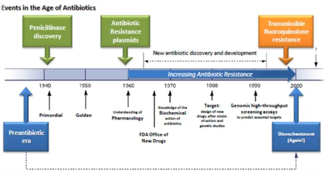
In 1928, Alexander Fleming (1881–1955) discovered the bacteria-killing property of penicillin (Fleming, 1929). Although he had made an incredible discovery, he was unable to produce penicillin in a form useful to physicians. In 1941, two English scientists, Howard Florey (1898–1968) and Ernst Chain (1906–1979), developed a form of penicillin that could be used to fight bacterial infections in humans. In 1945, penicillin was available for widespread use. Fleming, Florey, and Chain shared the Nobel Prize in Medicine in 1945 for their work on penicillin (Davies & Davies, 2010).
While Fleming was working on penicillin, Gerhard Domagk (1895–1964) discovered, in 1935, the first sulfa drug, sulfonamide. However, these drugs had some serious problems, especially kidney failure, and scientists continued their search for other antibiotics. The next breakthrough was in 1944, when Selman
INTRODUCTION:CHAPTER 2
found naturally in soil and it was proved to be a cure for many intestinal diseases. In the late 1940s and early 1950s, new antibiotics were introduced, including chloramphenicol and tetracycline, and the age of antibiotic chemotherapy came into full being (Figure 1).
Figure 1 - History of antibiotic discovery and concomitant development of antibiotic resistance. Adapted from Davies & Davies (2010).
2.2. Antibiotic Targets
Antibiotics generally target processes or structures that are essential for bacterial growth, survival or both simultaneously; the most successful compounds are those that interfere with the construction of the bacterial cell wall, the synthesis of protein, or the replication and transcription of DNA. Relatively few clinically useful agents act at the level of the cell membrane or by interfering with specific metabolic processes of the bacterial cell (Figure 2).
Their specificity for bacterial targets means that eukaryotic cells in the host are not greatly affected (Betina, 1983). Antibiotics can be classified according to their antimicrobial spectrum of activity, mechanism of action, source (e.g., produced by Streptomyces spp.), mechanism of production (natural, synthetic, or semi-synthetic drugs), or chemical structure.
Figure 2 - Antibiotic targets and mechanisms of resistance (Wright, 2010).
2.2.1. Inhibition of Cell Wall Synthesis
Bacterial cell wall contain murein or peptidoglycan, which is essential in maintaining cell wall structure. Cell wall synthesis inhibitors, such as β-lactams or glycopeptides, block the ability of microorganisms to synthesize their cell wall by inhibiting the synthesis of peptidoglycan. The interruption of the normal crosslinking can be mediated by penicillins, that inhibit the transpeptidase activity, or by vancomycin, that sequester the substrate by complexation of the D-Ala-D-Ala termini of peptidoglycans; these antibiotics show synergy when used in combination.
In Chapter 4, the inhibition of cell wall synthesis mechanism will be described in more detail upon the elucidation of the mechanism of action of β-lactamases.
2.2.2. Inhibition of Protein Synthesis
Many antibiotics work by binding to bacterial ribosomes, inhibiting the protein synthesis machinery (translation) in the cell. Examples of antibiotics that bind to
INTRODUCTION:CHAPTER 2
the 30S ribosomal subunit are aminoglycosides and tetracyclines, which prevent the binding of tRNA (Brodersen et al, 2000; Carter et al, 2000). Macrolide antibiotics, such as erythromycin, bind to the 50S ribosomal subunit and block the exit tunnel of the bacterial ribosome (Schlünzen et al, 2001).
2.2.3. Alteration of Cell Membranes
Polymyxins consists of a cationic cyclic peptide with a fatty acid chain. The interaction between the cationic peptide and the bacterial cell membrane causes disruption of the membrane and increases the permeability of cell components (Daugelavicius et al, 2000; Newton, 1956). These antibiotics are mostly effective on Gram-negative bacteria because they contain a definite cell membrane.
On the other hand, the biological activity of ionophore antibiotics is related to their ability to disrupt the flow of ions either into or out of cells, i.e. they can disrupt the ionic imbalance by allowing ions to penetrate the cell membrane as ion-ionophore complexes or via the formation of ion channels (Bergen & Bates, 1984; Bilgili & Kart, 2008). Gram-positive bacteria appear to be particularly sensitive to the effect of ionophores perturbing normal ion transport.
2.2.4. Inhibition of Nucleic Acid Synthesis
Differences between the enzymes used to synthesize nucleic acids in prokaryotes and eukaryotes provide the means for selective action of antibiotics that take their effect by inhibiting nucleic acid synthesis. Antibiotics of the rifamycin family inhibit RNA synthesis by binding to the RNA polymerase, which is responsible for transcribing bacterial DNA to RNA (Campbell et al, 2001; Floss & Yu, 2005). Antibiotics of the (fluoro)quinolone group interfere with DNA synthesis by inhibiting the activity of topoisomerase II (DNA gyrase), an enzyme involved in DNA replication(Hooper & Rubinstein, 2003; Shen et al, 1989).
2.2.5. Competitive Inhibitors
Also referred to as anti-metabolites or growth factor analogs, these antibiotics competitively inhibit the important metabolic pathways occurring inside the bacterial cell. The most important in this class are sulfonamides and trimethoprim. Amino acid and purine synthesis in bacteria is dependent on tetrahydrofolate,
which is a folic acid derivative. Bacteria are unable to absorb preformed folic acid and need to synthesize it by means of two key enzymes involved in folate synthesis (DHFR, a dihydrofolate reductase and DHFS, a dihydrofolate synthase) which are inhibited by sulfamethoxazole and trimethoprim, respectively. Overproduction of these enzymes causing resistance to these drugs is recognized in several pathogenic organisms. These agents act at separate stages in the pathway of folic acid synthesis and therefore they have a synergistic effect (Murray et al, 2005; Walsh, 2000).
2.3. The β-lactam antibiotics
2.3.1. Structure
β-Lactam antibiotics are among the most commonly prescribed drugs. They are composed of an isolated ring (monobactam), or associated in bicyclic ring structures in other classes (Table 1). Overall, side chain modifications within groups alter the pharmacokinetic and antibacterial properties of different β-lactam antibiotics. For example, modifications at position 7 of cephalosporins increase penetration into the periplasmatic space and stability against β-lactamases, but may reduce antibiotic activity (Donowitz & Mandell, 1988a; Donowitz & Mandell, 1988b).
β-Lactam antibiotics are indicated for the prophylaxis and treatment of bacterial infections caused by susceptible organisms. Although there are several classification schemes for antibiotics based on bacterial spectrum (broad versus narrow) or type of activity (bactericidal vs. bacteriostatic), the most useful is based on chemical structure (Table 1).
β-Lactams can range from very narrow spectrum to very broad spectrum depending on the subgroups. The ones with the broadest spectrum, third and fourth generation cephalosporins, can inactivate both negative and Gram-positive bacteria (Murray et al, 2005).
INTRODUCTION:CHAPTER 2
Table 1 - Classification scheme for β-lactam antibiotics, based on chemical structure.
Class Subclass Example Chemical struture
Penams Penams - Penicillin - Aminopenicillins - Ureidopenicillin - Carboxypenicillin - Penicillinase-stable penicillins
Benzylpenicillin, the gold
standard type of penicillin
Carbapenams (3S,5R)-carbapenam;
carbapenams exist primarily as biosynthetic intermediates to the carbapenem antibiotics Oxapenams or clavams
- β-lactam/β-lactamase inhibitor combinations
Clavulanic acid
Penems Penems Faropenem
Carbapenems Imipenem
Cephems Cephamycins1 Cefoxitin
Cephalosporins(oral and parenteral) - 1st generation - 2nd generation3 - 3rd generation2,3 - 4th generation2,3 Cefalotin (1st generation)
Carbacephems (oral) Loracarbef
Oxacephems (parenteral) Flomoxef; oxacephems are
synthetic compounds and have not been discovered in nature.
Monobactams3 Aztreonam; monobactams
have a second thiazole ring which is not fused to the β-lactam ring
1.
Cephamycins contain a methoxyl group and are thus also called 7-α-methoxy-cephalosporins.
2.
Cephalosporins from 3rd and 4th generations are also referred to as ―extended-spectrum cephalosporins‖.
3.
Cephalosporins from 2nd (cefuroxime), 3rd (cefotaxime, ceftazidime, ceftriaxone) and 4th (cefepime) generations, and aztreonam are also oxyimino-β-lactams. With the exception of aztreonam, these antibiotics are also called oxyimino-cephalosporins.
Among β-lactams, carbapenems are antibiotics with a broad spectrum of antibacterial activity, compared to other β-lactams such as penicillins and cephalosporins. These antibiotics were originally developed from thienamycin, a naturally-derived product of Streptomyces cattleya (Kahan et al, 1983), and they are often used for treatment of resistant isolates of P. aeruginosa and A. baumannii that have become increasingly resistant to the broad-spectrum cephalosporins used in hospital settings (Livermore & Woodford, 2006). Carbapenems (imipenem, meropenem, ertapenem, and doripenem) have proven to be more stable against the action of many β-lactamases, due to their unusual stereochemistry across the C5-C6 bond and the presence of the α-hydroxyethyl group (Table 1).
Clavulanic acid, discovered as a natural product of the bacterium Streptomyces clavigularis, was the first β-lactamase inhibitor to be introduced into clinical practice (Reading & Cole, 1977). Modifications to its structure were later made in improvement of its activity, producing two additional inhibitors, sulbactam and tazobactam. They are called ―suicide inhibitors‖ because they irreversibly bind to β-lactamase (Bonomo & Rice, 1999; Knowles, 1985).
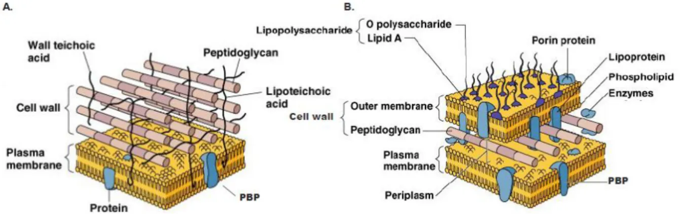
2.3.2. Mechanism of action
β-lactam antibiotics inhibit the growth of sensitive bacteria by inactivating enzymes that are involved in the third stage of cell wall synthesis and are located in the bacterial cell membrane. In Gram-positive bacteria, peptidoglycan is the main constituent of the cell wall, appearing as a heteropolymer of complex composition that provides strength to the wall, to which the cytoplasmic membrane is juxtaposed. It is in these structures, in the outer leaflet, that the targets of -lactam antibiotics – the PBP (Penicillin-Binding Proteins) – are inserted (Figure 3) (Murray et al, 2005; Scheffers & Pinho, 2005). By contrast, Gram-negative cell walls have a more complex structure than do those of Gram-positive organisms. On the outside of the cytoplasmic membrane, in whose outer leaflet PBPs are located, there is the periplasm, which contains a thin layer of peptidoglycan, with less cross-linking than in Gram-positive cells; Gram-negative bacteria are thus mechanically weaker than Gram-positive cells. Beyond the peptidoglycan of the Gram-negative cell wall lies an outer membrane, which has protein channels -
INTRODUCTION:CHAPTER 2
porins - that act as transporters through its surface (Figure 3).
Figure 3 – Graphical illustration of Gram-positive (A) and Gram-negative (B) cell wall structures. The Gram-positive cell wall is thicker than that of Gram-negative bacteria, compensating for the absence of a second (outer) bilayer membrane. Adapted from Tortora et al (2010)
Peptidoglycan is composed of alternating chains of N-acetylglucosamine (NAG) and N-acetylmuramic acid (NAM) linked by β-(1,4)-glycoside units (Figure 4). The carboxyl group of muramic acid is frequently replaced by an amino acid chain composed of four amino acids. The most common are L-alanine, alanine, D-glutamic acid, D-glutamine and L-lysine or diaminopimelic acid (DAP). This is the only biological structure that contains D-amino acids and it is the target of numerous antibacterial antibiotics.
Figure 4 - acetylglucosamine (NAG) and N-acetylmuramic acid (NAM) chains forming the structure of peptidoglycan (Paustian & Roberts, 2011).
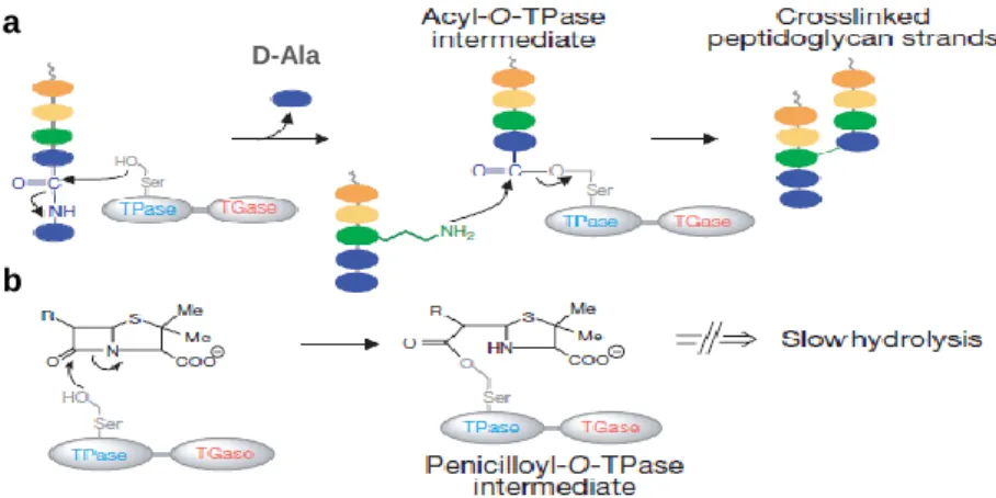
Synthesis of peptidoglycan occurs in three phases: assembly of precursor in the cytoplasm, transport across the inner membrane, and polymerization. Peptidoglycan synthesis starts in the cytoplasm by binding the peptide chains to one of the sugar molecules. After translocation to the outside of the cell, peptide
chains from adjacent glycan chains are cross-linked to each other by a peptide bond exchange (transpeptidation) between the free amine of the amino acid in the third position of the pentapeptide (e.g., lysine) and the D-alanine at the fourth position of the other peptide chain, releasing the terminal D-alanine of the precursor (Figure 5a). These reactions are catalyzed by transpeptidase and carboxypeptidase enzymes, which are only sensitive to β-lactam antibiotics (Araki et al, 1966).
Figure 5 – Schematic representation of the biosynthesis of the cell wall. a) The final step comprises the cross-linking between two peptidoglycan isolates and release of the terminal D-Alanine (D-Ala) of the precursor; and b) the inhibition of transpeptidase activity by penicillins through the formation of a slowly hydrolyzing covalent acyl-enzyme intermediate. Adapted from Walsh (2000).
β-lactam antibiotics inhibits the cell wall synthesis because the highly reactive CO-N bond in the β-lactam ring of the β-lactam molecule lies in exactly the same position as the CO-N bond in D-alanyl-D-alanine, which is the target of transpeptidation (Tipper & Strominger, 1965) (Figure 6). The β-lactam-enzyme complex acts as a competitor to the formation of the normal acylated enzyme (Figure5b). The β-lactam-enzyme complex is very stable, and its formation culminates with the inactivation of the PBP’s functions (Georgopapadakou & Liu, 1980; Ghuysen, 1988). This interference with the normal cross-linking in the cell wall results in cellular lysis.
Figure 6 – β-lactams (a) acts as a substrate analog, binding to the substrate-anchoring site normally occupied by D-alanyl-D-alanine (b).
D-Ala
b a
INTRODUCTION:CHAPTER 3
Chapter 3. Antibiotic Resistance in Gram-negative Bacteria
Chance has always played a part in the evolution of bacteria, and the emergence of antibiotic resistance is one of the many aspects of this phenomenon (Courvalin, 2005). Hence, once an antibiotic is proven to be effective and enters widespread human therapeutic use, its days are numbered. Clinically significant antibiotic resistance appears in periods of months to years (Davies, 1996; Davies & Davies, 2010; Demain & Sanchez, 2009).
Gram-negative bacteria are usually more resistant to the antibiotic action than Gram-positive (Russell & Day, 1996). This is due to the presence of a double membrane structure that prevents antibiotics, like penicillin G, from accessing the target in the cell wall. Generally, Gram-negative microorganisms exhibit two types of antibiotic resistance mechanisms, i.e. intrinsic and acquired resistance (Livermore, 2003):
Innate or intrinsic resistance is an inherent feature of a species, resulting in the lack of activity of an antibiotic or antibiotic class; it is usually controlled by the microorganism’s own genetic background, i.e. all bacteria of the same specie may lack the appropriate antibiotic target or possess natural antibiotic resistance mechanisms that avoid the agent to reach the target (Table 2). In this case, the microorganism is resistant to an antibiotic without record of previous exposure; for example, most Gram-negative organisms are intrinsically resistant to vancomycin and teicoplanin, because their outer membrane is impermeable to large glycopeptide molecules (Arthur & Courvalin, 1993; Reynolds, 1989).
"The greatest possibility of evil in self-medication is the use of too small doses so that instead of clearing up infection, the microbes are educated to resist penicillin".
Table 2 – Examples of intrinsic resistance phenotypes. Organism Intrinsic Resistance
All Enterobacteriaceae penicillin G, macrolides, lincosamides, streptogramins,
glycopeptides, fusidic acid, linezolid, mupirocin Klebsiella spp.
Citrobacter koseri
aminopenicillins, carboxypenicillins
Proteus vulgaris Proteus penneri
aminopenicillins, carboxypenicillins, cefuroxime, colistin, nitrofurantoin, tetracyclines
Proteus mirabilis colistin, nitrofurantoin, tetracyclines
Serratia marcescens aminopenicillins, amoxicillin-clavulanic acid, 1st and 2nd gen cephalosporins, colistin
Enterobacter spp. aminopenicillins, amoxicillin-clavulanic acid, 1st gen cephalosporins, cefoxitin
Citrobacter freundii aminopenicillins, amoxicillin-clavulanic acid, 1st gen cephalosporins, cefoxitin, nitrofurantoin
Pseudomonas aeruginosa aminopenicillins, carboxypenicillins, amoxicillin- clavulanic acid, 1st and 2nd gen cephalosporins, cefotaxime, ceftriaxone, nalidixic acid, aminoglycosides, colistin, nitrofurantoin, fosfomycin, chloramphenicol, imipenem and trimethoprim
Salmonella spp. 1st and 2nd gen cephalosporins, cefuroxime (active in
vitro, not active in vivo), aminoglycosides (in vivo)
Morganella morgannii aminopenicillins, amoxicillin-clavulanic acid, 1st and 2nd gen cephalosporins, cefoxitin, tetracyclines, fosfomycin, colistin, nitrofurantoin
Acinetobacter baumannii Acinetobacter calcoaceticus
aminopenicillins, 1st and 2nd gen cephalosporins, nitrofurantoin, fosfomycin, chloramphenicol and trimethoprim
Stenotrophomonas maltophilia
ureidopenicillins, carboxypenicillins, 1st and 2nd gen cephalosporins, imipenem, cefotaxime, aztreonam, aminoglycosides, tetracyclines, fosfomycin
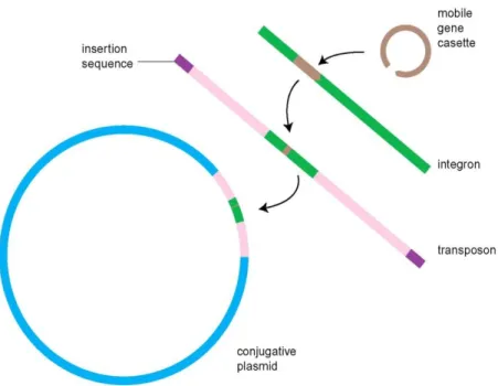
Extrinsic or acquired resistance is the result of a series of genetic changes, through the horizontal acquisition of foreign genetic information from bacteria cohabitating the same environment (mediated by plasmids and/or transposons, which may contain integron sequences) or by mutation in structural or regulatory housekeeping genes, such as gyrA, parC and rpoB, (Courvalin, 2005). Sometimes, genetic changes result in diminished activity, but not complete loss of antibiotic effectiveness.