Daniela Filipa Firmino Rosa
Graduation in Biochemistry
Bringing the biorefinery concept to the health-care
facilities: use of industrial by-products against
multi-drug resistant bacteria
Dissertation to obtain the Master degree in Biochemistry
Supervisor: Doctor Paula Cristina Castro Parreira e Guerra, CEBAL
Co-supervisor: Doctor Andreia Patrícia Henriques Ascenso, Faculdade de
Farmácia - UL
Jury:
President: Prof. Doctor José Ricardo Franco Tavares Examiner: Prof. Doctor Maria Luísa Lopes de Castro e Brito Vowel: Doctor Paula Cristina Castro Parreira e Guerra
Bringing the biorefinery concept to the health-care facilities: use of industrial by products against multi-drug resistant bacteria
Copyright © DANIELA FILIPA FIRMINO ROSA, Faculdade de Ciências e Tecnologia, Universidade Nova de Lisboa.
A Faculdade de Ciências e Tecnologia e a Universidade Nova de Lisboa têm o direito, perpétuo e sem limites geográficos, de arquivar e publicar esta dissertação através de exemplares impressos reproduzidos em papel ou de forma digital, ou por qualquer outro meio conhecido ou que venha a ser inventado, e de a divulgar através de repositórios científicos e de admitir a sua cópia e distribuição com objetivos educacionais ou de investigação, não comerciais, desde que seja dado crédito ao autor e editor.
i
Agradecimentos
A todos aqueles que de uma forma direta ou indireta contribuíram para a concretização deste trabalho quero deixar o meu sincero agradecimento.
Em primeiro lugar, como não poderia deixar de ser, quero deixar o meu maior e mais sincero agradecimento à Doutora Paula Parreira, por ter aceite ser minha orientadora neste trabalho, pelo apoio incondicional prestado, pelas palavras de incentivo, dedicação, paciência e sincera amizade. Foi um enorme privilégio tê-la como orientadora!
Quero agradecer à Doutora Fátima Duarte por me ter aberto as portas do CEBAL há dois anos atrás, pela confiança que em mim depositou para integrar no seu grupo de investigação, onde pude desenvolver conhecimento e competências na área de investigação. Pelo incentivo em ingressar no mestrado e pela constante contribuição no decorrer deste trabalho de investigação. Muito obrigada!
Ao CEBAL por me ter dado todas as condições para a realização deste trabalho, bem como aos projetos Neukbark, RefinOlea e ValBioTecCynara por terem cedido as biomassas a utilizar neste trabalho de investigação. A todos os investigadores que constituem o CEBAL agradeço pelo espírito de entreajuda, com especial agradecimento à Teresa Brás pelo apoio incondicional e preocupação; à Olinda Guerreiro, mestre da estatística, pela disponibilidade e ajuda na análise estatística deste trabalho; à Ângela Guerra e Ana Paulino pelas palavras de incentivo e companhia no nosso laboratório 15; a todas pela amizade e, claro, pelos nossos lanchinhos. Ao Miguel Ferro pelas sessões de verão no HPLC há um ano atrás e à Flávia Fernandes pelo contributo no último ensaio do meu trabalho. Não posso deixar de agradecer à maltinha da bioinformática, Anabel Usié, Brígida de Meireles, Daniel Gaspar e Pedro Barbosa, e à Margarida Pereira, pelos cafés matinais, pelos momentos de descontração e pelas after work sessions.
Quero agradecer à FF-UL por me ter dado a possibilidade de desenvolver o meu trabalho nas suas instalações. À Professora Doutora Andreia Ascenso, agradeço por me ter recebido com a maior simpatia e me ter dado todo o apoio e conhecimento para desenvolver uma das etapas deste trabalho. A todos os docentes e investigadores, em especial à Dra. Ana Salgado, pela contribuição, palavras de apoio e simpatia, muito obrigada.
À Diretora da Escola Superior Agrária do Instituto Politécnico de Beja, Doutora Maria Margarida Pereira, por ter concedido o espaço para a realização da análise sensorial. Ao Professor Nuno Bartolomeu Alvarenga, pela disponibilidade para realizar a análise reológica da Formulação, pela ajuda e pelo conhecimento transmitido.
ii Ao Coordenador do Mestrado em Bioquímica, Professor Doutor Ricardo Franco, agradeço a oportunidade e o privilégio que tive em frequentar este Mestrado, que muito contribuiu para o enriquecimento da minha formação académica e científica. Agradeço também a todos os colegas que tive o prazer de conhecer no primeiro ano do Mestrado, com especial agradecimento à Carla Varela que tanto me acompanhou, pelos momentos de descontração, por me ter mostrado Almada, pela boa disposição, pela amizade; à Ana Diniz pela constante preocupação, por arranjar sempre um tempinho na sua agenda super preenchida para estar comigo, pela sincera amizade; à Filipa Trovão por estar sempre disponível para ajudar, por nunca se ter esquecido de mim depois de ter voltado para a minha cidade, pelo incentivo, pela amizade; à Sara Silva pela ajuda na minha integração na faculdade, pelos passeios até à Costa da Caparica que ajudavam tanto a desanuviar, pelas longas tardes e noites de estudo no Edifício VII, pela amizade; à Raquel Costa pela preocupação e incentivo. Às minhas colegas de casa, Filipa Pereira e Filipa Gomes, agradeço por todos os bons momentos que passamos juntas e pela vossa amizade.
Às minhas amizades de Évora, agradeço à Maria Miguel Castro pela amizade sincera, constante preocupação e apoio, mesmo quando eu desapareço do mapa, e à Ana Antão por me querer levar por bons caminho quando eu a quero levar para os maus. Para além disso, só tenho que agradecer por me acompanhar, me apoiar e me motivar sempre que preciso, pela amizade para a vida.
Agradeço às minhas amizades mais sinceras, às minhas manas de coração, Ana Raquel Barata, Marisa Mendes, Daniela Bica, Cláudia Silva, Tânia Lampreia, Vanessa Gonçalves e Adriana Silva, pelo apoio incondicional em todos os altos e baixos. “Os amigos não se escolhem,
reconhecem-se”… Obrigado!
À minha família, em especial aos meus pais, ao meu irmão e à minha avó Rosária, um enorme obrigada por acreditarem sempre em mim e pelo apoio incondicional que sempre me deram. Espero que esta etapa, que agora termino, possa de alguma forma retribuir e compensar todo o carinho, apoio e dedicação que, constantemente, me oferecem. A eles, dedico todo este trabalho.
Financial Support:
This work was supported by the Program Alentejo 2020, through the European Fund for Regional Development under the scope of ValBioTecCynara – Economic valorization of Cardoon (Cynara cardunculus): study of natural variability and biotechnological applications (ALT20-03-0145-FEDER-000038).
iii
Abstract
Multi-drug resistant bacteria (MDRB) are a public health issue worldwide, being alternatives to the conventional failing antibiotherapy required. This fact has turned attention to bioactive compounds with antimicrobial activity features. These bioactives can be extracted from several biomasses sources, namely industrial by-products, within a biorefinery concept.
The main objective of this Thesis was the design of a novel antiseptic formulation (AF), based on natural plant extracts, obtained from forest/agriculture industries by-products.
The antimicrobial activities of three extracts from different sources were studied:
Eucalyptus nitens total bark (ENTB) extract, Cynara cardunculus leaves (CcL) extract and dry
olive pomace (DOP) extract. Those that presented better antibacterial activity were chosen to be included in the AF. Studies regarding formulation design and its antibacterial performance were made.
ENTB extract was the one with better anti-MDRB performance, particularly against
Staphylococcus spp., with minimal inhibitory concentrations between 64 and 2048 µg/mL, being
included in the AF. The antibacterial activity of ENTB extract-based-AF presented promising results, achieving a 97±2% bacterial growth inhibition after exposure to 45% of AF.
In conclusion, the developed AF presents potential to be further investigated, namely for hand sanitation within healthcare environment, but additional adjustments should be executed, namely in order to turn it more appellative to users.
Keywords: Multi-drug resistant bacteria, healthcare-associated infections, antimicrobial activity,
v
Resumo
As bactérias multirresistentes a antibióticos (BMRA) são um problema de saúde pública a nível mundial, sendo necessárias alternativas à antibioterapia convencional, cuja eficácia tem decrescido nas últimas décadas. Este facto atraiu a atenção para compostos bioativos com atividade antimicrobiana. Estes bioativos podem ser extraídos a partir de diversas biomassas, nomeadamente subprodutos industriais, dentro do conceito de biorrefinaria.
O principal objetivo da presente Tese foi o desenvolvimento de uma nova formulação antisséptica (FA), com base em extratos naturais de plantas, obtidos a partir de subprodutos da indústria florestal/agrícola.
Para tal, foi estudada a atividade antimicrobiana de três extratos de diferentes origens: extrato da casca total de Eucalyptus nitens (ECTEN); extrato de folhas de Cynara cardunculus (cardo) e; extrato de bagaço de azeitona seco. Aqueles que apresentaram melhor atividade antimicrobiana foram escolhidos para serem incluídos na FA. Estudos de desenvolvimento da formulação e desempenho antimicrobiano da mesma foram efetuados.
O ECTEN foi o que apresentou melhor desempenho anti-BMRA, particularmente contra
Staphylococcus spp., com concentrações mínimas inibitórias entre 64 e 2048 µg/mL, sendo
incluído na FA. A atividade antibacteriana da FA à base de ECTEN apresentou resultados promissores, alcançando 97±2% de inibição de crescimento bacteriano após exposição a 45% de FA.
Em conclusão, a FA desenvolvida apresenta potencial para prosseguir para investigações futuras, nomeadamente para a higienização das mãos em contexto hospitalar ou unidades de cuidados de saúde, mas ajustes adicionais deverão ser feitos, nomeadamente de forma a tornar a FA mais apelativa para os utilizadores.
Palavras-chave: Bactérias multirresistentes a antibióticos, infeções nosocomiais, atividade
vii
Table of contents
Agradecimentos... i
Abstract ... iii
Resumo ... v
Table of contents ... vii
List of figures ... xi
List of tables ... xiii
List of abbreviations ... xv
1 – Introduction ... 1
1.1 – Infectious diseases: the main players and the challenges in the 21st century ... 2
1.1.1 – Bacterial antibiotic resistance: intrinsic vs acquired mechanisms ... 2
1.1.1.1 – Intrinsic mechanisms ... 2
1.1.1.2 – Acquired mechanisms ... 3
1.1.2 – Mechanisms of acquired resistance to antibiotics ... 3
1.1.2.1 – Inhibition of cell wall synthesis ... 4
1.1.2.2 – Protein synthesis ... 5
1.1.2.3 – Nucleic acid synthesis ... 5
1.1.3 – Drug-resistant bacteria in health-care facilities ... 7
1.1.3.1 – ESKAPE ... 7
1.1.3.2 – Nosocomial infections (NI) ... 7
1.2 – Phytotherapy: use of bioactive compounds as therapeutic tools ... 9
1.2.1 – Phenolic compounds ... 10
1.2.2 – Terpenes ... 11
1.2.2.1 – Triterpenes ... 12
1.2.2.2 – Sesquiterpenes... 13
1.3 – Biorefinery ... 15
1.3.1 – Eucalyptus spp. and paper production ... 16
1.3.2 – Olive and olive oil production ... 18
1.3.3 – Cynara cardunculus – artisanal cheese production and other applications ... 19
1.4 – Pharmaceutical formulation ... 20
viii
1.4.2 – Quality control and stability study ... 22
2 – Objectives ... 25
3 – Material & Methods ... 27
3.1 – Plant material and extraction method ... 28
3.1.1 – Eucalyptus spp. ... 28
3.1.2 – C. cardunculus ... 28
3.1.3 – Dry olive pomace ... 28
3.1.3.1 – DOP phenolic extraction ... 28
3.1.3.1.1 – Determination of total phenolic content ... 29
3.1.3.1.2 – Quantification of hydroxytyrosol, tyrosol and oleuropein by high-performance liquid chromatography-UV/Vis (HPLC-UV/Vis) ... 29
3.2 – Bacterial strains and growth ... 30
3.3 – Antibacterial activity assays ... 31
3.3.1 – Antibiotics antibacterial activity assay ... 31
3.3.1.1 – Minimal inhibitory concentrations (MIC) ... 31
3.3.2 – TAs and ENTB extract for antibacterial activity assays ... 32
3.3.2.1 – Minimal bactericidal concentration (MBC) determination ... 33
3.3.2.2 – Time-kill assay ... 33
3.3.2.3 – Synergistic assays (ENTB extract plus antibiotics) ... 33
3.3.3 – CcL extract antibacterial activity assay ... 34
3.3.4 – DOP extract antibacterial activity assay ... 34
3.4 – Quantification of reducing sugars content ... 34
3.4.1 – ENTB extract ... 34 3.4.2 – CcL extract ... 34 3.4.3 – DOP extract ... 35 3.5 – Formulation ... 35 3.5.1 – Formulation design ... 35 3.5.2 – Formulation characterization ... 35 3.5.2.1 – Organoleptic characteristics ... 35
3.5.2.2 – Identification and quantification of the active substances ... 36
3.5.2.3 – pH determination ... 36
ix
3.5.2.5 – Microbiological assay ... 36
3.5.3 – Antimicrobial activity of the formulations ... 36
3.6 – Statistical analysis ... 37
4 – Results & Discussion ... 39
4.1 – Plant material extraction ... 40
4.2 – Antibacterial activity assays ... 41
4.2.1 – Antibiotics ... 41
4.2.2 – ENTB extract ... 43
4.2.2.1 – MIC determination ... 43
4.2.2.2 – MBC determination ... 48
4.2.2.3 – Time-kill assay ... 48
4.2.2.4 – Synergistic activity (antibiotic plus extract) ... 50
4.2.3 – Cynara cardunculus leaf (CcL) extract ... 52
4.2.4 – DOP extract ... 54
4.3 – Reducing sugars quantification in the extract ... 56
4.4 – Formulation Design ... 56
4.4.1 – Preformulation and characterization studies ... 56
4.4.2 – Antibacterial activity of formulations A, B and C ... 59
4.4.3 – Stability studies ... 60
4.4.4 – Sensorial evaluation ... 62
5 – Conclusion & Future perspectives ... 65
6 – References ... 67
7 – Appendix... 77
xi
List of figures
Figure 1.1. Illustration of structural differences in Gram-positive and Gram-negative bacterial cell
wall. ... 3
Figure 1.2. Peptidoglycan synthesis. ... 4
Figure 1.4. Chemical structures of oleuropein, hydroxytyrosol and tyrosol.. ... 11
Figure 1.5. Pentacyclic triterpenic acids. ... 12
Figure 1.6. Sesquiterpene lactones basic structure. ... 14
Figure 1.7. Biorefinery concept. ... 16
Figure 1.8. Eucalyptus spp. in Portugal. ... 17
Figure 1.9. Olive tree in Portugal... 18
Figure 1.10. Geographic distribution of Cynara carcunculus in Portugal. ... 19
Figure 1.11. Morphological appearance of each variant of Cynara cardunculus. ... 20
Figure 1.12. Representative scheme of main stages of formulation design. ... 22
Figure 3.1. Microbroth dilution method. ... 31
Figure 4.1. MTT assay results for ENTB extract against Staphylococcus spp.. ... 45
Figure 4.2. ENTB extract antibacterial effect in bacteria cell growth. ... 46
Figure 4.3. Time-kill curves for ENTB extract for sub-MIC values. ... 49
Figure 4.4. MTT assay results for CcL extract. ... 53
Figure 4.5. CcL extract antibacterial effect against S. aureus ATCC 43300. ... 53
Figure 4.6. Visual appearance of the three formulations designed... 58
Figure 4.7. Flow curves of formulations B and C at different shear rates. ... 58
Figure 4.8. Antibacterial effect of the formulations designed. ... 59
Figure 4.9. Quantification of TAs in formulation C over time represented as ratio concentration : initial concentration (C/C0). ... 61
xiii
List of tables
Table 1.1. Antibiotics target and resistance mechanisms. ... 6
Table 1.2. Bioactive compounds and its biological activities. ... 15
Table 1.3. Administration routes and dosage forms associated ... 21
Table 1.4. Examples of quality control parameters of a pharmaceutical formulation ... 23
Table 3.1. MDRB panel used. ... 30
Table 3.2. Ingredients of the initial formulation. ... 35
Table 4.1. DOP extraction yield (%) and total phenolic content expressed as milligrams of Gallic acid (GAE) equivalents per gram of dry weight (DW). ... 40
Table 4.2. Extract phenolic content in hydroxytyrosol, tyrosol and oleuropein (DW) and its respective retention times. ... 40
Table 4.3. MIC of antibiotics against MDRB ... 41
Table 4.4. MICs of ENTB extract and standard compounds of TAs ... 43
Table 4.5. MBC of ENTB extract against MDRB ... 48
Table 4.6. MIC (expressed in µg/mL) of antibiotic, ENTB extract (MIC) and ENTB extract together with antibiotics (MICa) against Staphylococcus spp.. ... 51
Table 4.7. CcL extract MIC against MDRB. ... 52
Table 4.8. MIC for DOP extract and for standard compounds of phenolic compounds hydroxytyrosol (H), tyrosol (T) and oleuropein (O). ... 54
Table 4.9. Reducing sugars (RS) content in xylose (Xyl) and glycose (Gly) in ENTB, DOP and CcL extracts. ... 56
Table 4.10. Qualitative and quantitative composition of ENTB formulations. ... 57
Table 4.11. Quality control of ENTB formulations. ... 57
Table 4.12. Stability study of formulation C at different temperatures: room temperature (RT) and 4°C. ... 60
Table 4.13. Initial concentration (C0) of TAs: betulinic acid (BA), betulonic acid (BOA), oleanolic acid (OA) and ursolic acid (UA) at 0.7% used for HPLC analysis, and 0.1% used in the formulation.. ... 61
xv
List of abbreviations
AF – Antiseptic formulation BA – Betulinic acid
BOA – Betulonic acid
CcL – Cynara cardunculus leaf
CDC – Centers for Diseases Control and Prevention DMSO – Dimethyl sulfoxide
DNS – 3,5-dinitrosalicylic DOP – Dry olive pomace
ECDC – European Centre for Disease Prevention and Control ENTB – Eucalyptus nitens total bark
ESKAPE – vancomycin-resistant Enterococcus, MRSA, β-lactamase of extended spectrum producer Klebsiella, imipenem-resistant Acinetobacter, imipenem-resistant Pseudomonas and third generation cephalosporin-resistant Enterobacter
GC – Growth control
HAI – Healthcare-associated infections
HPLC-UV/Vis – High Liquid Chromatography with UV/Vis detector ICU – Intensive care unit
MBC – Minimum bactericidal concentration MDRB – Multidrug-resistant bacteria MIC – Minimum inhibitory concentration MHA – Muller Hinton agar
MHB – Muller Hinton broth
MRSA – Methicillin-resistant Staphylococcus aureus MSSA – Methicillin-sensitive Staphylococcus aureus
MTT – (3-(4,5-dimethylthiazol-2-yl)-2,5-diphenyltetrazolium bromide NI – Nosocomial infections OA – Oleanolic acid PBP – Penicillin-biding proteins PW – Peptone water RS – Reducing sugars RT – Room temperature SC – Solvent control SNC – Solution control StC – Sterility control TAs – Triterpenic acids UA – Ursolic acid
USA – United States of America WHO – World Health Organization
1
1 – Introduction
2
1.1
– Infectious diseases: the main players and the challenges in
the 21
stcentury
Bacteria are one of the main players in what concerns infectious diseases, to which features of high morbidity and mortality are associated. (Zaffiri, Gardner, and Toledo-Pereyra 2012) For centuries, the only available resource to treat infections were plants, known to have, among other health benefits, antimicrobial activity. (Ríos and Recio 2005)
In the early 20th century, respiratory infections, like pneumonia and tuberculosis, were the leading cause of death. (Sabin 1970) In order to minimize infection spreading, public health measures were taken, such as: protection of food and water supplies, improvement of personal hygiene and introduction of a vaccination program. (Lederberg 2000) The revolutionary discovery of antibiotics as new therapeutic agents was the turning point in the treatment of infectious diseases caused by bacteria, allowing to save an uncountable number of lives. (Davies, J, & Davies, D. 2010)
Selman Waksman defined “antibiotic” as any class of organic molecules able to inhibit or kill
bacterial cells through specific interactions, with low toxicity to the mammalian host. (Davies, J, &
Davies, D. 2010) In 1928, Alexander Fleming discovered the first effective antibiotic: penicillin. In 1940s it was introduced in the clinical practice, providing quick and complete treatment of previously incurable bacterial diseases. (Davies, J, & Davies, D. 2010) However, antibiotics efficiency has been shadowed since the beginning by the ability of bacteria to develop resistance, and effectiveness of antimicrobial agents, such as antibiotics, has been decreasing over the years. (Lewis 2013)
According to Instituto Nacional Ricardo Jorge (INSA, Portugal), resistant bacteria are defined
as immunes (not affected) to the bacteriostatic (inhibits cell proliferation) or bactericidal (destruction of the bacterial population) effect of antibacterial agents at its therapeutic dosage.
(Jorge n.d.)
1.1.1 – Bacterial antibiotic resistance: intrinsic vs acquired mechanisms
The resistance mechanism to antimicrobial agents, such as antibiotics, can be either intrinsic or acquired.
1.1.1.1 – Intrinsic mechanisms
In intrinsic resistance mechanism, the microorganism has the innate ability to resist to a class of antimicrobial compounds. A good example of intrinsic resistance is Pseudomonas aeruginosa, a Gram-negative bacteria that is naturally resistant to β-lactam antibiotics, due to the presence of a multidrug efflux system and the production of enzymes, β-lactamases, which have the ability to hydrolyze the β-lactamic ring. (Lewis 2013) β-lactam antibiotics act on the peptidoglycan cell wall synthesis, which in Gram-negative is more difficult to access, since peptidoglycan is present in the periplasmic space, between the inner and outer lipid membranes, creating a natural
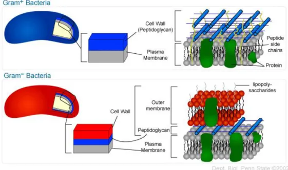
3 barrier.(Lewis 2013)In Gram-positive bacteria, the lack of an outer membrane, as well as the presence of a thick cell wall, composed by the peptidoglycan layer, (Figure 1.1), makes them more susceptible to antibiotics.(Levy and Marshall 2004)
Figure 1.1. Illustration of structural differences in Gram-positive and Gram-negative bacterial cell wall. (State 2002)
1.1.1.2 – Acquired mechanisms
In acquired resistance, the microorganisms develop the ability to resist to the antibiotic’s action by genetic material alterations, namely mutations or genetic material transference, through mobile elements, such as plasmids, bacteriophages, transposons, and others. (Levy and Marshall 2004) Antibiotics misuse, which exerts selective pressure upon bacterial populations, contributes to the growing appearance of resistance among bacterial strains. According to the Centers for Disease Control and Prevention (CDC, USA), the misuse of antibiotics is largely caused by the unnecessary or inappropriate antibiotics prescription in 50% of the cases, both in medicine and agriculture fields. (“Health Policy Brief: Antibiotic Resistance” 2015)
1.1.2 – Mechanisms of acquired resistance to antibiotics
There are several pathways by which bacteria may acquire resistance to antibiotics. The transference of genetic material is a common event, leading to a cumulative acquisition of resistance genes to different antibiotics, in a determined bacterial population. (Lowy 2003) For example, methicillin-resistant Staphylococcus aureus (MRSA) acquired resistance to β-lactam antibiotics through horizontal gene transference of a resistance gene (mecA), which is responsible for the synthesis of a class of penicillin-binding proteins (PBP), which are not sensitive to antibiotic inhibition. (Lowy 2003)
4 Since the discovery of the first effective broad-spectrum antibiotic, able to be used against different bacteria in therapeutic doses, others have been developed and refined, in order to target specific bacteria genre and with defined action mechanisms against bacterial cells. (Davies, J, & Davies, D. 2010) Therefore, antibiotics can be grouped in three major categories, according to their mechanism of action: cell wall synthesis inhibition; protein synthesis inhibition or nucleic acid synthesis inhibition. (Levy and Marshall 2004; McDermott, Walker, and White 2003)
1.1.2.1 – Inhibition of cell wall synthesis
Cell wall is a distinctive bacterial characteristic and, therefore, it is an excellent target for selectively kill or inhibit bacteria in a mammalian organism. The most commonly used inhibitors of cell wall biosynthesis are β-lactams (penicillins and cephalosporins) and glycopeptides (vancomycin and teicoplanin). (Tenover 2006)
Figure 1.2. Peptidoglycan synthesis: (1) synthesis of precursors in the cytoplasm; (2) transport of lipid-bound precursors across the cytoplasmic membrane; (3) insertion of glycan units into the cell wall; and (4) transpeptidation linking and maturation.(McDermott, Walker, and White 2003) (Adapted from Pinho et al.(2013))
β-lactams interact with PBP, the enzymes responsible for the generation of the mature
peptidoglycan. On the other hand, glycopeptides bind peptidoglycan side chains, blocking the transglycosylation and transpeptidation reactions necessary to add new subunits to the growing peptidoglycan chain (Figure 1.2). (McDermott, Walker, and White 2003)
The interaction with Gram-negative and Gram-positive bacteria is different: in Gram-positive bacteria, the target is more accessible in the outer layer, while in Gram-negative bacteria the drug needs to be transported through the outer membrane transport system via proteins (porins). (McDermott, Walker, and White 2003) Due to their low permeability, glycopeptides cannot be
1
2 3
5 transported, therefore having limited spectrum of action against Gram-positive bacteria. (McDermott, Walker, and White 2003)
The resistance mechanisms to β-lactams are: i) mutations in the target PBP; ii) acquisition of new PBPs with decreased affinity for the drug; iii) production of one or more β-lactamases that inactivate the drug; iv) changes in cell wall porins, which limit the drug movement to the target site; and v) active efflux of the drug out of the cell by energy-dependent pumps. (McDermott, Walker, and White 2003) The resistance mechanism responsible for glycopeptides ineffectiveness is the alteration of the amino acid chain target. (McDermott, Walker, and White 2003)
1.1.2.2 – Protein synthesis
Protein synthesis is a vital process for cell survival and multiplication. Several types of antibacterial agents target the bacterial protein synthesis, by binding to either the 30S or 50S subunits of the ribosomes, (Lewis 2013) which leads to the disruption of the normal cellular metabolism, resulting in death or growth inhibition. Aminoglycosides (streptomycin and gentamicin), chloramphenicol, macrolides (erythromycin) and tetracyclines (tetracycline, doxycycline, minocycline) act at the protein synthesis level. (McDermott, Walker, and White 2003) The resistance mechanisms associated to these antibiotics are linked to the expression of enzymes able to inactive them, either by phosphorylation, adenylation or acetylation; efflux pumps or target modification, namely at the ribosome level. (Avent et al. 2011) Gram-negative bacteria are intrinsically resistant to macrolides. (McDermott, Walker, and White 2003)
1.1.2.3 – Nucleic acid synthesis
DNA and RNA are the basic keys for the replication of all living forms, including bacteria. Some antibiotics act by binding to nucleotides (sulfonamides) or nucleic acids (quinolones and rifamycins), which are involved in the process of DNA or RNA synthesis, causing interference in the normal cellular processes and, ultimately, compromising bacterial multiplication and survival. (McDermott, Walker, and White 2003) Sulfonamides block the formation of nucleotide precursors, by competing with the active site of the enzyme. (McDermott, Walker, and White 2003) In nucleic acid synthesis, antibiotics usually act through specific binding to RNA polymerase or DNA topoisomerase, like rifamycin and quinolones, respectively. (McDermott, Walker, and White 2003) The resistance mechanism associated to sulfonamides is the acquisition of an enzyme with low affinity to sulfonamide. (Zaffiri, Gardner, and Toledo-Pereyra 2012) Rifamycin is also enzymatically altered, while quinolones have three different resistance mechanisms: target modification through mutation of topoisomerase genes; decreased permeability of bacterial cell wall or activation of efflux pump. (McDermott, Walker, and White 2003)
In summary, the success of penicillin encouraged the discovery and development of several other molecules with antimicrobial activity, either from natural or synthetic origin, against
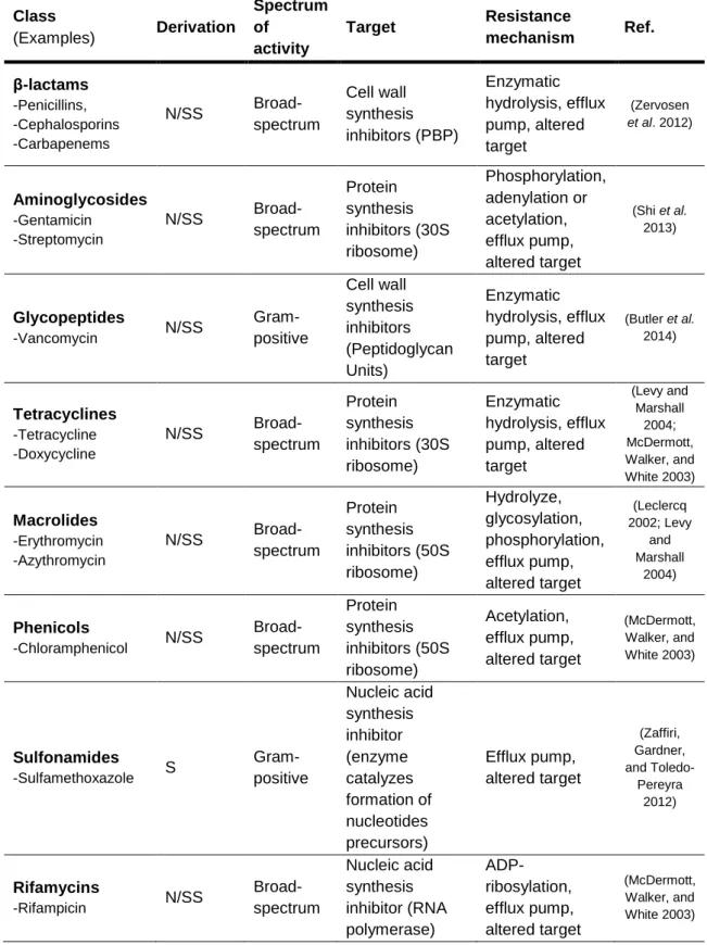
Gram-6 negative, Gram-positive or both, with different action mechanisms, which are summarized in Table 1.1.
Table 1.1.Antibiotics target and resistance mechanisms.
Class (Examples) Derivation Spectrum of activity Target Resistance mechanism Ref. β-lactams -Penicillins, -Cephalosporins -Carbapenems N/SS Broad-spectrum Cell wall synthesis inhibitors (PBP) Enzymatic hydrolysis, efflux pump, altered target (Zervosen et al. 2012) Aminoglycosides -Gentamicin -Streptomycin N/SS Broad-spectrum Protein synthesis inhibitors (30S ribosome) Phosphorylation, adenylation or acetylation, efflux pump, altered target (Shi et al. 2013) Glycopeptides -Vancomycin N/SS Gram-positive Cell wall synthesis inhibitors (Peptidoglycan Units) Enzymatic hydrolysis, efflux pump, altered target (Butler et al. 2014) Tetracyclines -Tetracycline -Doxycycline N/SS Broad-spectrum Protein synthesis inhibitors (30S ribosome) Enzymatic hydrolysis, efflux pump, altered target (Levy and Marshall 2004; McDermott, Walker, and White 2003) Macrolides -Erythromycin -Azythromycin N/SS Broad-spectrum Protein synthesis inhibitors (50S ribosome) Hydrolyze, glycosylation, phosphorylation, efflux pump, altered target (Leclercq 2002; Levy and Marshall 2004) Phenicols -Chloramphenicol N/SS Broad-spectrum Protein synthesis inhibitors (50S ribosome) Acetylation, efflux pump, altered target (McDermott, Walker, and White 2003) Sulfonamides -Sulfamethoxazole S Gram-positive Nucleic acid synthesis inhibitor (enzyme catalyzes formation of nucleotides precursors) Efflux pump, altered target (Zaffiri, Gardner, and Toledo-Pereyra 2012) Rifamycins -Rifampicin N/SS Broad-spectrum Nucleic acid synthesis inhibitor (RNA polymerase) ADP-ribosylation, efflux pump, altered target (McDermott, Walker, and White 2003)
7 Quinolones -Fluoroquinolone S Broad-spectrum Nucleic acid synthesis inhibitor (DNA topoisomerase) Acetylation, efflux pump, altered target (McDermott, Walker, and White 2003) N – Natural; SS – Semi-Synthetic; S – Synthetic
1.1.3 – Drug-resistant bacteria in health-care facilities
1.1.3.1 – ESKAPE
Multi-drug resistant bacteria (MDRB) are characterized by features of enhanced morbidity and mortality. (McDermott, Walker, and White 2003) The ESKAPE (vancomycin-resistant
Enterococcus, MRSA, β-lactamase of extended spectrum producer Klebsiella,
imipenem-resistant Acinetobacter, imipenem-imipenem-resistant Pseudomonas spp. and third generation cephalosporin-resistant Enterobacter) are a group of prevalent multidrug-resistant strains, that greatly concerns the scientific and health-care community, since they are increasingly present in health-care facilities, being associated to Nosocomial Infections (NI). (Cars, Hedin, and Heddini 2011) ESKAPE are highly resistant or even non-responsive to first line drugs recommended for their treatment, leading to reduced therapeutic options and to the need of more costly second and third line drugs. (Rice 2008)
In this antibiotic-resistance era, it is a challenging task to the scientific community, as well as to the pharmaceutical industry, to identify and develop new, effective, safe and broad-spectrum antibacterial drugs. (Brown and Wright 2016)
1.1.3.2 – Nosocomial infections (NI)
NI, also known as healthcare-associated infections (HAI), are defined as those acquired in/or
associated with health-care facilities and that were not actively present or in the incubation period when patient admission occurred. (Breathnach 2005) According to the World Health Organization
(WHO), at any given time, the NI prevalence ranges between 5.7% and 19.1%, in low- and middle-income countries.(World Health Organization 2015) NI are more prevalent in intensive care units (ICU), acute surgical and orthopedic wards. (World Health Organization 2015) The core risks associated to NI are intensive-care admission, bone marrow transplantation, blood transfusion and burn unit. (Zarb et al. 2012)
NI represent a burden to the 21st century society, reflected in a brutal socio-economical impact. NI lead to longer hospitalization periods, more intensive use of hospital’s human resources, increased risk for loss of quality of life and even death. (Zarb et al. 2012) The economical impact is not only related to the money spent in the above-mentioned scenarios, but it is also translated in the loss of working days and consequent production within the active population, extended to the patient’s relatives/caretakers. (Breathnach 2005; Fiorentino 2014)
8 As an example, in the USA, approximately 1.7 millions of patients contract a NI, with 100 000 having death as outcome, being the estimated annual costs associated to NI between 28 to 45 billions of USD. (Dick et al. 2015) Pneumonia and sepsis, the principal cause of infection related to the presence of external devices, like central lines and ventilators, are the most deadly and costly infections. (Dick et al. 2015) A 2012 study from the European Centre for Disease Prevention and Control (ECDC) highlighted that, per day, 81 089 patients get a NI, resulting in 3.5 million patients admitted to ICU annually. (Zarb et al. 2012) The costs related to these numbers are translated in 7 billion of € in Europe annually, including direct costs, and reflecting 16 million in extra days of hospital stay due to NI. (World Health Organization 2015)
The growing NI negative impact at different socio-economical levels has propelled the introduction of infection control programs. In Europe, in 2004, MRSA was the first bacteria that encouraged the creation of surveillance and control programs, in order to reduce resistant bacteria in hospital environment. (Breathnach 2005)
Recently, in Portugal, the number of deaths NI-related have increased, from 2973 in 2010 to 4606 in 2013. (Direção-Geral de Saúde 2014) Several control measures have been defined such as equipment, staff and patients disinfection, screening and monitoring of the occurrence of NI, restriction on the use of antibiotics, among others. (Breathnach 2005)
A 2014 study highlighted that the efforts performed by Health Agencies and the Government were able to decrease the incidence of some infections, such as pneumonia-associated with tracheal intubation in ICU, bacteremia associated with central venous catheter and infection associated with colon and rectal surgery. (Direção-Geral de Saúde 2014) However, there was no significant decrease in the number of deaths, with 4500 patients having NI as cause of death. (Direção-Geral de Saúde 2014) Regarding antibiotic consumption, there was a positive evolution, with 27% and 5% less consumption of quinolones in ambulatory and hospitals, respectively. Geral de Saúde 2014) Carbapenems use was reduced in 5% in hospitals. (Direção-Geral de Saúde 2014) Resistance rates associated to some MDRB, such as MRSA,
Enterococcus spp. and Acinetobacter spp. have began to decrease. (Direção-Geral de Saúde
2014) Nonetheless, there is still great concern regarding the Gram-negative microorganisms, being quinolones-resistant E. coli and carbapenems-resistant Klebsiella spp. the main players. (Direção-Geral de Saúde 2014) Over the last couple of years, outbreaks of NI by K. pneumoniae, which resulted in dozens of deaths, were reported. (Borja-Santos 2016; Direção-Geral de Saúde 2014) Every day, it is estimated that 12 patients die from NI caused by either E. coli or Klebsiella spp., a higher mortality rate than that of car crashes. (Madrinha 2016) However, despite all the efforts made, Portugal remains with one of the darkest European scenarios in what concerns NI. (Direção-Geral de Saúde 2014)
Among the several control measures implemented in hospitals, it was determined that the simplest but yet most effective preventive action is hand hygiene, with water and soap or with alcoholic-based solutions, allowing to prevent cross infections. However, adherence to this countermeasure is low among hospital staff for different reasons, such as: skin irritation due to disinfectants agents; forgetfulness; use of gloves; insufficient time for cleaning; lack of knowledge
9 of this effective measure; among others stated. (Pittet 2001) It is thought that the introduction of new disinfectants agents, with more attractive features, may improve the compliance to this simple but yet highly effective measure for NI prevention. (Pittet et al. 2000)
1.2
– Phytotherapy: use of bioactive compounds as therapeutic
tools
Plants have been used in folk medicine for centuries, from a trial and error strategy, in an initial form of crude drugs such as tinctures, teas, poultices, powders, infusions, to more recent and advanced formulations, tested with scientific methods. (Balunas and Kinghorn 2005; Gurib-Fakim 2006) Plants produce a wide range of bioactive compounds with diverse physiological and functional roles, such as defense mechanisms, pigments that attract pollinators of flowers, UV protection mechanisms (flavonoid, anthocyanin, etc.) and oxidative stress (phenolic compounds). (Simões, Bennett, and Rosa 2009) In defense mechanisms, a wide range of compounds that exhibit a huge chemical diversity are present, such as glycosteroids, flavonoids, terpenes and isoflavones. (Simões, Bennett, and Rosa 2009) Few infections occur in plants, which may be linked to the presence of a highly effective innate defense mechanism. (Abreu, McBain, and Simões 2012) The scientific advance in the phytotherapy field allowed to further uncover plants biological potential, highlighting its benefits and therapeutic actions associated to their chemical composition (bioactive compounds). (Gurib-Fakim 2006) A bioactive compound is usually a plant’s secondary metabolite, able to trigger pharmacological and/or toxicological effects in humans and/or animals, such as: anti-inflammatory; antioxidant; antibacterial; antifungal; antitumor among others. (Simões, Bennett, and Rosa 2009) They can act individually, additively or synergistically and have several biological activities attributed. (Gurib-Fakim 2006)
The decrease of conventional antibiotics effectiveness and the high costs to pharmaceutical industries for the development of new drugs, has contributed to the renewed interest in phytopharmaceuticals. (Abreu, McBain, and Simões 2012) An example of a commercialized drug obtained from natural sources is TAXOL®, and its derivative paclitaxel, cancer and anti-malaria drugs, which are synthesized from the bark of the yew tree (Taxus brevifolia). (Expósito
et al. 2009)
Bioactive compounds antimicrobial performance against bacterial cells has been previously studied, demonstrating activity against a broad array of pathogenic microorganisms, including MDRB. (Simões, Bennett, and Rosa 2009) Despite the antimicrobial potential of bioactive compounds, increased by its diversity and structural complexity, none is currently used as antibiotic. (Gibbons 2004) Nonetheless, there is evidence that bioactive compounds are able to potentiate antibiotic action against MDRB, a promising result for inclusion in the currently available antibiotherapy. (Abreu, McBain, and Simões 2012) Their mechanisms of action are not yet fully understood, which may also be linked to the delay of their complete introduction in the pharmaceutical field, but evidence points that cell wall degradation, damage in cytoplasmic
10 membrane and membrane proteins, cellular content output, cytoplasm coagulation and depletion of the proton motive force, may be responsible for the antibacterial effect. (Cetin-Karaca and Newman 2015)
From the wide range of bioactive compounds reported in the literature, (Cowan 1999) only those with relevance within the scope of this Master Thesis will be presented in the following sections.
1.2.1 – Phenolic compounds
Phenolic compounds are secondary metabolites derived from pentose phosphate, shikimic acid and phenylpropanoid pathways. (Balasundram, Sundram, and Samman 2006) Structurally, they have an aromatic ring with one or more hydroxyl substituents attached. (Gurib-Fakim 2006) The molecule complexity can go from the most simple to the highly polymerized compounds. (Gurib-Fakim 2006) The most abundant phenolic compounds are conjugated with mono- and polysaccharides, attached to one or more phenolic groups, and they may be functionalized with esters and methyl esters groups. (Balasundram, Sundram, and Samman 2006) All these connections possibilities demonstrate the variety of phenolic compounds classes that may be found in nature. (Gurib-Fakim 2006) They are associated with several biological activities, namely allergic effect, atherogenic, inflammatory, antitumor, antimicrobial, antioxidant, anti-thrombotic, vasodilating and cardioprotective. (Balasundram, Sundram, and Samman 2006; Gurib-Fakim 2006) Phenols can be found in a great variety of fruits, vegetables, nuts, seeds, stems and flowers as well as in teas, wine and honey. (Balasundram, Sundram, and Samman 2006) They also have an important role in plants physiology and morphology, as well as in defense mechanisms and sensorial characteristics. (Balasundram, Sundram, and Samman 2006; Cetin-Karaca and Newman 2015)
Oleuropein, hydroxytyrosol and tyrosol, being the last two degradation products from oleuropein hydrolysis, are phenolic compounds that belong to a more specific group, named secoiridoids. Oleuropein structure consists in three subunits: elenolic acid, glucose and hydroxytyrosol. Hydroxytyrosol and tyrosol structures are based in a phenylethyl alcohol, wherein the hydroxytyrosol has one more hydroxyl group than tyrosol (Figure 1.4). (Omar 2010a; Rodrigues, Pimentel, and Oliveira 2015)
11
Figure 1.3. Chemicalstructures of oleuropein, hydroxytyrosol and tyrosol. (Adapted from
Rodrigues et al. (2015))
Oleuropein, and its hydrolysis products, have been associated to a strong antimicrobial activity against both Gram-negative and Gram-positive bacteria, as well as against Mycoplasma spp. (Omar 2010b) It is accepted that the antibacterial effect may be due to damage to the bacterial membrane and/or disruption of the cell peptidoglycan. (Omar 2010b) Other authors suggest that the interference in protein synthesis and stimulation of the phagocytose response of the immune system are possible mechanisms underlying the antibacterial activity. (Omar 2010b)
These three compounds can be found in olive tree (Olea europaea L.), mainly in leaves and fruits (peel, pulp and seeds), varying their concentration according to the ripeness state. (Wichers, Soler-rivas, and Espı 2000) Oleuropein is the most abundant, due to its role in defense mechanism, and can reach concentrations of 140 mg/g of green olive dry weight and 60-90 mg/g of leaves dry weight. (Wichers, Soler-rivas, and Espı 2000) In olive, the concentration of oleuropein decreases according with the ripening state, while hydroxytyrosol and tyrosol concentrations increase. (Wichers, Soler-rivas, and Espı 2000)
1.2.2 – Terpenes
Terpenes are natural hydrocarbons with cyclic or acyclic chains isoprene-derived from secondary metabolism. (Cowan 1999) Isoprene units (C5) are the building blocks in terpenes biosynthesis, forming structures of monomers, dimers or polymers, being its natural precursor dimethylallyl pyrophosphate (DMAPP) and its isomer isopentenyl pyrophosphate (IPP). (Dewick 2002) Hemiterpene (C5) is the simplest terpene. Adding more isoprene units results in terpenes (C10), diterpenes (C20), triterpenes (C30), tetraterpenes (C40) and sesquiterpenes (C15). (Dewick 2002)
These compounds are commonly associated to plants fragrances, also known as the essential oils fraction. (Cowan 1999) Over the years, their biological activities have been highlighted, such as antimicrobial (antibacterial, antifungal, antiviral and protozoa), anti-inflammatory effect, anti-ulcerogenic, anti-carcinogenic, hepatocellular and cardioprotective
12 effect. (Cowan 1999) The terpenes of interest in this study are triterpenes and sesquiterpene compounds.
1.2.2.1 – Triterpenes
Triterpenic compounds are constituted by six isoprene units, which can be acyclic or form mono-, bi-, tri-, tetra- or pentacyclic structures. (Cowan 1999) Compounds with relevant biological activity belong to the tetracyclic triterpenes (dammarane and euphane) and pentacyclic triterpenes (oleanane, ursane and lupane) classes. (Dewick 2002) Pentacyclic triterpenes have been widely studied, due to their various pharmacological effects, biological activities (specially antibacterial and antitumor) and low toxicity, which confers high potential for its use as multi-target therapeutic tools. (Dzubak et al. 2006; Jäger et al. 2009) These compounds are widely distributed and may be isolated from peel of fruits, leaves and bark of the plant stems. (Jäger et al. 2009) According to Jäger et al. (2009), after the screening of 39 plants, it was possible to identify pentacyclic triterpenes in all but their concentration was higher in the dry extracts of the following plants: flat trunk bark (betulinic acid), olive leaves and pomace, clove flowers and mistletoe shoots (oleanolic acid), any apple pulp (ursolic acid) and equal amount of the three triterpenic acids in rosemary leaves. (Jäger et al. 2009)
Betulinic, betulonic, oleanolic and ursolic acids are triterpenic acids (TAs) that belong to the lupane (first two), oleanane and ursane family, with pentacyclic structure (Figure 1.5).
Figure 1.4. Pentacyclic triterpenic acids: (1) ursolic acid; (2) oleanolic acid; (3) betulinic acid and (4) betulonic acid. (Adapted from Muffler, et al. (2011))
13 These TAs have been extensively studied, mainly due to their antimicrobial activity against pathogenic bacteria, such as MDRB, demonstrating promising results in synergistic studies with conventional antibiotics. (Fontanay et al. 2008) Ursolic and oleanolic acids exhibit antibacterial effect mainly against Gram-positive bacteria. (Wolska et al. 2010) The minimal inhibitory concentrations (MIC) of oleanolic and ursolic acids against Staphylococcus spp. range from 8 to 64 µg/mL(Fontanay et al. 2008; Gibbons 2004) and between 3 µg/mL and 64 µg/mL (Fontanay
et al. 2008; Wang et al. 2016; Wolska et al. 2010), respectively. Moreover, when conjugated with
antibiotics, they are capable to influence the susceptibility of multidrug-resistant S. aureus, S.
epidermidis and L. monocytogenes to ampicillin and oxacillin. (Kurek et al. 2012) It is thought that
the primary target of these compounds is the bacterial cell wall, causing autolysis of the cell, while they also may influence the bacterial gene expression responsible for formation and maintenance of biofilms. (Kurek et al. 2012; Wolska et al. 2010) In a study performed by Chung et al. (2011), betulinic acid exhibited antibacterial effect against MRSA with a MIC of 64 µg/mL. (Chung, Navaratnam, and Chung 2011) Furthermore, synergistic studies using betulinic acid plus methicillin and vancomycin individually demonstrated a decrease in bacterial growth, in both combinations. (Chung, Navaratnam, and Chung 2011) Lastly, betulonic acid was described as presenting antibacterial effect against E. faecalis and S. aureus, diminishing bacterial growth in 74% and 51%, respectively. (Haque et al. 2014)
1.2.2.2 – Sesquiterpenes
Sesquiterpenes are formed from three isoprene units (C5). (Dewick 2002) Due to their long chain and presence of an additional double bond, the number of possible modes of cyclization increases, which is translated in a wide variety of mono-, bi-, and tricyclic structures. Post-synthesis modifications may also occur, such as glycosylation and oxidation, which gives further diversity to the compounds. (Chadwick et al. 2013) Sesquiterpene lactones are derived from an oxidation reaction in the C3 of the side chain, forming a lactone. (Dewick 2002) The more abundant classes of sesquiterpene lactones are: germacranolides; pseudoguaianolides; eudesmanolides and quaianolids, being germacranolides the most important regarding biological activities associated to humans (Figure 1.6).
14 Figure 1.5. Sesquiterpene lactones basic structure. (Adapted from Chaturvedi (2011))
Sesquiterpene lactones have demonstrated benefits within human health improvement, with antitumor, anti-inflammatory, antibacterial, antifungal, antiviral, antiprotozoal, anthelmintics, antiulcer, molluscicide, hepatoprotective and antidepressant effects. (Amorim et al. 2013) In plants, these compounds have an important role in defense mechanisms against stress situations and as predator repellent. (Chadwick et al. 2013) Sesquiterpene lactones have inhibitory activity against Gram-positive and Gram-negative bacteria. (Chaturvedi 2011) For instance, vernodalin and vernolide induce higher inhibitory effect in Gram-positive (MRSA), while helenalin act preferably against Mycobacterium tuberculosis and Corynebacterium diptheriae, among other Gram-negative bacteria. (Chaturvedi 2011) Inula helenium is currently used as an antiseptic of the urinary tract. (Chaturvedi 2011) Cynaropicrin revealed bactericidal effect, acting as an inhibitor of the bacterial cell wall formation. (Bachelier, Mayer, and Klein 2006)
Sesquiterpene lactones are present in a diverse family of plants such as Cactaceae,
Solanaceae, Araceae, Euphorbiaceae, etc., being more abundant in Asteraceae family, the more
diverse and abundant plants family in the world. (Chadwick et al. 2013) They are also present in dietary consumption of fruits and vegetables, such as lettuce and chicory, infusions and as additive in alcoholic drinks, conferring the bitter taste. (Chadwick et al. 2013)
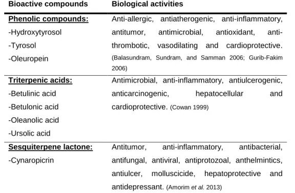
15 The bioactive compounds classes with relevance to this Thesis and respective biological activities are summarized in Table 1.2.
Table 1.2. Bioactive compounds and its biological activities.
Bioactive compounds Biological activities Phenolic compounds:
-Hydroxytyrosol -Tyrosol -Oleuropein
Anti-allergic, antiatherogenic, anti-inflammatory, antitumor, antimicrobial, antioxidant, anti-thrombotic, vasodilating and cardioprotective.
(Balasundram, Sundram, and Samman 2006; Gurib-Fakim 2006) Triterpenic acids: -Betulinic acid -Betulonic acid -Oleanolic acid -Ursolic acid
Antimicrobial, anti-inflammatory, antiulcerogenic, anticarcinogenic, hepatocellular and cardioprotective. (Cowan 1999)
Sesquiterpene lactone:
-Cynaropicrin
Antitumor, anti-inflammatory, antibacterial, antifungal, antiviral, antiprotozoal, anthelmintics, antiulcer, molluscicide, hepatoprotective and antidepressant. (Amorim et al. 2013)
1.3 – Biorefinery
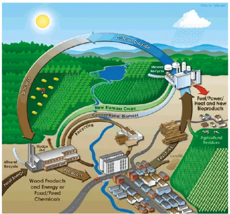
Fossil fuels are used worldwide for energy and chemical production, being oil the most used. It is estimated that 84 million barrels of oil are used per day only in the transport sector, and this number tends to increase. (Cherubini 2010) Moreover, approximately 4% off the refined oil is widely used for chemical and plastic production. (Cherubini 2010) These processes have been burdening the environment, with air, water and soil pollution, causing climate changes, being recognized that an active intervention and paradigm shift is required. (Fernando et al. 2006) Therefore, the search for alternatives to fossil fuels is mandatory but still a huge challenge. The recycling concept is well established in the society, and thus, it can be extended to industries, that while using raw materials of plant origin, generate large amounts of waste (biomass), which can be further reused. (Fernando et al. 2006) In this context, the biorefinery concept has emerged, being defined as the sustainable processing of biomass in a marketable products spectrum and
energy. (Fernando et al. 2006) Thus, a biorefinery should consist in a unit (or several)
incorporating the equipment and technology required for biomass processing considered as waste (wood, grass, corn, etc.) and its basic components (carbohydrates, proteins, triglycerides,
16 etc.) into biofuel, electricity and chemicals, in the concept of exhausting its resources completely (Figure 1.7). (Cherubini 2010)
The "power supply" of biorefineries consist in raw carbon-derived materials, supplied from four main sectors: agriculture (crop and waste fields), forestry industries (waste and leftovers processes), housing (solid waste and municipal wastewater) and aquaculture (algae). (Cherubini 2010)
Figure 1.6. Biorefinery concept. (Herrera 2004)
In the present Thesis three biomasses, and respective extracts, from different sources, were used: 1) forestry – Eucalyptus spp. total bark, from pulp and paper industry; 2) agriculture – olive pomace, from olive oil production and; 3) Cynara cardunculus (cardoon), as an endogenous resource from the Alentejo region, mainly used in artisanal cheese production.
1.3.1 – Eucalyptus spp. and paper production
Eucalyptus spp. is the main raw material used for pulp and paper production in the paper
industry, an economical sector with high economical relevance in the Iberian Peninsula. (Domingues et al. 2010) Eucalyptus urograndis, E. grandis, E. maidenii, E. globulus and E. nitens are the main species (Figure 1.8a). (Domingues et al. 2010)
17
Figure 1.7. Eucalyptus spp. in Portugal: a – Geographic distribution of Eucalyptus spp. in Portugal; b – Relationship between evolution of Eucalyptus spp. area and the Eucalyptus spp. pulp production, reaching 812 000 ha in 2010. (Adapted from: Source CELPA and ICNF, I.P.)
This industry generates large quantities of sub-products, approximately 1.0 x 10⁵ ton/year, such as bark and wood waste (leaves, branches, fruits, etc.), which are usually left in the forest to nourish the soil or are burned for energy. (Domingues et al. 2011; Domingues et al. 2012) It is an industry that is constantly increasing its production levels (Figure 1.8b) and, therefore, the implementation of the biorefinery concept would allow to completely reuse the waste (biomass), as well as to increase the economical potential of the sector.(Domingues et al. 2011)
The main compounds found in this biomass are phytosterols, such as β-sitosterol, lignans and botulin.(Domingues et al. 2010) It is described in the literature that the outer bark of
Eucalyptus spp. is the richer fraction in triterpene compounds, as well as monoterpenes and
sesquiterpenes, small amounts of fatty acids and aromatic compounds. (Domingues et al. 2010) The main TAs present in Eucalyptus spp. are the ursolic, oleanolic, betulinic, betulonic, 3-acetylursolic and 3-acetyloleanolic acids. (Domingues et al. 2010) According to Pereira et al. (2014) studies, E. globulus-derived extracts showed promising results in antimicrobial activity, either isolated or in combination with conventional antibiotics. (Pereira et al. 2014)
18
1.3.2 – Olive and olive oil production
Olive trees (Olea europaea L.) are native from the Mediterranean region, but during the last decade, its cultivation worldwide has increased, due to the recognized benefits of olive oil consumption in human health (Figure 1.9a). (Romero-García et al. 2014) In 2013, 2.67 million tons of olive oil were produced for human consumption in Europe. (Romero-García et al. 2014) In addition to oil, olives are also a product of interest for consumption. (Romero-García et al. 2014) In Portugal, between 2000 and 2009, olive oil production increased approximately 63%, especially in the Alentejo region, representing 56% of the total national production. (Ramos et al. 2013) In 2013, 999 853 hectoliters of olive oil were produced, having 689 261 hectoliters been produced in the Alentejo region (Figure 1.9b). (Instituto Nacional de Estatísitca (INE) 2014)
Figure 1.8.Olive tree in Portugal: a – Geographic distribution of olive trees in Portugal.
(Adapted from: source INE, RA 2009) b – Olive oil production, between 2009 and 2013, reached an approximated average value of 800 000 hl. (Adapted from: source INE, EA 2013)
In Alentejo, olive oil extraction is made through a centrifugation system in two phases, which generates a large amount of a residue named olive pomace, with high water content and composed mainly of skin, pulp and stone pieces of olive fruit. (Ramos et al. 2013) Every year, it is estimated that 400 000 ton of this residue are generated, which is subsequently subjected to drying at high temperatures, resulting in another residue, dry olive pomace that is mainly used for energy production (combustion). (Ramos et al. 2013) Phenolic compounds can be extracted from both residues. (Ramos et al. 2013) According to Ramos et al. (2013), dry olive pomace revealed
19 higher phenolic content than olive pomace, being hydroxytyrosol the more abundant. (Ramos et
al. 2013) Other phenolic compounds were identified in dry olive pomace extract, namely
HT-1-glucoside, tyrosol, oleuropein aglycone isomers, verbascoside, oleuropein and de(carboxymethyl)oleuropein aglycone isomer in aldehyde form. (Ramos et al. 2013)
1.3.3 – Cynara cardunculus – artisanal cheese production and other applications
Cynara cardunculus (cardoon) can be found in the Mediterranean region and its distribution
in Portugal is represented in Figure 1.10.
Figure 1.9. Geographic distribution of Cynara carcunculus in Portugal. (Marabuto et al. 2016)
C. cardunculus is divided in three taxa: two domestic forms, the artichoke (var. scolymus L.)
20 Figure 1.10. Morphological appearance of each variant of Cynara cardunculus: (a) var. sylvestris , (b), var. scolymus L. and (c) var. altilis. (Source: CEBAL)
It has rigorous growth conditions, demanding high temperatures, high salinity and low precipitation. (Falleh et al. 2008) C. cardunculus is associated to a wide variety of applications, such as in the Mediterranean diet in Spain, Italy, France and south of Portugal; in artisanal cheese production and in pulp industry. (Falleh et al. 2008; Velez et al. 2012) Recently, it has shown promising results for biodiesel production. (Velez et al. 2012) The valorization of C. cardunculus is mainly linked to the lignocellulosic fraction and the high value compounds that constitute it. (Ramos et al. 2013) The major compounds have been identified by Ramos et al. (2013), being among them a sesquiterpene lactone (diacylcynaropicrin), four pentacyclic triterpenes (β- and α-amyrin, lupenyl and ψ- taraxasteryl acetate) and four sterols (stigmasterol, 24-methylenecholesterol, campesterol and Δ5-avenasterol). (Ramos et al. 2013) Cynaropicrin and ψ- taraxasteryl acetate are the sesquiterpene lactone and pentacyclic triterpene, respectively, found in higher concentrations. (Ramos et al. 2013) These compounds are responsible for the C.
cardunculus pharmacological effects, which has led to an increase in its cultivation and biological
activity studies. (Velez et al. 2012)
1.4 – Pharmaceutical formulation
1.4.1 – Formulation Design
Drugs are usually included in formulated preparations, varying in complexity according to the type and amount of excipients/additives added. (Aulton 2002) These vehicles provide different characteristics to formulations, depending on the final preparation or dosage form desired, such as solubility, thickness, preservation, emulsion, among others. (Aulton 2002)
There is a large variety of dosage forms where the drug can be incorporated, generally chosen according to the more convenient and best administration route and also in order to obtain maximum effectiveness and, consequently, have the maximum therapeutic response. (Aulton 2002) Various administration routes and associated dosage forms are shown in Table 1.3.
21
Table 1.3. Administration routes and dosage forms associated. (Aulton 2002)
Administration route Dosage forms
Oral Solutions, syrups, suspensions, emulsions,
gels, powders, granules, capsules, tablets
Rectal Suppositories, ointments, creams, powders,
solutions
Topical Ointments, creams, pastes, lotions, gels, solutions, topical aerosols
Parenteral
Injections (solution, suspension, emulsion forms), implants, irrigation and dialysis solutions
Respiratory Aerosols (solution, suspension, emulsion, powder forms) - sprays; foams
Nasal
Solutions, inhalators Eye
Solutions, ointments, creams Ear
Solutions, suspensions, ointments, creams
Among these dosage forms, solutions for topical administration are those with relevance for this Thesis. According to pharmaceutical terms, solutions are liquid preparations that contain one
or more chemical substances dissolved in a suitable solvent or mixture of mutually miscible solvents. (Ansel, Popovich, and Allen 1989) Generally, topical solutions are formulated in an
aqueous vehicle and it is necessary the addition of co-solvents, among other excipients, to enhance both stability and solubility of the drug. (Ansel, Popovich, and Allen 1989) Liquid dosage forms, as topical solutions, are mainly used for local application and present several advantages, such as ease of administration and faster absorption, since the drug is already dissolved and, therefore, it is easily available for skin absorption. However, solutions can also present disadvantages, such as: the lower chemical stability of the formulation components, as they are more susceptible to hydrolysis; as well as the fact that this may be a suitable medium for microorganisms proliferation. (York 2000)
In a technical way, a pharmaceutical formulation can be defined as the set of operations that
aims to create a physical system which contains the active substance in order to meet the specifications of the formulation and ensure the maintenance of efficacy and safety of the active substance (Figure 1.12). (Sousa e Silva 2013)
22 Figure 1.11. Representative scheme of main stages of formulation design.(Sousa e Silva 2013)
This process is performed in order to select, optimize and evaluate the pharmaceutical preparations obtained. (Sousa e Silva 2013) The final product must be stable, efficacious, well-conditioned, attractive, easy to administer, and safe. (Allen 2010)
1.4.2 – Quality control and stability study
The stability of pharmaceutical formulations is defined as the ability of a formulation,
appropriately packed, to maintain the physical, chemical, microbiological, therapeutic and toxicological characteristics in compliance with its specifications. Environmental conditions such
as heat, light and humidity and chemical factors, such as oxidation, reduction, hydrolysis, among others, may play a very important role in stability.
There are official guidelines for quality control that must be followed in order to ensure that pharmaceutical formulations maintain their quality, effectiveness and safety. This quality standard is periodically published in pharmacopeias of major pharmaceutical manufacturing and exporting countries. The U.S. Pharmacopeia, European Pharmacopoeia, International Pharmacopoeia, published by the WHO, and the British Pharmacopoeia are widely used. (WHO 2012)
23 The main characteristics that should be well established and controlled in a formulation are: identity, purity, drug assay, uniformity of dosage form, and stability (Table 1.4). These characteristics can be affected cumulatively through the several steps of the manufacturing process, including the starting materials, errors in manufacturing process, packaging, transportation and storage conditions. (WHO 2012)
Table 1.4. Examples of quality control parameters of a pharmaceutical formulation. (WHO
2012)
Quality Control Description
Identity The identity test should confirm the presence of the active substance.
Purity
The ingredients used should not have potentially harmful contaminants, microorganisms or other products from cross-contamination.
Drug assay
The pharmaceutical formulation should contain the declared amount of the active substance. Most pharmacopoeias specify an average content range of active substance as well as of by-products of degradation that may be harmful.
Uniformity of dosage forms
Consistency, color, shape and size of certain formulation forms should not vary between doses. The lack of uniformity may suggest problems in other quality parameters, and reflect a lack of good manufacturing practices.
Stability
Stability studies allow obtaining information of better conservation conditions and predicting expiration date. They are performed under normal storage conditions, called real-time assays, where physical, chemical and microbiological parameters are evaluated over time.
25