305
Revista da Sociedade Brasileira de Medicina Tropical 45(3):305-308, may-jun, 2012
INTRODUCTION
Article/Artigo
1. Pós-Graduação em Medicina e Saúde Humana, Escola Bahiana de Medicina e Saúde Pública, Salvador, BA. 2. Faculdade de Medicina, Escola Bahiana de Medicina e Saúde Pública, Salvador, BA. 3. Faculdade de Medicina, Universidade Federal da Bahia, Salvador, BA. 4. Laboratório Avançado de Saúde Pública, Centro de Pesquisa Gonçalo Muniz, Fundação Oswaldo Cruz, Salvador, BA.
Address to: Dra. Maria Fernanda Rios-Grassi. Lab. Avançado de Saúde Pública/CPqGM/FIOCRUZ.
Rua Waldemar Falcão 121, Candeal, 40296-710 Salvador, BA, Brasil. Phone: 55 71 3176-2213
e-mail: [email protected]
Received in 22/08/2011
Accepted in 20/12/2011
Evidence of a higher prevalence of HPV infection in HTLV-1-infected
women: a cross-sectional study
Evidência de maior prevalência de infecção pelo HPV em mulheres infectadas pelo HTLV-1:
um estudo de corte transversal
Sônia Sampaio Lôpo
1, Paula Matos Oliveira
1, Iuri Usêda Santana
2, Geisa Barbosa Pena
2, Maria Betânia Torrales
3,
Rita Elizabeth Mascarenhas
2,4, Bernardo Galvão-Castro
1,4and Maria Fernanda Rios Grassi
1,4ABSTACT
Introduction: HTLV-1 infection increases susceptibility to other infections. Few studies have addressed the co-infection between HPV and HTLV-1 and the immune response involved in this interaction. he aim of this study was to determine the prevalence of cervical HPV infection in HTLV-1-infected women and to establish the risk factors involved in this co-infection. Methods: A cross-sectional study was carried out in Salvador, Brazil, between September 2005 and December 2008, involving 50 HTLV-1-infected women from the HTLV Reference Center and 40 uninfected patients from gynecological clinic, both at the Bahiana School of Medicine. HPV infection was assessed using hybrid capture. HTLV-1 proviral load was quantiied using real-time polymerase chain reaction (PCR). Results: he mean age of HTLV-1-infected women (38 ± 10 years) was similar to that of the control group (36 ± 13 years). he prevalence of HPV infection was 44% in the HTLV-1-infected group and 22.5% in uninfected women (p = 0.03). HTLV-1-infected women had lower mean age at onset of sexual life (17 ± 3 years versus 19 ± 3 years; p = 0.03) and greater number of lifetime partners compared with the control group (4 ± 3 versus 2 ± 1; p < 0.01). In the group of HTLV-1-infected patients, there was neither diference in HTLV-1 proviral load between HPV-infected women and the uninfected. Conclusions: he prevalence of HPV infection was higher in HTLV-1-infected women. Further studies should be performed to evaluate the progression of this co-infection. Keywords: HTLV-1. HPV. Proviral load. Brazil.
RESUMO
Introdução: A infecção peloHTLV-1 aumenta a susceptibilidade para outras infecções. Poucos estudos avaliaram a co-infecção entre HPV/HTLV-1 e a resposta imune envolvida nesta interação. O objetivo deste trabalho é determinar a prevalência de infecção cervical pelo HPV em mulheres infectadas pelo HTLV-1 e estabelecer os fatores de risco envolvidos nesta co-infecção. Métodos: Um estudo de corte transversal foi conduzido em Salvador, Brasil, entre setembro de 2005 e dezembro de 2008, envolvendo 50 mulheres infectadas pelo HTLV-1, acompanhadas no Centro de Referência de HTLV e 40 mulheres não infectadas, acompanhadas no Serviço de Ginecologia, ambos na Escola Bahiana de Medicina. A infecção pelo HPV foi conirmada pela Captura Híbrida. A carga proviral do HTLV-1 foi quantiicada pelo PCR em tempo real. Resultados: A média de idade das mulheres infectadas pelo HTLV-1 (38±10 anos) foi semelhante ao do grupo controle (36±13 anos). A prevalência de infecção pelo HPV foi 44% nas mulheres infectadas pelo HTLV-1 e de 22,5% no grupo controle (p=0,03). Mulheres infectadas pelo HTLV-1 informaram menor idade de início de vida sexual (17±3 anos versus
19±3 anos; p=0,03) e maior número de parceiros sexuais, em relação ao grupo controle (4±3
versus 2±1; p<0,01). No grupo de mulheres infectadas pelo HTLV-1, não se observou diferença entre a carga proviral do HTLV-1 entre as mulheres infectadas pelo HPV e as não infectadas. Conclusões: A prevalência de infecção pelo HPV foi maior em mulheres infectadas pelo HTLV-1. Novos estudos devem ser realizados para avaliar a progressão desta co-infecção. Palavras-chaves: HTLV-1. HPV. Carga proviral. Brasil.
Human T-cell lymphotropic virus type-1
(HTLV-1) infects approximately 10-20 million
people around the world
1. In Brazil, the highest
(1.7%) prevalence of infection is in Salvador, a
city with about three million people, located in the
northeast of Brazil. he infection is more frequent
in women, reaching 10% of prevalence in women
above ity years
2. HTLV-1 is the etiologic agent
of adult T-cell leukemia/lymphoma
3,
HTLV-1-associated myelopathy/tropical spastic paraparesis
(HAM/TSP)
4,5, and HTLV-1-associated uveitis
6. In
addition, a higher prevalence of infectious diseases,
such as infective dermatitis
7,8, tuberculosis
9,10,
strongyloidiasis
11-13, and Norwegian scabies
14-16, has
also been described in HTLV-1-infected individuals.
Human papillomavirus (HPV) is the etiologic
agent of one of the most prevalent sexually
transmited diseases in the world and is also directly
related to cervical cancer
17, the third most common
cancer in Brazil. he HPV types 16 and 18 are the
most frequent types found in cervical squamous
cell carcinoma, accounting for more than 70% of
cases
18. A higher frequency of HPV infection and
an increased risk of cervical cancer are found in
immunosuppressed women, as can be observed in
human immunodeficiency virus (HIV)-infected
patients
19-21. he studies that assessed the association
of HPV infection in HTLV-1 patients have not
reached a consensus on the epidemiological aspects
of this co-infection
22-25. he aim of this study was
to determine the prevalence of HPV infection in
HTLV-1-infected women in the City of Salvador,
Bahia, Brazil, and to establish the risk factors
involved in this co-infection.
METHODS
306
RESULTS
Lôpo SS et al - HPV infection in HTLV-1-infected womenTABLE 1 -Sociodemographic characteristics and sexual antecedents (n = 90).
HTLV-1-infected patients Healthy control p
(n = 50) (n = 40) value
Age (years) 37.8 ± 10.4 35.6 ± 13 0.37
Civil status 0.32
long-time relationship 29 (58.0) 19 (47.5)
single/widow 21 (42.0) 21 (52.5)
Smoking 3 (6.0) 5 (12.5) 0.45
Study (years) 7.8 ± 3.6 8.4 ± 2.2 0.38
<8 48.0 47.5
>8 52.0 52.5
Age at irst sexual intercourse (years) 17± 3.4 18.6 ± 3.3 0.03
Numbers of sexual partners 4.1 ± 3.2 1.9 ± 1.1 <0.01
Parity 2.3 ± 2.6 1.6 ± 1.4 0.18
Abortions 1 ± 1.2 0.5 ± 0.7 0.02
HTLV-1: human T-cell lymphotropic virus type-1. Categorical variables were expressed as n (%). Quantitative variables were described as mean and standard deviation.
women. All patients were followed at the Bahia School of Medicine
and Public Health (EBMSP), Salvador, Brazil. HTLV-1-infected
patients were selected from the HTLV Reference Center that has
provided comprehensive care to a total of 1,050 patients since 2002.
Of these, 50% are regularly seen at least twice a year (75% women).
Uninfected controls were selected at the Gynecological Outpatient
Clinic, located on the same campus. Patients were invited and
sequentially included in the study upon their medical visit. Inclusion
criteria were positive serology for HTLV-1 (HTLV-1 group), tested
negative for HTLV-1 (control group), sexually active, and met
criteria for sample collection for Pap smear. Patients with positive
serology for HIV, pregnant women, transplant recipients, patients on
chemotherapy or steroids, or those with diseases that compromise
the immune system were excluded.
he patients answered a questionnaire that assessed demographics,
medical, sexual, and gynecological data. Ater the interview, physical
and gynecological examinations were performed. Specimens for
Papanicolaou smears were collected from ectocervix and endocervix
using an Ayres spatula and cytobrush, respectively. Squamous cell
abnormalities seen in the Papanicolaou smears were classiied as
low-grade or high-low-grade squamous intraepithelial lesions, in accordance
with the Bethesda System
26. he cervical samples were collected using
a sterile swab and stored in universal collection medium (UCM)
solution. he hybridization was performed using the commercial kit
supplied by the Digene HPV (São Paulo, Brazil), according to the
protocol speciied by the manufacturer. he samples were processed
in the laboratory of clinical analysis DNA (Salvador, Bahia).
he populations of CD4+ and CD8+ (cluster of diferentiation)
lymphocytes were analyzed using flow cytometry (FACScan),
ater labeling cells with monoclonal antibodies and CD3-FITC
(luorescein isothiocyanate) CD4-PE (phycoerythrin) and
anti-CD8-PerCP (peridinin chlorophyll protein). he HTLV-1 proviral
load was measured using the technique of real-time polymerase chain
reaction (PCR) ater obtaining mononuclear cells from peripheral
blood by density gradient and DNA extraction by the commercial
kit (QIAGEN, Germany) previously described
27.
Categorical variables were expressed as absolute values and
percentage. Quantitative variables were described as mean and
standard deviation or as median and interquartile interval. he
Chi-square or the Fischer’s exact tests were used to compare categorical
variables between groups. he independent Student’s t-test was used
to compare continuous variables between groups. he association
between HPV infection and the presence of HTLV-1 were veriied,
along with others known risk factors for HPV infection. Unadjusted
odds ratio (OR) were calculated to screen for inclusion in an initial
multivariate model. Variables that exhibited at least a moderate
association with the outcome in the presence of these design variables
were considered for inclusion in the inal models. he analysis was
performed using the SPSS sotware (Statistical Package for the Social
Sciences), version 17.0.
Ethical considerations
he project was approved by the Ethics Research Center from
Gonçalo Muniz
Center,
Fundação Oswaldo Cruz
, Bahia. All patients
read and signed an informed consent before being included in the
study.
he mean age of the HTLV-1-infected group was similar to that
of the control group. HTLV-1-infected patients had a lower age of
irst sexual intercourse (17 years
versus
18.6 years; p = 0.03), a higher
number of sexual partners (4.1
versus
1.9; p < 0.01), and a higher
number of abortions (1
versus
0.5; p = 0.02) compared with the
control group
(Table 1)
.
The HPV prevalence was higher among HTLV-1-infected
women than in the control group (44%
versus
22.5%; p = 0.03)
307
Rev Soc Bras Med Trop 45(3):305-308, may-jun, 2012
TABLE 2 -HPV infection and cytological indings (n = 90).
HTLV-1-infected patients Healthy control p (n = 50) (n = 40) value
HPV infection 22 (44.0) 9 (22.5) 0.03
HPV group
group A 2 (9.1) 0 (0.0) 1.00
group B 12 (54.5) 7 (77.8) 0.41
group A+B 8 (36.4) 2 (22.2) 0.67
Cytological indings 1.00
normal or inlammatory 49 (98.0) 40 (100.0)
HSIL 1 (2.0) 0 (0.0)
Categorical variables were expressed as n (%). Quantitative variables were described as mean and standard deviation; HTLV-1: human T-cell lymphotropic virus type-1; HSIL: High-grade squamous intraepithelial lesion.
TABLE 3 -Adjusted odds ratiofor potential risk factors for HPV infection (n = 90).
Variables Adjusted OR 95% CI p value
HTLV-1 infection 2.7 0.8-8.2 0.07
First sexual intercourse 1.0 0.8-1.2 0.63
Numbers of sexual partners 1.1 0.9-1.4 0.09
Age 0.9 0.8-0.9 0.03
Parity 0.9 0.6-1.2 0.57
HPV: human papillomavirus; HTLV-1: human T-cell lymphotropic virus type;
OR: odds ratio; CI: conidence interval.
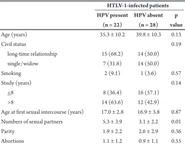
TABLE 4 - Possible risk factors associated with HTLV-1 and HPV co-infection (n = 50).
HTLV-1-infected patients HPV present HPV absent p
(n = 22) (n = 28) value
Age (years) 35.3 ± 10.2 39.8 ± 10.3 0.13
Civil status 0.19
long-time relationship 15 (68.2) 14 (50.0)
single/widow 7 (31.8) 14 (50.0)
Smoking 2 (9.1) 1 (3.6) 0.57
Study (years) 0.14
<8 8 (36.4) 16 (57.1)
>8 14 (63.6) 12 (42.9)
Age at irst sexual intercourse (years) 17.0 ± 2.8 16.9 ± 3.8 0.87 Numbers of sexual partners 5.3 ± 3.9 3.1 ± 2.2 0.01
Parity 1.9 ± 2.2 2.6 ± 2.9 0.36
Abortions 1.1 ± 1.2 0.9 ± 1.1 0.55
HTLV-1: human T-cell lymphotropic virus type; HPV: human papillomavirus. Categorical variables were expressed as n (%). Quantitative variables were described as mean and standard deviation.
infection among HTLV-infected and uninfected groups
when women were stratiied into age ranges (15-25,
26-35, 36-45, 46-55, and 56-65 years). Four positive cases of
HPV infection (18.2%) were observed in HTLV-infected
women with 46-55 years, whereas none was found in the
control group at same age (p = 0.31) (data not shown).
The OR for HPV infection in HTLV-1-infected
patients was 2.7 (95% conidence interval, CI: 0.8-8.2;
p = 0.07) ater adjusting the variables. he OR of age for
HPV infection was 0.9 (95% CI: 0.8-0.9; p = 0.03), and
the number of sexual partners showed an OR of 1.1%
(95% CI: 0.9-1.4, p = 0.09)
(Table 3)
. he
HTLV-1-infected patients with HPV co-infection showed a higher
number of sexual partners compared with the
HTLV-1-infected patients not HTLV-1-infected with HPV (5.3
versus
2.6;
p = 0.01). There were no observed differences regarding other
possible risk factors associated with this co-infection
(Table 4)
.
Regarding immunological features, there was no diference in
the HTLV-1 proviral load in patients with HPV infection compared
with the group without HPV infection (16,000 copies/10
6peripheral
blood mononuclear cell [PBMC]
versus
6,114 copies/10
6PBMC;
p = 0.42). No diference was observed in the proportion of T-CD4+
lymphocytes in the HTLV-1 and HPV-co-infected patients compared
with HTLV-1-infected patients (43.8
±
10.6%
versus
48.9
±
8.6%;
p = 0.1) (data not shown).
DISCUSSION
he present study indicated a higher prevalence of cervical HPV
infection in HTLV-1-infected women. he HTLV-1 infection and the
number of sexual partners appeared to be associated with increased
risk for HPV infection, despite the absence of statistical diference
in this study. Only the age of patients had a signiicant association
with HPV infection: younger patients had an increased risk for HPV
infection. In spite of the higher frequency of cervical HPV infection
in HTLV-1-infected women, there was no occurrence of cervical cancer in these patients.
A few studies have assessed the association between HTLV-1 and cervical HPV infection. In Japan, a cross-sectional study22 observed higher prevalence of HTLV-1 infection in patients with cervical cancer, assigning a threefold higher risk for this malignancy. In Jamaica, a case-control study suggested that HTLV-1 infection was a risk factor for progression of cervical intraepithelial neoplasia23. However, another study performed in that same area did not conirm this association24. Moreover, a study conducted in Mexico failed
to ind a relation between HTLV-1 infection and risk for cervical
cancer
25.
As this is a pilot cross-sectional study, involving a small sample
size, it was not possible to estimate the prevalence of cervical cancer
or premalignant lesions. The absence of cervical cancer in this
population could also be explained by the young age of patients,
although cervical cancer may occur from the third decade of life;
the peak age-speciic cervical cancer is around the sixth decade of
life
28. Also, it was not possible to identify the HPV types involved
in the infections or to determine whether this population has lower
tissue viral clearance.
The HTLV-1 infection induces a spontaneous proliferation
of lymphocyte subsets. As a result, HTLV-1-infected individuals
have an increased population of activated lymphocytes, an
308
ACKNOWLEDGMENTS
he authors declare that there is no conlict of interest.
CONFLICT OF INTEREST
FINANCIAL SUPPORT
REFERENCES
his study indicated that HTLV-1-infected patients are more
susceptible to HPV infection than healthy women and the number of
sexual partners seems to be involved with the risk for HPV infection.
hus, these patients could beneit from gynecological visits at shorter
intervals. Longitudinal studies should be conducted, following a
higher number of women infected with HTLV-1 to conirm the HPV
prevalence and observe the onset of cervical abnormalities.
he authors thank Dr. Raymond Césarie for providing HTLV/
Albumina clones and Vivianna Olavarria for the technical assistance
in the HTLV proviral load measurements.
his study was supported by the
Fundacão de Amparo à Pesquisa
da Bahia
(FAPESB).
1. Dourado I, Alcantara LC, Barreto ML, Teixeira MG, Galvão-Castro B. HTLV-I in the general population of Salvador, Brazil: a city with African ethnic and sociodemographic characteristics. J Acquir Immune Defic Syndr 2003; 34:527-531.
2. Proietti FA, Carneiro-Proietti AB, Catalan-Soares BC, Murphy EL. Global epidemiology of HTLV-I infection and associated diseases. Oncogene 2005; 24:6058-6068.
3. Uchiyama T, Yodoi J, Sagawa K, Takatsuki K, Uchino H. Adult T-cell leukemia: clinical and hematologic features of 16 cases. Blood 1977; 50:481-492. 4. Gessain A, Barin F, Vernant JC, Gout O, Maurs L, Calender A, et al. Antibodies to
human T-lymphotropic virus type-I in patients with tropical spastic paraparesis. Lancet 1985; 2:407-410.
5. Osame M, Usuku K, Izumo S, Ijichi N, Amitani H, Igata A, et al. HTLV-I associated myelopathy, a new clinical entity. Lancet 1986; 1:1031-1032.
6. Rathsam-Pinheiro RH, Boa-Sorte N, Castro-Lima-Vargens C, Pinheiro CA, Castro-Lima H, Galvao-Castro B. Ocular lesions in HTLV-1 infected patients from Salvador, State of Bahia: the city with the highest prevalence of this infection in Brazil. Rev Soc Bras Med Trop 2009; 42:633-637.
7. LaGrenade L, Hanchard B, Fletcher V, Cranston B, Blattner W. Infective dermatitis of Jamaican children: a marker for HTLV-I infection. Lancet 1990; 336:1345-1347.
8. Oliveira MF, Brites C, Ferraz N, Magalhaes P, Almeida F, Bitencourt AL. Infective dermatitis associated with the human T cell lymphotropic virus type I in Salvador, Bahia, Brazil. Clin Infect Dis 2005; 40:e90-96.
9. Marinho J, Galvao-Castro B, Rodrigues LC, Barreto ML. Increased risk of tuberculosis with human T-lymphotropic virus-1 infection: a case-control study. J Acquir Immune Deic Syndr 2005; 40:625-628.
10. Moreira Jr ED, Ribeiro T, Swanson P, Sampaio Filho C, Melo A, Brites C, et al. Seroepidemiology of human T-cell lymphotropic virus type I/II in northeastern Brazil. J Acquir Immune Deic Syndr 1993; 6:959-963.
11. Nakada K, Kohakura M, Komoda H, Hinuma Y. High incidence of HTLV antibody in carriers of Strongyloides stercoralis. Lancet 1984; 1:633. 12. Robinson RD, Lindo JF, Neva FA, Gam AA, Vogel P, Terry SI, et al.
Immuno-epidemiologic studies of Strongyloides stercoralis and human T lymphotropic virus type I infections in Jamaica. J Infect Dis 1994;169:692-696.
13. Sato Y, Shiroma Y, Kiyuna S, Toma H, Kobayashi J. Reduced efficacy of chemotherapy might accumulate concurrent HTLV-1 infection among
strongyloidiasis patients in Okinawa, Japan. Trans R Soc Trop Med Hyg 1994; 88:59.
14. Blas M, Bravo F, Castillo W, Castillo WJ, Ballona R, Navarro P, et al. Norwegian scabies in Peru: the impact of human T cell lymphotropic virus type I infection. Am J Trop Med Hyg 2005; 72:855-857.
15. Brites C, Weyll M, Pedroso C, Badaro R. Severe and Norwegian scabies are strongly associated with retroviral (HIV-1/HTLV-1) infection in Bahia, Brazil. AIDS 2002; 16:1292-1293.
16. Takeshita T, Takeshita H. Crusted (Norwegian) scabies in a patient with smoldering adult T-cell leukemia. J Dermatol 2000; 27:677-679.
17. Almonte M, Albero G, Molano M, Carcamo C, Garcia PJ, Perez G. Risk factors for human papillomavirus exposure and co-factors for cervical cancer in Latin America and the Caribbean. Vaccine 2008; 26 (suppl 11):L16-36.
18. Munoz N, Bosch FX, Sanjose S, Herrero R, Castellsague X, Shah KV, et al. Epidemiologic classiication of human papillomavirus types associated with cervical cancer. N Engl J Med 2003; 348:518-527.
19. Nappi L, Carriero C, Betocchi S, Herrero J, Vimercati A, Putignano G. Cervical squamous intraepithelial lesions of low-grade in HIV-infected women: recurrence, persistence, and progression, in treated and untreated women. Eur J Obstet Gynecol Reprod Biol 2005; 121:226-232.
20. Levi JE, Kleter B, Quint WG, Fink MC, Canto CL, Matsubara R, et al. High prevalence of human papillomavirus (HPV) infections and high frequency of multiple HPV genotypes in human immunodeiciency virus-infected women in Brazil. J Clin Microbiol 2002; 40:3341-3345.
21. Sanjose S, Palefsky J. Cervical and anal HPV infections in HIV positive women and men. Virus Res 2002; 89:201-211.
22. Miyazaki K, Yamaguchi K, Tohya T, Ohba T, Takatsuki K, Okamura H. Human T-cell leukemia virus type I infection as an oncogenic and prognostic risk factor in cervical and vaginal carcinoma. Obstet Gynecol 1991; 77:107-110. 23. Strickler HD, Ratray C, Escofery C, Manns A, Schifman MH, Brown C, et al.
Human T-cell lymphotropic virus type I and severe neoplasia of the cervix in Jamaica. Int J Cancer 1995; 61:23-26.
24. Castle PE, Escofery C, Schachter J, Ratray C, Schifman M, Moncada J, et al. Chlamydia trachomatis, herpes simplex virus 2, and human T-cell lymphotrophic virus type 1 are not associated with grade of cervical neoplasia in Jamaican colposcopy patients. Sex Transm Dis 2003; 30:575-580.
25. Gongora-Biachi A, Gonzalez-Martinez P, Castro-Sansores C, Bastarrachea-Ortiz J. Infection with HTLV virus type I-II in patients with cervico-uterine cancer in the Yucatan peninsula, Mexico. Ginecol Obstet Mex 1997; 65:141-144. 26. Solomon D, Davey D, Kurman R, Moriarty A, O’Connor D, Prey M, et al. he
2001 Bethesda System: terminology for reporting results of cervical cytology. JAMA 2002; 287:2114-2119.
27. Dehee A, Cesaire R, Desire N, Lezin A, Bourdonne O, Bera O, et al. Quantitation of HTLV-I proviral load by a TaqMan real-time PCR assay. J Virol Methods 2002; 102:37-51.
28. Goldie SJ, Grima D, Kohli M, Wright TC, Weinstein M, Franco E. A comprehensive natural history model of HPV infection and cervical cancer to estimate the clinical impact of a prophylactic HPV-16/18 vaccine. Int J Cancer 2003 106:896-904.
29. Rios Grassi MF, Mascarenhas RE, Galvao-Castro B. Immunossupressions of HTLV-1-infected individuals: possible immunological mechanisms. Gazeta Médica da Bahia 2009; 79:56-60.
30. Mascarenhas RE, Brodskyn C, Barbosa G, Clarencio J, Andrade-Filho AS, Figueiroa F, et al. Peripheral blood mononuclear cells from individuals infected with human T-cell lymphotropic virus type 1 have a reduced capacity to respond to recall antigens. Clin Vaccine Immunol 2006; 13:547-552.
31. Alves DB, Tozeti IA, Gato FA, Cassandri F, Ferreira AM, Fernandes CES, et al. CD4 and CD8 T lymphocytes and NK cells in the stroma of the uterine cervix of women infected with human papillomavirus. Rev Soc Bras Med Trop 2010; 43:425-429.
32. Santana IU, Gomes AD, Lyrio LD, Rios Grassi MF, Santiago MB. Systemic lupus erythematosus, human papillomavirus infection, cervical pre-malignant and malignant lesions: a systematic review. Clin Rheumatol 2010; 30:665-672.