Relation between the external branch of the superior laryngeal
Relation between the external branch of the superior laryngeal
Relation between the external branch of the superior laryngeal
Relation between the external branch of the superior laryngeal
Relation between the external branch of the superior laryngeal
nerve and the superior thyroid artery: A study in 101 nerves
nerve and the superior thyroid artery: A study in 101 nerves
nerve and the superior thyroid artery: A study in 101 nerves
nerve and the superior thyroid artery: A study in 101 nerves
nerve and the superior thyroid artery: A study in 101 nerves
Relações do ramo externo do nervo laríngeo superior com a artéria tireóidea
Relações do ramo externo do nervo laríngeo superior com a artéria tireóidea
Relações do ramo externo do nervo laríngeo superior com a artéria tireóidea
Relações do ramo externo do nervo laríngeo superior com a artéria tireóidea
Relações do ramo externo do nervo laríngeo superior com a artéria tireóidea
superior: Estudo em 101 nervos
superior: Estudo em 101 nervos
superior: Estudo em 101 nervos
superior: Estudo em 101 nervos
superior: Estudo em 101 nervos
JOÃO BOSCO BOTELHO, TCBC-AM1; JOSÉ CARDOSO NETO2; GECILDO SORIANODOS ANJOS3; DIEGO MONTEIRODE CARVALHO4; YANNADA SILVA DE MELO5; EMANUEL GOMESDOS SANTOS JÚNIOR5; EDUARDO FERNANDESDA SILVA JÚNIOR5
A B S T R A C T A B S T R A C T A B S T R A C T A B S T R A C T A B S T R A C T
Objective: Objective: Objective: Objective:
Objective: This paper aims to propose a protocol with the topographical relations of the ESLN and the superior thyroid artery in order to minimize the number of flaws in the proposals found in the literature. Methods:Methods:Methods:Methods:Methods: the cervical regions of 57 human cadavers from the Forensic Medicine Institute were dissected bilaterally, with photographic identification of the ESLNs and their relation to the superior thyroid artery. These data were used to propose the following classification: Type 1: unidentified ESLN; Type 2: the ESLN crosses the superior thyroid artery 1.5 cm above the upper pole of the thyroid gland; Type 3: the ESLN crosses the superior thyroid artery between 1 and 1.5cm above the upper pole of the thyroid gland; Type 4: the ESLN crosses the superior thyroid artery less than 1cm above the upper pole of the thyroid gland; Type 5: the ESLN crosses lateral-medially below the upper pole of the thyroid gland. As a supplement, types 2, 3 and 4 received the subclassification: A – the ESLN was partially or totally enclosed in the perimuscular or intramuscular areolar connective tissue, in the inferior pharyngeal constrictor muscle, in more than half of its path after crossing the superior thyroid artery; B – the ESLN was not enclosed in either of these tissues. Results:Results:Results:Results: Type 1, 11.88%; Type 2, 13.86%; TypeResults: 3, 34.65%; Type 4, 38.61%; Type 5, 0.99%. Conclusion:Conclusion:Conclusion:Conclusion:Conclusion: the majority of the ESLN nearest to the upper pole of the gland (types 3 and 4) followed the perimuscular or intramuscular superficial connective tissue in the upper pole of the thyroid gland, which, in this study, disobliges the systematic identification of the nerve in thyroidectomies (p=0.075).
Key words Key words Key words Key words
Key words: Thyroidectomy. Superior laryngeal nerve.
Research study for the Programa de Apoio à Iniciação Científica (Undergraduate Research Support Program), UEA, conducted at the Forensic Medicine Institute (IML) of Amazonas state, ORL-CCF Services of the HAJ/UEA and Hospital Santa Júlia (HSJ) and PPGBIOTEC.
1. Chief, Otorhinolaryngology–Cervicofacial Surgery Service (ORL-CCF), Hospital Adriano Jorge of the Universidade do Estado do Amazonas (HAJ/ UEA), AM, Brazil; 2. Advisor, Graduate Program in Biotechnology (PPGBIOTEC) of the Universidade Federal do Amazonas (UFAM), AM, Brazil; 3. Doctoral student, PPGBIOTEC, UFAM, AM, Brazil; 4. Medical student, grantee of the Fundação de Amparo à Pesquisa do Amazonas, AM, Brazil; 5. Medical student, UEA, AM, Brazil.
INTRODUCTION
INTRODUCTION
INTRODUCTION
INTRODUCTION
INTRODUCTION
T
he surgical diseases of the thyroid gland, especially large goiters (LG), frequent in goitrogenic areas such as the state of Amazonas, Brazil, represent an important publich health issue.Undoubtedly, the size of LGs, many of them weighing over 100 g, could increase the likelihood of intraoperative accidents1-3, even with an experienced surgeon using a broad access such as the U-incision, which affords better technical conditions for the identification of the contiguous anatomical structures.
The superior laryngeal nerve (SLN) is a branch of the vagus nerve (cranial nerve X). The SLN arises from the skull base, follows a descending path as far as the proximity of the horn of the hyoid bone, and divides into two branches: the internal or superior (ISLN) and the external or inferior (ESLN).
The ISLN enters the larynx after perforating the thyroid membrane and is related to the sensory
innervation of the supraglottic region. At that location, the nerve may form anastomoses with the branches of the recurrent laryngeal nerve and thus give rise to Galen’s Anastomosis.
The ESLN travels on the inferior pharyngeal constrictor muscle or pierces it following a cranio-cau-dal path, obliquely, as far as the level where it innervates the cricothyroid muscle. It is a motor nerve, as it keeps the vocal folds under tension. Through anastomoses with the recurrent laryngeal nerve, the ESLN also supplies m o t o r i n n e r v a t i o n t o t h e t h y r o a r y t e n o i d a n d interarytenoid muscles.
Other variations of the ESLN worth mentioning: branches to the thyroid, to the pharyngeal plexus, perforating the cricothyroid membrane and, more rarely, a descending cardiac branch4-8.
Because of the contiguity with the superior thyroid artery, iatrogenic lesions of the ESLN in thyroidectomies are mentioned in the literature9-12. It is reasonable to assu-me that iatrogenic injuries, which may involve the superior parathyroid glands, are influenced by anatomical variations of the nerve in relation to the short vertical segment of the superior thyroid artery before it penetrates the gland, since the superior thyroid vein usually lies more laterally in relation to the cranial border of the thyroid lobe; thus, farther from the nerve13-17.
The aims of this study are (1) to elaborate a surgical protocol of the relations of the ESLN to the supe-rior thyroid artery; (2) contribute to the prevention of iatrogenesis in the ligation of that artery, and (3) contribute data to the classification schemes described in the literature.
METHODS
METHODS
METHODS
METHODS
METHODS
The data were collected from the dissection of the suprahyoid and infrahyoid regions in 57 fresh cadavers, not fixed with formaldehyde, at the IML-AM from December 2007 through September 2008.
Inclusion criteria: Inclusion criteria: Inclusion criteria: Inclusion criteria:
Inclusion criteria: fresh cadavers of either sex, all ages and ethnic groups.
Exclusion criteria: Exclusion criteria: Exclusion criteria: Exclusion criteria:
Exclusion criteria: fresh cadavers with neck traumas associated or not with the causa mortis and those with goiters of any type.
The dissections were conducted with the cadavers in the supine position and the neck extended over a shoulder roll: (1) Median longitudinal incision from the mentum to the jugular notch; (2) Dissection of the visceral aspect of the platysma muscle; (3) Section of the cranial insertions of the sternohyoid and sternothyroid muscles; (4) Lateral retraction of the sternocleidomastoid muscle; (5) Exposure of the superior pole of the thyroid gland and the superior thyroid artery; (6) Identification of the ESLN, dissected cephalad; (7) Photographic record of the relations of the ESLN to the cranial border of the thyroid lobe and the superior thyroid artery, using a tape measure in milimeters.
The data obtained from the dissection of 101 ESLNs, all documented with photographs, comprising a little over 1,000 images, were analyzed statistically based on the inclusion of the anatomical variations in the type categories of the new classification proposed herein:
Type 1: Unidentified nerve.
Type 2: ESLN crosses the superior thyroid artery at a distance greater than 1.5 cm from the cranial border of the thyroid gland lobe;
Type 3: ESLN crosses the superior thyroid artery between 1 cm and 1.5 cm from the cranial border of the thyroid gland lobe.
Type 4: ESLN crosses the superior thyroid artery less than 1cm from the cranial border of the gland lobe.
Type 5: ESLN crosses latero-medially below the cranial border of the thyroid lobe.
Types 2, 3 and 4 ESLNs were subdivided into: intramuscular or A: ESLN partly or totally enclosed in the perimuscular or intramuscular superficial fascia , in the in-ferior pharyngeal constrictor muscle, for over half of its path after crossing the superior thyroid artery ; extramuscular or B: ESLN visible, therefore dissectable, for over half of its path after crossing the superior thyroid artery .
Data analysis was carried out through the MINITAB statistical software, using the paired t-test and the chi-square (x² ) test, as well as descriptive statistical analysis.
Approved by the Research Ethics Committee of the Escola Superior de Ciências da Saúde, UEA, AM, Brazil.
RESULTS
RESULTS
RESULTS
RESULTS
RESULTS
The study comprised 101 relations between the ESLN and the superior thyroid artery in 57 fresh cadavers, not fixed with formaldehyde.
The administrative records of the IML-AM informed ages between 15 and 68 years, with a mean of 32 years. Distribuition by sex was 50 men and 7 women.
The statistical analysis of this sample showed the following: pardo individuals, predominantly male, ages ranging between 21 and 35 years. (figures 1, 2 and 3).
The 101 dissected ESLNs were studied according to the new classification system proposed in the present study (Table 1 and Figures 4, 5, 6).
The distal segment of the ESLN was found enclosed in the areolar connective tissue or in the substance of the inferior pharyngeal constrictor muscle in half of the lobes dissected (Table 1 and Figure 4).
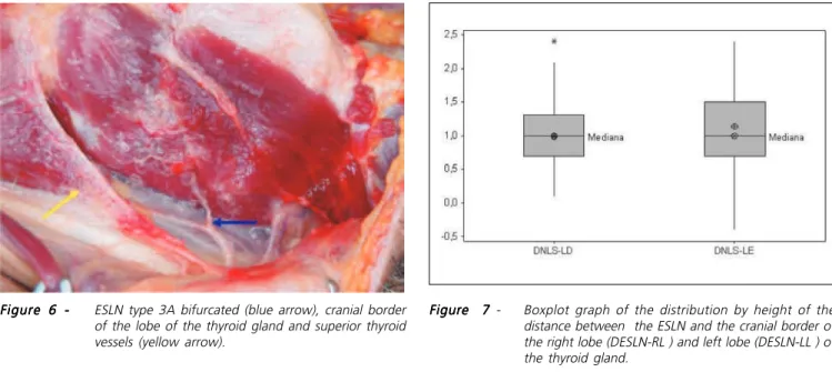
With few cases described in the literature, 3 (2.97%) out of the 101 ESLNs exhibited a distal bifurcation along its descending path (Figure 6).
The evaluation of the distances between the cranial borders of the thyroid lobes and the intersections of the ESLNs with the superior thyroid arteries, both to the right and to the left, resulted in similar maximum and minimum values, and no statistically significant difference was found (p=0.144) in the paired t-test (Table 2). This finding is in agreement with Ozlugedik13 (Figure 7).
Thus, surgical safety is enhanced, and iatrogenic injuries are prevented, if the ligation of the superior thyroid artery is performed with a margin of safety, under direct vision of the short vertical path of the artery, before it enters the thyroid lobe (Figure 8).
DISCUSSION
DISCUSSION
DISCUSSION
DISCUSSION
DISCUSSION
that ratio does not coincide with the prevalence found in clinical practice. On the other hand, stressing the importance of using different samples from those referenced in clinical trials, specifically regarding anatomical variations of the SLN, Furlan10 pointed out that no difference exists between men and women.
The studies published by Kierner et al 18 in 1998, and Furlan et al19 in 2003, regarding the relations of the ESLN to the a superior thyroid artery, corroborated the data obtained by Cernea et al12, 14 in 1992.
Since we could not find similar results to those described by Cernea12, particularly as to the variation in which the ESLN crosses the lobe of the thyroid below the entry of the superior thyroid artery, in 89 partial or total thyroidectomies performed at the Otorhinolaryngology and Cervicofacial Surgery Services of the HAJ/UEA and HSJ in Manaus between 2005-2007, with photographic records of all the ligations of the superior thyroid arteries (Figure 8), the choice was made for an anatomical study of the ESLN in fresh cadavers.
The present study, conducted in 2008, comprising 101 superior laryngeal nerves, did not confirm the results obtained by Cernea et al14, Kierner et al18 and Furlan et al19. In addition, it became evident that it is necessary to add further data to the proposed classification schemes that have been published. For that reason, the new parameters obtained herein culminated in the need for a novel classification that would better define the relations of the ESLN to the superior thyroid artery (Table 1).
Overall, in agreement with the literature, no statistically significant relationship was found between the ESLN variations and the side of the neck, sex or ethnic group10, 19.
The results were analyzed statistically according to the parameters of the new classification system, and showed the following: when the ESLNs are analyzed in relation to the distance from the cranial border of the lobe to the intersection of the nerve with the superior thyroid artery, 87.12% of the ESLNs are types 2, 3 and 4 (Tables 1 and 2 ); as for the presence or absence of an intramuscular path or in the perimuscular areolar connective tissue
(subtypes A and B), especially for those ESLNs closer to the cranial border of the lobe (types 3 and 4), the intramuscular path is evident in 60% of the type-3 ESLNs and in 64.10% of type 4 (Table 1); in the analysis by the chi-square test (x2), the variables distance of the intersection of the nerve with the superior thyroid artery and intramuscular path in
Figure 2 Figure 2 Figure 2
Figure 2 Figure 2 – Frequency by age (n=57). Figure 1
Figure 1 Figure 1
Figure 1 Figure 1 – Sample distribution by skin color (n=57).
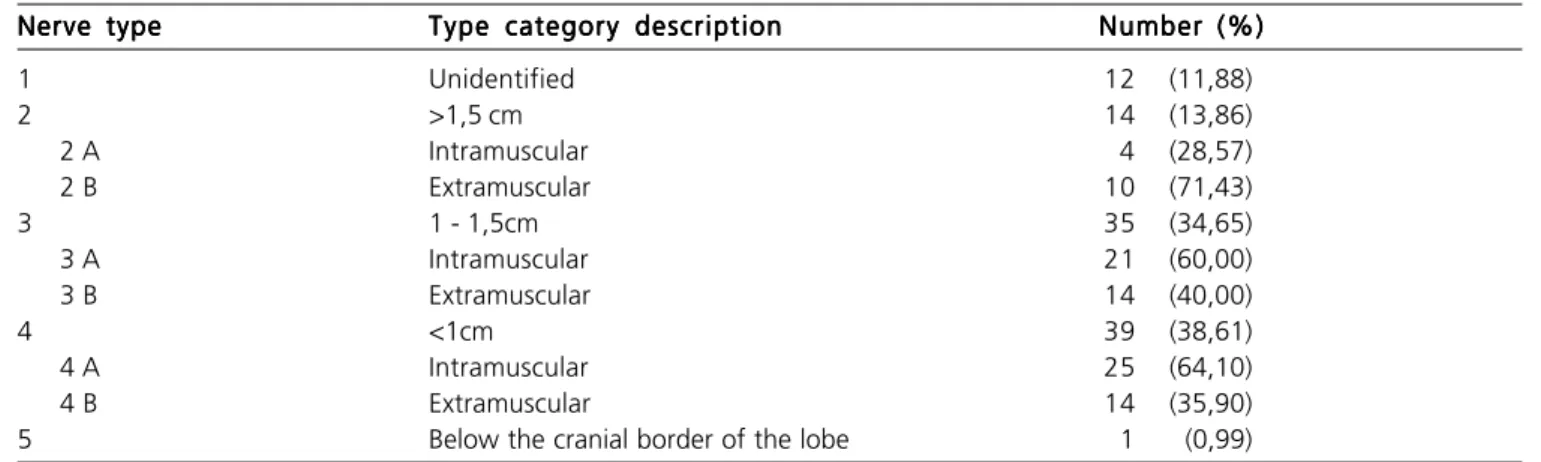
Table 1 – Table 1 –Table 1 –
Table 1 –Table 1 – Results (n=101).
Nerve type Nerve typeNerve type
Nerve typeNerve type Type category descriptionType category descriptionType category descriptionType category descriptionType category description Number (%)Number (%)Number (%)Number (%)Number (%)
1 Unidentified 12 (11,88)
2 >1,5 cm 14 (13,86)
2 A Intramuscular 4 (28,57)
2 B Extramuscular 10 (71,43)
3 1 - 1,5cm 35 (34,65)
3 A Intramuscular 21 (60,00)
3 B Extramuscular 14 (40,00)
4 <1cm 39 (38,61)
4 A Intramuscular 25 (64,10)
4 B Extramuscular 14 (35,90)
5 Below the cranial border of the lobe 1 (0,99)
the inferior pharyngeal constrictor proved to be independent (p=0.075).
the ESLN with the superior thyroid artery , but also the relation of the nerve to other topographical structures, especially the inferior pharyngeal constrictor muscle and its areolar connective tissue, included in the new classification system (table 1).
The divergence from the results of Cernea et al14, Kierner18 and Furlan et al19 refers to the high likelihood of the ESLN crossing the thyroid gland below the cranial border of the lobe. Our results, which identified one case (0.99%) of ESLN at that location, are statistically comparable to those obtained by Naidoo20, who does not describe the nerve in that position at all. Thus, the results much differ from those published by Cernea14 in 1992, who found six (20%) in 30 dissections; by Kierner18 in 1998, who identified 14 (28%) in 62 lobes, and Furlan19 , who in 2003 identified, out of 72 dissections, 16 (22%) ESLNs crossing the gland below the cranial border of the lobe.
Therefore, based on those authors12,14,18,19, supported by the results obtained by them, the previous identification of the ESLN is recommended before the ligation of the superior thyroid artery in thyroidectomies, in order to prevent iatrogenic injuries.
However, because the differences between the findings by Cernea12,14, Furlan and Kierner19and the data obtained in the present study, as well as in Naidoo’s20, are statistically significant and do matter in the context of a specific surgical decision – whether or not to identify the ESLN prior to the ligation of the su-perior thyroid artery –, it is necessary to take them into careful consideration.
Based on the findings of the present study, there is no indication for mandatory systematic identification of the ESLN over the course of partial or total thyroidectomies. This position becomes even more sustainable if the ligation of the superior thyroid artery is performed under direct vision of the segment before it penetrates the gland.
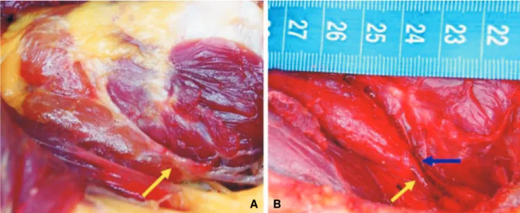
Figure 5 - Figure 5 - Figure 5 - Figure 5 -
Figure 5 - ESLN (blue arrow) and cranial border of the lobe of the thyroid gland (yellow arrow). A:A:A:A: type 1 (unidentified ESLN); B: A: B: B: B: B: type 5... Figure 4
-Figure 4 -Figure 4 Figure 4
-Figure 4 - ESLN (blue arrow) and cranial border of the lobe of the thyroid gland (yellow arrow). A:A:A:A:A: type 2A; B: B: B: B: B: type 2B;;;;; C:
C: C: C:
C: type 3A; D: ; D: ; D: ; D: type 3B; E: ; D: E: E: E: E: type 4A, and F: F: F: F: F: type 4B. Figure 3
Figure 3 Figure 3 Figure 3
Figure 3 – Frequency by sex (n=57).
Figure 7 Figure 7Figure 7
Figure 7Figure 7 - Boxplot graph of the distribution by height of the distance between the ESLN and the cranial border of the right lobe (DESLN-RL ) and left lobe (DESLN-LL ) of the thyroid gland.
Figure 6 -Figure 6 -Figure 6
-Figure 6 -Figure 6 - ESLN type 3A bifurcated (blue arrow), cranial border of the lobe of the thyroid gland and superior thyroid vessels (yellow arrow).
Table 2 -Table 2 -Table 2
-Table 2 -Table 2 - Descriptive statistics for the identified ESLNs (n=89).
V a r i a b l e V a r i a b l eV a r i a b l e
V a r i a b l eV a r i a b l e NNNNN Mean (cm)Mean (cm)Mean (cm)Mean (cm)Mean (cm) Standard Deviation Minimum (cm)Standard DeviationStandard DeviationStandard DeviationStandard Deviation Minimum (cm)Minimum (cm)Minimum (cm)Minimum (cm) Median (cm)Median (cm)Median (cm)Median (cm)Median (cm) Máximum (cm)Máximum (cm)Máximum (cm)Máximum (cm)Máximum (cm)
DNLSE-LD* 42 0,98 0,47 0,10 1,00 2,40
DNLSE-LE** 47 1,14 0,60 -0,40 1,00 2,40
*DESLN-RL – Distance separating the ESLN from the cranial border of the right lobe of the thyroid . **DESLN-LL – Distance separating the ESLN from the cranial border of the left lobe of the thyroid.
R E S U M O R E S U M O R E S U M O R E S U M O R E S U M O
Objetivo Objetivo Objetivo Objetivo
Objetivo: Este trabalho objetivou construir um protocolo das relações anatômicas topográficas do ramo externo do nerve laríngeo superior (SLNE) com a artéria tireóidea superior, para minimizar as falhas das propostas descritas na literatura. Métodos: Métodos: Métodos: Métodos: ForamMétodos: dissecadas bilateralmente as regiões cervicais de 57 cadáveres frescos, no Instituto Médico-Legal do Estado do Amazonas, com identificação fotográfica dos SLNE e respectivas relações com a artéria tireóidea superior. A partir dos dados obtidos elaborou-se classificação: Tipo1: SLNE não identificado; Tipo 2: SLNE cruza a artéria tireóidea superior a distância maior de 1,5cm do limite cranial do lobe da glândula tireóide; Tipo 3: SLNE cruza a artéria entre 1cm e 1,5cm do limite cranial do lobe da glândula tireóide; Tipo 4: SLNE cruza a artéria a menos de 1cm do limite cranial do lobe da glândula; Tipo 5: SLNE cruza, látero-medialmente, abaixo do limite cranial do lobe tireóideo. Como suplemento classificatório, os tipos 2, 3 e 4 receberam subclassificação: A - quando se encontrava parcial ou totalmente envolto no tecido conjuntivo superficial peri-muscular ou intra-muscular, no constritor inferior da faringe, em mais da metade de seu percurso, após cruzar a artéria tireóide superior; B – quando se encontrava fora destes tecidos. Resulta-Resulta-Resulta-Resulta- Resulta-dos:
dos: dos: dos:
dos: Tipo 1, 11,88%; Tipo 2, 13,86%; Tipo 3, 34,65%; Tipo 4, 38,61% e Tipo 5, 0,99%. ConclusãoConclusãoConclusãoConclusão: Os SLNE mais próximos aoConclusão limite cranial (Tipos 3 e 4) percorriam, em sua maioria, o tecido frouxo peri-muscular ou intramuscular, no pólo superior da tireóide, o que neste estudo, desobrigam identificação sistemática do nerve nas tireoidectomias (p=0,075).
Descritores: Descritores: Descritores: Descritores:
Descritores: Tireoidectomia. Nerve laríngeo superior.
REFERENCES
REFERENCES
REFERENCES
REFERENCES
REFERENCES
1. Botelho JB. Patologia da glândula tireóide. In: Botelho JB. Otorrinolaringologia e cirurgia de cabeça e pescoço para estudan-tes. Manaus: Universidade Federal do Amazonas; 2000. p. 325-98. 2. Botelho JBL. Pathologie de la glande thyroïde. In: Botelho JBL, Gehanno P. Otorhinolaryngologie et chirurgie cervico-faciale à l‘usage dês étudiants. Paris: Edk; 2002. p. 86-9.
3. Botelho JB. Incisão em U: nova via de acesso aos bócios de grande volume. Prêmio e Medalha Franz Escher. Centenário de Kocher. 94º Congresso Suíço de Otorrinolaringologia e Cirurgia Cervico-facial. Berna. 2007.
4. Morton RP, Whitfield P, Al-Ali S. Anatomical and surgical considerations of the external branch of the superior laryngeal nerve: a systematic review. Clin Otolaryngol. 2006; 31(5):368-74. 5. Tiago RSL, Munhoz MSL, Faria FP, Guilherme A. Aspectos histomorfométricos do nerve laríngeo superior. Rev Bras Otorrinolaringol. 2002; 68(2):157-65.
6. Testut L, Jacob O. Tratado de anatomia topográfica. Barcelona: Salvat. 1952. p.709.
7. Testut L, Latarjet A. Tratado de anatomia humana. Barcelona: Salvat.1959. p.1052-60.
9. Olthoff A, Schiel R, Kruse E. The supraglottic nerve supply: an anatomic study with clinical implications. Laryngoscope. 2007; 117(11):1930-3.
10. Furlan JC, Cordeiro AC, Brandão LG. Study of some “Intrinsic risk factors” that can enhance a iatrogenic injury of the external branch of the superior laryngeal nerve. Otolaryngol Head Neck Surg. 2003; 128(3):396-400.
11. Aina EM, Hisham AN. External laryngeal nerve in thyroid surgery: is the nerve stimulator necessary?. Eur J Surg. 2001; 167(9):662-5. 12. Cernea CR, Ferraz AR, Furlani J, Monteiro S, Nishio S, Hojaij FC et al. Identification of the external branch of the superior laryngeal nerve during thyroidectomy. Am J Surg. 1992; 164(6):634-9. 13. Ozlugedik S, Acar HI, Apaydin N, Tekdemir I, Elhan A, Comert A.
Surgical anatomy of the external branch of the superior laryngeal nerve. Clin Anat. 2007; 20(4):387-91.
14. Cernea CR, Ferraz AR, Nishio S, Dutra A Jr, Hojaij FC, dos Santos LR. Surgical anatomy of the external branch of the superior laryngeal nerve. Head Neck.1992; 14(5):380-3.
15. Friedman M, LoSavio P, Ibrahim H. Superior laryngeal nerve identification and preservation in thyroidectomy. Arch Otolaryngol Head Neck Surg. 2002; 128(3):296-303.
16. Botelho JB, Anjos GS, Gomes Filho JM, Pires GP, Ferreira DM et al. Relações anatômicas das glândulas paratireóides cervicais com a tireóide: estudo em 53 tireoidectomias. Rev Col Bras Cir. [periódico na internet] 2008; 35(2). Disponivel em URL: http://www.scielo.br/ rcbc
17. Botelho JB, Cançado ARS, Souza EA. Importância anatomocirúrgica das características macroscópicas, localização e suprimento vascular das glândulas paratireóides cervicais. Rev Col Bras Cir. 2004; 31(2):132-8.
18. Kierner AC, Aigner M, Burian M. The external branch of the supe-rior laryngeal nerve: its topographical anatomy as related to surgery of the neck. Arch Otolaryngol Head Neck Surg. 1998; 124(3)301-3.
19. Furlan JC, Brandão LG, Ferraz AR, Rodrigues AJ. JR. Surgical anatomy of the extralaryngeal aspect of the superior laryngeal nerve. Otolaryngol Head Neck Surg. 2003; 129(1):79-82. 20. Naidoo D, Boon JM, Mieny CJ, Becker PJ, van Schoor AN. Relation
of the external branch of the superior laryngeal nerve to the superior pole of the thyroid gland: an anatomical study. Clin Anat. 2007; 20(5):516-20.
Received in 07/10/2008
Accepted for publication in 12/12/2008 Conflict of interest: none
Financial source: FAPEAM
How to cite: How to cite: How to cite: How to cite: How to cite:
Botelho JB, Cardoso Neto J, Anjos CS, Carvalho DM, Melo YS, Santos Júnior EG, Silva Júnior EF. Relations of the external branch of the superior laryngeal nerve to the superior thyroid artery: a study of 101 nerves. Rev Col Bras Cir. [periódico na Internet] 2009; 36(2). Disponível em URL: http://www.scielo.br/rcbc
Correspondence address: Correspondence address: Correspondence address: Correspondence address: Correspondence address: João Bosco Lopes Botelho