Rev Bras Med Esporte _ Vol. 13, Nº 1 – Jan/Fev, 2007
1e
1. Graduanda em Fisioterapia pela FMRP-USP.2. Fisioterapeuta. Mestre em Ortopedia, Traumatologia e Reabilitação do Aparelho Locomotor FMRP-USP.
3. Fisioterapeuta. Professora Doutora do Departamento de Biomecânica, Medicina e Reabilitação do Aparelho Locomotor.
4. Médico. Professor Doutor do Departamento de Biomecânica, Medicina e Reabilitação do Aparelho Locomotor.
Received in 4 / 7 / 05. Final version received in 22 / 9 / 05. Approved in 22 / 5 / 06.
Correspondence to: Débora Bevilaqua-Grossi, Faculdade de Medicina de Ribeirão Preto-USP, Campus Universitário – 14049-900 – Ribeirão Preto, SP – Brasil. Tel./fax: (16) 602-4413 /633-0336. E-mail: [email protected]
Analysis of the medium frequency of the electromyographic
signal of individuals with lesion of the anterior cruciate ligament
in isometric exercises of open and closed kinetic chain
Letícia Maciel Pizzato1, Juliano Coelho Arakaki2, Rodrigo Antunes Vasconcelos2, Guilherme
de Carvalho Sposito1, Anamaria Siriani de Oliveira3, Cleber J. Paccola4 and Débora Bevilaqua Grossi3
O
RIGINALA
RTICLEKeywords: Knee. Electromyography. Rehabilitation. ENGLISH VERSION
ABSTRACT
Subjects with injury of the anterior cruciate ligament (ACL) have shown relevant functional alterations in the knee muscles. There-fore, it is extremely important to characterize these alterations, as well as to emphasize an efficient rehabilitation protocol for these subjects and consequently return them to physical activities. The purpose of this study was to investigate the medium frequency (Fmed) of the electromyographic signal in ACL subjects with le-sions during isometric exercises in open (OKC) and closed kinetic chain (CKC). Forty subjects (with and without lesion) performed knee extension during maximal voluntary isometric contraction on Leg Extension and Leg Press at 30o, 60o and 90o of knee flexion.
The results showed smaller Fmed values for ACL deficient sub-jects when compared with counter lateral and control groups in OKC exercises (p < 0,05). However, there was not significant dif-ference in CKC exercises between groups (p > 0,05), not showing thus, this kind of injury. Therefore, the Fmed can be considered an efficient tool in the LCA injury characterization. Moreover, CKC ex-ercises seem to be the best alternative for rehabilitation of the ACL deficient subjects.
INTRODUCTION
Lesion of the anterior cruciate ligament (ACL) is one of the most serious and recurrent lesions during physical activities(1). Instability
in the knee joint occurs with ACL injury due to excessive internal rotation and tibial anterior translation, especially at the last exten-sion degrees, leading to limitations in the daily routine and sports activities of these individuals(2-3). The role of the periarticular
mus-cles becomes essential in order to compensate the stability loss, once the knee’s joint stability is dependent on the combination among ligament tension, congruency between the articular surfac-es and contraction of the periarticular musclsurfac-es(3-4).
Several studies have emphasized the important role played by the muscles in the knee joint stability, especially in individuals with ACL lesion, showing thus its role in the joint stability(3,5-7).
The main functional alterations in individuals with ACL lesion are the strength loss and the reduction of the muscle voluntary activity standard, being these alterations more evident in the thigh quadriceps muscle (QC)(8-10). When the ACL is injured, the lesion
affects not only the joint stability but also the neuromuscular per-formance, with consequent weakness of the TQ muscle due to the loss of the mechanoreceptors placed there. This lack of recep-tors suppresses the recruiting of the motor units during voluntary contraction and such blocking of the sensory inference results in the inactivation of the periarticular muscles(8-9,11).
Williams et al.(9), observed through surface electromyography
(SEMG), decrease in the voluntary muscle activation of the TQ muscle in individuals with ACL lesion compared with individuals without lesion during static and dynamic exercises. Later, the same authors verified weakness and significant atrophy of the quadri-ceps muscle in individuals with ACL unilateral lesion comparing the injured and contralateral limbs and failure in the voluntary acti-vation in both groups(10).
The aims of the rehabilitation protocols are the recovery of the muscular strength, reestablishment of the joint mobility, normal-ization of the neuromuscular control and return to sports activities with degrees similar to the ones prior to lesion. These aims are based on continuous knowledge of the functional deficits of the lower limbs in patients with ACL lesion, once the pre-surgery mus-cular strengthening facilitates the early return to sports practice after joint reconstruction surgical procedure(12-13), through the
com-bination of exercises in open (OKC) and closed (CKC) kinetic chains with the purpose to regain muscular strength(14-15).
Michelson et al.(14), observed faster recovery in patients post
ACL reconstruction, combining OKC and CKC, when compared with isolated exercises in CKC. Ross et al.(15), showed that the OKC and
CKC combination may be used in the ACL post-reconstruction re-habilitation without excessive tension on the joint and stress on the patellofemoral joint.
Such exercises have been evaluated through the RMS analysis of the SEMG signal in order to verify the voluntary activation pat-tern of the knee muscles, both in normal individuals and others with ACL lesion(16-19).
Nonetheless, the SEMG presents other tolls that have been lit-tle explored in the literature that analyses individuals with ACL le-sion as medium frequency (Fmed). The Fmed of the electromyo-graphic signal is a variable that divides the power spectrum in two equal regions(20), and can be used for physiological muscular
fa-tigue(21) and type II fiber atrophy detection(22) with consequent
vari-ation in the velocity of conduction in the muscular fiber(23).
Moreover, the Fmed depends on the muscular characteristic, that is, the type of predominant fiber in each muscle. Concerning the VL muscle, a directly proportional relation among the torque increase, contraction intensity and the Fmed seems to occur, find-ing high indices of the Fmed with increase of the contraction in-tensity for a few seconds of exertion in normal individuals(24).
2e
Rev Bras Med Esporte _ Vol. 13, Nº 1 – Jan/Fev, 2007McHugh et al.(21) researching the Fmed in individuals with pre
and post surgical ACL lesion, found a remarkable reduction of the Fmed during isometric contraction in the injured limb compared with the contralateral limb during a fatigue protocol. Even without a fatigue protocol, McNair et al.(22) also found significant reduction
of the Fmed in individuals with ACL lesion during isometric con-traction.
Therefore, the studies revealed that the Fmed is influenced by the ACL lesion and that the rehabilitation protocols are designated to the combination of different types of exercises. Nevertheless, studies analyzing the Fmed have been focusing more the exercis-es performance in OKC. Bexercis-esidexercis-es that, the behavior of the Fmed in CKC is not established in individuals with ACL lesion.
A better understanding of the Fmed behavior can guide us in order to establish the most efficient rehabilitation type.
Thus, the aim of this study was to analyze the Fmed behavior of the electromyographic signal of individuals with ACL lesion in iso-metric exercises of OKC and CKC.
MATERIALS AND METHODS
Sample
40 male volunteers participated in this study (31,1 ± 7,45 years, 174 ± 6,65 cm of height); twenty individuals with unilateral ACL lesion and twenty individuals without ACL lesion. The group with ACL lesion was subdivided in two groups of 20 volunteers each; injured ACL and contralateral to the ACL injured (ACL-C). The group without lesion, control group, was divided as well in two subgroups: dominant control (AD) and non-dominant control (AND).
The volunteers were informed on the aims of the work and signed the clarified formal and free consent form, approved by the Ethics and Human Research Committee of the Clinical Hospital of the Medicine School of Ribeirão Preto of the São Paulo University (HCFMRP-USP). The triage of the volunteers with ACL lesion was performed at the knee infirmary HCFMRP-USP. The lesion was also confirmed through clinical examinations and positive answer of the anterior drawer, pivot shift and Lachman specific tests.
ACL unilateral lesion, healthy contralateral limb and lesion time longer than six months were adopted as inclusion criteria. Volun-teers who presented bilateral ACL lesion, lower limbs fracture, combined joint lesions, pain, joint blocking or previous surgeries were excluded from the study.
Instruments
The OKC Leg extension apparel (Queens®, São Paulo, Brasil)
and the CKC Leg Press apparel (Nakagym®, São Paulo, Brasil), were
used for the isometric effort of the leg extension.
The myoelectric capture was performed through active elec-trodes of differential simple surface (EMG SYSTEM DO BRASIL®,
São José dos Campos, Brasil) consisted of two pure silver parallel rectangular bars of 10 x 2 mm, 10 mm spaced and steady on an acrylic capsule of 20 x 41 x 5 mm. These electrodes presented entrance prevention higher than 10 GΩ, minimum CMRR of 130 dB and gain of 20 times.
The SEMG signal analysis was through an 8 channel- signal con-ditioner module and analogical/digital conversing plaque of 12 bites of resolution of dynamic sample frequency of 1KHz (Myosystem Br-1, PROSECON®, Uberlândia, BRA). This signal conditioner
pre-sents Butterworth analogical filter of second order of low – pass of 500 Hz and high-pass of 20 Hz.
A stainless steel circular reference electrode was placed on the tibial anterior tuberosity of the limb to be tested with gel and adhe-sive tape in order to help in the reduction of the acquisition noise.
Experimental protocol
As a warm-up, the volunteers performed three alternated series of 30 seconds of active muscular stretching for the quadriceps and hamstring muscles, followed by three series of 20 repetitions of submaximal contractions in the OKC apparel with two minute-in-tervals between the series.
Later, the sites for the SEMG electrodes positioning was estab-lished. The SEMG electrodes positioning was performed accord-ing to Bevilaqua-Grossi et al.(28), for the long vast lateral (LVL), thigh
straight (TS), oblique vast medial (OVM) muscles and according to the SENIAM rules(29) for the thigh biceps (TB) and semitendinosus
muscles (ST).
The volunteers were accordingly placed on the leg extension seat with posterior chest support, 100° of hip flexion, 90° of knee flexion, upper limbs parallel to the chest and the limb that was not being tested remaining relaxed or in position comfortable for the volunteer. The volunteers were stuck to the apparel in order to avoid compensatory movements during the leg extension exertion (figure 1). The volunteers were accordingly placed on the leg press seat with upper limbs parallel to the chest for CKC exertion. The limb to be tested was leveled on an imaginary line between the antero superior iliac backbone-lateral femoral condyle-fibular mal-leoli in order to avoid hip rotation, abduction and adduction com-pensatory movements. The foot of the limb to be tested was placed on the resistance platform with tarsi-tibial angle at 90°. The limb that was not being tested remained relaxed or in position comfort-able for the volunteer (figure 2).
The volunteer was instructed to perform the leg extension move-ment during seven seconds of CIVM for the 30°, 60° and 90°
Rev Bras Med Esporte _ Vol. 13, Nº 1 – Jan/Fev, 2007
3e
gles. Three repetitions of the extension CIVM for each analyzed angle were performed and interval of two minutes was given for each volunteer between the contractions in order to avoid the muscular fatigue effects. The angles, apparels and limb to be test-ed order were random.
SEMG Fmed data analysis
The SEMG raw signals were digitally processed, being the 5 final seconds of each CIVM analyzed and filtered with pass-ribbon filter of 20-500 Hz. The Fmed data normalization in each angulation was performed through the CIVM of knee extension at 50° of flex-ion for the centers of the quadriceps muscles, in OKC and CKC as well. Conversely, for the centers of the hamstring muscle, the nor-malization was performed through the knee flexion CIVM at 30° of flexion in OKC.
Statistical analysis
The method of Variance Analysis (ANOVA) with repeated mea-sures and the technique of contrasts formation were used for the comparison between groups in the different types of exercises, OKC and CKC, whenever necessary. It was adopted as significance index p ≤ 0,05.
RESULTS
Significant statistical difference was not observed when com-paring dominant control group and non-dominant control group. Therefore, they will be considered as a single control group (A) (p > 0,05).
During exercises in OKC, the results revealed Fmed indices lower for the ACL group in relation to the other groups. In the exercises in CKC, no statistical difference was observed for the Fmed indi-ces in relation to the evaluated muscles between the different stud-ied groups (p > 0,05) (table 1).
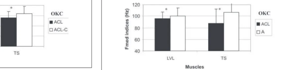
The Fmed indices were significantly lower for the TS (p = 0,02) and LVL (p = 0,03) muscles in the comparison between the ACL and ACL-C groups, (figure 3); while in the comparison between
the ACL and A groups, significant statistical difference was ob-served for the TS (p = 0,05) and TB (p < 0,001) muscles, (figure 4).
DISCUSSION
The results of this study revealed that the Fmed in the injured group presented indices significantly lower when compared with the contralateral group and control group for the VLV and TS mus-cles during the exercises in OKC.
These results may be derived from the changes in the activation of the thigh muscles of individuals with ACL lesion in order to avoid the excessive tibial anterior translation that occurs, especially in the end of the extension in OKC favored by the quadriceps muscle action. In this situation a reciprocal inhibition of this muscle occurs in order to avoid the increase of the tibia shearing in relation to the femur(8-9). Moreover, this quadriceps inhibition may be caused by
the loss of mechanoreceptors placed in the ACL(8-9,11) or type II
fibers atrophy(23). According to these results one may suggests that
the Fmed may be a parameter for the characterization of the ACL lesion, confirming the results by McNair et al.(23) who found
signif-icant decrease of the Fmed in the limbs with ACL lesion when analyzing the Fmed behavior in the VL muscle in individual with ACL lesion, comparing injured limb with contralateral healthy limb. Other studies that proposed to analyze the Fmed behavior also verified significant reduction of the Fmed when performing fatigue-induction protocols(22,26-28). Such decrease was given to the change
in the muscle fibers type due to the protocol. The aim of this study was to evaluate the Fmed behavior in individuals with chronic ADL lesion with no use of a fatigue-induction protocol, and even then, it was observed similar behavior between individuals with and with-out ACL lesion who performed this fatigue protocol. Such results reinforce the hypothesis that individuals with chronic ADL lesion present type II fibers atrophy in the quadriceps muscle.
Works that analyzed the Fmed behavior of the quadriceps and hamstring muscles simultaneously during exercises in OKC and CKC were not found in the literature. The results revealed that, probably as compensatory mechanism, in order to hold the
exces-TABLE 1
Fmed normalized indices of all groups and muscles in open kinetic chain (OKC) and closed kinetic chain (CKC) exercises
OKC CKC
ACL ACL-C A ACL ACL-C A
LVL 099,38 ± 13,30** 106,17 ± 11,77** 101,66 ± 13,15 099,23 ± 12,24 103,16 ± 20,80 098,02 ± 14,41 TS 095,74 ± 12,02** 103,33 ± 14,15** 098,21 ± 13,49 103,06 ± 17,75 110,72 ± 33,44 107,46 ± 20,81 OVM 105,64 ± 22,01** 103,38 ± 21,45** 105,11 ± 19,34 096,84 ± 14,76 092,18 ± 16,68 095,51 ± 17,43 TB 087,43 ± 25,43** 089,59 ± 27,65** 106,50 ± 20,16 084,86 ± 19,88 078,29 ± 19,88 090,13 ± 15,31 ST 080,29 ± 27,64** 087,70 ± 29,49** 087,22 ± 24,87 097,02 ± 27,31 099,32 ± 39,75 094,14 ± 30,12 LVL – long vast lateral, TS – thigh straight, OVM – oblique vast medial, TB – thigh biceps, ST – semi tendinosus.
** p ≤ 0,05 in the comparison between the ACL and ACL-C groups. ACL – injured ACL group, ACL-C – contralateral to ACL lesion group. ** p ≤ 0,05 in the comparison between the ACL and A. ACL groups – injured ACL group, A – control group.
Figure 4 – Fmed normalized indices of the thigh straight (TS) (p = 0,05) and thigh biceps muscles (TB) (p = 0,001) in the ACL and A groups (n = 40) in open kinetic chain (OKC). ACL –injured ACL group, A – control group. * p ≤ 0,05
Figure 3 – Fmed normalized indices of the long vast lateral (LVL) (p = 0,03) and thigh straight (TS) (p = 0,02) muscles in the ACL and ACL-C groups (n = 20) in open kinetic chain (OKC). ACL –injured ACL group, ACL-C –con-tralateral to ACL lesion group.
4e
Rev Bras Med Esporte _ Vol. 13, Nº 1 – Jan/Fev, 2007sive internal rotation and tibia anterior translation, the ACL lesion may trigger a higher activation of the hamstring muscles, especial-ly the TB in OKC, leading to an overload of this muscle due to its constant activation to keep the joint stability. This overload in the chronic lesion could lead to changes in fibers type, triggering hence, type II fiber atrophy and resulting in a decrease of the Fmed. Such explanation can explain the lower indices of the Fmed found in the TB muscle in OKC. Nonetheless, more studies are necessary in order to better understand the Fmed indices behavior of the ham-string muscles in individuals with ACL lesion.
Significant differences were not found in the Fmed indices be-tween the groups during the exercises in CKC. These results sug-gest that the exercise in CKC does not seem to induce a different response in the individual with ACL lesion, that is, the Fmed be-havior of the thigh muscles was not altered in the exercises in CKC, differently from the exercises in OKC, which due to their more selective nature, result in a higher stress for the knee joint(2,30).
The exercises in CKC are mentioned as safer due to the lower stress caused in the ACL, lower shearing force(2,30-31), higher joint
stability and for activating not only the specific muscles of the knee joint but other muscles of the lower limb as well(30,32). Moreover,
exercises in CKC seem to be similar to the functional activities, which are essential to the sports practice and /or recreational activ-ities return(31). The results of this work suggest that the use of
exercises in CKC seems to favor the joint stability in the rehabilita-tion of individuals with ACL lesion, especially in the initial lesions (31-32), minimizing the lesion effects on the muscular fatigue, once Fmed
alteration of the thigh muscles does not occur.
Among the study’s limitations, a relevant variable to be used along with the Fmed would be the muscular strength analysis. Such resource, besides acting as feedback for the volunteers during the CIVM, would offer a better control of the movement from the force exerted by each volunteer, once the velocity of the muscular fiber conduction is influenced by the contraction force. Moreover, al-though not being the aim of our study and being the Fmed an effi-cient tool in the muscular fatigue quantification, a fatigue-induc-tion protocol could have shown evidence on the difference between the groups and exercises.
CONCLUSION
Within the experimental conditions we may conclude that the Fmed presents lower indices for individuals with ACL lesion in OKC for the LVL, TS and TB muscles, suggesting that it is a parameter of the EMG signal able to characterize this kind of lesion.
The Fmed is influenced by the kind of exercises, whether OKC or CKC. The exercises in CKC seem to minimize the ACL lesion effects and can be more efficient in the rehabilitation of these indi-viduals when compared with exercises in OKC.
ACKNOWLEDGMENTS
We thank the Laboratory of Postural Analysis and Human Movement (LAPOMH) of the Medicine School of Ribeirão Preto – USP for the techni-cal support and the CNPq /PIBIC – USP and Fapesp – 2003 /01431-3 re-search support institutions for their financial support.
All the authors declared there is not any potential conflict of inter-ests regarding this article.
REFERENCES
1. Fu FH, Bennet CH, Lattermann C, Ma CB. Current trends in anterior cruciate ligament reconstruction. Part 1: Biology and biomechanics of reconstruction. Am J Sports Med. 1999;27:821-30.
2. Escamilla RF, Fleisig GS, Zheng N, Barrentine SW, Wilk KE, Andrews JR. Biome-chanics of the knee during closed kinetic chain and open kinetic chain exercises. Med Sci Sports Exerc. 1998;30(4):556-69.
3. Kvist J, Gillquist J. Anterior positioning of tibia during motion after anterior cru-ciate ligament injury. Med Sci Sports Exerc. 2001;33(7):1063-72.
4. Czerniecki J, Lippert F, Olerud JE. A biomechanical evaluation of tibiofemoral rotation in anterior cruciate ligament deficient knees during walking and run-ning. Am J Sports Med. 1988;16(4):327-31.
5. Markolf KL, Graff-Radford A, Amstutz HC. In vivo knee stability. A quantitative assessment using an instrumented clinical testing apparatus. J Bone Joint Surg Am. 1978;60:664-74.
6. Williams GN, Chmielewski T, Rudolph KS, Buchanan TS, Snyder-Mackler L. Dy-namic knee stability: current theory and implications for clinicians and scien-tists. J Orthop Sports Phys Ther. 2001;3:546-66.
7. Chmielewski TL, Rudolph KS, Snyder-Mackler L. Development of dynamic knee stability after acute ACL injury. J Electromyogr Kinesiol. 2002;12:267-74. 8. Williams GN, Barrance PJ, Snyder-Mackler L, Axe MJ, Buchanan TS. Specificity
of muscle action after anterior cruciate ligament injury. J Orthop Res. 2003;21(6): 1131-7.
9. Williams GN, Barrance PJ, Snyder-Mackler L, Buchanan TS. Altered quadriceps control in people with anterior ligament deficiency. Med Sci Sports Exerc. 2004; 1089-97.
10. Williams GN, Buchanan TS, Barrance PJ, Axe MJ, Snyder-Mackler L. Quadri-ceps weakness, atrophy, and activation failure in predicted noncopers after an-terior cruciate ligament injury. Am J Sports Med. 2005;33(3):402-7.
11. Konishi Y, Fukubayashi T, Takeshita D. Possible mechanism of quadriceps femo-ris weakness in patients with ruptured anterior cruciate ligament. Med Sci Sports Exerc. 2002;34(9):1414-8.
12. Fitzgerald GK, Axe MJ, Snyder-Mackler L. Proposed practice guidelines for non-operative anterior cruciate ligament rehabilitation of physically active individu-als. J Orthop Sports Phys Ther. 2000;30(4):194-203.
13. Tyler TF, McHugh MP. Neuromuscular rehabilitation of a female Olympic ice hock-ey player following anterior cruciate ligament reconstruction. J Orthop Sports Phys Ther. 2001;31(10):577-87.
14. Mikkelsen C, Werner S, Eriksson E. Closed kinetic chain alone compared to combined open and closed kinetic chain exercises for quadriceps strengthening after anterior cruciate ligament reconstruction with respect to return to sports: a prospective matched follow-up study. Knee Surg Sports Traumatol Arthrosc. 2000; 8(6):337-42.
15. Ross MD, Denegar CR, Winzenried JA. Implementation of open and closed ki-netic chain quadriceps strengthening exercises after anterior cruciate ligament reconstruction. J Strength Cond Res. 2001;15(4):466-73.
16. Beutler AI, Cooper LW, Kirkendall DT, Garrett WE Jr. Electromyographic analysis of single-leg, closed chain exercises: implications for rehabilitation after anterior cruciate ligament reconstruction. J Athl Train. 2002;37(1):13-8.
17. McHugh MP, Tyler TF, Browne MG, Gleim GW, Nicholas SJ. Electromyographic predictors of residual quadriceps muscle weakness after anterior cruciate liga-ment reconstruction. Am J Sports Med. 2002;30(3):334-9.
18. Heller BM, Pincivero DM. The effects of ACL injury on lower extremity activa-tion during closed kinetic chain exercise. J Sports Med Phys Fitness. 2003;43(2):180-8.
19. Kubo K, Tsunoda N, Kanehisa H, Fukunaga T. Activation of agonist and antago-nist muscles at different joint angles during maximal isometric efforts. Eur J Appl Physiol. 2004;91:349-52.
20. Stulen FB, De Luca CJ. Frequency parameters of the myoelectric signal as a mesure of muscle conduction velocity. IEEE Trans Biomed Eng. 1981;28(7):515-23.
21. McHugh MP, Tyler TF, Nicholas SJ, Browne MG, Gleim GW. Electromyographic analysis of quadriceps fatigue after anterior cruciate ligament reconstruction. J Orthop Sports Phys Ther. 2001;31(1):25-32.
22. McNair PJ, Wood GA. Frequency analysis of the EMG from the quadriceps of anterior cruciate ligament deficient individuals. Electromyogr Clin Neurophysiol. 1993;33(1):43-8.
23. Kupa, et al. Effects of muscle fiber type and size on EMG median frequency and condution velocity. J Appl Physiol. 1995;79:23-32.
24. Pincivero DM, Campy RM, Salfetnikov Y, Bright A, Coelho AJ. Influence of con-traction intensity, muscle, and gender on median frequency of the quadriceps femoris. J Appl Physiol. 2001;90(3):804-10.
25. Mannion AF, Dolan P. Relationship between myoelectric and mechanical mani-festations of fatigue in the quadriceps femoris muscle group. Eur J Appl Physiol Occup Physiol. 1996;74(5):411-9.
26. Masuda K, Masuda T, Sadoyama T, Inaki M, Katsuta S. Changes in surface EMG parameters during static and dynamic fatiguing contractions. J Electromyogr Kinesiol. 1999;9(1):39-46.
27. Masuda T, Kizuka T, Zhe JY, Yamada H, Saitou K, Sadoyama T, et al. Influence of contraction force and speed on muscle fiber conduction velocity during dynamic voluntary exercise. J Electromyogr Kinesiol. 2001;11(2):85-94.
28. Bevilaqua Grossi D, Pedro VM, Bérzin F. Análise funcional dos estabilizadores patelares. Acta Ortopédica Brasileira. 2004;12(2):99-104.
29. Hermes HJ, Freriks B, Disslhorst-Klug C, Rau G. European recommendations for surface electromyography – Results of the SENIAM Project. 1999. 30. Yack HJ, Collins CE, Whieldon TJ. Comparison of closed and open kinetic chain
exercise in the anterior cruciate ligament-deficient knee. Am J Sports Med. 1993; 21(1):49-54.
31. Beynnon BD, Johnson RJ, Fleming BC. The science of anterior cruciate liga-ment rehabilitation. Clin Orthop Relat Res. 2002;(402):9-20.