ORIGINAL ARTICLE
Acute and chronic administration of cannabidiol
increases mitochondrial complex and creatine kinase
activity in the rat brain
Samira S. Valvassori,
1Daniela V. Bavaresco,
1Giselli Scaini,
2Roger B. Varela,
1Emilio L. Streck,
2Marcos H. Chagas,
3Jaime E. C. Hallak,
3Antonio W. Zuardi,
3Jose´ A. Crippa,
3Joa˜o Quevedo
11Laboratory of Neurosciences and National Science and Technology Institute for Translational Medicine (INCT-TM), Graduate Program in
Health Sciences, Health Sciences Unit, Universidade do Extremo Sul Catarinense (UNESC), Criciu´ma, SC, Brazil.2Laboratory of
Experimental Pathophysiology and INCT-TM, Graduate Program in Health Sciences, Health Sciences Unit, UNESC, Criciu´ma, SC, Brazil.
3Department of Neurosciences and Behavior, Division of Psychiatry, Ribeira˜o Preto Medical School, Universidade de Sa˜o Paulo (USP), and
INCT-TM, Ribeira˜o Preto, SP, Brazil.
Objective:To investigate the effects of cannabidiol (CBD) on mitochondrial complex and creatine kinase (CK) activity in the rat brain using spectrophotometry.
Method:Male adult Wistar rats were given intraperitoneal injections of vehicle or CBD (15, 30, or 60 mg/kg) in an acute (single dose) or chronic (once daily for 14 consecutive days) regimen. The activities of mitochondrial complexes and CK were measured in the hippocampus, striatum, and prefrontal cortex.
Results: Both acute and chronic injection of CBD increased the activity of the mitochondrial complexes (I, II, II-III, and IV) and CK in the rat brain.
Conclusions:Considering that metabolism impairment is certainly involved in the pathophysiology of mood disorders, the modulation of energy metabolism (e.g., by increased mitochondrial complex and CK activity) by CBD could be an important mechanism implicated in the action of CBD.
Keywords: Cannabidiol; mitochondrial respiratory chain; creatine kinase
Introduction
AlthoughD9-tetrahydrocannabinol (D9-THC) is commonly accepted as the cannabinoid responsible for the main effects ofCannabis sativa, several reports have demon-strated that other components of the plant can influence its pharmacological activity.1One of these components is cannabidiol (CBD), which may constitute up to 40% of cannabis extracts2 and is devoid of the typical psycho-logical effects of cannabis in humans.3
Initially considered to be an inactive cannabinoid, CBD has been reported to have a wide range of pharmacolo-gical effects, many of which can be of great therapeutic interest.4 Around the year 2000, most research on the possible therapeutic effects of CBD was focused on its antiepileptic, sedative, anxiolytic, and antipsychotic effects.5,6In recent years, there has been a remarkable increase in CBD-related research, mainly stimulated by the discovery of its anti-inflammatory and neuroprotective effects. These studies have provided evidence of possible therapeutic effects of CBD in several conditions, including Parkinson’s disease, Alzheimer’s disease,
cerebral ischemia, diabetes, rheumatoid arthritis, other inflammatory diseases, nausea, and cancer.4
Although the mechanisms of action underlying these effects are little known, it has been established that CBD is an antagonist of CB1- and CB2-receptor agonists, an
enhancer of adenosine signaling, has action on the 5-HT1a receptor, and is a potent anti-oxidant.7 Furthermore, a number of studies have suggested mitochondria as targets for cannabinoids.8-10
Mitochondrial abnormalities have been implicated in neuroinflammatory and neurodegenerative conditions and in psychiatric conditions, including schizophrenia, bipolar disorder, and major depression.11,12Mitochondria are the principal source of adenosine triphosphate (ATP), produced through oxidative phosphorylation (OXPHOS). This process is carried out by the respiratory chain complexes I, II, III, and IV, and F1F0-ATP synthase.13 The central nervous system requires a high energy supply, due to its intense ATP-consuming processes. Thus, abnormal cellular energy metabolism may impair neuronal function and plasticity.14
Creatine kinase (CK) is an enzyme that catalyzes the reversible transphosphorylation of creatine by ATP, play-ing a key role in cellular energy bufferplay-ing and energy transport, particularly in cells with high and fluctuating energy requirements, including neurons. The CK system plays a significant role in the brain; a functional impairment
Correspondence: Joa˜o Quevedo, Laborato´rio de Neurocieˆncias, PPGCS, UNASAU, Universidade do Extremo Sul Catarinense, CEP 88806-000, Criciu´ma, SC, Brazil.
E-mail: [email protected]
Submitted May 14 2012, accepted Jan 20 2013. ß2013 Associac¸a˜o Brasileira de Psiquiatria
of this system leads to a deterioration in energy metabo-lism, a characteristic of many mental illnesses.15
The aim of the present study was to investigate the activities of mitochondrial respiratory chain complexes (I, II, II-III, and IV) and CK in the brain of rats after acute and chronic CBD administration.
Methods
Animals
Forty adult male Wistar rats (250-300 g) were kept on a 12-hour light/dark cycle (lights on at 7:00 a.m.) and constant temperature (22616C). They were housed in plastic cages (five per cage) with water and commercial lab chow ad libitum. All experiments were performed in accordance with the guidelines of the National Institute of Health and with the approval of our Institutional Animal Ethics Committee to minimize suffering and the number of animals sacrificed (protocol no. 51/2009).
Treatment
CBD (THC-Pharm, Frankfurt, Germany) was suspended in 2% polysorbate 80 (Tween 80) dissolved in 0.9% saline solution. The solutions were prepared immediately before use and were protected from light during the experimental session.
Three doses of CBD (15, 30, or 60 mg/kg body weight) in a volume of 1 mL/kg were administered intraperitone-ally in two treatments: I) acute, a single injection of CBD was administered (n=5 animals in each group); and II) chronic, CBD was administered once a day for 14 consecutive days (n=5 animals in each group). All control rats for both treatments received injections of 2% polysorbate 80 dissolved in saline (0.9% NaCl) in a volume of 1 mL/kg. The doses of CBD used in this study were based on previous studies.16,17 Different animals were used for acute and chronic experiments. Overall, the sample comprised 40 animals.
Brain preparations
Rats were sacrificed by decapitation 2 hours after CBD administration in the acute treatment and 24 hours after the last CBD administration in the chronic treatment. Brains were removed and transferred within 1 min to ice-cold isolation buffer (0.23 M mannitol, 0.07 M sucrose, 10 mM Tris-HCl, and 1 mM ethylenediaminetetraacetic acid [EDTA], pH 7.4). The prefrontal cortex, hippocampus, striatum, and cerebral cortex (n=5 animals per group) were dissected in ice-cold buffer in a Petri dish.
Activity of mitochondrial respiratory chain enzymes
The prefrontal cortex, hippocampus, striatum, and cere-bral cortex were homogenized (1:10, w/v) in SETH buffer (250 mM sucrose, 2 mM EDTA, 10 mM Trizma base, 50 IU/mL heparin, pH 7.4). The homogenates were centri-fuged at 800gfor 10 min and the supernatants were used to determine the activities of the mitochondrial respiratory
chain enzymes (complexes I, II, II-III, and IV). On the day of the assays, the samples were frozen and thawed thrice in hypotonic assay buffer to fully expose the enzymes to substrates and achieve maximal activities. NADH dehy-drogenase (complex I) was evaluated according to the method described by Cassina & Radi18 by the rate of NADH-dependent ferricyanide reduction at l = 420 nm. The activities of succinate-2,6-dichloroindophenol (DCIP)-oxidoreductase (complex II) and succinate-cyto-chrome c oxidoreductase (complex II-III) were deter-mined by the method described by Fischer et al.19 Complex II activity was measured by the decrease in absorbance due to the reduction of 2,6-DCIP at l = 600 nm. Complex II-III activity was measured by cytochrome c reduction from succinate at l = 550 nm. The activity of cytochrome c oxidase (complex IV) was assayed according to the method described by Rustin et al.,20 measured by the decrease in absorbance due to the oxidation of previously reduced cytochrome c at l = 550 nm. The activities of the mitochondrial respiratory chain complexes were calculated as nmol/min/mg protein.
CK activity assay
CK activity was measured in prefrontal, hippocampus, striatum, and cerebral cortex homogenates pre-treated with 0.625 mM lauryl maltoside. The reaction mixture consisted of 60 mM Tris-HCl, pH 7.5, containing 7 mM phosphocrea-tine, 9 mM MgSO4, and approximately 0.4-1.2mg protein in a final volume of 100mL. After 15 min of pre-incubation at 376C, the reaction was started by the addition of 0.3mmol of adenosine diphosphate (ADP) plus 0.08mmol of reduced glutathione. The reaction was stopped after 10 min by the addition of 1mmol ofp-(hydroxymercuri)benzoic acid. The creatine formed was estimated according to the colorimetric method of Hughes.21 The color was developed by the addition of 100mL 2%a-naphthol and 100mL 0.05% diacetyl in a final volume of 1 mL and analyzed spectrophotome-trically after 20 min at 540 nm. Results were expressed as nmol/min/mg protein.
Statistical analysis
All data are presented as mean6standard error (SEM). All data were considered parametric in accordance with the Shapiro-Wilk test for normality (p , 0.05) and the Levene test for homogeneity of variances among groups (p,0.05). Differences among experimental groups in the open field test and in the assessment of complex I, complex II, complex II-III, complex IV, and CK activity were determined by one-way analysis of variance (ANOVA), followed by Fisher’s post-hoc test when ANOVA was significant. P-values , 0.05 were consid-ered statistically significant.
Results
Complex I
cerebral cortex, whereas the 30 mg/kg dose increased activity of this enzyme only in the prefrontal cortex (Figure 1A). Repeated injections of CBD at doses of 30 and 60 mg/kg increased complex I activity in the prefrontal cortex and in the hippocampus; however, only the 60 mg/kg dose increased complex I activity in the striatum and cerebral cortex (Figure 1B).
Complex II
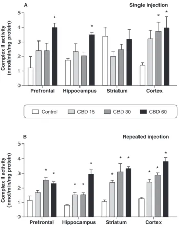
In the rats treated acutely with CBD 60 mg/kg, increased complex II activity was found in the hippocampus, prefrontal cortex, and cerebral cortex; the 30 mg/kg dose was associated with increased complex II activity in the cerebral cortex only (Figure 2A). The chronic administra-tion of CBD increased complex II activity in the hippocampus, striatum, and cerebral cortex at all doses, and in the prefrontal cortex at the higher doses (30 and 60 mg/kg; Figure 2B).
Complex II-III
A single injection of CBD at the dose of 60 mg/kg increased complex II-III activity in the prefrontal cortex, hippocampus, and cerebral cortex (Figure 3A); repeated injections of CBD at higher doses increased complex II-III activity in the prefrontal cortex and cerebral cortex (Figure 3B).
Complex IV
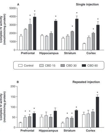
In rats receiving a single injection of CBD, increased complex IV activity was found in the prefrontal cortex, striatum, and cerebral cortex at the doses of 30 and 60 mg/kg, while increased activity of this enzyme in the hippocampus was observed only when CBD was administered at the highest dose (Figure 4A). In rats subjected to repeated injections of CBD, increased Figure 1 Mitochondrial respiratory chain complex I activity
in the prefrontal cortex, hippocampus, striatum, and cerebral cortex (n=5 for each group). Bars represent means; error bars represent standard error of means; * p , 0.05 vs. control group, according to ANOVA followed by Fisher’s least significant difference (LSD) test. A) Animals subjected to a single injection of cannabidiol (CBD) (15, 30, or 60 mg/ kg) or controls (2% polysorbate 80 in 0.9% saline). Rats were sacrificed by decapitation 2 hours after CBD adminis-tration in the acute treatment. B) Animals subjected to repeated injections (once a day for 14 consecutive days) of CBD (15, 30, or 60 mg/kg) or controls (2% polysorbate 80 in 0.9% saline).
complex IV activity was observed in the prefrontal and striatum at all doses. In the cerebral cortex, increased activity of this enzyme was found only when CBD was administered at the highest dose (Figure 4B).
CK activity
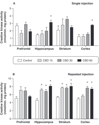
Acute treatment with CBD 60 mg/kg increased CK activity in the hippocampus and cerebral cortex, as compared with vehicle-treated animals. However, CK activity was not altered in the prefrontal cortex or striatum of rats following acute CBD administration (Figure 5A).
After chronic treatment with CBD, there was increased CK activity in the hippocampus and striatum at all doses of CBD administered. Finally, in the cerebral cortex, chronic administration of CBD at 30 and 60 mg/kg increased CK activity as compared with the control group. Conversely, chronic CBD administration did not alter CK activity in the prefrontal cortex of rats (Figure 5B).
Discussion
The precise mechanisms underlying the therapeutic effects of CBD are still unclear. It has been well established that CBD is an antagonist of CB1- and CB2
-receptor agonists.22It is also well known that alterations in brain metabolism are associated with various dis-orders. Energy impairment has been linked to neuronal death and neurodegeneration.23-26
In this work, we evaluated the activities of mitochon-drial respiratory chain complexes I, II, II-III, and IV) and CK in the brains of Wistar rats submitted to acute or chronic treatment of CBD. We observed that both the acute and chronic treatment of CBD increased the activity of mitochondrial enzymatic complexes in the brain of rats. A previous study reported that THC can decrease mitochondrial NADH oxidase activity, which is a non-specific measurement of mitochondrial complex I activ-ity.8 In rat heart mitochondria, the cannabinoid receptor
agonists anandamide, D-9-tetrahydrocannabidiol (THC), Figure 3 Mitochondrial respiratory chain complex II-III
activity in the prefrontal cortex, hippocampus, striatum, and cerebral cortex (n=5 for each group). Bars represent means; error bars represent standard error of means;*p,0.05 vs. control group, according to ANOVA followed by Fisher’s least significant difference (LSD) test. A) Animals subjected to a single injection of cannabidiol (CBD) (15, 30, or 60 mg/ kg) or controls (2% polysorbate 80 in 0.9% saline). Rats were sacrificed by decapitation 2 hours after CBD adminis-tration in the acute treatment. B) Animals subjected to repeated (once a day for 14 consecutive days) injections of CBD (15, 30, or 60 mg/kg) or controls (2% polysorbate 80 in 0.9% saline).
and HU-210 caused significant decreases in oxygen consumption and mitochondrial membrane potential, decreasing mitochondrial complexes I and II-III and inducing cell death.10A recent study has demonstrated that THC and CBD have distinct and often opposing effects on widely distributed neural networks, including the medial temporal lobe, prefrontal cortex, hippocampus, and striatum; these brain regions are rich in cannabinoid receptors and implicated in the pathophysiology of psychosis.27
This was the first study to evaluate the effects of CBD on the activity of mitochondrial respiratory chain com-plexes. CBD has low affinity for both known cannabinoid receptors, CB1 and CB2,22 and appears to have
therapeutic potential, with antiepileptic, anxiolytic, anti-inflammatory, and even antipsychotic effects.5,6 Some
studies show that the neuroprotective effects of CBD are associated with reduced intracellular calcium.3,28-30 It is known that mitochondria plays a fundamental role in cellular Ca2+homeostasis.31Ryan et al.32suggested that
intracellular Ca2+ regulation via CBD is achieved via mitochondrial uptake and release, which could potentially be achieved via the mitochondrial permeability transition pore or the mitochondrial Na+/Ca2+-exchanger.
Ca2+accumulation within mitochondria regulates intrin-sic functions of this organelle, including the main mitochondrial task, which is ATP production by oxidative phosphorylation. Three matrix dehydrogenases are acti-vated by Ca2+. Pyruvate dehydrogenase is regulated by a Ca2+-dependent phosphatase, anda-ketoglutarate
dehy-drogenase and isocitrate dehydehy-drogenase are regulated by direct binding of Ca2+to these enzymes. Stimulation of Ca2+-sensitive dehydrogenases increases NADH avail-ability and enhances the flow of electrons down the respiratory chain, thus increasing ATP synthesis.31 Therefore, we suggest that, in the present study, CBD may have increased Ca2+accumulation within mitochon-dria, increasing NADH accessibility and mitochondrial complex activity.
In the present study, chronic CBD treatment increased mitochondrial complex activity in more brain structures and at more CBD doses than acute treatment, depending on the complex evaluated. We suggest that the difference between the experimental protocols can be a cumulative effect of CBD in the chronic treatment regimen. As can be observed, the results presented herein are not consistent across the different steps of the mitochondrial respiratory chain. This might be explained by the fact that the mitochondrial respiratory chain complexes are different enzymes that respond differently to substances and injuries.33
Complex I is the first step of the respiratory mitochon-drial chain, and the most common site for mitochonmitochon-drial abnormalities. Complex I deficiency is usually found in progressive neurodegenerative disorder.33Complex II is
the second entry point of reducing equivalents into the respiratory mitochondrial chain via FADH, and it is the only complex that pumps protons across the inner mitochondrial membrane. Complex II inhibition causes an excessive generation of reactive oxygen species and, ultimately, cell death.34 Complex III represents a
con-fluence point for reducing equivalents from various dehydrogenases; it can catalyze the transfer of electrons from hydroxyquinones. Complex III deficiency is a severe, multisystem disorder, which is characterized by lactic acidosis, hypotonia, hypoglycemia, failure to thrive, encephalopathy, delayed psychomotor development, and early death.35,36Complex IV belongs to the heme-copper oxygen reductase superfamily, whose members catalyze the complete reduction of dioxygen to water and promote proton translocation across the mitochondrial or periplas-mic membrane, further contributing to the difference in electrochemical potential. Complex IV deficiency can interrupt the oxidative phosphorylation process, decreas-ing energy production for the cells to function properly.33 However, the effects of CBD vary depending on the brain region analyzed. The various regions of the central nervous system may respond differently.23Mitochondrial chain complexes were analyzed from different brain regions that, in part, represent different cell types. Figure 5 Creatine kinase activity in the prefrontal cortex,
Furthermore, in this homogeneous network of neurons, there is heterogeneity in terms of physiological and metabolic characteristics.37 Particularly in the prefrontal cortex and cerebral cortex, CBD always increased the activity of the mitochondrial respiratory chain complexes, in both experimental protocols, mainly at the highest dose. In the hippocampus, CBD administration at the highest tested dose increased the activity of mitochon-drial respiratory chain complexes II, II-III, and IV in acute treatment. Chronic CBD administration increased the activity of complexes I (30 and 60 mg/kg) and complex II activity (15, 30, and 60 mg/kg) in hippocampus. The CB1 receptor is expressed principally in the central nervous system, with the highest concentrations in the prefrontal cortex, anterior cingulate cortex, and hippocampus.38 CBD has modest antagonistic properties on the CB1 receptor. This effect of CBD on CB1 inhibits the reuptake and hydrolysis of anandamide, and exhibits neuroprotec-tive effects.39 A previous study demonstrated that exposure to anandamide increases oxidative phosphor-ylation in brain mitochondria.40 Perhaps the action of CBD on anandamide increases the activity of mitochon-drial complexes in the hippocampus and prefrontal cortex, as observed here. In the striatum, a single administration of CBD at higher doses (30 and 60 mg/ kg) increases the activity of complex IV. After chronic CBD administration, mainly at the higher doses, the activity of complexes I, II, and IV increases in striatum. The CB1 receptor is also densely expressed and functions presynaptically in the lateral striatum, which is directly related to movement.41,42Previous studies have suggested that the striatal actions of CBD may be implicated in its purported antipsychotic effects.27,43
In the present work, we also demonstrated that CK activity is increased by acute or chronic administration of CBD in the rat brain. Our results showed that acute CBD administration increased CK activity in the hippocampus and cerebral cortex at the dose of 60 mg/kg. Moreover, acute CBD administration did not alter CK activity in the prefrontal cortex and striatum. On the other hand, chronic administration of CBD increased CK activity in the hippocampus and striatum at all administered doses. In addition, chronic administration of CBD at high doses (30 and 60 mg/kg) increased CK activity in the cerebral cortex. The chronic administration of CBD did not alter CK activity in the prefrontal cortex.
The present study was the first to evaluate the effects of CBD on CK activity. There are no articles in the literature to help us explain the CBD-induced increase in CK observed herein. In a previous study that investigated the global molecular effects of cannabinoids on normal human astrocytes, using genomic and proteomic ana-lyses, treatment with D9-THC was demonstrated to upregulate brain CK protein.44 The presence of brain CK in Bergmann glial cells and astrocytes is likely related to the energy requirements for ion homeostasis in the brain, as well as for metabolite and neurotransmitter trafficking between glial cells and neurons. CK and its substrates, creatine and phosphocreatine, represent a complex cellular energy buffering and transport system,
linking sites of energy production (mitochondria) with sites of energy consumption.45 Creatine stabilizes mito-chondrial CK and reduces opening of the mitomito-chondrial transition pore.46 In a transgenic animal model of amyotrophic lateral sclerosis, creatine administration protected against increases in oxidative damage in the brain.47It has been widely shown that a decrease in CK activity is associated with a neurodegenerative pathway that results in neuronal loss following brain ischemia,48 neurodegenerative diseases,49 and other pathological
states.50Exogenous creatine supplementation has been shown to reduce neuronal cell loss in experimental paradigms of acute and chronic neurological diseases.51 In line with these findings, preliminary clinical trials have shown beneficial effects of therapeutic creatine supple-mentation.52
In conclusion, CBD administration increased complex I, II, II-III, IV, and CK activity in the brains of rats, mainly at the highest dose of CBD (60 mg/kg) and after a prolonged treatment (14 days), suggesting that one of the possible effects of CBD is modulation of brain energy metabolism.
Acknowledgements
This research was supported by grants from Conselho Nacional de Desenvolvimento Cientı´fico e Tecnolo´gico (CNPq), Fundac¸a˜o de Amparo a` Pesquisa e Inovac¸a˜o do Estado de Santa Catarina (FAPESC), and Fundac¸a˜o de Amparo a` Pesquisa do Estado de Sa˜o Paulo (FAPESP). This study was also sponsored by THC-Pharm (Frankfurt, Germany) and STI Pharmaceuticals Ltd. (Brentwood, UK), who kindly provided the CBD used.
Disclosure
The authors report no conflicts of interest.
References
1 Carlini EA, Santos M, Claussen U, Bieniek D, Korte F. Structure activity relationship of four tetrahydrocannabinols and the pharma-cological activity of five semi-purified extracts of Cannabis sativa. Psychopharmacologia. 1970;18:82-93.
2 Grlic L. A comparative study on some chemical and biological characteristics of various samples of cannabis resin. Bull Narc. 1962;14:37-46.
3 Zuardi AW, Shirakawa I, Finkelfarb E, Karniol IG. Action of cannabidiol on the anxiety and other effects produced by delta 9-THC in normal subjects. Psychopharmacology (Berl). 1982;76:245-50.
4 Zuardi AW. Cannabidiol: from an inactive cannabinoid to a drug with wide spectrum of action. Rev Bras Psiquiatr. 2008;30:271-80. 5 Cunha JM, Carlini EA, Pereira AE, Ramos OL, Pimentel C, Gagliardi
R, et al. Chronic administration of cannabidiol to healthy volunteers and epileptic patients. Pharmacology. 1980;21:175-85.
6 Zuardi AW, Crippa JA, Hallak JE, Moreira FA, Guimaraes FS. Cannabidiol, a Cannabis sativa constituent, as an antipsychotic drug. Braz J Med Biol Res. 2006;39:421-9.
7 Mechoulam R, Peters M, Murillo-Rodriguez E, Hanus LO. Cannabidiol--recent advances. Chem Biodivers. 2007;4:1678-92. 8 Bartova A, Birmingham MK. Effect of delta9-tetrahydrocannabinol on
and cell energetics. Am J Physiol Lung Cell Mol Physiol. 2003;284:L298-306.
10 Athanasiou A, Clarke AB, Turner AE, Kumaran NM, Vakilpour S, Smith PA, et al. Cannabinoid receptor agonists are mitochondrial inhibitors: a unified hypothesis of how cannabinoids modulate mitochondrial function and induce cell death. Biochem Biophys Res Commun. 2007;364:131-7.
11 Kato T, Takahashi S, Shioiri T, Inubushi T. Brain phosphorous metabolism in depressive disorders detected by phosphorus-31 magnetic resonance spectroscopy. J Affect Disord. 1992;26:223-30. 12 Mancuso M, Orsucci D, LoGerfo A, Calsolaro V, Siciliano G. Clinical features and pathogenesis of Alzheimer’s disease: involvement of mitochondria and mitochondrial DNA. Adv Exp Med Biol. 2010;685:34-44.
13 Orth M, Schapira AH. Mitochondria and degenerative disorders. Am J Med Genet. 2001;106:27-36.
14 Mattiasson G, Shamloo M, Gido G, Mathi K, Tomasevic G, Yi S, et al. Uncoupling protein-2 prevents neuronal death and diminishes brain dysfunction after stroke and brain trauma. Nat Med. 2003;9:1062-8. 15 Andres RH, Ducray AD, Schlattner U, Wallimann T, Widmer HR. Functions and effects of creatine in the central nervous system. Brain Res Bull. 2008;76:329-43.
16 Valvassori SS, Elias G, de Souza B, Petronilho F, Dal-Pizzol F, Kapczinski F, et al. Effects of cannabidiol on amphetamine-induced oxidative stress generation in an animal model of mania. J Psychopharmacol. 2011;25:274-80.
17 Jones G, Pertwee RG. A metabolic interaction in vivo between cannabidiol and 1 -tetrahydrocannabinol. Br J Pharmacol. 1972;45:375-7.
18 Cassina A, Radi R. Differential inhibitory action of nitric oxide and peroxynitrite on mitochondrial electron transport. Arch Biochem Biophys. 1996;328:309-16.
19 Fischer JC, Ruitenbeek W, Berden JA, Trijbels JM, Veerkamp JH, Stadhouders AM, et al. Differential investigation of the capacity of succinate oxidation in human skeletal muscle. Clin Chim Acta. 1985;153:23-36.
20 Rustin P, Lebidois J, Chretien D, Bourgeron T, Piechaud JF, Rotig A, et al. Endomyocardial biopsies for early detection of mitochondrial disorders in hypertrophic cardiomyopathies. J Pediatr. 1994;124:224-8.
21 Hughes BP. A method for the estimation of serum creatine kinase and its use in comparing creatine kinase and aldolase activity in normal and pathological sera. Clinica Chimica Acta. 1962;7:597-603. 22 Pertwee RG. Pharmacological and therapeutic targets for D9 tetrahydrocannabinol and cannabidiol. Euphytica. 2004;140:73-82. 23 Sullivan PG, Rabchevsky AG, Waldmeier PC, Springer JE.
Mitochondrial permeability transition in CNS trauma: cause or effect of neuronal cell death? J Neurosci Res. 2005;79:231-9.
24 Volz HR, Riehemann S, Maurer I, Smesny S, Sommer M, Rzanny R, et al. Reduced phosphodiesters and high-energy phosphates in the frontal lobe of schizophrenic patients: a (31)P chemical shift spectroscopic-imaging study. Biol Psychiatry. 2000;47:954-61. 25 Clark JB. N-acetyl aspartate: a marker for neuronal loss or
mitochondrial dysfunction. Dev Neurosci. 1998;20:271-6.
26 Stork C, Renshaw PF. Mitochondrial dysfunction in bipolar disorder: evidence from magnetic resonance spectroscopy research. Mol Psychiatry. 2005;10:900-19.
27 Bhattacharyya S, Atakan Z, Martin-Santos R, Crippa JA, McGuire PK. Neural mechanisms for the cannabinoid modulation of cognition and affect in man: a critical review of neuroimaging studies. Curr Pharm Des. 2012;18:5045-54.
28 Lastres-Becker I, Molina-Holgado F, Ramos JA, Mechoulam R, Fernandez-Ruiz J. Cannabinoids provide neuroprotection against 6-hydroxydopamine toxicity in vivo and in vitro: relevance to Parkinson’s disease. Neurobiol Dis. 2005;19:96-107.
29 Esposito G, De Filippis D, Maiuri MC, De Stefano D, Carnuccio R, Iuvone T. Cannabidiol inhibits inducible nitric oxide synthase protein expression and nitric oxide production in beta-amyloid stimulated PC12 neurons through p38 MAP kinase and NF-kappaB involve-ment. Neurosci Lett. 2006;399:91-5.
30 Iuvone T, Esposito G, Esposito R, Santamaria R, Di Rosa M, Izzo AA. Neuroprotective effect of cannabidiol, a non-psychoactive
component from Cannabis sativa, on beta-amyloid-induced toxicity in PC12 cells. J Neurochem. 2004;89:134-41.
31 Rizzuto R, De Stefani D, Raffaello A, Mammucari C. Mitochondria as sensors and regulators of calcium signalling. Nat Rev Mol Cell Biol. 2012;13:566-78.
32 Ryan D, Drysdale AJ, Lafourcade C, Pertwee RG, Platt B. Cannabidiol targets mitochondria to regulate intracellular Ca2+ levels. J Neurosci. 2009;29:2053-63.
33 Lenaz G, Genova ML. Structure and organization of mitochondrial respiratory complexes: a new understanding of an old subject. Antioxid Redox Signal. 2010;12:961-1008.
34 Grimm S. Respiratory chain complex II as general sensor for apoptosis. Biochim Biophys Acta. 2013;1827:565-72.
35 Fellman V. The GRACILE syndrome, a neonatal lethal metabolic disorder with iron overload. Blood Cells Mol Dis. 2002;29:444-50. 36 V Visapaa I, Fellman V, Vesa J, Dasvarma A, Hutton JL, Kumar V,
et al. GRACILE syndrome, a lethal metabolic disorder with iron overload, is caused by a point mutation in BCS1L. Am J Hum Genet. 2002;71:863-76.
37 Sonnewald U, Hertz L, Schousboe A. Mitochondrial heterogeneity in the brain at the cellular level. J Cereb Blood Flow Metab. 1998;18:231-7.
38 Pertwee RG, Ross RA. Cannabinoid receptors and their ligands. Prostaglandins Leukot Essent Fatty Acids. 2002;66:101-21. 39 Roser P, Vollenweider FX, Kawohl W. Potential antipsychotic
properties of central cannabinoid (CB1) receptor antagonists. World J Biol Psychiatry. 2010;11:208-19.
40 Costa B, Colleoni M. Changes in rat brain energetic metabolism after exposure to anandamide or delta(9)-tetrahydrocannabinol. Eur J Pharmacol. 2000;395:1-7.
41 Herkenham M, Lynn AB, de Costa BR, Richfield EK. Neuronal localization of cannabinoid receptors in the basal ganglia of the rat. Brain Res. 1991;547:267-74.
42 Rodriguez JJ, Mackie K, Pickel VM. Ultrastructural localization of the CB1 cannabinoid receptor in mu-opioid receptor patches of the rat Caudate putamen nucleus. J Neurosci. 2001;21:823-33.
43 Guimaraes VM, Zuardi AW, Del Bel EA, Guimaraes FS. Cannabidiol increases Fos expression in the nucleus accumbens but not in the dorsal striatum. Life Sci. 2004;75:633-8.
44 Bindukumar B, Mahajan SD, Reynolds JL, Hu Z, Sykes DE, Aalinkeel R, et al. Genomic and proteomic analysis of the effects of cannabinoids on normal human astrocytes. Brain Res. 2008;1191:1-11.
45 Hemmer W, Wallimann T. Functional aspects of creatine kinase in brain. Dev Neurosci. 1993;15:249-60.
46 O’Gorman E, Beutner G, Wallimann T, Brdiczka D. Differential effects of creatine depletion on the regulation of enzyme activities and on creatine-stimulated mitochondrial respiration in skeletal muscle, heart, and brain. Biochim Biophys Acta. 1996;1276:161-70. 47 Klivenyi P, Ferrante RJ, Matthews RT, Bogdanov MB, Klein AM, Andreassen OA, et al. Neuroprotective effects of creatine in a transgenic animal model of amyotrophic lateral sclerosis. Nat Med. 1999;5:347-50.
48 Tomimoto H, Yamamoto K, Homburger HA, Yanagihara T. Immunoelectron microscopic investigation of creatine kinase BB-isoenzyme after cerebral ischemia in gerbils. Acta Neuropathol. 1993;86:447-55.
49 David S, Shoemaker M, Haley BE. Abnormal properties of creatine kinase in Alzheimer’s disease brain: correlation of reduced enzyme activity and active site photolabeling with aberrant cytosol-mem-brane partitioning. Brain Res Mol Brain Res. 1998;54:276-87. 50 Streck EL, Amboni G, Scaini G, Di-Pietro PB, Rezin GT, Valvassori
SS, et al. Brain creatine kinase activity in an animal model of mania. Life Sci. 2008;82:424-9.
51 Sestili P, Martinelli C, Bravi G, Piccoli G, Curci R, Battistelli M, et al. Creatine supplementation affords cytoprotection in oxidatively injured cultured mammalian cells via direct antioxidant activity. Free Radic Biol Med. 2006;40:837-49.