09 artigo 532
ORIGINAL ARTICLE
1 – Resident Physician in the Department of Orthopedics and Traumatology, Santa Casa de Misericórdia de São Paulo, São Paulo, SP, Brazil.
2 – Trainee Physician in the Knee Surgery Group, Department of Orthopedics and Traumatology, Santa Casa de Misericórdia de São Paulo, São Paulo, SP, Brazil. 3 – Resident Physician in the Musculoskeletal Radiology Group, Department of Imaging Diagnostics, Santa Casa de Misericórdia de São Paulo, São Paulo, SP, Brazil. 4 – Attending Physician in the Musculoskeletal Radiology Group, Department of Imaging Diagnostics, Santa Casa de Misericórdia de São Paulo, São Paulo, SP, Brazil. 5 – Attending Physician in the Knee Surgery Group, Department of Orthopedics and Traumatology, Santa Casa de Misericórdia de São Paulo, São Paulo, SP, Brazil. 6 – Physician and Head of the Knee Surgery Group, Department of Orthopedics and Traumatology, Santa Casa de Misericórdia de São Paulo, São Paulo, SP, Brazil. Work performed in the Department of Orthopedics and Traumatology (DOT) and the Department of Imaging Diagnostics, Santa Casa de Misericórdia de São Paulo, FCMSCSP. Correspondence: Rua Doutor Cesário Mota Júnior 112, Vila Buarque, 01221-020 São Paulo, SP, Brazil. E-mail: [email protected]
Work received for publication: June 2, 2011; accepted for publication: August 4, 2011.
STUDY ON THE PATELLOFEMORAL jOINT USING MAGNETIC
RESONANCE IMAGING: MORPHOLOGICAL VARIATION
OF THE MEDIAL PATELLOFEMORAL LIGAMENT
Alfredo dos Santos Netto1, Marcelo Botelho Soares de Brito1, Fabrício Roberto Severino2, Leila Rodrigues Andrade Campos3, Marcelo Astolfi Caetano Nico4, Victor Marques de Oliveira5, Nilson Roberto Severino6
AbSTRACT
Objectives: To study the measurements and anatomical rela-tionships of the patellofemoral joint using magnetic resonance imaging, and to evaluate the variation in the morphology of the medial patellofemoral ligament (MPFL) according to patients’ heights and ages and the variation in measurements on other structures that are known to be involved in predispo-sition to patellar instability. Method: Twenty-three knees (18 patients) underwent magnetic resonance imaging and their interepicondylar distance, patellar height, trochlear depth, ventral trochlear prominence, trochlear groove angle, lateral facet tilt, lateral patellar tilt and size of the lateral and medial
INTRODUCTION
Detailed study of the anatomy of the knee is funda-mental for understanding the infirmities that affect it. Among the abnormalities that involve the knee joint, disorders of the extensor apparatus are one of the most frequently encountered problems in orthopedic practice(1).
Instability of the extensor apparatus, represented by recurrent dislocation and subluxation of the patella, con-sists of displacement of the patella, generally laterally, which occurs at some point of the flexion and extension movement of the knee(1,2).
This instability may affect both athletes and non--athletic individuals, causing pain and incapacity. It
in-The authors declare that there was no conflict of interest in conducting this work
This article is available online in Portuguese and English at the websites: www.rbo.org.br and www.scielo.br/rbort
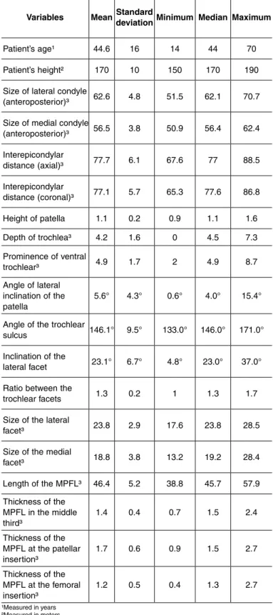
facets and their ratio were measured. These measurements were compared with the length and thickness of the MPFL. Results: The average length of the MPFL was 46.4 mm, while the average thicknesses of its patellar insertion, middle third and femoral insertion were, respectively, 1.7 mm, 1.4 mm and 1.2 mm. The thickness of the MPFL correlated positi-vely with the lateral condyle and interepicondylar distance measurements, and negatively with the patients’ ages. Con-clusion: The morphology of the MPFL varies with the inte-repicondylar distance and the lateral condyle distance, and with patients’ ages.
Keywords – Patella; Patellar Ligament; Joint Instability; Knee
volves great biomechanical complexity and frequently necessitates surgical intervention to treat it(2).
It occurs most frequently in individuals with one or more anatomical abnormalities that predispose to-wards dislocation or subluxation, such as trochlear or patellar dysplasia, high patella, rotational deviations of the lower limbs, muscle dysplasia (especially of the vastus medialis obliquus), retinacular dysplasia and generalized laxity of the ligaments(2-5).
2010 and January 2011.
Patients who had undergone any previous surgi-cal intervention in the knee under examination, and patients with a history of patellar instability, were excluded from the study.
The images were obtained from the Department of Imaging Diagnostics, Santa Casa de Misericórdia de São Paulo, using a 1.5 T magnetic resonance ma-chine (Intera, Philips), a specific eight-channel coil and proton density-weighted sequences in three planes (sagittal, coronal e axial), with and without fat satura-tion, using the following parameters: TE 1642; TE 30; matrix 512×256; FOV, 16×16; and slice thickness 3.5 mm, with intervals of 0.3 mm). The image analyses and all the measurements and correlations necessary were done on workstations with the Agfa PACS/RIS system, by two radiologists who were specialists in radiology of the musculoskeletal system, who analyzed the images together and simultaneously.
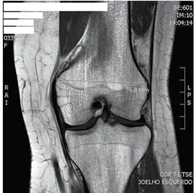
The following measurements were obtained from the MRI: anteroposterior distances along the late-ral and medial femolate-ral condyles, measured on their greatest anteroposterior axes (Figure 1); interepicon-dylar distance, measured on axial and coronal slices (Figures 2 and 3), at the level of the confluence of the insertions of the medial collateral ligament, adductor magnus and MPFL; height of the patella, measured on the joint surface on a sagittal slice (Caton-Des-champs method); depth of the trochlea, measured on an axial slice, 3 cm above the joint surface; ventral trochlear prominence, measured on a sagittal slice; angle of lateral inclination of the patella (angle be-tween the lateral facets of the patella and trochlea), measured on the axial slice, 3 cm above the joint surface; angle of the sulcus of the trochlea, measu-red on the axial slice, 3 cm above the joint surface; inclination of the lateral facet (angle of inclination of the lateral facet in relation to the coronal plane tangential to the posterior region of the femoral con-dyles); measurement of the size of the patellar facets on the axial slice; ratio between the trochlear facets (lateral over medial), measured on the axial slice, 3 cm above the joint surface; Wiberg classification of the patella; and measurements on the MPFL regar-ding its total length, thickness in the middle third, thickness at the patellar insertion and thickness at the femoral insertion (Figure 4).
retinaculum .
Among these structures, it has been accepted that the MPFL is the primary restrictor against lateral pa-tellar displacement(1,6,7-9). It is recognized as the most
important structure in restricting against lateral dis-placement or subluxation of the patella, and tearing of the MPFL is frequently cited as the essential lesion in lateral displacement of the patella. This theory is increasingly accepted, based both on biomechanical and on clinical factors(2,7-9).
Studies have demonstrated that the MPFL is res-ponsible for 50 to 60% of the restriction of lateral displacement of the patella promoted by soft tissues, when the knee is in a position of 0 to 20 degrees of flexion(1,2,7,10).
The MPFL is a very variable structure that is loca-ted in a layer below the vastus medialis obliquus mus-cle. It has insertions at variable levels of the medial
fe-moral epicondyle and medial edge of the patella(6,9,11).
There is insufficient information about how the MPFL relates to other anatomical structures to de-termine whether it would be possible to correlate its morphological alterations with variations in the struc-tures of the knee, such as the shape of the sulcus of the trochlea and whether there is any association between the presence of dysplastic abnormalities (in bones or other ligaments) and the presence of abnormalities of
ligament morphology(6,12).
ObJECTIVE
To study the measurements and anatomical re-lationships of the patellofemoral joint in magnetic resonance examinations, and to assess whether the variations in MPFL morphology have any association with patients’ height and age, and with variations in the measurements of other structures in the knee.
METHODS
Figure 1 – Measurement of the anteroposterior distanced of the lateral condyle.
Figure 2 – Measurement of the interepicondylar distance (axial).
Figure 3 – Measurement of the interepicondylar distance (coronal).
Figure 4 – Measurement of the thickness of the MPFL in its three thirds. The MPFL was characterized as a band of greater focal and continuous thickness in relation to the other portions of the medial patellar retinaculum, presenting a low signal in proton density se-quences, extending from the medial edge of the upper half of the patella to the posterior portion of the medial epicondyle of the femur.
The MPFL was characterized as a band of greater focal and continuous thickness in relation to the other portions of the medial patellar retinaculum. It presen-ted a low signal in the proton density sequences, and extended from the medial margin of the upper half of the patella to the posterior portion of the medial epicondyle of the femur. The course and femoral and patellar insertions of the MPFL were better assessed in axial sequences. The course and insertion of the MPFL were better observed in coronal sequences than in sagittal sequences.
2.6 mm
1.5 mm
1.3 mm
The results were organized by means of tables and graphs, and were then subjected to statistical tests to analyze and validate the results from this study.
To perform the general descriptive analysis, the mean, standard deviation, minimum value, median and maximum value of each of the measurements made were calculated.
measurements made in this study, Pearson’s correlation coefficient was used, except for comparisons with the angle of lateral inclination of the patella, for which Spearman’s comparison coefficient was used.
The significance level used was 5% (p-value ≤ 0.05).
RESULTS
The results revealed that the mean length and thickness of the middle third of the MPFL among the patients studied were, respectively, 46.4 mm and 1.4 mm, while the thickness of the MPFL at its femoral and patellar insertions presented dimensions of 1.2 mm and 1. 7 mm, in this order (Table 1).
In relation to the Wiberg classification, 11 patients (48%) fitted into the morphology of type I, while 12 patients (52%) were placed in the type II morpholo-gical category (Table 2).
No statistically significant relationship was obser-ved between the length of the MPFL and any of the other measurements (Table 3).
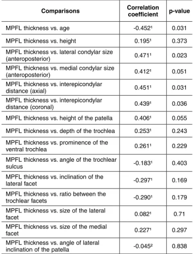
It was observed that the thickness of the MPFL in its middle third had positive relationships with the in-terepicondylar distance in the axial and coronal planes and with the measurement of the anteroposterior size of the lateral condyle; and a negative relationship with the patient’s age (Table 4).
DISCUSSION
The presence of the MPFL was observed in the MRI examinations of the knees of all the patients included in our study, as suggested by the majority of published papers in the literature(7,8,13-15). However,
some studies that involved dissection of a cadaver failed to find this ligament in all specimens because of its small dimensions and the difficulty of dissection. Thus, from these studies, it was suggested that the
MPFL did not exist in some knees(9,16).
The studies evaluating of the anatomy of the MPFL are diverse, but almost all of them use dissection of specimens and manual measurements as the study
method. Among these, Tuxøe et al(15) observed that
the mean length of the MPFL was 53 mm; Aragão et al(9) found a mean length of 55.6 mm; Philippot et al(8)
found a mean length of 57.7 mm; and Nomura et al(7)
dissected 20 knees and found a mean MPFL length of 58.8 mm, which was the same as found by LeGrand
Table 2 – General descriptive analysis for the categorical variable “Wiberg”.
Wiberg Total (%)
I 11 48.00%
II 12 52.00%
Variables MeanStandard
deviationMinimum MedianMaximum
Patient’s age¹ 44.6 16 14 44 70
Patient’s height² 170 10 150 170 190 Size of lateral condyle
(anteroposterior)³ 62.6 4.8 51.5 62.1 70.7 Size of medial condyle
(anteroposterior)³ 56.5 3.8 50.9 56.4 62.4 Interepicondylar
distance (axial)³ 77.7 6.1 67.6 77 88.5 Interepicondylar
distance (coronal)³ 77.1 5.7 65.3 77.6 86.8 Height of patella 1.1 0.2 0.9 1.1 1.6 Depth of trochlea³ 4.2 1.6 0 4.5 7.3 Prominence of ventral
trochlear³ 4.9 1.7 2 4.9 8.7
Angle of lateral inclination of the
patella 5.6° 4.3° 0.6° 4.0° 15.4°
Angle of the trochlear
sulcus 146.1° 9.5° 133.0° 146.0° 171.0° Inclination of the
lateral facet 23.1° 6.7° 4.8° 23.0° 37.0° Ratio between the
trochlear facets 1.3 0.2 1 1.3 1.7
Size of the lateral
facet³ 23.8 2.9 17.6 23.8 28.5
Size of the medial
facet³ 18.8 3.8 13.2 19.2 28.4
Length of the MPFL³ 46.4 5.2 38.8 45.7 57.9 Thickness of the
MPFL in the middle
third³ 1.4 0.4 0.7 1.5 2.4
Thickness of the MPFL at the patellar
insertion³ 1.7 0.6 0.9 1.5 2.7
Thickness of the MPFL at the femoral
insertion³ 1.2 0.5 0.4 1.3 2.7
et al . Higuchi et al analyzed the MPFL by means of MRI examination and found a mean MPFL length of 52 mm, while the mean length of this ligament found in the present study was 46.4 mm (range: 38.8-57.9 mm). This suggests that manual measurements on cadavers (which are less precise) find higher va-lues. This may be due to difficulty in separating out the ligament during the dissection in order to make the measurement.
This suspicion becomes even more sustainable through our assessment of the ligament thickness measurements in the middle third, which would be difficult to achieve with extreme precision with
unaided eyes. Both Nomura et al(7) and LeGrand et al(2)
found a mean thickness of 0.44 mm for the ligament in their studies on cadavers, while the mean thickness of the MPFL in its middle third in the present study was 1.4 mm (range: 0.7-2.4 mm), which suggests that dissection of the ligament in specimens excludes from the measurement a good part of the structure that works towards medially stabilizing the patella.
Unfortunately, Higuchi et al(14) did not make thickness
measurements in their study: such measurements would have provided a better basis for our reasoning. We also made thickness measurements on the MPFL at its femoral and patellar insertions, with values of 1.2 mm (range: 0.4-2.7 mm) and 1.7 mm (range: 0.9-2.7 mm), respectively. These measurements were not made on specimens because of the difficulty in performing dissections to measure the ligament at its insertion sites.
One limitation of our study was the impossibili-ty of evaluating the width of the MPFL using MRI, which has been done in several studies on
anatomi-cal dissection of cadavers(7,9). Because of the oblique
layout and small dimensions, this measurement was impaired on conventional MRI slices. We chose not to produce special slices during the examination so that we would not modify the study protocol, which would have diminished the reproducibility and applicability of this study.
In 1941, Wiberg(17) showed that the shape of the
pa-tella is also important in containing it within the
troch-lear sulcus. Like in the study by Higuchi et al(14), who
analyzed the MPFL by means of MRI examinations on healthy individuals and found that the patella was Wi-berg type I or II in all cases(17), the patellae of all the
patients in the present study were classified as presenting
Table 3 – Relationship between length of the MPFL and each of the variables.
Comparisons Correlation coefficient p-value
MPFL length vs. age -0.012¹ 0.955
MPFL length vs. height 0.373¹ 0.08
MPFL length vs. interepicondylar
distance (axial) 0.267¹ 0.218
MPFL length vs. interepicondylar
distance (coronal) 0.259¹ 0.232
MPFL length vs. height of the patella 0.293¹ 0.174
MPFL length vs. depth of the trochlea 0.014¹ 0.948
MPFL length vs. prominence of the
ventral trochlea 0.400¹ 0.059
MPFL length vs. angle of the trochlear
sulcus -0.150¹ 0.494
MPFL length vs. inclination of the lateral
facet -0.055¹ 0.803
MPFL length vs. ratio between the
trochlear facets 0.181¹ 0.409
MPFL length vs. size of the lateral facet 0.060¹ 0.785
MPFL length vs. size of the medial facet -0.101¹ 0.648
MPFL length vs. angle of lateral
inclination of the patella 0.171² 0.435
Table 4 – Relationship between MPFL thickness in the middle third and each of the variables.
Comparisons Correlation coefficient p-value
MPFL thickness vs. age -0.452¹ 0.031 MPFL thickness vs. height 0.195¹ 0.373 MPFL thickness vs. lateral condylar size
(anteroposterior) 0.471¹ 0.023
MPFL thickness vs. medial condylar size
(anteroposterior) 0.412¹ 0.051
MPFL thickness vs. interepicondylar
distance (axial) 0.451¹ 0.031
MPFL thickness vs. interepicondylar
distance (coronal) 0.439¹ 0.036
MPFL thickness vs. height of the patella 0.406¹ 0.055 MPFL thickness vs. depth of the trochlea 0.253¹ 0.243 MPFL thickness vs. prominence of the
ventral trochlea 0.261¹ 0.229
MPFL thickness vs. angle of the trochlear
sulcus -0.183¹ 0.403
MPFL thickness vs. inclination of the
lateral facet -0.297¹ 0.169
MPFL thickness vs. ratio between the
trochlear facets -0.290¹ 0.179
MPFL thickness vs. size of the lateral
facet 0.082¹ 0.71
MPFL thickness vs. size of the medial
facet 0.227¹ 0.297
MPFL thickness vs. angle of lateral
none of the patients had complaints of instability. A positive association was found between the thi-ckness of the MPFL in its middle third and the inte-repicondylar distance (in the axial and coronal planes) and between MPFL thickness and measurement of the anteroposterior size of the lateral condyle. No studies evaluating these relationships were found in the lite-rature, but it is plausible and to be expected that in knees of larger diameter, all the anatomical structures would have larger dimensions, and not just the MPFL.
No relationship was observed between the length or thickness of the MPFL and the patients’ height. However, a negative association between MPFL thi-ckness in the middle third and age was observed. No studies evaluating these relationships were found in
consequence of the effects of the natural process of aging, reflected in the fibers of the MPFL.
CONCLUSION
The mean length of the MPFL was 46.4 mm, while the mean thickness of the ligament in its middle third was 1.4 mm.
A positive association was found between the thick-ness of the MPFL in its middle third and the interepi-condylar distance (both along the coronal axis and in the axial plane) and between MPFL thickness and the measurement of the anteroposterior size of the lateral condyle; and a negative association between MPFL thickness in the middle third and the patient’s age.
REFERENCES
1. Amis AA. Current concepts on anatomy and biomechanics of patellar stability. Sports Med Arthrosc. 2007;15(2):48-56.
2. LeGrand AB, Greis PE, Dobbs RE, Burks RT. MPFL reconstruction. Sports Med Arthrosc Rev. 2007;15(2):72-7.
3. Salzmann GM, Weber TS, Spang JT, Imhoff AB, Schöttle PB. Comparison of native axial radiographs with axial MR imaging for determination of the trochlear morphology in patients with trochlear dysplasia. Arch Orthop Trauma Surg. 2010;130(3):335-40.
4. Pfirrmann CW, Zanetti M, Romero J, Hodler J. Femoral trochlear dysplasia: MR findings. Radiology. 2000;216(3):858-64.
5. Biedert RM, Bachmann M. Anterior-posterior trochlear measurements of normal and dysplastic trochlea by axial magnetic resonance imaging. Knee Surg Sports Traumatol Arthrosc. 2009;17(10):1225-30.
6. Amis AA, Firer P, Mountney J, Senavongse W, Thomas NP. Anatomy and bio-mechanics of the medial patellofemoral ligament. Knee. 2003;10(3):215-20.
7. Nomura E, Inoue M, Osada N. Anatomical analysis of the medial patellofemoral ligament of the knee, especially the femoral attachment. Knee Surg Sports Traumatol Arthrosc. 2005;13(7):510-5.
8. Philippot R, Chouteau J, Wegrzyn J, Testa R, Fessy MH, Moyen B. Medial patellofemoral ligament anatomy: implications for its surgical reconstruction. Knee Surg Sports Traumatol Arthrosc. 2009;17(5):475-9.
9. Aragão JA, Reis FP, de Vasconcelos DP, Feitosa VL, Nunes MA. Metric me-asurements and attachment levels of the medial patellofemoral ligament: an
anatomical study in cadavers. Clinics (Sao Paulo). 2008;63(4):541-4.
10. Guerrero P, Li X, Patel K, Brown M, Busconi B. Medial patellofemoral ligament injury patterns and associated pathology in lateral patella dislocation: an MRI study. Sports Med Arthrosc Rehabil Ther Technol. 2009;1(1):17.
11. Kaba R, Mashru S, Sooriakumaran P. Why do orthopaedic surgeons ignore the medial patellofemoral ligament? Int J Surg. 2004;2(2):101-3.
12. Balcarek P, Ammon J, Frosch S, Walde TA, Schüttrumpf JP, Ferlemann KG, Lill
H, Stürmer KM, Frosch KH. Magnetic resonance imaging characteristics of the
medial patellofemoral ligament lesion in acute lateral patellar dislocations con-sidering trochlear dysplasia, patella alta, and tibial tuberosity-trochlear groove distance. Arthroscopy. 2010;26(7):926-35.
13. Camanho GL, Viegas AC. Estudo anatômico e artroscópico do ligamento fe-moropatelar medial. Acta Ortop Bras. 2003;11(3):145-9.
14. Higuchi T, Arai Y, Takamiya H, Miyamoto T, Tokunaga D, Kubo T. An analysis of the medial patellofemoral ligament length change pattern using open-MRI. Knee Surg Sports Traumatol Arthrosc. 2010;18(11):1470-5.
15. Tuxøe JI, Teir M, Winge S, Nielsen PL. The medial patellofemoral ligament: a dissection study. Knee Surg Sports Traumatol Arthrosc. 2002;10(3):138-40.
16. Reider B, Marshall JL, Koslin B, Ring B, Girgis FG. The anterior aspect of the knee joint. J Bone Joint Surg Am. 1981;63(3):351-6.