Expression and Characterization of a Potent
Long-Acting GLP-1 Receptor Agonist,
GLP-1-IgG2
σ
-Fc
Yi Yang1,2, Fang Chen2, Deyou Wan2, Yunhui Liu2, Li Yang2, Hongru Feng2, Xinling Cui2, Xin Gao2*, Haifeng Song1,2*
1Anhui Medical University, Hefei, Anhui, China,2Research Center of Pharmacokinetics, Academy of Military Medical Sciences, Beijing, China
*bapklab@yahoo.com(HS);gaox_amms@126.com(XG)
Abstract
Human GLP-1 (glucagon-like peptide-1) can produce a remarkable improvement in glyce-mic control in patients with type 2 diabetes. However, its clinical benefits are limited by its short half-life, which is less than 2 min because of its small size and rapid enzymatic inacti-vation by dipeptidyl peptidase IV. We engineered GLP-1-IgG2σ-Fc, a 68-kDa fusion protein
linking a variant human GLP-1 (A8G/G26E/R36G) to a human IgG2σconstant heavy-chain.
A stably transfected Chinese hamster ovary cell line was obtained using electroporation. Western blotting showed that the expressed protein was immunoreactive to both GLP-1 and IgG antibodies. GLP-1-IgG2σ-Fc stimulated insulin secretion from INS-1 cells in a
dose- and glucose-dependent manner and increased insulin mRNA expression. The half-life of GLP-1-IgG2σ-Fc in cynomolgus monkeys was approximately 57.1±4.5 h. In the
KKAy mouse model of diabetes, one intraperitoneal injection of GLP-1-IgG2σ-Fc (1 mg/kg)
reduced blood glucose levels for 5 days. A 4-week repeat-administration study identified sustained effects on blood glucose levels. Oral glucose tolerance tests conducted at the beginning and end of this 4-week period showed that GLP-1-IgG2σ-Fc produced a stable
glucose lowering effect. In addition, KKAy mice treated with GLP-1-IgG2σ-Fc showed
sta-tistically significant weight loss from day 23. In conclusion, these properties of GLP-1-IgG2σ-Fc demonstrated that it represented a potential long-acting GLP-1 receptor agonist
for the treatment of type 2 diabetes.
Introduction
Type 2 diabetes mellitus (T2DM) is a progressive chronic disease characterized by hyperglyce-mia. The prevalence of this condition has increased in both developing and developed coun-tries. The common classes of glucose-lowing agents include basal insulin, sulfonylureas, thiazolidinediones, dipeptidyl peptidase IV (DPP-IV) inhibitors, and glucagon-like peptide-1 (GLP-1) receptor agonists. Hypoglycemia is the major adverse effect of traditional anti-diabetic drugs. In contrast, preclinical studies indicated that the insulinotropic effect of GLP-1 was
strictly glucose-dependent, which reduces the risk of hypoglycemia [1].
a11111
OPEN ACCESS
Citation:Yang Y, Chen F, Wan D, Liu Y, Yang L, Feng H, et al. (2016) Expression and Characterization of a Potent Long-Acting GLP-1 Receptor Agonist, GLP-1-IgG2σ-Fc. PLoS ONE 11
(5): e0156449. doi:10.1371/journal.pone.0156449
Editor:Christian Holscher, University of Lancaster, UNITED KINGDOM
Received:December 23, 2015
Accepted:May 13, 2016
Published:May 27, 2016
Copyright:© 2016 Yang et al. This is an open access article distributed under the terms of the
Creative Commons Attribution License, which permits unrestricted use, distribution, and reproduction in any medium, provided the original author and source are credited.
Data Availability Statement:All relevant data are within the paper and its Supporting Information files.
Funding:This work was supported by the Natural Science Foundation of China (Haifeng Song; 81272701). The funders had no role in study design, data collection and analysis, decision to publish, or preparation of the manuscript.
GLP-1 is an incretin hormone encoded by the proglucagon gene. This 30-amino acid
pro-tein exerts its biological effects by activating a G-propro-tein-coupled receptor [2]. GLP-1 improves
glycemic control in patients with T2DM by reducing their postprandial and fasting glucose lev-els. Its major pharmacological activities include the promotion of first- and second-phase insu-lin secretion, the suppression of glucagon activity under hyperglycemic conditions, and delaying the gastric emptying rate. Although GLP-1 provides effective weight control and
blood glucose reduction, it is rapidly degradedin vivoand has a plasma elimination half-life
(t1/2) of 2 min because of its rapid enzymatic degradation by DPP-IV and subsequent kidney
clearance [3,4]. The biologically active forms of GLP-1 are GLP-1(7–37) and GLP-1(7–36)
NH2. DPP-IV preferably cleaves N-terminal Ala8-Gln9 dipeptide sequences, which converts
GLP-1(7–37) and GLP-1(7–36)NH2to the inactive peptides, GLP-1(9–37) and GLP-1(9–36)
NH2. Extension of the t1/2has become a key issue for research relating to GLP-1. DPP-IV
inhibitors (sitagliptin, vildagliptin, saxagliptin, alogliptin, and linagliptin) and GLP-1 receptor
agonists have therefore been developed to extend the t1/2. Clinical research confirmed that the
use of DPP-IV inhibitors or GLP-1 receptor agonists significantly reduced fasting and
post-prandial blood glucose, HbA1c, andβ-cell function. DPP-IV inhibitors and GLP-1 receptor
agonists are mainly used to treat poorly controlled T2DM after sulfonylureas or thiazolidine-diones have been employed. Their use is associated with a lower incidence of hypoglycemic
events, and with good clinical safety and tolerability [5].
It is worth noting that more than 80% of people with T2DM are overweight or obese. There is robust research evidence indicating that obesity is a key determinant of insulin secretion and resistance to the effects of insulin. A modest reduction in body weight may therefore be benefi-cial for hyperglycemic control. According to the new guidelines of the American Diabetes Association and the European Association for the Study of Diabetes, GLP-1 receptor agonists
are the only commonly used anti-diabetic agents shown to reduce body weight [6].
Five novel GLP-1 receptor agonists (exenatide, liraglutide, albiglutide, semaglutide, and dulaglutide) have been approved for the treatment of T2DM. Structural modification mainly
includes cleavage site replacement [7] and the addition of a macromolecular protein or an
ali-phatic chain [8,9]. These molecules can retain the biological activities of GLP-1, while showing
a prolonged t1/2[10]. The use of GLP-1 receptor agonists can cause gastrointestinal reactions
and thyroid C cell proliferation [11,12]. There is no research evidence for a correlation
between these adverse reactions and the t1/2. However, antibodies and allergic reactions are
fre-quently reported by research studies involving GLP-1 receptor agonists [13,14]. To reduce
immunogenicity and prolong t1/2, GLP-1-IgG2σ-Fc was constructed by fusing the human
IgG2σconstant heavy-chain with a natural GLP-1 variant (A8G/G26E/R36G). The Fc region
of IgG2σhas a t1/2of 16 days [15]. This longer t1/2is based on reduced renal clearance and
FcRN-mediated receptor recycling [16]. IgG2σis an Fc variant of IgG2 (V234A/G237A/P238S/
H268A/V309L/A330S/P331S). In pre-clinical studies, IgG2σshowed minimal binding to Fcγ
Rs and activation of immune responses, as compared to other previously well-characterized
‘muted’Fc variants, including aglycosylated IgG1, IgG2m4, and IgG4 ProAlaAla (Dulaglutide
is a fusion protein that includes a GLP-1 variant and IgG4 ProAlaAla Fc.) [15]. This
GLP-1-IgG2σ-Fc fusion protein was therefore predicted to show a longer t1/2and lower immune
activity. We expressed the GLP-1-IgG2σ-Fc fusion protein and tested its bioactivity bothin
vitroandin vivoin order to investigate whether this represented a potential long-acting GLP-1
receptor agonist for the treatment of T2DM. GLP-1-IgG4-Fc was used as a positive control drug in this study instead of dulaglutide, which has yet to be marketed in China. GLP-1-IgG4-Fc, dulaglutide, and GLP-1-IgG2σ-Fc all include the same GLP-1 variant (A8G/G26E/
R36G), fused to IgG4, IgG4 ProAlaAla (S228P/L234A/L235A), or IgG2σ(V234A/G237A/
immunoactivity, and bioactivities of GLP-1-IgG4-Fc were likely to be more similar to those of dulaglutide than to those of liraglutide or exenatide.
Materials and Methods
Materials
Cell culture medium and serum were purchased from Invitrogen (Shanghai, China). GLP-1-IgG4-Fc fusion protein used as a positive control drug and was obtained from the Institute of Radiation Medicine, Academy of Military Medical Sciences (Beijing, China). The rat and mouse ultrasensitive insulin enzyme-linked immunosorbent assay (ELISA) kits were purchased from Alpco (Beijing, China). All other reagents, unless indicated, were purchased from Sigma-Aldrich (St Louis, MO, USA).
Animals and Ethics Statement
This study was approved by the Institutional Animal Care and Use Committee at the animal center, Academy of Military Medical Sciences (IACUC: E20150602). Cynomolgus monkeys
aged 3–4 years old (2.8–3.2 kg; animal numbers: 1009548, 1202501, and 1202473) were
obtained from the Experimental Animal Center at the Beijing Sharing Institute of Biological Resources Co. Ltd. The monkeys were maintained in stainless steel cages (L × W × H:
800 × 700 × 750 mm) at a temperature of 20 ± 2°C, with 50–60% relative humidity, a 12-h
light-dark cycle with artificial illumination from 0700 to 1900, and a room air exchange rate of
12 times/h. Animals hadad libitumaccess to water and fresh supplies of 300 g of standard
monkey diet, while supplemental fruit and vegetables were offered twice daily. All monkeys were allowed to socialize by being housed in pairs during the day from approximately 09:00 to 15:00. Seasonal produce, seeds, and cereal were offered as supplements for environmental enrichment. During the experiment, the monkeys were housed individually with toys (such as mirrors, wooden trunks, and balls) and monitored daily by the animal care staff for any behav-ioral changes or illnesses. To minimize suffering, monkeys were lightly anesthetized with 5% inhaled isoflurane prior to their manipulation by veterinary staff. During the study period and prior to drawing blood, the monkeys were evaluated by a veterinary surgeon to determine whether the procedure should be carried out or discontinued. Since no significant trauma was involved in this study, only a brief muscle twitch (restricted to the stimulated limb) was expected. None of the cynomolgus monkeys was sacrificed and no complications resulting from manipulation were reported. Cynomolgus monkeys were placed back into their colony after the experiment and their use in other experiments was determined by the Institutional Animal Care and Use Committee at the animal center.
Male KKAy mice (10 weeks old) were purchased from Hua Fukang Biological Technology
Co. Ltd. (Beijing, China) and maintained in a controlled environment (20 ± 2°C, 50–60%
humidity) with a 12-h light-dark cycle (lights on at 07:00 and off at 19:00) and free access to water and food. During this study, blood samples were obtained by tail-vein prick. This method involves only slight trauma and was selected to minimize the potential suffering of these mice. The procedure would have been discontinued if the mice became stressed. All manipulations were performed under analgesia (2% inhaled isoflurane) to minimize suffering. Eighteen KKAy mice were used in this study and none of these became ill or died prior to its conclusion.
At the end of the experiment, mice were euthanized by CO2asphyxiation.
Cell culture
INS-1 cells (passage 20–26) were obtained from the China Infrastructure of Cell Line Resources
and maintained in RPMI 1640 medium containing 10% fetal bovine serum, 1 mM sodium
pyruvate, 10 mM HEPES, and 25 nMβ-mercaptoethanol at 37°C in an atmosphere of
humidi-fied air (95%) and CO2(5%).
Plasmid construction
A sequence encoding GLP-1-IgG2σ-Fc was designed using the nucleotide sequences of GLP-1
and IgG2σand synthesized. This sequence was then digested by the restriction enzymes,
Hin-dIII andEcoRI (New England Biolabs, Ipswich, MA, USA), and inserted into the mammalian
SGLs expression vector (obtained from the Research Center of Pharmacokinetics, Beijing, China). The recombinant SGLs-GLP-1-IgG2σ-Fc plasmid was verified by DNA sequencing
(forward primer: 50
-CAGGACCACGTCGTGCCAGT-30
).
Expression and purification of GLP-1-IgG2
σ
-Fc
This was conducted as described previously [17]. Chinese hamster ovary cells (passage 13–19)
were obtained from the China Infrastructure of Cell Line Resources and expanded in CD-CHO complete medium (Invitrogen, Carlsbad, CA, USA) containing 8 mM glutamine. Stably expressing clones were obtained by electroporation with the SGLs-GLP-1-IgG2σ-Fc plasmid (Gene Pulser Xcell Electroporation System; BioRad, CA, USA). Highly expressing clones were selected based on SDS-PAGE and ELISA analyses using a horseradish peroxidase (HRP)-con-jugated goat anti-human IgG monoclonal antibody at a 1:5000 dilution (catalog number 31413; Pierce, Rockford, IL, USA). Stable cell lines were cultured continuously for 12 days with rotation at 225 rpm at 37°C. The expression medium was then harvested and filtered. The GLP-1-IgG2σ-Fc fusion protein was purified from the medium by protein A affinity chroma-tography (Hi-Trap protein A column; GE Healthcare, Piscataway, NJ, USA). The retained pro-tein was washed with 10 mM phosphate-buffered saline (PBS; 1 mL/min flow rate) and eluted with 100 mM sodium citrate-buffered saline (pH 3.0). All of these purification steps were car-ried out at 4°C. The sodium citrate-buffered saline was removed using an Amicon Ultra-4 ultrafiltration tube with a molecular weight cut-off of 10000 Da (Millipore, Bedford, MA, USA)
at 4500 ×gand 4°C for 45 min. The products were characterized by high-performance liquid
chromatography, quantified using a bicinchoninic acid protein assay kit, and stored in 10 mM PBS (pH 7.4) at -80°C.
Western blotting
Purified GLP-1-IgG2σ-Fc fusion protein was resolved by SDS-PAGE (10%) and transferred to nitrocellulose membranes. One membrane was probed with the HRP-conjugated goat anti-human IgG monoclonal antibody (1:5000; Pierce) for 60 min at 37°C. Another was probed with biotin-conjugated GLP-1 monoclonal antibody (1:5000, catalog number ABS033-10B-005; Thermo Fisher, Fremont, CA, USA) for 60 min at 37°C, followed by HRP-conjugated streptavi-din (1:100000; catalog number AS-60668; Anaspec, San Jose, CA, USA) for 60 min at 37°C. After washing 6 times with PBS containing 0.05% Tween-20 (PBST), the protein bands were visualized using soluble 3,30
,5,50
-tetramethylbenzidine (TMB) (CWBIOTECH, Beijing, China).
In vitro
activity of GLP-1-IgG2
σ
-Fc
INS-1 cells were plated in 96-well assay plates at a concentration of 5 × 10⁴cells/well. After a
precondition the cells. This medium was then replaced with 200μL RPMI 1640 supplemented with 2.2 mM or 16.8 mM glucose and GLP-1-IgG2σ-Fc (1 nM, 10 nM, or 100 nM) for 2 h. The amount of insulin released into the media was then evaluated using a rat ultrasensitive insulin ELISA kit.
Total RNA was also isolated from INS-1 cells incubated with GLP-1-IgG2σ-Fc using Trizol.
For cDNA synthesis, 0.5μg of total RNA was reverse-transcribed using a RevertAid First
Strand cDNA Synthesis Kit (Thermo, USA). Real-time PCR (RT-PCR) was performed using a
KAPA SYBR1FAST qPCR Kit and the fluorescent signal was detected by a Roche
LightCy-cler196 system (Sweden) [18]. The following oligonucleotide primer pairs (forward and
reverse) were used to amplify rat insulin: 50
-CACCCAAGTCCCGTCGTGAAGT-30
and 50
-GA
TCCACAATGCCACGCTTCTG-30
; and rat glyceraldehyde 3-phosphate dehydrogenase: 50
-CCCACTCCTCCACCTTTGAC-30
and 50
-TCTTCCTCTTGTGCTCTTGC-30
.
Pharmacokinetics in cynomolgus monkeys
Pharmacokinetic studies of GLP-1-IgG2σ-Fc were carried out in adult male cynomolgus mon-keys (n = 3). The monmon-keys were lightly anesthetized with 5% inhaled isoflurane, weighed, and placed on a heated primate chair to maintain normal body temperature. The heart rate, respira-tory rate, blood pressure, and breathing pattern were continuously monitored. After fixation of the foreleg, blood (0.6 mL) was collected from the foreleg vein immediately pre-administration (time 0), and at 2, 4, 8, 12, 48, 72, 96, 192, 240, and 288 h after a single subcutaneous injection of GLP-1-IgG2σ-Fc (0.1 mg/kg) using a retained needle and a 1-mL syringe. After the collec-tion of blood, the animal was returned to the recovery cage and monitored until it was able to sit up. Plasma samples were obtained by centrifugation and stored at -70°C in polyethylene
tubes containing 10μL of DPP-IV inhibitor (Millipore, Milford, MA, USA). The
GLP-1-IgG2σ-Fc levels were determined by sandwich ELISA, using a mouse anti-GLP-1 monoclonal antibody (1:500; catalog number ab121086; Abcam, Cambridge, UK), a secondary HRP-conju-gated goat anti-human IgG monoclonal antibody (1:20000; Pierce), and TMB as the chromo-genic reagent. Standard curves were prepared for GLP-1-IgG2σ-Fc in cynomolgus monkey
plasma. The ELISA assay range was approximately 0.66–1000 ng/mL. Concentrations of
GLP-1-IgG2σ-Fc were calculated using Watson LIMS v.7.3.0.01 (Thermo Scientific Inc.). Pharmaco-kinetic data were analyzed by noncompartmental methods using WinNonLin version 5.2.1 (Pharsight Inc., Mountain View, CA, USA).
Oral glucose tolerance test (OGTT)
Eighteen KKAy mice (10 weeks old; 34–37 g) were acclimated for 3 days and fasted overnight
(12 h) before they were randomly divided into three groups (n = 6/group).These groups received GLP-1-IgG2σ-Fc (1 mg/kg), GLP-1-IgG4-Fc (1 mg/kg), or saline by intraperitoneal injection 30 min prior to the OGTT. Glucose was dissolved in saline and administered by oral gavage at a dose of 2 g/kg. Blood samples were obtained by tail-vein prick at pre-determined time points (-30, 0, 10, 20, 30, 60, and 90 min) and glucose levels were measured using a
glu-cose meter (One Touch1Ultra; LifeScan, NJ, USA) [19].
Acute and long-term pharmacodynamics
After a 3-day acclimatization period, 10-week-old male KKAy mice were injected
intraperito-neally with GLP-1-IgG2σ-Fc (1 mg/kg), GLP-1-IgG4-Fc (1 mg/kg), or saline (200μL). Blood
glucose levels were measured using a glucose meter at 0, 1, 2, 3, 4, 5, 6, and 7 days (08:00). For the long-term study, KKAy were acclimatized for 3 days prior to intraperitoneal
three days for 4 weeks. Blood glucose was measured using a glucose meter before these injec-tions. Body weight and food intake were recorded every day. At the end of this treatment period, the mice had 10 drug administration-free days before they were subjected to the OGTT test described above. Blood samples were obtained by tail-vein prick at pre-determined time points (-30, 0, 10, 20, 30, 60, and 90 min) and glucose levels were measured using a glucose meter. Blood samples (obtained at -30 and 30 min) were centrifuged at 3000 rpm for 5 min at 4°C to isolate plasma. Insulin levels were evaluated using a mouse ultrasensitive insulin ELISA kit (Alpco, Beijing, China).
Statistical analysis
Concentrations of GLP-1-IgG2σ-Fc and insulin were calculated using a four-parameter algo-rithm. The area under the blood glucose concentration-time curve (AUC) in the OGTT was
calculated using the following algorithm: AUC (mM120 min) = (BG-30+ BG0) × 15 + (BG0
+ BG10) × 5 + (BG10+ BG20) × 5 + (BG20+ BG30) × 5 + (BG30+ BG60) × 15 + (BG60+ BG90) ×
15. BG-30, BG0, BG10, BG20, BG30, BG60, and BG90represent the blood glucose levels at -30, 0, 10, 20, 30, 60, and 90 min, respectively. All data were expressed as means ± the standard error of the mean (SEM), and differences between groups in assays were determined by one-way
ANOVA followed by Tukey’s post hoc multiple comparison test using GraphPad Prism 5
soft-ware. A p-value of less than 0.05 was regarded as statistically significant.
Results
Construction, expression, and purification of GLP-1-IgG2
σ
-Fc
DNA encoding GLP-1-IgG2σ-Fc (918 bp) was synthesized and inserted into the mammalian
SGLs expression vector (7956 bp) (Fig 1A). DNA sequencing demonstrated that the insertion
sequence was correct. The expressed GLP-1-IgG2σ-Fc protein included a DPP-IV-protected
GLP-1 variant (A8G/G26E/R36G) linked to the human IgG2σconstant heavy-chain by a
pep-tide linker (GGGSGGGSGGGS) (Fig 1B) [20]. Analysis of CHO-S culture medium by
SDS-PAGE indicated that these cells secreted the highest level of GLP-1-IgG2σ-Fc on day 12 (Fig 1C). Western blotting analysis confirmed that the expressed protein showed good antige-nicity to both GLP-1 and human IgG antibodies and had an apparent molecular weight of 68
kDa (Fig 1D). After one-step purification using protein A affinity chromatography, this
expressed GLP-1-IgG2σ-Fc was used in subsequent experiments.
In vitro
activity of GLP-1-IgG2
σ
-Fc
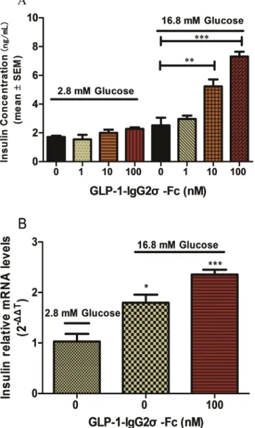
Tests of glucose-induced insulin secretion were performed using INS-1 cells exposed to various
concentrations of GLP-1-IgG2σ-Fc. As shown inFig 2A, GLP-1-IgG2σ-Fc stimulated insulin
secretion in a glucose-dependent manner. In cells incubated with medium containing 2.8 mM glucose, there were no statistically significant differences between each group. However, cells incubated with medium containing 16.8 mM glucose secreted significantly more insulin in the presence of 10 nM (5.2 ± 0.4 ng/mL) and 100 nM (7.3 ± 0.3 ng/mL) GLP-1-IgG2σ-Fc, as
com-pared with control cells (2.5 ± 0.5 ng/mL) (p<0.05). To investigate this effect on insulin
secre-tion further, mRNA was isolated from the INS-1 cells exposed to 16.8 or 2.2 mM glucose and
100 nM GLP-1-IgG2σ-Fc and insulin mRNA expression was evaluated by RT-PCR (Fig 2B).
GLP-1-IgG2
σ
-Fc has an extended t
1/2and produced sustained glucose
reduction
The pharmacokinetics of GLP-1-IgG2σ-Fc were studied in cynomolgus monkeys after a single
dose of 0.1 mg/kg. The t1/2of GLP-1-IgG2σ-Fc was approximately 57.1 ± 4.5 h and it could be
detected for more than 14 days after a single injection (Fig 3andTable 1).
The blood glucose level in KKAy mice showed a sustained reduction after a single dose of 1
mg/kg GLP-1-IgG2σ-Fc (Fig 4). Mice treated with GLP-1-IgG2σ-Fc showed significantly lower
levels of postprandial glucose from days 1 to 5, as compared to the vehicle group. The GLP-1-IgG4-Fc-treated group only showed a statistically significant hypoglycemic effect on the first two days post-administration. Based on this result and on pre-clinical research involving
dula-glutide, the administration frequency was set to three days [21].
GLP-1-IgG2
σ
-Fc reduced postprandial glucose and body weight in
KKAy mice
KKAy mice were treated with intraperitoneal GLP-1-IgG2σ-Fc once every three days for 4 weeks. During this period, GLP-1-IgG2σ-Fc significantly reduced the plasma glucose levels (Fig 5A). The AUC from days 1 to 28 was significantly larger in the vehicle-treated group than
in the 1 mg/kg GLP-1-IgG2σ-Fc-treated group (p<0.001;Fig 5B). KKAy mice treated with
GLP-1-IgG2σ-Fc had a significantly lower body weight from day 23, while the group treated
with GLP-1-IgG4-Fc showed no significant difference, as compared to the vehicle group (Fig
5C). The vehicle group consumed ~1.5-fold more food than the GLP-1-IgG2σ-Fc group and
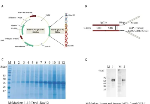
Fig 1. Generation of the GLP-1-IgG2σ-Fc expression vector.(A) DNA encoding GLP-1-IgG2σ-Fc was synthesized and inserted into a mammalian expression vector, SGLs. (B) GLP-1-IgG2σ-Fc comprises a pair
of human GLP-1 variants (A8G/G26E/R36G) and a human IgG2σconstant heavy-chain, with a molecular
mass of 68 kDa. (C) SDS-PAGE analysis of cell culture medium indicated that stably transfected cells secreted the highest amount of GLP-1-IgG2σ-Fc on day 12. (D) Western blotting analysis confirmed that the
expressed 68-kDa protein showed good antigenicity to both GLP-1 and human IgG.
~1.3-fold more food than the GLP-1-IgG4-Fc group. In addition, the GLP-1-IgG2σ-Fc group consumed significantly less food than the GLP-1-IgG4-Fc group.
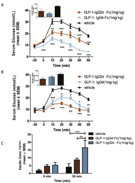
OGTT tests were performed in KKAy mice at the beginning and end (10 days after the last dose) of a 4-week treatment with GLP-1-IgG2σ-Fc, GLP-1-IgG4-Fc, or saline to investigate the effects on postprandial glucose levels. Compared with the vehicle group, mice treated with either GLP-1-IgG4-Fc or GLP-1-IgG2σ-Fc showed reduced glucose levels. Calculation of the AUC showed that mice treated with GLP-1-IgG4-Fc showed a reduction of ~61.5% before treatment and of ~24.9% after treatment. GLP-1-IgG2σ-Fc produced a greater hypoglycemic effect after treatment, with a reduction of ~31.2% before treatment and ~43.5% after treatment (Fig 6A–6D). Insulin levels were tested after 4 weeks of treatment to further investigate the Fig 2. Insulin synthesis and secretion in INS-1 cells.(A) INS-1 cells were plated in 96-well plates with glucose-free RPMI 1640 for 120 min before media supplemented with the indicated concentrations of glucose and GLP-1-IgG2σ-Fc were added for 2 h. Insulin levels were measured by ELISA. (B) Insulin mRNA
levels in the INS-1 cells treated as indicated were determined by RT-PCR. Values are means±SD (n = 6). All
INS-1 cell experiments were repeated for four times. A representative result of multiple independent experiments is presented.*p<0.05 versus vehicle-treated cells in 16.8-mM glucose media.
effect of GLP-1 fusion proteins in KKAy mice. The groups treated with GLP-1-IgG4-Fc and GLP-1-IgG2σ-Fc showed no significant improvements in basal insulin levels prior to gastric perfusion of glucose. As compared with vehicle-treated mice, those treated with
GLP-1-IgG4-Fc or GLP-1-IgG2σ-Fc showed significant improvements in insulin secretion at 30 min (Fig 6C).
Discussion
GLP-1-Fc fusion proteins can be used to produce molecules that retain the glycemic control
properties of GLP-1, with longer t1/2values. We successfully engineered GLP-1-IgG2σ-Fc, a
68-kDa fusion protein linking variant human GLP-1 (A8G/G26E/R36G) with a human IgG2σ constant heavy-chain. We expressed this in a eukaryotic expression system because this is the only established system that is suitable for the expression of complex recombinant proteins
with human-like glycoforms that are bioactive in humans [22]. The protein yield of 1.2 g from
1 L culture medium, continuously cultured for 12 days (data not shown), confirmed that this mammalian expression system was a highly efficient producer of GLP-1-IgG2σ-Fc.
Consistent with our previous observations using native GLP-1 and GLP-1 receptor agonists, incubation of INS-1 cells with GLP-1-IgG2σ-Fc resulted in concentration-dependent effects on
insulin release [23]. Further mRNA analyses of these cells indicated that GLP-1-IgG2σ-Fc also
increased insulin synthesis. This confirmed that the method used to purify the expressed GLP-1-IgG2σ-Fc did not inhibit its bioactivity. The results also confirmed that GLP-GLP-1-IgG2σ-Fc stimulated insulin secretion in a glucose-dependent manner.
GLP-1-IgG4-Fc (comprising native GLP-1 fused with IgG4) was used as a positive control
in the presentin vivostudy. The t1/2of GLP-1-IgG4-Fc in cynomolgus monkeys was
approxi-mately 37.5 ± 4.9 h (data not shown), while the t1/2of GLP-1-IgG2σ-Fc was 57.1 ± 4.5 h. This
represented a significant improvement over native GLP-1 (t1/2<5 min). Other GLP-1 agonists
have shown t1/2values of 51.6 ± 3.2 h in monkeys (dulaglutide), 11–13 h in humans
Fig 3. Pharmacokinetics in cynomolgus monkeys.Plasma fusion protein levels were determined by ELISA following administration of 0.1 mg/kg GLP-1-IgG4-Fc.
doi:10.1371/journal.pone.0156449.g003
Table 1. Pharmacokinetic parameters in cynomolgus monkeys.
Cmax(ng/mL) Tmax(h) AUC0-1(hng/mL) t1/2(h) CL/F (mL/h/kg) Vd (mL/kg) MRT (h) Monkey 701.0±52.2 6.7±1.3 48353.2±3228.6 57.1±4.5 2.1±0.1 172.9±22.3 68.6±2.0
Pharmacokinetic parameters (mean±SEM) were calculated using the plasma levels of GLP-1-IgG2σ-Fc in three cynomolgus monkeys at each time-point
following administration of 0.1 mg/kg. Cmax, maximum plasma concentration; Tmax, time of maximum plasma concentration; AUC, area under the curve; t1/2, elimination half-life; CL/F, clearance; Vd, volume of distribution; MRT, mean residence time.
(liraglutide), and 2.4 h in humans (exenatide BID). Due to species differences, dulaglutide (a
human GLP-1 variant fused with a human IgG4 variant) showed a longer t1/2in humans than
in monkeys (120 h versus 51.6 ± 3.2 h, respectively) and further research will be required to
determine whether GLP-1-IgG2σ-Fc also has a longer t1/2in humans.
GLP-1 and GLP-1 receptor agonists improve glycemic control in T2DM by reducing
post-prandial glucose levels [24]. The present study used ten-week-old male KKAy mice as anin
vivomodel of T2DM because these mice develop non-insulin-dependent diabetes mellitus
spontaneously at the age of 12 weeks [25–27]. Although KKAy, db/db, and ob/ob mice are all
Fig 4. Acute pharmacodynamics.KKAy mice received a single intraperitoneal injection of GLP-1-IgG2σ-Fc
(1 mg/kg), GLP-1-IgG4-Fc (1 mg/kg), or saline. Blood glucose levels were measured at the indicated time-points.*p<0.05;**p<0.01;***p<0.001, versus the vehicle-treated group.
doi:10.1371/journal.pone.0156449.g004
Fig 5. Long-term pharmacodynamics.(A) Blood glucose was measured prior to the administration of the indicated treatments to KKAy mice every three days for 4 weeks. (B) The area under the curve (AUC) of the data presented in (A). (C) Mouse body weights were recorded every day. (D) Food intake of each group. *p<0.05;**p<0.01;***p<0.001, versus the vehicle-treated group;#p<0.05, versus the
GLP-1-IgG4-Fc-treated group.
used as genetic mouse models of T2DM, KKAy mice were the least expensive of these and were readily available. GLP-1-IgG2σ-Fc produced a more sustained hypoglycemic effect in KKAy mice than did GLP-1-IgG4-Fc. An intraperitoneal injection of GLP-1-IgG2σ-Fc (1 mg/kg) reduced blood glucose levels for 5 days, while GLP-1-IgG4-Fc affected this for 2 days. Based on this observation and on previous studies of dulaglutide, we elected to administer these Fig 6. OGTT in KKAy mice.(A) OGTT in KKAy mice at the beginning of the 4-week treatment period. Glucose (2 g/kg) was administered 30 min before injection of GLP-1-IgG2σ-Fc (1 mg/kg), GLP-1-IgG4-Fc (1
mg/kg), or saline (200μL). The area under the curve (AUC) for the glucose levels is shown in the upper left
panel. (B) OGTT in KKAy mice at the end of the 4-week treatment period. The AUC for the glucose levels is shown in the upper left panel. (C) Insulin levels were tested at 0 and 30 min during the OGTT test;*p<0.05;
**p<0.01;***p<0.001 versus the vehicle-treated group.
treatments once every three days during the present long-term pharmacodynamics study. Our findings suggested that GLP-1-IgG2σ-Fc produced a stable hypoglycemic and insulin secretion effect during the treatment period. Notably, the drug resistance observed in animals treated with GLP-1-IgG4-Fc was not seen in those treated with GLP-1-IgG2σ-Fc. Bioactivity is often reduced by neutralizing antibodies, which can be generated at different levels, depending on
the immunogenicity of the protein drug [28]. Several studies of GLP-1 receptor agonists have
reported the formation of antibodies and exenatide treatment was associated with antibody
generation in 74% of patients [29]. Liraglutide, which shares 97% of its amino acid sequence
with the endogenous human protein, also generated antibodies in 4–13% of patients [30,31].
The variation present in IgG2 completely eliminates its affinity for FcγRs and the C1q
comple-ment protein, which induce antibody-dependent cellular cytotoxicity and phagocytosis, as well
as complement-dependent cytotoxicity. These‘silent’properties of IgG2σrender it superior to
IgG4 for use in therapeutic non-immunostimulatory fusion proteins. In addition, the GLP-1 variant used in the present study is a close structural homolog of native human GLP-1, with three amino acid substitutions (A8G/G26E/R36G) that confer protection from DPP-IV
cleav-age. The longer t1/2and stable glucose-lowering effects observed in ourin vivostudy indicated
that GLP-1-IgG2σ-Fc did not trigger a significant level of antibody production and was not metabolized rapidly. This suggested that GLP-1-IgG2σ-Fc may be appropriate for therapeutic application as a GLP-1 receptor agonist.
Their effect on weight loss is another important property of GLP-1 receptor agonists [32].
In the present pharmacodynamic study, 4-week treatment with GLP-1-IgG2σ-Fc resulted in significant weight loss from day 23, as compared to the vehicle group. The food intake result also supported this conclusion and indicated that GLP-1-IgG2σ-Fc may reduce weight by inhibiting feeding. However, it is not possible to extrapolate these pre-clinical findings to infer effects in humans. For example, dulaglutide includes the same GLP-1 variant as GLP-1-IgG2σ-Fc and produces significant weight reduction in db/db mice, but not in humans.
Conclusions
The hypoglycemic effects of 1-IgG2σ-Fc were sustained for longer than those of GLP-1-IgG4-Fc in KKAy mice. Pharmacokinetic and pharmacodynamic analyses of
GLP-1-IgG2σ-Fc confirmed that it had a prolonged t1/2and could reduce body weight. These superior
fea-tures indicated that GLP-1-IgG2σ-Fc could provide a potential long-acting GLP-1 receptor agonist for the treatment of T2DM.
Acknowledgments
We gratefully acknowledge Dr. Ling Zhang, Laboratory of Hematological Pharmacology, for his expert technical assistance.
Author Contributions
Conceived and designed the experiments: YY XG HS. Performed the experiments: YY FC DW YL LY HF XC. Analyzed the data: YY FC. Contributed reagents/materials/analysis tools: DW YL LY HF. Wrote the paper: YY.
References
2. Light PE, Manning Fox JE, Riedel MJ, Wheeler MB. Glucagon-like peptide-1 inhibits pancreatic ATP-sensitive potassium channels via a protein kinase A- and ADP-dependent mechanism. Molecular endo-crinology (Baltimore, Md). 2002; 16(9):2135–44. Epub 2002/08/29. doi:10.1210/me.2002-0084PMID: 12198249.
3. Murage EN, Gao G, Bisello A, Ahn JM. Development of potent glucagon-like peptide-1 agonists with high enzyme stability via introduction of multiple lactam bridges. Journal of medicinal chemistry. 2010; 53(17):6412–20. Epub 2010/08/07. doi:10.1021/jm100602mPMID:20687610.
4. Baggio LL, Drucker DJ. Biology of incretins: GLP-1 and GIP. Gastroenterology. 2007; 132(6):2131–57.
Epub 2007/05/15. doi:10.1053/j.gastro.2007.03.054PMID:17498508.
5. Pratley RE, Gilbert M. Targeting Incretins in Type 2 Diabetes: Role of GLP-1 Receptor Agonists and DPP-4 Inhibitors. The review of diabetic studies: RDS. 2008; 5(2):73–94. Epub 2008/09/17. doi:10. 1900/rds.2008.5.73PMID:18795210; PubMed Central PMCID: PMCPmc2556445.
6. Inzucchi SE, Bergenstal RM, Buse JB, Diamant M, Ferrannini E, Nauck M, et al. Management of hyper-glycemia in type 2 diabetes: a patient-centered approach: position statement of the American Diabetes Association (ADA) and the European Association for the Study of Diabetes (EASD). Diabetes care. 2012; 35(6):1364–79. Epub 2012/04/21. doi:10.2337/dc12-0413PMID:22517736; PubMed Central
PMCID: PMCPmc3357214.
7. Siegel EG, Gallwitz B, Scharf G, Mentlein R, Morys-Wortmann C, Folsch UR, et al. Biological activity of GLP-1-analogues with N-terminal modifications. Regulatory peptides. 1999; 79(2–3):93–102. Epub
1999/04/01. PMID:10100921.
8. Huang YS, Chen Z, Chen YQ, Ma GC, Shan JF, Liu W, et al. Preparation and characterization of a novel exendin-4 human serum albumin fusion protein expressed in Pichia pastoris. Journal of peptide science: an official publication of the European Peptide Society. 2008; 14(5):588–95. Epub 2007/11/
13. doi:10.1002/psc.942PMID:17994612.
9. Lorenz M, Evers A, Wagner M. Recent progress and future options in the development of GLP-1 recep-tor agonists for the treatment of diabesity. Bioorganic & medicinal chemistry letters. 2013; 23(14):4011–
8. Epub 2013/06/08. doi:10.1016/j.bmcl.2013.05.022PMID:23743288.
10. Levin D, Golding B, Strome SE, Sauna ZE. Fc fusion as a platform technology: potential for modulating immunogenicity. Trends in biotechnology. 2015; 33(1):27–34. Epub 2014/12/10. doi:10.1016/j.tibtech. 2014.11.001PMID:25488117.
11. Astrup A, Carraro R, Finer N, Harper A, Kunesova M, Lean ME, et al. Safety, tolerability and sustained weight loss over 2 years with the once-daily human GLP-1 analog, liraglutide. International journal of obesity (2005). 2012; 36(6):843–54. Epub 2011/08/17. doi:10.1038/ijo.2011.158PMID:21844879;
PubMed Central PMCID: PMCPmc3374073.
12. Macconell L, Brown C, Gurney K, Han J. Safety and tolerability of exenatide twice daily in patients with type 2 diabetes: integrated analysis of 5594 patients from 19 placebo-controlled and comparator-con-trolled clinical trials. Diabetes, metabolic syndrome and obesity: targets and therapy. 2012; 5:29–41.
Epub 2012/03/01. doi:10.2147/dmso.s28387PMID:22375098; PubMed Central PMCID: PMCPmc3287409.
13. Russell-Jones D. The safety and tolerability of GLP-1 receptor agonists in the treatment of type-2 diabe-tes. International journal of clinical practice. 2010; 64(10):1402–14. Epub 2010/08/19. doi:10.1111/j. 1742-1241.2010.02465.xPMID:20716148.
14. Buse JB, Garber A, Rosenstock J, Schmidt WE, Brett JH, Videbaek N, et al. Liraglutide treatment is associated with a low frequency and magnitude of antibody formation with no apparent impact on glyce-mic response or increased frequency of adverse events: results from the Liraglutide Effect and Action in Diabetes (LEAD) trials. The Journal of clinical endocrinology and metabolism. 2011; 96(6):1695–
702. Epub 2011/04/01. doi:10.1210/jc.2010-2822PMID:21450987.
15. Vafa O, Gilliland GL, Brezski RJ, Strake B, Wilkinson T, Lacy ER, et al. An engineered Fc variant of an IgG eliminates all immune effector functions via structural perturbations. Methods (San Diego, Calif). 2014; 65(1):114–26. Epub 2013/07/23. doi:10.1016/j.ymeth.2013.06.035PMID:23872058. 16. Lobo ED, Hansen RJ, Balthasar JP. Antibody pharmacokinetics and pharmacodynamics. Journal of
pharmaceutical sciences. 2004; 93(11):2645–68. Epub 2004/09/25. doi:10.1002/jps.20178PMID: 15389672.
17. Chung HS, Oh JY, Yoo SB, Lee SM, Cho HS. The N-terminal alanine-extended GLP-1/IgG-Fc fusion protein confers resistance to DPP-IV and reduces serum glucose level in db/db mice. Regulatory pep-tides. 2011; 170(1–3):1–3. Epub 2011/05/31. doi:10.1016/j.regpep.2011.05.002PMID:21621561. 18. Buteau J, Roduit R, Susini S, Prentki M. Glucagon-like peptide-1 promotes DNA synthesis, activates
phosphatidylinositol 3-kinase and increases transcription factor pancreatic and duodenal
homeobox gene 1 (PDX-1) DNA binding activity in beta (INS-1)-cells. Diabetologia. 1999; 42(7):856–
19. Amiram M, Luginbuhl KM, Li X, Feinglos MN, Chilkoti A. A depot-forming glucagon-like peptide-1 fusion protein reduces blood glucose for five days with a single injection. Journal of controlled release: official journal of the Controlled Release Society. 2013; 172(1):144–51. Epub 2013/08/10. doi:10.1016/j. jconrel.2013.07.021PMID:23928357; PubMed Central PMCID: PMCPmc3834218.
20. Hashimoto Y, Ikenaga T, Tanigawa K, Ueda T, Ezak I, Imoto T. Expression and characterization of human rheumatoid factor single-chain Fv. Biological & pharmaceutical bulletin. 2000; 23(8):941–5.
Epub 2000/08/30. PMID:10963300.
21. Glaesner W, Vick AM, Millican R, Ellis B, Tschang SH, Tian Y, et al. Engineering and characterization of the long-acting glucagon-like peptide-1 analogue LY2189265, an Fc fusion protein. Diabetes/metab-olism research and reviews. 2010; 26(4):287–96. Epub 2010/05/27. doi:10.1002/dmrr.1080PMID: 20503261.
22. Maccani A, Landes N, Stadlmayr G, Maresch D, Leitner C, Maurer M, et al. Pichia pastoris secretes recombinant proteins less efficiently than Chinese hamster ovary cells but allows higher space-time yields for less complex proteins. Biotechnology journal. 2014; 9(4):526–37. Epub 2014/01/07. doi:10. 1002/biot.201300305PMID:24390926; PubMed Central PMCID: PMCPmc4315903.
23. MacDonald PE, El-Kholy W, Riedel MJ, Salapatek AM, Light PE, Wheeler MB. The multiple actions of GLP-1 on the process of glucose-stimulated insulin secretion. Diabetes. 2002; 51 Suppl 3:S434–42.
Epub 2002/12/12. PMID:12475787.
24. Meier JJ. GLP-1 receptor agonists for individualized treatment of type 2 diabetes mellitus. Nature reviews Endocrinology. 2012; 8(12):728–42. Epub 2012/09/05. doi:10.1038/nrendo.2012.140PMID: 22945360.
25. Liu M, Wu K, Mao X, Wu Y, Ouyang J. Astragalus polysaccharide improves insulin sensitivity in KKAy mice: regulation of PKB/GLUT4 signaling in skeletal muscle. Journal of ethnopharmacology. 2010; 127 (1):32–7. Epub 2009/10/06. doi:10.1016/j.jep.2009.09.055PMID:19800959.
26. Zhang J, Xue C, Zhu T, Vivekanandan A, Pennathur S, Ma ZA, et al. A tripeptide Diapin effectively low-ers blood glucose levels in male type 2 diabetes mice by increasing blood levels of insulin and GLP-1. PloS one. 2013; 8(12):e83509. Epub 2014/01/05. doi:10.1371/journal.pone.0083509PMID:
24386218; PubMed Central PMCID: PMCPmc3873933.
27. Hou GJ, Li CN, Liu SN, Huan Y, Liu Q, Sun SJ, et al. Long-term treatment with EXf, a peptide analog of Exendin-4, improves beta-cell function and survival in diabetic KKAy mice. Peptides. 2013; 40:123–32.
Epub 2013/01/29. doi:10.1016/j.peptides.2013.01.010PMID:23353893.
28. Rossman HS. Neutralizing antibodies to multiple sclerosis treatments. Journal of managed care phar-macy: JMCP. 2004; 10(3 Suppl B):S12–9. Epub 2004/07/16. PMID:15253685.
29. Drucker DJ, Buse JB, Taylor K, Kendall DM, Trautmann M, Zhuang D, et al. Exenatide once weekly ver-sus twice daily for the treatment of type 2 diabetes: a randomised, open-label, non-inferiority study. Lan-cet (London, England). 2008; 372(9645):1240–50. Epub 2008/09/11. doi:10.1016/s0140-6736(08) 61206-4PMID:18782641.
30. Russell-Jones D, Vaag A, Schmitz O, Sethi BK, Lalic N, Antic S, et al. Liraglutide vs insulin glargine and placebo in combination with metformin and sulfonylurea therapy in type 2 diabetes mellitus (LEAD-5 met+SU): a randomised controlled trial. Diabetologia. 2009; 52(10):2046–55. Epub 2009/08/19. doi: 10.1007/s00125-009-1472-yPMID:19688338; PubMed Central PMCID: PMCPmc2744824.
31. Zinman B, Gerich J, Buse JB, Lewin A, Schwartz S, Raskin P, et al. Efficacy and safety of the human glucagon-like peptide-1 analog liraglutide in combination with metformin and thiazolidinedione in patients with type 2 diabetes (LEAD-4 Met+TZD). Diabetes care. 2009; 32(7):1224–30. Epub 2009/03/
18. doi:10.2337/dc08-2124PMID:19289857; PubMed Central PMCID: PMCPmc2699702.
32. Mayo KE, Miller LJ, Bataille D, Dalle S, Goke B, Thorens B, et al. International Union of Pharmacology. XXXV. The glucagon receptor family. Pharmacological reviews. 2003; 55(1):167–94. Epub 2003/03/