J of Evolution of Med and Dent Sci/ eISSN- 2278-4802, pISSN- 2278-4748/ Vol. 4/ Issue 25/ Mar 26, 2015 Page 4261
UTILITY OF A DUAL IMMUNOSTAIN LIKE P53 AND CK20 TO AID IN THE
DIAGNOSIS AND CATEGORIZATION OF NEOPLASTIC BLADDER BIOPSIES
Dipkana Das1, Ranjan Kumar Dey2, Subhasish Saha3, Tushar Kanti Das4HOW TO CITE THIS ARTICLE:
Dipkana Das, Ranjan Kumar Dey, Subhasish Saha, Tushar Kanti Das. Utility of a dual immunostain like P53 and CK20 to Aid in the Diagnosis and Categorization of Neoplastic Bladder Biopsies. Journal of Evolution of Medical and Dental Sciences 2015; Vol. 4, Issue 25, March 26; Page: 4261-4269, DOI: 10.14260/jemds/2015/616
ABSTRACT: INTRODUCTION: Urothelial neoplasms contribute significantly to the overall human cancer burden with approximately 260,000 new cases per year worldwide. Although over 50% cases, the lesions are diagnosed by biopsy at an early stage but the prognosis is sometimes difficult to assess because of heterogeneity, multifocality and tumor recurrence. AIMS AND OBJECTIVE: To evaluate the scope of immunohistochemical expression of mutated p53 & CK20 in human urothelial neoplasms with regard to grade, stage and outcome of the patients. MATERIALS AND METHODS: In this study fifty three 5 consecutive patients were taken from May’ to April’ 2013. Histopathological examinations were performed and the grading was done according to the WHO/ISUP (2004) consensus classification of urothelial neoplasms. Immunohistochemical staining for p53 & CK20 were performed. RESULTS: 11 cases were found to be of high grade papillary urothelial carcinoma,9 cases of invasive urothelial carcinoma, 29 cases of low grade papillary urothelial carcinoma and 4 patients with papillary urothelial neoplasms of low malignant potential (PUNLMP). Unfortunately not a single benign lesion was received in the study period. Mean percent positivity of p53 in case of high grade carcinoma is 75.45% with a p value of 0.001 and mean percent positivity of CK20 in case of high grade carcinoma is 84.55% with a p value of 0.001; both are highly significant.
KEYWORDS: Papillary neoplasia, Urothelial neoplasms, Transitional cell carcinoma, p53, CK20.
INTRODUCTION: Cancer of the urinary bladder accounts for about 3.2% of all cancers worldwide and is considerably more common in males than in females (ratio is 3.5:1). Urothelial carcinoma is the 7th most common cancer with addition of 260,000 new cases in men and 76,000 cases in women
per year.1 As per the Indian cancer registry data in men, it is the 9th most common cancer accounting
for 3.9% of all cancer cases.2 The median age at presentation is 69 years in case of male & 71 years in
case of female.3
Both urothelial dysplasia (UD) and carcinoma in situ (CIS) are precursor lesions of invasive carcinoma and their presence is associated with a high risk of progression and recurrence.4-6 UD is
defined as "appreciable loss of polarity with nuclear rounding and crowding and cytologic atypia that is not severe enough to merit a diagnosis of CIS."7
J of Evolution of Med and Dent Sci/ eISSN- 2278-4802, pISSN- 2278-4748/ Vol. 4/ Issue 25/ Mar 26, 2015 Page 4262 With loss of function of p53, DNA damage goes unrepaired, mutation accumulate in dividing cells and the cell marches along one way traffic leading to malignant transformation.8 Mutated p53
may bind to wild type p53 and change it to mutated conformation.9 p53 immunohistochemistry has
been suggested as a prognostic marker of malignancy.
The cytokeratin family consists of at least 20 types of cytoplasmic intermediate filaments found in epithelial cells.10 Expression of these cytoskeletal proteins is determined by cell type, stage
of development, and differentiation. Cytokeratin 20 is a low-molecular weight cytokeratin, is specifically expressed in the superficial and in some of the intermediate cells of the normal urothelium and is associated with recurrence in the bladder.11 CK20 is a protein that is encoded by
the KRT20 gene. It is a type-1 cytokeratin & major cellular protein of mature enterocytes and goblet cells also. This protein is commonly found in transitional cell cancer, colorectal cancer and merkel cell cancer. Anti-Cytokeratin 20 is useful in the differentiation of specific types of simple epithelial cells of the urinary tract as well as normal and malignantly transformed epithelia. CK20 is the cytoplasmic marker and a known component on the cytokeratin subgroup of intermediate filament. It has been suggested that CK20 may be an important tool for detecting and identifying these types of cancer and their metastases, either by immunohistochemistry or by reverse transcription–polymerase chain reaction analysis.
AIMS AND OBJECTIVE: To evaluate the scope of immunohistochemical expression of mutated p53 & CK20 in human urothelial neoplasms with regard to grade, stage and outcome of the patients. Many studies in the literature have attempted to identify specific and sensitive biomarkers for evidence of bio- molecular mechanisms involved in urothelial carcinogenesis. Highlighting the relationship between these mechanisms and alternative pathways between tumor initiation and progression is still major objectives of research programs.
MATERIALS AND METHODS: It was a prospective study of 53 consecutive patients of urothelial neoplasms attending the department of Urosurgery from May, 2012 to April, 2013. A transurethral resection of the bladder tumor (TURBT) was performed using Glycine (1.5%) as an irrigant. A deep biopsy was taken separately to include the detrusor muscle. Suspicious lesions were biopsied. None of the patients had repeat TURBT. The patients were informed and consent was obtained. Urothelial neoplasms of different grades were diagnosed according to the world health organisation/ International Society of Urological Pathology (WHO/IUSP) consensus Classification of urothelial neoplasms of the urinary bladder.12 Anti-p53 mouse monoclonal antibody (clone-DO7, Cell Marque,
Rocklin, CA, USA) used which reacted with mutant as well as wild form of p53 and Ck20 mouse monoclonal antibody (Clone-Ks20.8, Cell Marque, Rocklin, CA, USA) used and reacted with intermediate filament of cytoplasm and cell membrane. The immunohistochemical stains were applied on 4 µm sections of formalin fixed paraffin embedded tissue. Epitope retrieval was done in conjunction with a pressure cooker. Immunostaining was evaluated by counting 500 tumor cells for each case at high power. Colon carcinoma cells were taken as positive control for CK20 and ductal carcinoma breast taken as positive control for p53 immuno-stains.
J of Evolution of Med and Dent Sci/ eISSN- 2278-4802, pISSN- 2278-4748/ Vol. 4/ Issue 25/ Mar 26, 2015 Page 4263 Statistical analysis done by ANOVA showing p53 staining, maximum mean percentage
positivity is found in case of high grade urothelial carcinoma that is 75.45% and minimum in PUNLMP that is 9% and p value is 0.001 which is highly significant. In case of CK20 staining maximum mean percentage positivity is found in non-invasive high grade urothelial carcinoma (84.55%) and minimum percentage positivity found in PUNLMP (09%). It shows F=27.008 and p value is 0.001 which is again highly significant.
RESULTS: 53 cases of urothelial carcinoma were examined those were presented in the Urosurgery department by 1 year. Among cases of urothelial carcinoma, age at presentation range from 33 years to 78 years (Mean: 51.9 years) 49(92.45%) were male patients, whereas 4(7.54%) were female patients (Table-1, Histogram-1). So, Male: Female ratio is12.25:1. From the above data it was observed that 49/53 were male patients and among them 43 were addicted to smoking or tobacco chewing. There has been a strong relationship between smoking and development of urothelial carcinoma.
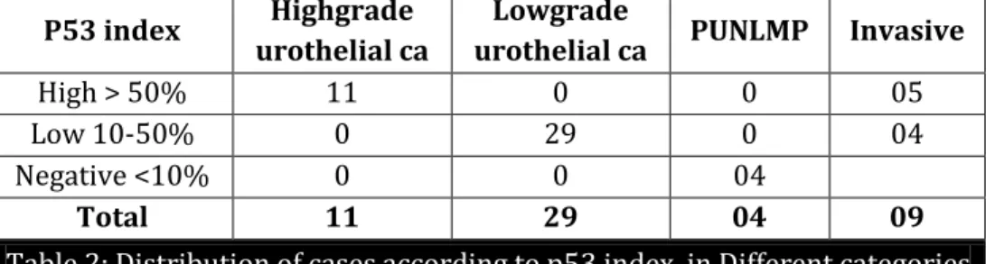
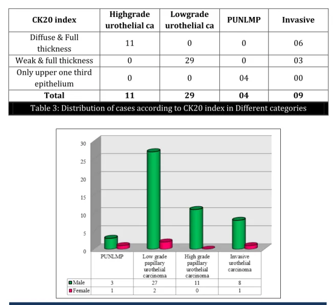
All specimens underwent for histopathological and immunohistochemical analysis. Papillary urothelial neoplasms of low malignant potential (PUNLMP) was found in 4 cases, papillary urothelial carcinoma low grade 29 cases and papillary urothelial carcinoma high grade 11 cases and 9 cases of invasive urothelial carcinoma. All cases of PUNLMP showed negative p53 reactivity and CK20 positive in upper one third epithelium including umbrella cells. All the low grade carcinomas had <50% cells range 20-45% cells were immunoreactive for both the stains and high grade carcinomas showed >50% nucleus range 60-95% were immunoreactive for p53 and full thickness and diffuse cytoplasm and cell membrane positivity for CK20. Distribution of cases according to p53index and distribution of cases according to CK20 index in different categories are explained in table2 and table 3 respectively. 55% of the neoplasms of our study are of low grade followed by 21% of high grade papillary neoplasm; invasive urothelial neoplasms ranked after that. Expression of CK20 and p53 were diffuse & strongly positive for high grade lesions. Mean percent positivity of p53 in case of high grade carcinoma is 75.45% with a p value of 0.001 and mean percent positivity of CK20 in case of high grade carcinoma is 84.55% with a p value of 0.001; both are highly significant.
DISCUSSION: Most cases of bladder cancer are superficial at the time of diagnosis (Stage Ta-T1). The recurrence rate of superficial tumors can be as high as 70%, with 10-15% progressing to muscle invasive disease.13 The T2-T4 urothelial carcinoma of bladder is considered as poor prognosis.14
In the present study 53 consecutive patients irrespective of their gender who admitted Urology indoor of R. G. Kar Medical College & Hospital with the diagnosis of urinary bladder tumor were incorporated. Bladder cancer presents with painless hematuria in 80-85% of the patients. Practically, nearly all patients with cystoscopically detectable bladder cancer have at least microhematuria if enough urine samples are tested. This high incidence may be due to lack of screening for microscopic hematuria in form of dipstick method.
Transitional cell carcinoma (TCC) is the most common variant accounting for 90% of bladder cancer in the world literature.15 In our study 100% of the patients had TCC and in one case we found
TCC with squamous differentiation.
J of Evolution of Med and Dent Sci/ eISSN- 2278-4802, pISSN- 2278-4748/ Vol. 4/ Issue 25/ Mar 26, 2015 Page 4264 presenting in their 40-60 years of age group with a mean age of 52 years. It is less common below the age of 40 years but in our study we found three cases in the age group of 30s. It is seen that male: female ratio is 12.25:1. Cigarette smokers have at least four times higher incidence of bladder cancer16, because a higher number of mutations occurred in smokers. In the above study, 54.71% that
is 29 out of 53 patients were smokers and about 75% patients that are 39 out of 53 cases were taking tobacco in any form. Mutation study could not be performed because of limited resources.
Younger individuals present more frequently with low-grade and low-stage tumors than their elderly counterparts and behave in an indolent fashion.17-18 The result of our study is dissimilar that
of the above fact. It has been observed that lowest mean ± SD (48.78±9.230) is found in case of infiltrating carcinoma. This is contemporary to the common belief in malignancy that biological behaviour of malignancy is more aggressive in younger age group.
Carme Mallorfe et al19 studied immunohistochemical panel composed of p53, Ki-67, and CK20
is useful for confirming the presence of dysplastic changes in the urothelium. The positivity was always strong and in many cases, in a high percentage of cells (Ranging from 50–90%). Unfortunately we could not find any isolated case of dysplasia or carcinoma –in-situ (CIS), possibly because most of patients presented late in the course of their disease in our country.
McKenney20 and associates from Emory University described the use of immunostains for
cytokeratin 20 (CK20) (a cytoplasmic Antigen), p53 (a nuclear antigen), and the standard isoform of CD44 (expressed on the cell membrane) to address this problem. Their study included 21 cases of CIS, 15 urothelial biopsies with reactive atypia, and 10 normal ureters from nephrectomy specimens. Overexpression of p53 and CK20 were defined as follows: ">50% of the urothelium had to be moderately to strongly positive". In their study, they noted distinctly different patterns of expression of these markers in normal urothelium, reactive atypia, and CIS.
In normal urothelium, CK20 showed patchy expression only in the umbrella (Superficial) cell layer of the epithelium and p53 was negative or only focally weakly expressed (<10% of cells, weak expression). Cases of reactive atypia showed similar CK20 reactivity to normal urothelium (Apparent only in the umbrella cell layer), and p53 was at most patchy and weak, mostly in the basal cell layer.
In contrast, cases of CIS showed overexpression of CK20 (defined as > 50% of neoplastic cells) in 81% of cases, and p53 was over-expressed in 57% of the cases of CIS. The present study also revealed high positive expressions by both the stains in high grade as well as invasive carcinomas.
In another study Gonzalez et al21 in the year 2008 worked on the prognostic value of
combined p53 and surviving in PT1G3 urothelial carcinoma of the urinary bladder. Tumors with invasion of the lamina propria above the level of muscularis mucosa were sub classified as pT1a and tumors with infiltration of deep lamina propria beyond the muscularis mucosa were subclassified as pT1b and it was seen that patients with pT1b tumors had a significantly increased risk of local progression and metastasis. They reported that percentage of stained nuclei with p53 was higher for patients with tumor progression than for patients without tumor progression. As this is a one year study, progression as well as recurrence of disease cannot be assessed.
Yildiz et al22 investigated the utility and advantages of p53+CK20 dual immunohistochemistry
J of Evolution of Med and Dent Sci/ eISSN- 2278-4802, pISSN- 2278-4748/ Vol. 4/ Issue 25/ Mar 26, 2015 Page 4265 carcinoma cases exhibited moderate p53 positivity. Abnormal expression of CK 20 was found in 90% (9/10) of dysplasia, 89% (8/9) of CIS and 71% (5/7) of IC cases whereas the rest of the cases lacked abnormal CK20 expression.
Yin H studied23 that Immunostaining of CK20 marker was applied to 26 cases of CIS, 14 atypia
of unknown significance, 4 dysplasia, 6 normal, and 9 hyperplastic urothelium. CIS showed CK20 staining of deep urothelial cells in 23/26 CIS compared with restricted staining in surface cells in all non-neoplastic lesions. Among the cases of atypia, 3/14 displayed deep staining for CK20. In dysplasia similar findings were present in 1/4 and 2/4 cases, respectively. These findings suggest that CK20 is objective markers to distinguish CIS from non-neoplastic urothelium. In cases of "atypia of unknown significance" and "dysplasia," positivity for the marker should raise the possibility of CIS or pre-neoplastic change and identify those cases for follow-up.
In the current work no recurrence or progression occurred in any of the patients whose tumors had normal pattern of CK20 expression.
However, the group of abnormal expression experienced recurrence and progression rates of 94.7% and 100%, respectively. No statistical significant difference was detected; possibly due to the small number of cases showing normal expression. Desai et al24 found that tumors with normal CK20
expression recurred less frequently than tumors with abnormal pattern, but with insignificant statistical difference (44.9% vs. 63.2%, p = 0.06). However, Harnden et al25 revealed that disruption
of normal CK20 expression was highly significantly correlated with recurrent tumors.
CONCLUSION: Extensive search reveals that very little work has been done on this topic in India till date. We can conclusively state that papillary urothelial carcinoma of bladder is the most common histomorphological type in eastern India with significant male predominance. Association with tobacco smoking has been established in most of the patients. Though we do not know whether it is co-incidental or it has a cause effect relationship.
Papillary tumors which presents as low grade and in early age are less aggressive than non- papillary tumors - this conventional belief well corroborates with the results of our study.
Many authorities believe that pathological stage and invasion are most important predicators of survival as it is a cross sectional study we have hardly any scope to confirm it. Distinguishing CIS and dysplasia from reactive atypia is often difficult on the basis of histological features alone. CK20 & p53 are related either to neoplastic change or prognosis in urothelial proliferation. Changes in the expression of CK20 & p53 probably provide useful prognostic information in papillary urothelial carcinoma and may be an objective criterion in the diagnosis of urothelial dysplasia. More intense immunostain may predict more chances of recurrences. CK20 expression of bladder carcinoma may help to identify those patients at risk of developing disease progression and recurrence.
Above all we have some limitations like:
1) Studies on a small cohort of patients will not confirm the above findings.
2) To follow up patients in a restricted time period was outside the preview of our study. 3) Disease progression and recurrence of disease could not be incorporated into the study.
J of Evolution of Med and Dent Sci/ eISSN- 2278-4802, pISSN- 2278-4748/ Vol. 4/ Issue 25/ Mar 26, 2015 Page 4266 REFERENCES:
1. Eble JN, Sauter G, Epstein JI, Sesterhenn IA: World Health Organization Classification of Tumours: Pathology and Genetics of Tumours of the Urinary System and Male Genital Organs Lyon: IARC Press; 2004.
2. Kurkure AP. Cancer incidence and pattern in Urban Maharashtra. Consolidated report of the population based cancer registries, year 2001. Lynch CF, Cohen MB. Urinary System.Cancer1995; 75: 316-29.
3. Epstein JI, Amin MB, Reuter VR, Mostofi FK: The World Health Organization/International Society of Urological Pathology consensus classification of urothelial (transitional cell) neoplasms of the urinary bladder. Bladder Consensus Conference Committee. Am J Surg Pathol 1998, 22: 1435-48.
4. Cheng L, Cheville JC, Neumann RM, Bostwick DG: Natural history of urothelial dysplasia of the bladder. Am J Surg Pathol 1999, 23: 443-7.
5. Lynch CF,Cohen MB.Urinary System.Cancer1995;75:316-29
6. Cheng L, Cheville JC, Neumann RM, Leibovich BC, Egan KS, Spotts BE, BostwickDG: Survival of patients with carcinoma in situ of the urinary bladder. Cancer 1999, 85:2469-74.
7. Isil Z Yildiz et al: Utility of a dual immunostain cocktail comprising of p53 and CK20 to aid in the diagnosis of non-neoplastic and neoplastic bladder biopsies. Diagnostic Pathology 2009, 4: 35. 8. Stricker TP, Kumar V-Neoplasia. In:Kumar V, Abbas AK, Fausto N, Aster JC,editors. Robbins and
Cotran Pathologic Basis of Disease.8th ed. Philadelphia: Elsevier.2010; 259-30.
9. Hainut p, Milner J. Interaction of heat-shock protein 70 with p53 translated in vitro: evidence for interaction with dimeric p53 and for the role in the regulation of p53 conformation. EMBO J.1992; 11: 3513-20.
10.Southgate J, Harnden P, Trejdosiewicz LK. Cytokeratin expression patterns in normal and malignant urothelium: a review of the biological and diagnostic implications. Histopathol.1999; 24: 657–64.
11.Harnden P, Mahmood N, Southgate J. Expression of cytokeratin 20 redefines urothelial papillomas of the bladder. Lancet.1999; 353: 974–77.
12.Lopez-Beltran A,Sauter G, GasserT, Hartmann A, Schmitz-Drager BJ, Helpap B, et al-Tumors of the Urinary System. In: Eble JN, Sauter G, Epstain JL, editors. World Health Organisation Classification of Tumors: Pathology and Genetics of Tumors of the Urinary System and Male Genital Organs. Lyon: IARC Press, 2004: 89-123.
13.Konety BR, Williams RD. Superficial transitional (Ta/T1/CIS) cell carcinoma of the bladder. BJU Int.2004; 94: 18-21.
14.Domanowska E, Jozwicki W, Domaniewski J, Golda R, Skok Z, Wis’niewska (,et al. Muscle-invasive urothelial cell carcinoma of the human bladder: Multidirectional differentiation and ability to matastasise. Human Pathol.2007; 3: 741-46.
15.Rabbani F; Cordon- Cardo C. Mutation of cell cycle regulators and their impact onsuperficial bladder cancer. Urol Clin North Am.2000; 27: 83-102.
16.Gupta P, Jain M, Kapoor R, Murugandanam K, Srivastava A, Mandhani A. Impact of age and gender on the clinicopathological caeracteristics of bladder cancer. Indian Journal of Urology.2009; 207-10.
J of Evolution of Med and Dent Sci/ eISSN- 2278-4802, pISSN- 2278-4748/ Vol. 4/ Issue 25/ Mar 26, 2015 Page 4267 18.Linn JF, Sesterhann I, Mostofi FK, Schoenberg M. The molecular characteristics of bladder cancer
in young patients. J Urol.1998; 159: 1493-96.
19.Mallofre C et al: Immunohistochemical Expression of CK20, P53, and Ki-67 as Objective Markers of Urothelial Dysplasia. Modern Pathology 16: 187-191, 2003.
20.McKenney JK et al: Discriminatory immunohistochemical staining of Urothelial Carcinoma in Situ and Non-Neoplastic Urothelium. An Analysis of Cytokeratin 20, P53, and CD44 Antigens. American Journal of Surgical pathology 25 (8): 1074-1078, 2001.20.
21.Gonzalez S, Aubert S, Kerdraon O, Haddad O, Fantoni J C, Biserte J, et al. Prognostic value of combined p53 and survivin in Pt1G3 urothelial carcinoma of the bladder. Am J Clinical Pathol.2008; 129: 232-37.
22.Isil Z Yildiz et al: Utility of a dual immunostain cocktail comprising of p53 and CK20 to aid in the diagnosis of non-neoplastic and neoplastic bladder biopsies. Diagnostic pathology, 4: 35: 1746-1596, 2009.
23.Yin H, He Q, Li T, Leong AS: Cytokeratin 20 and Ki-67 to distinguish carcinoma in situ from flat non-neoplastic urothelium. Appl Immunohistochem Mol Morphol 2006, 14(3): 260-5.
24.Desai S., Lim S., Jimenez R., Chun T., Keame T, Mckenney J, et al.: Relationship of cytokeratin 20 and CD44 protein expression with WHO/ISUP grade in pTa and pT1 papillary urothelial neoplasia. Modern Pathology, 2000; 13: 1315-1323.
25.Harnden P., Allam A., Joyce A., Patel A., Selby P. and Southgate J.: Cytokeratin 20 expression by noninvasive transitional cell carcinomas: potential for distinguishing recurrent from non-recurrent disease. Histopathology, 1995; 27: 169-174.
Different urothelial lesion Male(49) Female (04) Infiltrating ca(09) 08 (15.09%) 01(1.88%)
High grade (11) 11 (20.75%) 00 (0%) Low grade (29) 27 (50.94%) 02 (03.77%)
PUNLMP (04) 03 (05.66%) 01 (1.88%)
Table 1: Gender distributions in different grades of Urothelial Lesion
P53 index Highgrade urothelial ca
Lowgrade
urothelial ca PUNLMP Invasive
High > 50% 11 0 0 05
Low 10-50% 0 29 0 04
Negative <10% 0 0 04
Total 11 29 04 09
J of Evolution of Med and Dent Sci/ eISSN- 2278-4802, pISSN- 2278-4748/ Vol. 4/ Issue 25/ Mar 26, 2015 Page 4268
CK20 index Highgrade
urothelial ca
Lowgrade
urothelial ca PUNLMP Invasive Diffuse & Full
thickness 11 0 0 06
Weak & full thickness 0 29 0 03
Only upper one third
epithelium 0 0 04 00
Total 11 29 04 09
Table 3: Distribution of cases according to CK20 index in Different categories
1(a): H&E stained photomicrograph showing low grade urothelial carcinoma (4x). (b): Photomicrograph showing <50% neoplastic cells are positively stained by IHC p53 (10x). (c)
Photomicrograph showing <50% neoplastic cells are positively stained by IHC CK 20(40x).
J of Evolution of Med and Dent Sci/ eISSN- 2278-4802, pISSN- 2278-4748/ Vol. 4/ Issue 25/ Mar 26, 2015 Page 4269 2(a): H &E stained photomicrograph showing high grade urothelial carcinoma (40x). (b): Photomicrograph showing >50% neoplastic cells are positively stained by IHC p53 (10x). (c):
Photomicrograph showing >50% neoplastic cells are positively stained by IHC CK 20(40x).
3(a): H &E stained photomicrograph showing invasive urothelial carcinoma infiltrating the muscularis propria (10x). (b): Photomicrograph showing >50% neoplastic cells are positively stained by IHC p53and infiltrating around muscle fibres (10x). (c): Photomicrograph showing >50% neoplastic cells are positively stained by IHC CK 20and infiltrating around muscle fibres(10x).
AUTHORS:
1. Dipkana Das 2. Ranjan Kumar Dey 3. Subhasish Saha 4. Tushar Kanti Das
PARTICULARS OF CONTRIBUTORS: 1. Assistant Professor, Department of
Pathology, Kamineni Institute of Medical Sciences.
2. Associate Professor, Department of Uro-surgery, R. G. Kar Medical College & Hospital.
3. Assistant Professor, Department of Pathology, Kamineni Institute of Medical Sciences.
FINANCIAL OR OTHER
COMPETING INTERESTS: None
4. Professor & HOD, Department of Pathology, R. G. Kar Medical College & Hospital.
NAME ADDRESS EMAIL ID OF THE CORRESPONDING AUTHOR: Dr. Dipkana Das,
Kamineni Institute of Medical Sciences, Staff Quarter DII/3,
Narketpally, Nalagonda-508254, Andhra Pradesh.
E-mail: [email protected]