RBCCV 44205-1656 DOI 10.5935/1678-9741.20150033
MicroRNAs and mesenchymal stem cells: hope
for pulmonary hypertension
MicroRNAs e células-tronco mesenquimais: esperança para a hipertensão pulmonar
Zhaowei Zhu
1, MD, PhD; Zhenfei Fang
2, MD, PhD; Xinqun Hu
2, MD, PhD; Shenghua Zhou
11The Second Xiangya Hospital, Central South University, Huan Province,
P.R. China.
2Department of Cardiology, Second Xiangya Hospital of Central South
Uni-versity, Huan Province, P.R. China.
This study was carried out at the The Second Xiangya Hospital, Central South University, Huan Province, P.R. China.
No inancial support.
Correspondence address: Shenghua Zhou
The Second Xiangya Hospital, Central South University
Middle Ren-Min road, No.139 - Changsha, Huan Province, P.R. China Zip code: 410011
E-mail: [email protected]
Article received on December 22th, 2014
Article accepted on May 12th, 2015
Abstract
Pulmonary hypertension is a devastating and refractory dis-ease and there is no cure for this disdis-ease. Recently, microRNAs and mesenchymal stem cells emerged as novel methods to treat pulmonary hypertension. More than 20 kinds of microRNAs may participate in the process of pulmonary hypertension. It seems microRNAs or mesenchymal stem cells can ameliorate some symptoms of pulmonary hypertension in animals and even improve heart and lung function during pulmonary hy-pertension. Nevertheless, the relationship between mesenchy-mal stem cells, microRNAs and pulmonary hypertension is not clear. And the mechanisms underlying their function still need
to be investigated. In this study we review the recent indings in
mesenchymal stem cells - and microRNAs-based pulmonary hy-pertension treatment, focusing on the potential role of microR-NAs regulated mesenchymal stem cells in pulmonary hyperten-sion and the role of exosomes between mesenchymal stem cells and pulmonary hypertension.
Descriptors: Hypertension, Pulmonary. MicroRNAs. Mesen-chymal Stem Cell Transplantation.
Resumo
A hipertensão pulmonar é uma doença devastadora e refra-tária, para a qual não existe cura. Recentemente, microRNAs e células-tronco mesenquimais emergiram como novos métodos para tratar a hipertensão pulmonar. Mais de 20 tipos de mi-croRNAs podem participar no processo de hipertensão pul-monar. Ao que parece, microRNAs ou células-tronco mesenqui-mais podem atenuar alguns sintomas de hipertensão pulmonar em animais de e até mesmo melhorar a função cardíaca e do pulmão durante a hipertensão pulmonar. No entanto, a relação entre células-tronco mesenquimais, microRNAs e hipertensão pulmonar não é clara. E os mecanismos subjacentes a sua fun-ção ainda precisam ser investigados. Neste estudo, revisamos as descobertas recentes no tratamento da hipertensão pulmonar baseado em células-tronco mesenquimais e microRNAs, enfo-cando o papel potencial dos microRNAs para regular as célu-las-tronco mesenquimais na hipertensão pulmonar e o papel dos exossomos entre células-tronco mesenquimais e hipertensão pulmonar.
INTRODUCTION
Pulmonary hypertension (PH) is a devastating and refrac-tory disease which is deined by a resting mean pulmonary artery pressure at or above 25 mmHg[1]. Untreated chronic PH can cause a hemodynamic and pathophysiological vi-cious cycle leading to right ventricle (RV) failure and despite modern treatments, the 3-year survival remains less than 60%[2]. Although currently there is no cure for this disease, treatment has been improved during the past decade, offer-ing both relief from symptoms and prolonged survival. Re-cently, the regenerative method and gene therapy have been introduced to break the vicious cycle of PH. For example, transplantation of bone marrow-derived mesenchymal stem cells (MSCs) is emerging as a regenerative method to treat PH[3,4]. However, current evidence indicates that the eficacy of MSCs transplantation was unsatisfactory, due to the poor viability and massive death of the engrafted MSCs in the in-jured tissue. MicroRNAs are short endogenous, conserved, non-coding RNAs and important regulators involved in nu-merous facets of pathophysiologic processes. There is an obvious involvement of microRNAs in cell differentiation, neovascularization, apoptosis, and others. Nevertheless, the relationship between MSCs, microRNAs and PH is not clear. Here we review the recent indings in MSCs- and microR -NAs-based PH treatment, focusing on the potential role of microRNAs regulated MSCs in PH.
MSCS AND PH
MSCs are multipotent progenitor cells that were orig-inally identiied in the bone marrow stroma. MSCs have several favorable features for the transplantation therapy of pulmonary hypertension. Besides the ease of isolation and expansion in culture and their capacity to differentiate into multiple lineages, MSCs: have been shown to migrate to sites of injury; they have key interactions with the immune sys-tem and generate strong paracrine effects[5]. In addition, Firth et al.[6] identiied that a myoibroblast cell phenotype arising from transdifferentiation of differentiation of mesenchymal progenitor cells is predominant within endarterectomized tissues, contributing extensively to the vascular lesion/clot.
Abbreviations, acronyms & symbols
BMPR2 Bone morphogenetic protein receptor type II BMSCs Bone marrow stromal cells
CTEPH Chronic thromboembolic pulmonary hypertension HGF Hepatocyte growth factor
IGF Insulin-like growth factor MSCs Mesenchymal stem cells PH Pulmonary hypertension RV Right ventricle
These properties and indings make MSCs treatment a novel and promising approach for protection from and repair of PH. Recently, A number of animal studies taking use of monocro-taline or hypoxia induced animal model in pulmonary medi-cine have demonstrated that naive or gene-modiied mesen -chymal stem cells from bone marrow can ameliorate some of the symptoms of pulmonary hypertension. More interesting, both intratracheal and intravenous administration of MSCs can attenuate pulmonary hypertension in the aspects from endothelial dysfunction[7], alveolar loss and lung inlamma -tion[8] even to ventricle remodeling[7,9-11].
Further researches using gene-modiied mesenchymal stem cells treatment also seem successful. Recent stud-ies have found that eNOS[12] or prostacyclin synthase[13] or lung-speciic HO-1[14] modiied MSCs can not only offer ameliorating effects on PH-related RV impairment but also improve the prognosis and even survival time in PH animals. Although haven’t been applied to PH in clinic, all the studies above really provide us a hopeful prospect of MSCs trans-plantation therapy for PH.
However, the mechanisms of MSCs’ therapeutic efi -cacy are still unclear. Although a robust protection against lung injury on MSCs treatment was observed in most of the above-mentioned animal models, only a small fraction of ad-ministered MSCs were detected in the wall of the pulmonary vessels[15]. This observation suggested that engraftment and direct tissue repair were not the sole mechanisms of MSC therapeutic function, and paracrine mechanisms were con-templated.
It is known that MSCs can be mobilized from the total pool of bone marrow stromal cells (BMSCs) when inluenced by hypoxia or other injury factors[16]. After mobilization, it can localize into the injured tissue, and even few MSCs can fuse with cells from the host[10]. In addition to being mobi-lized into the circulation, MSCs have been shown to increase production of growth factors, such as VEGF, insulin-like growth factor (IGF), and hepatocyte growth factor (HGF), when under stress by TNF or hypoxia[17,18]. It is possible that transplanted MSCs may repair injured vascular endothelium by an action involving the release of factors that improve en-dothelial function or stimulate vascular growth in the injured lung[19,20], which can be partly conirmed by the inhibition of lung inlammation after systemic delivery of MSCs-condi -tioned media[8]. So, mechanisms for this protection may be not limited to tissue repair, such as engraftment and differ-entiation of MSCs into speciic lung cell types, but also in -clude paracrine factors[7,10,21]. Considering the few numbers of MSCs located in injury tissue, MSCs paracrine signaling maybe a primary mechanism accounting for the beneicial effects of MSCs on responses to injury such as PH.
are a kind of better-deined subclass of secreted membrane microvesicles, which are usually 30 to 100 nm in diameter. They have been isolated and characterized from various cell types, including MSCs. Recent study found MSCs-derived exosomes can exert a pleiotropic protective effect on the lung and inhibit pulmonary hypertension through suppression of hyperproliferative pathways, including STAT3-mediated sig-naling induced by hypoxia[22].
MICRORNAS AND PH
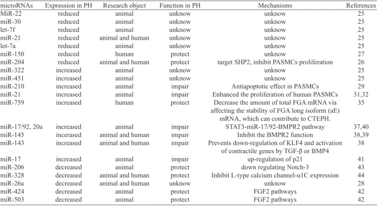
MicroRNAs (miRs) are small, non-coding RNAs reg-ulating gene expression at the post-transcriptional level by mRNA degradation or translational repression[23]. The hu-man genome has been estimated to contain up to 1000 miR-NAs[24]. Many miRNAs exhibit a tissue-speciic distribution and they appear to play a key role in cell function both under physiological and pathological conditions. Lots of in vivo and in vitro experiments related with functions of microR-NAs in PH have emerged recent years. From animal experi-ments to clinical trials, microRNA expression proiles in PH have been revealed. A range of miRNAs are dysregulated in the lungs of rats exposed to chronic hypoxic and the mono-crotaline model of PAH[25]. MiR-22, miR-30 and let-7f were down regulated, whereas miR-322 and miR-451 were up reg-ulated signiicantly during the development of PH in both hypoxic and monocrotaline models. miR-21 and let-7a were
signiicantly reduced only in monocrotalinetreated rats. miR-204 was consistently down regulated in pulmonary artery smooth muscle cells (PASMCs) from patients with PH and in cells from mice with PH[26]. Besides, circulating miR-150 and miR-26a levels are reduced in patients with poor survival in PH[27,28]. All the evidences above indicate a potential role of microRNAs in PH (Table 1).
As we all know, hypoxia is an important pathogenesis in PH. Inductions of microRNAs can be observed in SMCs cul-tured with hypoxia and in whole lungs of mice with chronic hypoxia-induced PH. miR-210 is the predominant miRNA induced by hypoxia, which has also been demonstrated by microarray analysis on human in hypoxic PASMCs and in whole lungs of hypoxic mice[29]. The induction of miR-210 is HIF-1α-dependent and triggers anti-apoptotic effects via directly targeting the transcription factor E2F3[29]. Previous-ly study performed on pulmonary artery endothelial cells (PAECs) found that miR-210 can provide an adaptation to hypoxic conditions by targeting Iron-Sulfur Cluster Assem-bly Proteins 1/2 (ISCU)[30].
MiR-21 can also be induced by hypoxia and overexpres-sion of miR-21 enhanced the proliferation of human PASMCs in vitro and the expression of cell proliferation associated proteins, such as proliferating cell nuclear antigen, cyclin D1, and Bcl-xL, which indicates that miR-21 plays an important role in the pathogenesis of chronic hypoxia-induced pulmo-nary vascular remodeling[31,32]. Previous study showed that
Table 1. Summary of microRNAs which may play a potential role in PH.
microRNAs MiR-22 miR-30 let-7f miR-21 let-7a miR-150 miR-204 miR-322 miR-451 miR-210 miR-21 miR-759 miR-17/92, 20a miR-145 miR-143 miR-17 miR-206 miR-328 miR-26a miR-424 miR-503
Expression in PH reduced reduced reduced reduced reduced reduced reduced increased increased increased increased increased increased incerased increased increased decreased decreased decreased decreased decreased Research object animal animal animal animal and human
animal human animal and human
animal animal animal animal human animal animal and human animal and human
animal animal animal and human animal and human
animal animal
Function in PH unknow unknow unknow unknow unknow protect protect unknow unknow impair impair protect impair impair impair impair protect protect unknow protect protect Mechanisms unknow unknow unknow unknow unknow unknow
target SHP2, inhibit PASMCs proliferation unknow
unknow
Antiapoptotic effect in PASMCs Enhanced the proliferation of human PASMCs
Decrease the amount of total FGA mRNA via affecting the stability of FGA long isoform (aE)
mRNA, which can contribute to CTEPH. STAT3-miR-17/92-BMPR2 pathway
Inhibit the BMPR2 function
Prevents down-regulation of KLF4 and activation of contractile genes by TGF-β or BMP4
up-regulation of p21 down regulating Notch-3
BMP-dependent signaling activation of miR-21 represses Rho-kinase activation in pulmonary artery endothelial cells, thus counteracting the Rho signaling in promoting pulmonary vascular pathology[33]. Besides, miR-21-null mice presented overexpression of RhoB and hyperaction of Rho-kinase activi-ty accompanied by exaggerated manifestation of PH[32].
Chronic thromboembolus is another leading cause of se-vere PH[34]. Chen et al.[35] investigated the involvement of miR-759 in chronic thromboembolic pulmonary hypertension (CTEPH). CTEPH is characterized by persistent pulmonary embolism that increases pulmonary vascular resistance, result-ing in pulmonary hypertension and subsequent right ventricu-lar heart failure. The 3′UTR of FGA was found to interact with miR-759, and a 28-bp deletion polymorphism at this site was found to be more frequent in patients with CTEPH.
Further studies have been investigated to elucidate the concrete mechanisms of microRNAs participating in PH those years. As we all know, bone morphogenetic protein receptor type II (BMPR2), a receptor for the transforming growth factor (TGF-) b family, plays an important role both in endothelial and vascular smooth muscle cells and vascular remodeling of the pulmonary arterial circulation[36]. Several Studies have been designed to identify miRNAs that could inhibit the translation of BMPR-II, and members of the miR-NA cluster 17/92 and miR-143, miR-145, miR-20a were identiied as the potential regulators[37-40]. All these microR-NAs inhibit the BMPR2 function, which were conirmed by experiments in either the patient vascular cells or the PH animal model. miR-145 and miR-143, two highly expressed miRNAs in SMCs, have been shown to play a pivotal role in the modulation of SMC phenotype. In particular, their expression is transcriptionally activated by both TGF-β and BMP4 and promotes a contractile phenotype in SMC by tar-geting the Kruppel-like factor-4 (KLF4)[38].
Besides the research in pathogenesis and mechanisms, there are also investigations in therapy eficacy of microR -NAs for PH in animal models. Pullamsetti et al.[41] demon-strated that inhibition of miR-17 improves heart and lung function in experimental PH by interfering with lung vascu-lar and right ventricuvascu-lar remodeling. The beneicial effects may be related to the up-regulation of p21. And recently, Kim et al.[42] found that reconstitution of 424 and miR-503 can ameliorate pulmonary hypertension in experimental models through FGF2 pathways.
Although recent studies found that most PH-related mi-croRNAs usually play a negative role in the pathogenesis process of PH, interestingly, there are still some microRNAs which can play a protective role in PH. 204 and miR-206 are two well researched microRNAs, both of which are down regulated in PASMCs from patients with PH or in cells from mice with PH. They all paticipate in the SMCs’ prolif-eration and apoptosis and even differentiation. miR-204 was consistently down regulated in PASMCs from patients with
PAH and in cells from mice with PAH[26]. miR-204 show a direct inluence on PASMC function and delivery of miR-204 mimics to the lungs of mice with PAH signiicantly can reduce disease severity. miR-206 can alleviate PAH through down regulating Notch-3 expression, which is key a factor in PAH development[43].
Besides, hypoxia produced a signiicant inhibition of miR-328 expression, which has been identiied as a strong candidate responsible for hypoxic pulmonary vasoconstric-tion. Overexpressing miR-328 in the transgenic mice remark-ably decreased the right ventricular systolic pressure and PA wall thickness under both normoxia and hypoxia. Through inhibiting L-type calcium channel-α1C expression the insu -lin growth factor 1 receptor, ultimately leading to apoptosis of pulmonary arterial smooth muscle cells[44].
POSSIBLE AND NOVEL LINK BETWEEN MSCS AND MIRNAS IN PH
It is well known that miRNAs have been implicated in many processes of stem cell functions, including cell pro-liferation, differentiation and apoptosis. Recent studies[45] suggest that mesenchymal stem cells have discrete miRNA expression proiles that can account for the intrinsic stem cell properties of self-renewal and pluripotency. Through certain modiied microRNAs, up or down regulation, there must be ways to enhance the viability of engrafted MSCs in the in-jured pulmonary tissue.
Exosomes have emerged as a novel media between kinds of cells. And exosomes based therapy has been conirmed by many researches. In consideration of its microRNAs-carried function, it is feasible to treat with PH by the microRNAs-car-ried exosomes secreted by MSCs. Recently, this hypothesis has been conirmed in a research that demonstrate MSCs can regulate neurite outgrowth by transfer of miR-133b to neural cells via exosomes[46].
In a word, either mimicking or antagonizing microRNA actions, MSCs functions can be regulated by microRNAs to enhance the properties of cell differentiation or anti-apoptosis. Considering that microRNAs can be delivered by exosomes secreted by MSCs, it is likely that overexpression of special microRNAs like miR-204/206/328 in MSCs will hopefully en-hance MSCs therapeutic eficacy for PH. So, microRNAs may be used as novel regulators in MSC-based therapy in PH and microRNAs-regulated MSCs transplantation may represent promising therapeutic strategy for PH patients in the future.
Authors’ roles & responsibilities
ZZ Study conception and design ZF Study conception and design
REFERENCES
1. Galiè N, Hoeper MM, Humbert M, Vachiery JL, Barbera JA, Beghetti M, et al. ; ESC Committee for Practice Guidelines (CPG). Guidelines for the diagnosis and treatment of pulmonary hypertension: the Task Force for the Diagnosis and Treatment of Pulmonary Hypertension of the European Society of Cardiology (ESC) and the European Respiratory Society (ERS), endorsed by the International Society of Heart and Lung Transplantation (ISHLT). Eur Heart J 2009;30(20):2493-537.
2. Humbert M, Sitbon O, Chaouat A, Bertocchi M, Habib G, Gressin V, et al. Survival in patients with idiopathic, familial, and anorexigen-associated pulmonary arterial hypertension in the modern management era. Circulation. 2010;122(2):156-63.
3. Jiang Y, Jahagirdar BN, Reinhardt RL, Schwartz RE, Keene CD, Ortiz-Gonzalez XR, et al. Pluripotency of mesenchymal stem cells derived from adult marrow. Nature. 2002;418(6893):41-9.
4. Zhao YD, Courtman DW, Deng Y, Kugathasan L, Zhang Q, Stewart DJ. Rescue of monocrotaline-induced pulmonary arterial hypertension using bone marrow-derived
endothelial-like progenitor cells: eficacy of combined cell and eNOS gene
therapy in established disease. Circ Res. 2005;96(4):442-50.
5. Myers TJ, Granero-Molto F, Longobardi L, Li T, Yan Y, Spagnoli A. Mesenchymal stem cells at the intersection of cell and gene therapy. Expert Opin Biol Ther. 2010;10(12):1663-79.
6. Firth AL, Yao W, Ogawa A, Madani MM, Lin GY, Yuan JX. Multipotent mesenchymal progenitor cells are present in endarterectomized tissues from patients with chronic thromboembolic pulmonary hypertension. Am J Physiol Cell Physiol. 2010;298(5):C1217-25.
7. Baber SR, Deng W, Master RG, Bunnell BA, Taylor BK, Murthy SN, et al. Intratracheal mesenchymal stem cell administration attenuates monocrotaline-induced pulmonary hypertension and endothelial dysfunction. Am J Physiol Heart Circ Physiol. 2007;292(2):H1120-8.
8. Aslam M, Baveja R, Liang OD, Fernandez-Gonzalez A, Lee C, Mitsialis SA, et al. Bone marrow stromal cells attenuate lung injury in a murine model of neonatal chronic lung disease. Am J Respir Crit Care Med. 2009;180(11):1122-30.
9. Guarita-Souza LC, Carvalho KAT, Rebelatto C, Senegaglia AC, Hansen P, Furuta M, et al. A comparação entre o transplante de células tronco mononucleares e mesenquimais no infarto do miocárdio. Rev Bras Cir Cardiovasc. 2005;20(3):270-8.
10. Spees JL, Whitney MJ, Sullivan DE, Lasky JA, Laboy M, Ylostalo J, et al. Bone marrow progenitor cells contribute to repair and remodeling of the lung and heart in a rat model of progressive pulmonary hypertension. FASEB J. 2008;22(4):1226-36.
11. Umar S, de Visser YP, Steendijk P, Schutte CI, Laghmani el H, Wagenaar GT, et al. Allogenic stem cell therapy improves
right ventricular function by improving lung pathology in rats with pulmonary hypertension. Am J Physiol Heart Circ Physiol. 2009;297(5):H1606-16.
12. Kanki-Horimoto S, Horimoto H, Mieno S, Kishida K, Watanabe F, Furuya E, et al. Implantation of mesenchymal stem cells overexpressing endothelial nitric oxide synthase improves right ventricular impairments caused by pulmonary hypertension. Circulation. 2006;114(1 Suppl):I181-5.
13. Takemiya K, Kai H, Yasukawa H, Tahara N, Kato S, Imaizumi T. Mesenchymal stem cell-based prostacyclin synthase gene therapy for pulmonary hypertension rats. Basic Res Cardiol. 2010;105(3):409-17.
14. Liang OD, Mitsialis SA, Chang MS, Vergadi E, Lee C, Aslam M, et al. Mesenchymal stromal cells expressing heme oxygenase-1 reverse pulmonary hypertension. Stem Cells. 2011; 29(1):99-107.
15. Anversa P, Perrella MA, Kourembanas S, Choi AM, Loscalzo J. Regenerative pulmonary medicine: potential and promise, pitfalls and challenges. Eur J Clin Invest. 2012;42(8):900-13.
16. Rochefort GY, Delorme B, Lopez A, Hérault O, Bonnet P, Charbord P, et al. Multipotential mesenchymal stem cells are mobilized into peripheral blood by hypoxia. Stem Cells. 2006;24(10):2202-8.
17. Wang M, Crisostomo P, Herring C, Meldrum KK, Meldrum DR. Human progenitor cells from bone marrow or adipose tissue produce VEGF, HGF and IGF-1 in response to TNF by a p38 MAPK-dependent mechanism. Am J Physiol Regul Integr Comp Physiol. 2006;291(4):R880-4.
18. Rehman J, Traktuev D, Li J, Merfeld-Clauss S, Temm-Grove CJ, Bovenkerk JE, et al. Secretion of angiogenic and antiapoptotic factors by human adipose stromal cells. Circulation. 2004;109(10):1292-8.
19. Kinnaird T, Stabile E, Burnett MS, Lee CW, Barr S, Fuchs S, et al. Marrow-derived stromal cells express genes encoding a broad spectrum of arteriogenic cytokines and promote in vitro and in vivo arteriogenesis through paracrine mechanisms. Circ Res. 2004;94(5):678-85.
20. Prockop DJ. Marrow stromal cells as stem cells for nonhematopoietic tissues. Science. 1997;276(5309):71-4.
21. Patel KM, Crisostomo P, Lahm T, Markel T, Herring C, Wang M, et al. Mesenchymal stem cells attenuate hypoxic pulmonary vasoconstriction by a paracrine mechanism. J Surg Res. 2007;143(2):281-5.
22. Lee C, Mitsialis SA, Aslam M, Vitali SH, Vergadi E, Konstantinou G, et al. Exosomes mediate the cytoprotective action of mesenchymal stromal cells on hypoxia-induced pulmonary hypertension. Circulation. 2012;126(22):2601-11.
24. Zhang L, Huang J, Yang N, Greshock J, Megraw MS, Giannakakis A, et al. microRNAs exhibit high frequency genomic alterations in human cancer. Proc Natl Acad Sci U S A. 2006;103(24):9136-41.
25. Caruso P, MacLean MR, Khanin R, McClure J, Soon E, Southgate
M, et al. Dynamic changes in lung microRNA proiles during the
development of pulmonary hypertension due to chronic hypoxia and monocrotaline. Arterioscler Thromb Vasc Biol. 2010;30(4):716-23.
26. Courboulin A, Paulin R, Giguère NJ, Saksouk N, Perreault T, Meloche J, et al. Role for miR-204 in human pulmonary arterial hypertension. J Exp Med. 2011;208(3):535-48.
27. Rhodes CJ, Wharton J, Boon RA, Roexe T, Tsang H, Wojciak-Stothard B, et al. Reduced miR-150 is associated with poor survival in pulmonary arterial hypertension. Am J Respir Crit Care Med. 2013;187(3):294-302.
28. Schlosser K, White RJ, Stewart DJ. miR-26a linked to pulmonary hypertension by global assessment of circulating extracellular MicroRNAs. Am J Respir Crit Care Med. 2013;188(12):1472-5.
29. Gou D, Ramchandran R, Peng X, Yao L, Kang K, Sarkar J, et al. miR-210 has an antiapoptotic effect in pulmonary artery smooth muscle cells during hypoxia. Am J Physiol Lung Cell Mol Physiol. 2012;303(8):L682-91.
30. Chan SY, Zhang YY, Hemann C, Mahoney CE, Zweier JL, Loscalzo J. MicroRNA-210 controls mitochondrial metabolism during hypoxia by repressing the iron-sulfur cluster assembly proteins ISCU1/2. Cell Metab. 2009;10(4):273-84.
31. Yang S, Banerjee S, Freitas Ad, Cui H, Xie N, Abraham E, et al. miR-21 regulates chronic hypoxia-induced pulmonary vascular remodeling. Am J Physiol Lung Cell Mol Physiol. 2012;302(6):L521-9.
32. Parikh VN, Jin RC, Rabello S, Gulbahce N, White K, Hale A, et al. MicroRNA-21 integrates pathogenic signaling to control pulmonary hypertension: results of a network bioinformatics approach. Circulation. 2012;125(12):1520-32.
33. Connolly MJ, Aaronson PI. Key role of the RhoA/Rho kinase system in pulmonary hypertension. Pulm Pharmacol Ther. 2011;24(1):1-14.
34. Jatene FB, Pêgo-Fernandes PM, Poveda S, Monteiro R, Cukier A, Mady C, et al. Tratamento cirúrgico da embolia pulmonar crônica: análise da experiência inicial. Rev Bras Cir Cardiovasc. 1995;10(2):70-6.
35. Chen Z, Nakajima T, Tanabe N, Hinohara K, Sakao S, Kasahara Y, et al. Susceptibility to chronic thromboembolic pulmonary hypertension may be conferred by miR-759 via its targeted
interaction with polymorphic ibrinogen alpha gene. Hum Genet.
2010;128(4):443-52.
36. Rabinovitch MJ. Molecular pathogenesis of pulmonary arterial hypertension. Clin Invest. 2012;122(12):4306-13.
37. Brock M, Trenkmann M, Gay RE, Michel BA, Gay S, Fischler M, et al. Interleukin-6 modulates the expression of the bone morphogenic protein receptor type II through a novel STAT3-microRNA cluster 17/92 pathway. Circ Res. 2009;104(10):1184-91.
38. Davis-Dusenbery BN, Chan MC, Reno KE, Weisman AS, Layne MD, Lagna G, et al. down-regulation of Kruppel-like factor-4 (KLF4) by microRNA-143/145 is critical for modulation of vascular smooth muscle cell phenotype by transforming growth factor-beta and bone morphogenetic protein 4. J Biol Chem. 2011;286(32):28097-110.
39. Caruso P, Dempsie Y, Stevens HC, McDonald RA, Long L, Lu R, et al. A role for miR-145 in pulmonary arterial hypertension: evidence from mouse models and patient samples. Circ Res. 2012;111(3):290-300.
40. Brock M, Samillan VJ, Trenkmann M, Schwarzwald C, Ulrich S, Gay RE, et al. AntagomiR directed against miR-20a restores functional BMPR2 signalling and prevents vascular remodelling in hypoxia-induced pulmonary hypertension. Eur Heart J. 2014;35(45):3203-11.
41. Pullamsetti SS, Doebele C, Fischer A, Savai R, Kojonazarov B, Dahal BK, et al. Inhibition of microRNA-17 improves lung and heart function in experimental pulmonary hypertension. Am J Respir Crit Care Med. 2012;185(4):409-19.
42. Kim J, Kang Y, Kojima Y, Lighthouse JK, Hu X, Aldred MA, et al. An endothelial apelin-FGF link mediated by miR-424 and miR-503 is disrupted in pulmonary arterial hypertension. Nat Med. 2013;19(1):74-82.
43. Jalali S, Ramanathan GK, Parthasarathy PT, Aljubran S, Galam L, Yunus A, et al. Mir-206 regulates pulmonary artery smooth muscle cell proliferation and differentiation. PLoS One. 2012;7(10):e46808.
44. Guo L, Qiu Z, Wei L, Yu X, Gao X, Jiang S, et al. The microRNA-328 regulates hypoxic pulmonary hypertension by targeting at insulin growth factor 1 receptor and L-type calcium
channel-α1C. Hypertension. 2012;59(5):1006-13.
45. Guo L, Zhao RC, Wu Y. The role of microRNAs in self-renewal and differentiation of mesenchymal stem cells. Exp Hematol. 2011;39(6):608-16.