ISSN 0100-879X
BIOMEDICAL SCIENCES
AND
CLINICAL INVESTIGATION
www.bjournal.com.br
www.bjournal.com.br
Volume 44 (10) 966-1069 October 2011
Institutional Sponsors
The Brazilian Journal of Medical and Biological Research is partially financed by
Faculdade de Medicina de Ribeirão Preto Campus
Ribeirão Preto
Ex plor e H igh - Pe r for m a n ce M S Or bit r a p Te ch n ology I n Pr ot e om ics & M e t a bolom ics
analit icaw eb.com .br S C I E N T I F I C
Braz J Med Biol Res, October 2011, Volume 44(10) 1000-1005
doi: 10.1590/S0100-879X2011007500108
Reference value of brachial-ankle pulse wave velocity for the
eastern Chinese population and potential influencing factors
Reference value of brachial-ankle pulse
wave velocity for the eastern Chinese
population and potential influencing
factors
Zi-Sheng Ai
1,
Jue Li
1, Zhong-Min Liu
2, Hui-Min Fan
2, Dai-Fu Zhang
3,
Yun Zhu
1, Li-Juan Zhang
1, Wen-Qing Zhu
1and Yan Bao
11Department of Preventive Medicine, College of Medicine, Tongji University, Shanghai, China 2Department of Cardiac Surgery, 3Department of Cardiology, East Hospital, Tongji University, Shanghai, China
Abstract
The present study was conducted to obtain reference values for brachial-ankle pulse wave velocity (baPWV) and to evaluate
influencing factors of baPWV according to gender. Using automatic devices, baPWV was measured simultaneously in 2095
subjects. A total of 647 healthy subjects, none of whom presented atherosclerotic risk factors, were analyzed in the present study. Two different statistical methods were used to obtain reference values for baPWV according to subject gender and age. The association between baPWV value and gender, as well as other features, were analyzed. For male subjects, multiple stepwise analysis showed that age, systolic blood pressure (SBP), heart rate (HR), and plasma levels of triglycerides (TG) were independent predictors of baPWV. For female subjects, age, SBP, HR, and plasma levels of uric acid (UA) were inde-pendent predictors of baPWV. In male subjects, the upper limits of baPWV values were 1497.43/1425.00, 1518.67/1513.25, 1715.97/1726.50, 1925.20/1971.90, and 2310.18/2115.00 cm/s, obtained using two different statistical methods for the age ranges of 30-39, 40-49, 50-59, 60-69, and 70 and older, respectively. For females, the upper limits of baPWV values were 1426.70/1411.13, 1559.15/1498.95, 1733.50/1739.00, 1958.63/1973.78, and 2720.80/2577.00 cm/s for the age ranges of
30-39, 40-49, 50-59, 60-69, and 70 and older, respectively. Aging is the most important influencing factor for baPWV value and its
effect is more prominent in females. The reference values of baPWV according to age and gender may be useful for the clinical diagnosis and preventive therapy of cardiovascular diseases.
Key words: Pulse wave velocity; Reference value; Age; Gender; Influencing factor
Introduction
Correspondence: Zi-Sheng Ai, Department of Preventive Medicine, College of Medicine, Tongji University, Shanghai, 200092 China. Fax: +86-21-6598-6270. E-mail: azs1966@126.com
Received March 31, 2011. Accepted August 10, 2011. Available online August 26, 2011. Published October 10, 2011.
Arteries are channels that transmit blood at high pressure to peripheral vascular beds (1). Arterial elasticity dysfunction is an important marker of cardiovascular risk and arterial stiffness plays a key role in the pathophysiology of the cardiovascular system (2-4). Increased arterial stiffness parallels structural changes in the medial layer of the elastic arteries (mainly aorta and major arterial conduits), and is largely the result of
progressive elastic fiber degeneration. An increase in stiffness
related to arterial wall composition occurs with aging, and is accelerated in patients with hypertension (5,6).
In recent years, great emphasis has been placed on the role of arterial stiffness in the development of cardiovascular (CV) diseases. Arterial stiffness has an important, independent predictive power with respect to cardiovascular mortality, coronary events and several atherosclerotic diseases (2,7).
Measurement of pulse wave velocity (PWV) in human subjects has been proposed as one method to diagnose and evaluate distensibility of large arteries and to assess arterial stiffness. PWV, which is inversely related to arterial wall distensibility, offers a simple and potentially useful approach for evaluat-ing cardiovascular disease (8,9). In-depth studies regardevaluat-ing PWV have been conducted and PWV is now considered to be an independent predictor of CV risk and prognosis. PWV
not only reflects systemic arteriosclerosis but also relates to
cardiovascular risk factor (10), and ischemic heart disease in type II diabetes mellitus (10,11). The aortic PWV is the “gold standard” marker for measuring arterial stiffness, and is widely
used to estimate vascular stiffness and “vascular health”(1).
Reference value of baPWV and influencingfactors 1001
measurement (2).
Brachial-ankle pulse wave velocity (baPWV) has been developed as a simple noninvasive index of arterial stiffness (12-15) and has been reported to be correlated with
carotid-femoral PWV (12,15). The baPWV measurement is a reflection of the flexibility of the aorta and medium arteries. This method
is simple and reproducible, saves time, and is also suitable for large-scale population screening and follow-up. Therefore, in order to adopt baPWV for routine clinical use and render it a powerful tool for early diagnosis of atherosclerosis in medical
institutions, it is vital to determinereference value of baPWV
in healthy subjects. The purpose of the present study was
to investigate major influencingfactors of baPWV in healthy eastern Chinese people and to determine the reference val-ues of baPWV in healthy people in different age groups and gender.
Material and Methods
Study population
A total of 2095consecutive subjects ranging in age from
30 to 85 years, from the Pudong New Districts of Shanghai, China, were enrolled in this study between July and August 2009. Informed consent was obtained from all subjects. We analyzed the cross-sectional epidemiological data at the end of the study. Of the 2095 volunteers, 647 were considered to
behealthy anddid not present any atherosclerotic risk factors.
These subjectswere separated according to gender and age
for analysis of baPWV. A ‘healthy subject’ was defined by
the following criteria: blood pressure <140/90 mmHg, fasting blood glucose (FBG) <6.1 mM, total cholesterol (TC) <5.9 mM,
triglycerides (TG) <1.70 mM, maleenzymatic uric acid (UA)
<428 µM, female enzymaticUA <357 µM, body mass index
(BMI) <25, no medication, no history of CV diseases, and no history of smoking (12,16).
Measurement of baPWV
baPWV was measured in 2095 subjects using an
au-tomated device (Model BP203RPE-II [VP-1000], Omron,
Japan). baPWV, systolic blood pressure (SBP), diastolic blood pressure (DBP), electrocardiogram, and heart sounds were recorded simultaneously. None of the participants took any medications on the day of the examination. Measurements were performed in the supine position. Waveforms were ob-tained from volume plethysmographic sensors in cuffs on the right brachium and both ankles. The instrument automatically recorded the time intervals (Tba) between the wave at the right brachium and at both ankles. The distance between the sampling points of baPWV was calculated using the following equation: baPWV = (La - Lb) / Tba, where Lb represents the length from the suprasternal notch to the right brachium (Lb) = 0.2195 x height (cm) - 2.0734, and La represents the length from the suprasternal notch to the ankle (La) = 0.8129 x height (cm) + 12.328.
The baPWV was calculated as the distance between
re-cording sites measured over the surface of the body, divided by the time interval of the pressure waves between the feet (17,18). This device automatically and simultaneously mea-sures bilateral baPWV and brachial and ankle blood presmea-sures. In our analysis, we used the mean value of the bilateral baPWV, and left brachial blood pressure.
Laboratory measurements
Fasting venous blood samples were drawn, and TC, TG, high-density lipoprotein cholesterol (HDL-C), low-density lipoprotein cholesterol (LDL-C), plasma levels of UA, high sensitivity C-reactive protein (hs-CRP), FBG, and hemoglobin A1c (HbA1c) were measured using standard methods.
Statistical analysis
Continuous variables are reported as means ± SD or median (quartile range) according to the characteristics of the data. The Kolmogorov-Smirnov test was applied to as-sess data distribution. Differences between variables were
compared using the independent samples t-test, while the
Mann-Whitney U-test was used to assess data that were not normally distributed. Linear correlations were determined using
Spearman’s rank-order correlation coefficient, and stepwise
multiple regression analysis was performed to determine fac-tors associated with baPWV. Potential risk facfac-tors, including age, blood pressure, heart rate, serum lipid, FBG, plasma level of UA, hs-CRP, and BMI were assessed in multiple analyses. The average values of the right and left baPWV were used in the statistical analysis. The arctan transformed baPWV value was used to determine the reference range of baPWV, and the upper limit was calculated by adding the mean value to +1.64 x SD, followed by the application of tangent transformation. The percentile method was also used to deduce the reference range of baPWV. Statistical calculations were performed using
the SPSS software, version 14.0 (SPSS, USA). P values ≤0.05 were considered to be statistically significant.
Results
Clinical characteristics
The clinical characteristics of the healthy subjects are shown in Table 1. The total number of subjects consisted of 702 men and 1393 women. The male subjects had a mean age of 58.12 ± 10.86 years (range, 30-87 years) and female subjects had a mean age of 57.14 ± 9.25 years (range, 30-88
years). There was no significant difference in pulse pressure,
ankle-brachial index, BMI, hs-CRP, or HbA1c (%) between genders. In contrast, males had higher values with respect to age, SBP, DBP, mean blood pressure, baPWV, height, weight,
waist, TG, UA, TC/HDL-C ratio, and FBG (male vs female,
P < 0.05). In females, TC, HDL-C, and LDL-C values were
significantly higher (female vs male, P < 0.05).
BaPWV and influencingfactors
between baPWV and other clinical variables revealed that, in
males, age, SBP, heart rate, and TG were significant variables
and, in females, age, SBP, heart rate and UA were positively associated with baPWV (Table 2).
The relationship between age and baPWV by gender
The Mann-Whitney U-test was used to analyze baPWV val-ues according to gender. The results revealed that the baPWV
values differed significantly between genders (Z = -2.781, P =
0.005). The Kruskal-Wallis H-test was used to compare baPWV
values among different age groups by gender, and the results showed that the values of baPWV among different age groups
were also statistically significant (H = 49.810, P = 0.000 in
male versus H = 120.120, P = 0.000 in female). The changes
in age and baPWV for both males and females are displayed in Figure 1. Lines within boxes represent median values. The upper and lower boundaries of the boxes indicate the 25th and 75th percentiles, and bars above and below the boxes indicate minimum and maximum values, respectively.
In the estimation of the regression curve (the relationship between age and baPWV) both genders demonstrated a
quadratic curve: baPWV (male) = 0.1354 x age2 - 3.8846
x age + 1203.1 (R2 = 0.2736, P < 0.001; Rs = 0.487,P <
0.001); baPWV (female) = 0.2237 x age2 - 9.6129 x age
+ 1220.8 (R2 = 0.2642, P < 0.001; Rs = 0.549,P < 0.001).
In addition to the value of the standardized coefficientin multiple regression analysis, the Spearman correlation
coefficient between age and baPWV also showed that the
effect of age on baPWV was greater in females than that in males.
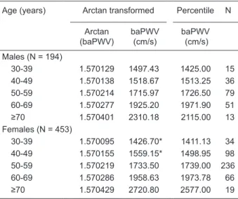
Determination of reference values of baPWV by two kinds of methods
In order to obtain reference values for baPWV, we applied the percentile method and arctan transformation method to the different age groups of both genders. The results are listed by gender in Table 3. In contrast to the fe-male population, the arctan transformation method revealed that healthy male subjects had a higher baPWV value when less than 40 years of age. Similarly, the percentile method revealed that healthy male subjects had a higher baPWV value when less than 50 years of age.
Discussion
The change in arterial function and structure is the main implication for clinical CV events. To date, the methods for evaluating arterial function are very limited; therefore,
the clinical significance of noninvasive evaluation of early
vascular diseases has attracted more attention. Increas-ing arterial stiffness is a pathological state of vascular damage, and is closely associated with atherosclerotic cardiovascular diseases, such as coronary artery disease (19,20). Pulse wave velocity is widely used as an indicator of arterial stiffness, and it has been shown that PWV is also
Table 1. Clinical characteristics of male and female participants of this study (N = 2095).
Variables Male (N = 702) Female (N = 1393)
Age (years) 58.12 ± 10.86 57.14 ± 9.25* SBP (mmHg) 136.29 ± 20.58 132.36 ± 22.47* DBP (mmHg) 77.99 ± 11.89 74.52 ± 11.52* MBP (mmHg) 97.43 ± 13.72 93.80 ± 17.65* PP (mmHg) 58.30 ± 14.59 57.84 ± 13.70 baPWV (cm/s) 1556.25 (431.00) 1482.00 (420.00)*
ABI 1.11 ± 0.08 1.10 ± 0.08
Height (cm) 168.11 ± 6.16 157.35 ± 5.69* Weight (kg) 68.82 ± 10.10 59.50 ± 9.14* BMI (kg/m2) 24.32 ± 3.10 24.03 ± 3.41 Waist (cm) 88.80 ± 8.86 84.65 ± 8.78* Laboratory parameters
TC (mM) 4.90 ± 0.94 5.34 ± 0.97* TG (mM) 1.94 ± 1.60 1.71 ± 1.20* HDL-C (mM) 1.16 ± 0.29 1.37 ± 0.32* LDL-C (mM) 2.88 ± 0.79 3.22 ± 1.12* UA (µM) 366.67 ± 78.42 290.32 ± 67.94* hs-CRP (g/L) 2.06 ± 2.85 2.10 ± 3.30
T/HR 4.42 ± 1.29 4.06 ± 1.09*
FBG (mM) 5.52 ± 1.46 5.35 ± 1.25* HbA1c (%) 6.02 ± 2.22 5.95 ± 1.64
Data are reported as means ± SD, except for baPWV. SBP = systolic blood pressure; DBP = diastolic blood pressure; MBP = mean blood pressure; PP = pulse pressure; baPWV = brachial/ ankle pulse wave velocity; ABI = ankle-brachial index; BMI = body mass index; TC = plasma levels of total cholesterol; TG = plasma levels of triglycerides; HDL-C = plasma levels of high-density poprotein cholesterol; LDL-C = plasma levels of low-density li-poprotein cholesterol; UA = plasma levels of uric acid; hs-CRP = high sensitivity C-reactive protein; T/HR = TC and HDL-C ratio; FBG = fasting blood glucose; HbA1c = hemoglobin A1c. *P < 0.05, compared to males (independent samples t-test).
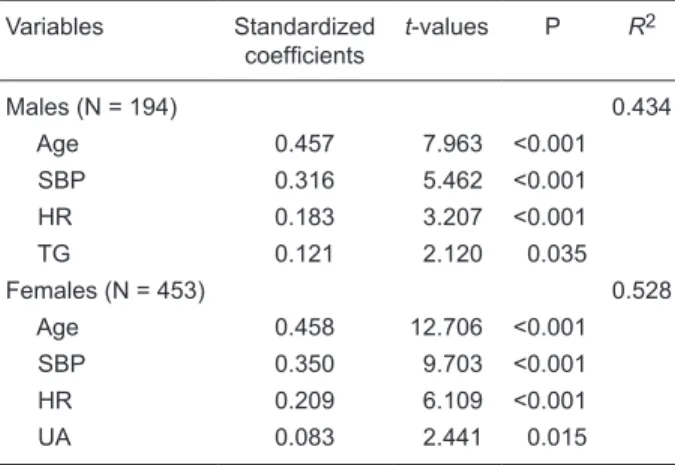
Table 2. Step-wise multiple regression analysis using the baPWV of healthy subjects as the dependent variable.
Variables Standardized
coefficients t-values P R
2
Males (N = 194) 0.434
Age 0.457 7.963 <0.001
SBP 0.316 5.462 <0.001
HR 0.183 3.207 <0.001
TG 0.121 2.120 0.035
Females (N = 453) 0.528
Age 0.458 12.706 <0.001
SBP 0.350 9.703 <0.001
HR 0.209 6.109 <0.001
UA 0.083 2.441 0.015
Reference value of baPWV and influencingfactors 1003
an important predictor of risk and prognosis of CV disease
(2,12,14,15,21). Considering the relationship between
flow-mediated dilation of the brachial artery, carotid intima-media thickness and PWV, the combination of these measures
may be of clinical significance (22).
Some studies have demonstrated that conventional ath-erosclerotic risk factors such as smoking, obesity, diabetes
mellitus, and dyslipidemia influence PWV (23-26). In order
to eliminate these confounding variables, the present study was carried out on a population of ‘healthy subjects’ who did not present any atherosclerotic risk factors. The result of the present study showed that the older the subject, the greater the upper limit value of baPWV, especially in people over 60 years. Males had a higher average and upper limit values than females at less than 50 years of age. Above 50 years of age, females had higher baPWV.
It was also reported that marked arterial stiffness was fit -ted with non-linear quadratic equation over 55 years of age (27). Although the present data did not meet normality
requirements sufficient for common data transformation
(P = 0.000), the data met the requirement of normality for arctan transformation (P = 0.200). In the present study, the normal distribution method via arctan transformation and the percentile method were used to determine the reference value of baPWV in different age groups by gender, and the upper limit of normal distribution and 95th percentile were considered for the formulation of said value. The normal value of baPWV will primarily aid in determining the degree of atherosclerosis, and in facilitating primary screening of potential cardiovascular and cerebrovascular diseases. Therefore, it is necessary to quickly and accurately detect patients with increased arterial stiffness in order to avoid or delay the occurrence and development of cardiovascular and cerebrovascular events.
For the elderly, who have no clinical symptoms of car-diac or cerebrovascular disease, increasing age induces pathological changes in the structure and function of the arterial wall. Age, blood pressure and other unknown factors are causes of increased arterial stiffness (16,28,29). The present study revealed that baPWV increased with age in both males and females, with no exception. In recent years, clinical studies have shown that decreased aortic compli-ance was an independent risk of CV disease mortality. In the present study, multivariate analysis demonstrated that age was a more important determinant of baPWV in females than in males independent of blood pressure variables.
Although we could not confirm the menopausal status of
each individual, the results in Table 3 show that menopause
was an important factor influencing arterial stiffness in
healthy females. Considering that levels of estrogens and
androgens in females and males fluctuate with age, it is possible that the influence of age on the arterial tree may
have contributed to this result. We also observed that the baPWV of both males and females was positively correlated with heart rate. Despite this result, an inherent correlation
between the increase in heart rate and the reduction of arterial elasticity is not clear. A high heart rate shortens the time available for recoil, which can lead to arterial
stiffen-ing. Epidemiological and clinical studies have confirmed
a relationship between increased serum uric acid and the
Figure 1. Box plots showing the median and dispersion of bra-chial-ankle pulse wave velocity (baPWV) distribution according to different age groups by gender.
Table 3. Reference brachial-ankle pulse wave velocity (baPWV) values for men and women.
Age (years) Arctan transformed Percentile N
Arctan (baPWV)
baPWV (cm/s)
baPWV (cm/s)
Males (N = 194)
30-39 1.570129 1497.43 1425.00 15 40-49 1.570138 1518.67 1513.25 36 50-59 1.570214 1715.97 1726.50 79 60-69 1.570277 1925.20 1971.90 51
≥70 1.570401 2310.18 2115.00 13
Females (N = 453)
30-39 1.570095 1426.70* 1411.13 34 40-49 1.570155 1559.15* 1498.95 98 50-59 1.570219 1733.50 1739.00 236 60-69 1.570286 1958.63 1973.78 66
≥70 1.570429 2720.80 2577.00 19
*P < 0.001, compared to males (Mann-Whitney U-test).The 95th
incidence and mortality of CV disease (30). Hyperuricemia is an independent indicator of carotid-femoral pulse wave velocity (cfPWV) and carotid-radial pulse wave velocity. We observed that baPWV in females correlated positively with UA, and, in males, a correlation between baPWV and TG was observed.
Recently, with the development of noninvasive mea-surement techniques and the increased incidence of car-diovascular diseases, PWV for noninvasive measurement,
which can effectively reflect arterial stiffness, has become
a hot topic in cardiovascular research. cfPWV is the most studied technique, and its reproducibility and validity have
been well verified. However, cfPWV can only reflects the flexibility of aortic arteries, and the measurement method is relatively complicated, while baPWV reflects the elasticity
of both the aorta and middle arteries and the measurement method is simple and time-saving. Based on these crite-ria, baPWV has greater application prospects compared
to cfPWV. The detection of PWV can help identify early changes in vascular structure and function in a high-risk cohort with cardiovascular diseases. The assessment of the damage in arterial structure and function is of great value
in determining high-risk patients and judging the efficacy
of treatments.
This study showed that there was a significant indepen -dent correlation between baPWV and age, SBP, heart rate,
and TG in men, while there was a significantly independent
correlation between baPWV and age, SBP, heart rate, and UA in women. These results suggest that baPWV may be used to identify the development of atherosclerosis and the risk of CV diseases, and offer guidance for treatment.
Aging is the most important influencing factor of baPWV,
and this effect is more prominent in females. The reference values of baPWV according to gender and age may be valuable for clinical treatment and preventive medicine in cardiovascular disease.
References
1. Gkaliagkousi E, Douma S. The pathogenesis of arterial stiff-ness and its prognostic value in essential hypertension and cardiovascular diseases. Hippokratia 2009; 13: 70-75. 2. Laurent S, Boutouyrie P, Asmar R, Gautier I, Laloux B, Guize
L, et al. Aortic stiffness is an independent predictor of all-cause and cardiovascular mortality in hypertensive patients. Hypertension 2001; 37: 1236-1241.
3. Asmar R, Rudnichi A, Blacher J, London GM, Safar ME. Pulse pressure and aortic pulse wave are markers of cardio-vascular risk in hypertensive populations. Am J Hypertens 2001; 14: 91-97.
4. Agabiti-Rosei E, Porteri E, Rizzoni D. Arterial stiffness, hy-pertension, and rational use of nebivolol. Vasc Health Risk Manag 2009; 5: 353-360.
5. Avolio AP, Deng FQ, Li WQ, Luo YF, Huang ZD, Xing LF, et al. Effects of aging on arterial distensibility in populations with high and low prevalence of hypertension: comparison between urban and rural communities in China. Circulation 1985; 71: 202-210.
6. Avolio AP, Chen SG, Wang RP, Zhang CL, Li MF, O’Rourke MF. Effects of aging on changing arterial compliance and left ventricular load in a northern Chinese urban community. Circulation 1983; 68: 50-58.
7. Mattace-Raso FU, van der Cammen TJ, Hofman A, van Popele NM, Bos ML, Schalekamp MA, et al. Arterial stiffness and risk of coronary heart disease and stroke: the Rotterdam Study. Circulation 2006; 113: 657-663.
8. Lee NB, Park CG. Reproducibility of regional pulse wave velocity in healthy subjects. Korean J Intern Med 2009; 24: 19-23.
9. Maple-Brown LJ, Piers LS, O’Rourke MF, Celermajer DS, O’Dea K. Increased arterial stiffness in remote Indigenous Australians with high risk of cardiovascular disease. J Hy-pertens 2007; 25: 585-591.
10. Sato H, Hayashi J, Harashima K, Shimazu H, Kitamoto K. A population-based study of arterial stiffness index in relation to cardiovascular risk factors. J Atheroscler Thromb 2005;
12: 175-180.
11. Hatsuda S, Shoji T, Shinohara K, Kimoto E, Mori K, Fuku-moto S, et al. Regional arterial stiffness associated with ischemic heart disease in type 2 diabetes mellitus. J Athero-scler Thromb 2006; 13: 114-121.
12. Yamashina A, Tomiyama H, Takeda K, Tsuda H, Arai T,
Hi-rose K, et al. Validity, reproducibility, and clinical significance
of noninvasive brachial-ankle pulse wave velocity measure-ment. Hypertens Res 2002; 25: 359-364.
13. Yamashina A, Tomiyama H, Arai T, Hirose K, Koji Y, Hiraya-ma Y, et al. Brachial-ankle pulse wave velocity as a Hiraya-marker of atherosclerotic vascular damage and cardiovascular risk. Hypertens Res 2003; 26: 615-622.
14. Munakata M, Ito N, Nunokawa T, Yoshinaga K. Utility of auto-mated brachial ankle pulse wave velocity measurements in hypertensive patients. Am J Hypertens 2003; 16: 653-657. 15. Sugawara J, Hayashi K, Yokoi T, Cortez-Cooper MY, DeVan
AE, Anton MA, et al. Brachial-ankle pulse wave velocity: an index of central arterial stiffness? J Hum Hypertens 2005; 19: 401-406.
16. Tomiyama H, Yamashina A, Arai T, Hirose K, Koji Y,
Chika-mori T, et al. Influences of age and gender on results of
noninvasive brachial-ankle pulse wave velocity measure-ment - a survey of 12517 subjects. Atherosclerosis 2003; 166: 303-309.
17. Khot UN, Khot MB, Bajzer CT, Sapp SK, Ohman EM, Brener SJ, et al. Prevalence of conventional risk factors in patients with coronary heart disease. JAMA 2003; 290: 898-904. 18. Hennekens CH. Increasing burden of cardiovascular
dis-ease: current knowledge and future directions for research on risk factors. Circulation 1998; 97: 1095-1102.
19. Park SM, Seo HS, Lim HE, Shin SH, Park CG, Oh DJ, et al. Assessment of arterial stiffness index as a clinical parameter for atherosclerotic coronary artery disease. Circ J 2005; 69: 1218-1222.
Reference value of baPWV and influencingfactors 1005
atherosclerosis and peripheral artery diseases in urban Chinese patients. Hypertens Res 2008; 31: 1079-1085. 21. Boutouyrie P, Tropeano AI, Asmar R, Gautier I, Benetos A,
Lacolley P, et al. Aortic stiffness is an independent predic-tor of primary coronary events in hypertensive patients: a longitudinal study. Hypertension 2002; 39: 10-15.
22. Kobayashi K, Akishita M, Yu W, Hashimoto M, Ohni M, Toba K. Interrelationship between non-invasive measurements
of atherosclerosis: flow-mediated dilation of brachial artery,
carotid intima-media thickness and pulse wave velocity. Atherosclerosis 2004; 173: 13-18.
23. Levenson J, Simon AC, Cambien FA, Beretti C. Cigarette smoking and hypertension. Factors independently associ-ated with blood hyperviscosity and arterial rigidity. Arterio-sclerosis 1987; 7: 572-577.
24. Toto-Moukouo JJ, Achimastos A, Asmar RG, Hugues CJ, Safar ME. Pulse wave velocity in patients with obesity and hypertension. Am Heart J 1986; 112: 136-140.
25. Airaksinen KE, Salmela PI, Linnaluoto MK, Ikaheimo MJ, Ahola K, Ryhanen LJ. Diminished arterial elasticity in
diabe-tes: association with fluorescent advanced glycosylation end
products in collagen. Cardiovasc Res 1993; 27: 942-945. 26. Muramatsu J, Kobayashi A, Hasegawa N, Yokouchi S.
Hemodynamic changes associated with reduction in total cholesterol by treatment with the HMG-CoA reductase inhibi-tor pravastatin. Atherosclerosis 1997; 130: 179-182. 27. Lajemi M, Labat C, Gautier S, Lacolley P, Safar M, Asmar
R, et al. Angiotensin II type 1 receptor-153A/G and 1166A/C gene polymorphisms and increase in aortic stiffness with age in hypertensive subjects. J Hypertens 2001; 19: 407-413. 28. Boutouyrie P, Laurent S, Briet M. Importance of arterial
stiff-ness as cardiovascular risk factor for future development of new type of drugs. Fundam Clin Pharmacol 2008; 22: 241-246.
29. McEniery CM, Yasmin, Hall IR, Qasem A, Wilkinson IB, Cockcroft JR. Normal vascular aging: differential effects on
wave reflection and aortic pulse wave velocity: the
Anglo-Cardiff Collaborative Trial (ACCT). J Am Coll Cardiol 2005; 46: 1753-1760.