The secondary alcohol and aglycone
metabolites of doxorubicin alter
metabolism of human erythrocytes
1Istituto di Biochimica e Biochimica Clinica, Facoltà di Medicina e Chirurgia,
Università Cattolica del Sacro Cuore, Rome, Italy
2CNR, Istituto di Chimica del Riconoscimento Molecolare, Rome, Italy
F. Misiti1,
B. Giardina1,2,
A. Mordente1 and
M.E. Clementi2
Abstract
Anthracyclines, a class of antitumor drugs widely used for the treat-ment of solid and hematological malignancies, cause a cumulative dose-dependent cardiac toxicity whose biochemical basis is unclear. Recent studies of the role of the metabolites of anthracyclines, i.e., the alcohol metabolite doxorubicinol and aglycone metabolites, have suggested new hypotheses about the mechanisms of anthracycline cardiotoxicity. In the present study, human red blood cells were used as a cell model. Exposure (1 h at 37ºC) of intact human red blood cells to doxorubicinol (40 µM) and to aglycone derivatives of doxorubicin (40 µM) induced, compared with untreated red cells: i) a ~2-fold stimulation of the pentose phosphate pathway (PPP) and ii) a marked inhibition of the red cell antioxidant enzymes, glutathione peroxidase (~20%) and superoxide dismutase (~60%). In contrast to doxorubicin-derived metabolites, doxorubicin itself induced a slighter PPP stimu-lation (~35%) and this metabolic event was not associated with any alteration in glutathione reductase, glutathione peroxidase, catalase or superoxide dismutase activity. Furthermore, the interaction of hemo-globin with doxorubicin and its metabolites induced a significant increase (~22%) in oxygen affinity compared with hemoglobin incu-bated without drugs. On the basis of the results obtained in the present study, a new hypothesis, involving doxorubicinol and aglycone me-tabolites, has been proposed to clarify the mechanisms responsible for the doxorubicin-induced red blood cell toxicity.
Correspondence
F. Misiti
Istituto di Biochimica e Biochimica Clinica
Facoltà di Medicina e Chirurgia Università Cattolica del Sacro Cuore L.go F. Vito, No. 1
00168 Rome Italy
Fax: +39-6-305-3598
E-mail: f.misiti@uniserv.ccr.rm.cnr.it
Received June 2, 2003 Accepted September 23, 2003
Key words
•Anthracyclines •Doxorubicinol •Doxorubicin •Aglycone metabolite •Pentose phosphate pathway •[13C]-NMR spectroscopy •Hemoglobin
Introduction
Doxorubicin (DOX), an anthracycline drug widely used for the treatment of solid and hematological malignancies (1), causes cumulative dose-dependent cardiac toxicity characterized by irreversible dilated cardio-myopathy and congestive heart failure (2).
of DOX-induced toxicity have implicated DOX metabolites other than semiquinone free radicals. Recently, it has been demon-strated that in human cardiac cytosol, DOX is primarily converted to a secondary alco-hol metabolite, i.e., doxorubicinol (DOXol) and/or to a broad panel of aglycones (see Figure 1) by an NADP(H)-dependent
mech-anism (6). In vivo and in vitro studies have
demonstrated that DOXol may be involved in several mechanisms of toxicity in heart
tissue, which include inhibition of the Ca2+/
Mg2+ ATPase of sarcoplasmic reticulum,
the F0-F1 proton pump of mitochondria and
the Na+-K+ ATPase and Na+-Ca2+ exchanger
of sarcolemma (7,8). Toxic effects on a num-ber of mitochondrial functions (9,10) have been reported for the aglycone derivatives of DOX.
Moreover, in vitro and in vivo studies
have shown that several parameters indica-tive of red blood cell function and integrity might be negatively affected by anthracycline drugs. In fact, changes in membrane ionic permeability (11), alteration of sodium trans-porters (12), inhibition of inositol lipid me-tabolism (13), increase in lipid peroxidation (14,15), and alterations of membrane struc-ture (16) have been reported. However, al-though several studies on the effects of an-thracyclines on red blood cells have been performed, none of them were focused on the effects of the anthracycline metabolites other than semiquinone free radicals. Earlier reports have identified secondary alcohol and aglycone metabolites of DOX in human plasma as the major metabolites following intravenous administration of this drug (17,18). Thus, it seemed appropriate to in-vestigate the interaction of DOX metabolites with intact red blood cells and the conse-quent effects on their energy metabolism. A complete understanding of the functional alterations induced in human red blood cells not only by DOX but also by its main me-tabolites might lead to the development of new therapeutic uses of this drug.
Material and Methods
Chemicals
[2-13C]-Glucose was purchased from
Cambridge Isotopes Laboratories (Andover, MA, USA). DOX hydrochloride and DOXol were obtained by Pharmacia & Upjohn (Erlangen, Germany) and were dissolved in distilled water. The derived aglycones were prepared by heating the parent compounds at 100ºC for 30 min in the presence of 2 N acetic acid and extracting the cleavage prod-ucts with chloroform (19) and were dis-solved in dimethyl sulfoxide (DMSO).
Pu-rity was checked by [1H]-NMR (data not
shown). All other reagents were of the high-est grade available and were obtained from Sigma (St. Louis, MO, USA).
Figure 1. Chemical structures of doxorubicin (DOX), doxorubicinol (DOXol), aglycone-DOX (agly-DOX) and aglycone-DOXol (agly-DOXol).
CHCH2OH
CHCH2OH
CCH2OH
OCH3
CCH2OH
OCH3
OH
OH H
OH O O
O O O
CH3
NH2
HO
O
O
O
OH OH OH
OH
OH
OH OH
O
O
OH O
O
O O
OH OH
OH H
HO
OCH3
CH3
OH
OCH3
NH2
DOXol agly-DOXol
DOX agly-DOX
Preparation of red blood cells
Heparinized fresh human blood was ob-tained from consenting informed healthy donors and processed immediately. Plasma
was separated by centrifugation at 2500 g for
5 min. Erythrocytes were washed four times with HEPES buffer (25 mM Hepes, 1 mM
NaH2PO4, 110 mM NaCl, 5 mM KCl, and 2
mM MgCl2), pH 7.4, and 290 ± 5 mOsm/kg,
measured with an Osmostat OM-6020 appa-ratus (Daiichikagakuco Ltd., Kyoto, Japan). The buffy coat together with part of the upper erythrocyte layer was removed and discarded after each washing step. After the washing procedure, the packed cells were gently resuspended with the HEPES buffer to obtain a 50% (v/v) hematocrit. Subse-quently, they were divided into 5 aliquots: four were incubated with the drugs, i.e., DOX, DOXol, aglycone-DOX (agly-DOX) and aglycone-DOXol (agly-DOXol), and one was left untreated (control). The concentra-tions of the drugs were 40 µM. After 1 h at 37ºC in the dark, cell suspensions were washed twice with HEPES washing buffer
(2500 g for 3 min at 4ºC) and used for the
metabolic determinations. At the end of in-cubation, hemolysis was determined by meas-uring spectrophotometrically (Cary 3E, Varian, Palo Alto, CA, USA) the hemoglo-bin (Hb) concentration in the supernatant solutions obtained by centrifugation at 2500
g for 5 min at 4ºC. Methemoglobin (MetHb)
levels were determined spectrophotometri-cally in lysed cells according to the method of Zijlstra et al. (20).
Determination of the pentose phosphate pathway
The pentose phosphate pathway (PPP)
was determined by [13C]-NMR spectroscopy
performed on lysates of cells incubated with
[2-13C]-glucose. The following procedure
was used to perform [13C]-NMR
measure-ments: both drug-treated and untreated
in-tact red blood cells were suspended in HEPES buffer (50%, v/v) and the cell suspensions
were incubated with 15 mM [2-13C]-glucose
for 6 h at 37ºC in the dark, as reported by Misiti et al. (21). At the end of the incubation time, samples were checked for pH and MetHb concentration and then immediately mixed with an equal volume of cold 12%
HClO4 solution (w/w). Denatured material
was discarded after centrifugation (2000 g,
10 min, 4ºC) and the acid supernatant was
transferred to the NMR tube for [13C]-NMR
determinations. Experiments were performed using autoclaved plastic ware, glassware and buffers. Each step of sample preparation was performed in a sterile chamber to avoid sample contamination.
[13C]-NMR measurements
[13C]-NMR experiments were performed
with a Gemini 300 Varian spectrometer, op-erating at 74.62 MHz and 25ºC, using a 5-mm diameter tube. A 45º pulse, an acquisi-tion time of 0.8-s and 1-s recovery between pulses were used. Spectra were broadband decoupled from protons and a line broaden-ing of 1 Hz was applied before Fourier trans-formation. A sealed capillary containing a
50% 2H
2O/methanol (v/v) solution was
in-serted into the NMR tube. Methanol reso-nance (49.9 ppm) was used as chemical shift reference. The PPP activity was assessed by measuring the ratio between the signals cor-responding to lactate molecules labeled at C-3 (methyl group) and at C-2 (methyne group)
generated from the metabolism of [2-13
C]-glucose (22).
Glutathione peroxidase, superoxide dismu-tase, glutathione reductase and catalase activities
peroxidase (GPx) was determined in a coupled assay in which the rate of tert-butyl hydroperoxide-dependent NADP(H) oxida-tion at 340 nm was monitored by the method of Beutler (23). Superoxide dismutase (SOD) was assayed by monitoring the autoxidation of 6-hydroxydopamine at 490 nm by the method of Heikkila (24). Glutathione reduc-tase (GR) was assayed spectrophotometri-cally by measuring the rate of NADP(H) oxidation at 340 nm (25). Catalase (CAT) activity was assayed using hydrogen perox-ide as a substrate (23).
Determination of oxygen equilibrium curves for hemoglobin
Human Hb (HbA) was purified by lysing a packed preparation of washed human eryth-rocytes, followed by the addition of 2 vol-umes of cold hypotonic buffer. The stroma
was removed by centrifugation at 12,000 g
for 30 min and HbA purity was checked by isoelectrofocusing in 8 M urea. Purity was always higher than 97%. Organic and inor-ganic phosphates (i.e., stripped Hb) were removed by passing the hemolysate first through a Sephadex G-25 column,
equili-brated with 10 mM Tris buffer, pH 8.0, containing 0.1 M NaCl, and then through a column of mixed-bed ion-exchange resin (BioRad AG 501X8, Hercules, CA, USA).
Oxygen binding isotherms were deter-mined by the tonometric method (26) at a protein concentration of 5 mg/ml. The study of the functional properties was performed in 0.1 M HEPES buffer plus 100 mM NaCl and 10 mM 2,3-diphosphoglyceric acid at pH 7.4 and 37ºC. The drugs were used at 10 µM concentration. The formation of MetHb after each measurement was less than 5%.
Titration of the sulfhydryl groups in Hb samples was performed according to the method of Boyer (27).
Statistical analysis
The significance of differences between the control and experimental groups was
determined by the two-tailed Student t-test.
Results
Effect of the drugs on the metabolic pathways of red blood cells
When human erythrocytes were incubated for 1 h in the dark at 37ºC in HEPES buffer in the presence or absence of 40 µM DOX, 40 µM DOXol, 40 µM agly-DOX and 40 µM agly-DOXol, a higher PPP glucose flux was observed in the drug-treated red blood cells compared to control cells. In particular, as shown in Figure 2, DOX caused a 35% PPP flux increase, whereas DOXol, agly-DOX and agly-agly-DOXol caused a 60, 45 and 110% increase in PPP flux, respectively, compared to control. No changes of PPP flux were observed (data not shown) when red blood cells were incubated with DMSO for 1 h at 37ºC in the dark, rouling out the possibility that DMSO itself might be re-sponsible for the observed metabolic modi-fications.
PPP flux as % control
250
200
150
100
50
0
DOX DOXol
agly-DOX (6)*
agly-DOXol (6)**
(6)*
(6)**
○ ○ ○ ○ ○ ○ ○ ○ ○ ○ ○ ○ ○ ○ ○ ○ ○ ○
Figure 2. DOX, DOXol, agly-DOX and agly-DOXol stimulate the pentose phosphate pathway (PPP) in human red blood cells. Human red blood cells were
in-cubated for 1 h at 37ºC in the
presence and absence of 40 µM DOX, 40 µM DOXol, 40 µM agly-DOX and 40 µM agly-agly-DOXol. Af-ter drug treatment, intact red blood cells were washed and
in-cubated with [2-13C]-glucose at
37ºC for 6 h. The PPP activity
was assessed by measuring the ratio between the signals corre-sponding to lactate molecules
labeled at C-3 (methyl group) and at C-2 (methyne group) generated from the metabolism of
[2-13C]-glucose. Data are reported as percent of controls (no drugs, control cells = 100 ±
5%, N = 12) and as means ± SEM of the number of observations in separate experiments indicated in parentheses. The absolute value of PPP flux in the absence of any drug was
2.70 ± 0.15 nmol min-1 ml-1 RBC (N = 12). The statistical significance of the effect of DOX,
DOXol, agly-DOX and agly-DOXol treatment was determined by the paired Student t-test
Effect of the drugs on red blood cell enzymes
To test the possibility that the alteration of erythrocyte metabolism following exposure to DOX, DOXol, agly-DOX and agly-DOXol might be linked to variation in GPx, SOD, GR or CAT activity, we performed an analysis of the activity of these enzymes in hemolysates. Figure 3 reports the effects of DOX, DOXol, agly-DOX and agly-DOXol on intracellular GPx activity. As can be seen, all DOX metabo-lites tested decreased GPx activity. In the pres-ence of DOXol, agly-DOX and agly-DOXol, the inhibition of GPx activity was 19, 17 and 23%, respectively, of that observed in control cells. In contrast, in intact red blood cells exposed to DOX, GPx activity was unaltered. Although we employed a conventional GPx assay system that used GR as a coupling en-zyme, DOX, DOXol, DOX and agly-DOXol did not affect the assay system (see Figure 3). Figure 4 shows the effects of DOX, DOXol, agly-DOX and agly-DOXol on intra-cellular SOD. SOD activity showed signifi-cant differences between controls and drug-treated red blood cells. In erythrocytes ex-posed to DOXol, agly-DOXol and agly-DOX, the inhibition of SOD activity was 60% of control. Contrary to DOX-derived metabo-lites, DOX itself had no significant effects on SOD activity. Figure 4 reports the effects of DOX, DOXol, agly-DOXol and agly-DOX on intracellular CAT. As can be seen, CAT activ-ity was not significantly affected after 1 h of incubation with DOX, DOXol or its corre-sponding aglycone metabolites. These results are consistent with the suggestion that DOX and its metabolites may have different targets within the red blood cell.
Oxygen equilibrium measurements
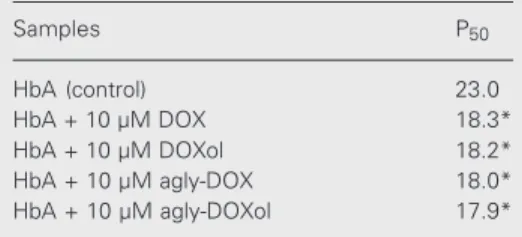
Table 1 reports the change in P50 for Hb
induced by DOX, DOXol, DOX and agly-DOXol at pH 7.4 and 37ºC. It is evident that the presence of the drugs caused a 22% in-crease in oxygen affinity compared to control.
Enzymatic activity as % control
120 100 80 60 40 0 20
DOX DOXol
agly-DOX
agly-DOXol (6)*
(8)
(6) (8) (6)
(6)* (8)
(6)*
○ ○ ○ ○ ○ ○ ○ ○ ○ ○ ○ ○ ○ ○ ○ ○ ○ ○
Figure 4. Effect of DOX, DOXol, agly-DOX and agly-DOXol on su-peroxide dismutase (closed umns) and catalase (open col-umns) activity of human red blood cells. Human red blood cells were incubated for 1 h at
37ºC in the presence and
ab-sence of 40 µM DOX, 40 µM DOXol, 40 µM agly-DOX and 40 µM agly-DOXol, before hemoly-sis and enzymatic activity deter-minations as described in Mate-rial and Methods. Data are re-ported as percent of control ac-tivity (no drugs, control cells = 100 ± 10%, N = 8) and as means ± SEM of the number of observations in separate experiments indicated in parentheses. The statistical significance of the effect of DOX, DOXol, agly-DOX and agly-DOXol
treat-ment was determined by the paired Student t-test (*P < 0.01). For abbreviations, see
legend to Figure 1.
Enzymatic activity as % control
140 120 100 80 60 0
DOX DOXol
agly-DOX (5)* agly-DOXol (8) (8) (8) 40 20 (8) (7)* (8) (6)*
Figure 3. Effect of DOX, DOXol, agly-DOX and agly-DOXol on glu-tathione peroxidase (closed col-umns) and glutathione reduc-tase (open columns) activity of human red blood cells. Human red blood cells were incubated
for 1 h at 37ºC in the presence
and absence of 40 µM DOX, 40 µM DOXol, 40 µM agly-DOX and 40 µM agly-DOXol, before hem-olysis and enzymatic activity de-terminations as described in Material and Methods. Data are reported as percent of control activity (no drugs, control cells = 100 ± 7%, N = 6) and as means ± SEM of the number of observations in separate experiments indicated in parentheses. The statistical significance of the effect of DOX, DOXol, agly-DOX and agly-DOXol
treat-ment was determined by the paired Student t-test (*P < 0.05). For abbreviations, see
legend to Figure 1.
○ ○ ○ ○ ○ ○ ○ ○ ○ ○ ○ ○ ○ ○ ○ ○ ○ ○ This suggests that DOX and its main me-tabolites may interact with Hb modifying its binding to oxygen, and consequently caus-ing reduced tissue oxygenation. To test the possibility that alteration of Hb oxygen bind-ing properties followbind-ing exposure to DOX, DOXol, agly-DOX and agly-DOXol might be linked to the oxidation of thiol groups, we titrated the ß-93 cysteines of HbA treated with
the drugs using ρ-hydroxymercuribenzoate
and aglycones the -SH groups were not ti-tratable, demonstrating a specific modifica-tion of this amino acid group by the drugs.
Effects of drugs on Pmethemoglobin levels and hemolysis degree
MetHb levels determined in intact red cells after treatment with anthracycline drugs and their metabolites were 6.3 ± 4.3, and the values for control cells were 5.6 ± 6.0%, N = 6, P = 0.3. MetHb levels did not change
significantly even during [2-13C]-glucose
in-cubation (6 h at 37ºC). Percent hemolysis was always less than ~2%.
Discussion
The present study clearly demonstrates that not only DOX but also its main metabo-lites, i.e., the secondary alcohol DOXol and aglycone derivatives of DOX, dramatically affect the red blood cell energy metabolism, stimulating the PPP. These results are in agreement with previous studies that reported a dose-dependent activation of PPP in intact human erythrocytes after exposure to adria-mycin and daunoadria-mycin exposure (28). Hen-derson et al. (28) proposed that DOX could
undergo cyclic oxidation-reduction within the red blood cell acting as a mediator of electron transfer from NADP(H) via
semi-quinone to oxyhemoglobin. Thus, the direct
oxidation of NADP(H) by DOX could ac-count for the larger portion of the PPP stim-ulation that we observed in intact red blood cells following DOX exposure. Therefore, the cited investigators proposed that the re-active oxygen species generated during the oxidation-reduction cycle of anthracyclines might oxidize intracellular glutathione and provide further PPP stimulation. Bates and
Winterbourn (29) proposed that O2-• and
a-driamycin semiquinone may be generated directly by reaction of adriamycin with
oxy-hemoglobin. However, although many in
vi-tro studies devoted to the understanding of
the biochemistry have emphasized the toxic effects of anthracyclines within the red blood cells, the molecular basis of these effects is not fully understood. Alternative mechan-isms of DOX-induced toxicity have impli-cated DOX metabolites other than semi-quinone free radicals. This is the case for DOXol, a hydroxy metabolite formed upon a two-electron reduction of the C-13 carbonyl group in the side chain of DOX (7,8,30) and for the aglycone metabolites (9,10). Thus, in the present study, we used red blood cells as a cell model in order to acquire more infor-mation on the biological activity of DOXol, agly-DOX and agly-DOXol. It should also be noted that, although the concentrations of the drugs used in this study would not be achievable in the blood stream in normal
human usage, in many earlier in vitro studies
concerning the effects of anthracyclines or other drugs on red blood cell, the drugs were used at similar or higher concentrations (13,16,28,29,31). Here, we show that both DOXol and the aglycone derivatives of DOX increased the PPP flux in human red blood cells. It is important to remark that DOXol as well as its corresponding aglycone metabo-lite were at least 1.8 times more potent than the parent compound in the stimulation of
Table 1. Oxygen affinity of hemoglobin A in the absence and in the presence of doxorubicin (DOX), doxorubicinol (DOXol), aglycone-DOX (agly-DOX) and aglycone-DOXol (agly-DOXol).
Samples P50
HbA (control) 23.0
HbA + 10 µM DOX 18.3*
HbA + 10 µM DOXol 18.2*
HbA + 10 µM agly-DOX 18.0*
HbA + 10 µM agly-DOXol 17.9*
Hemoglobin A (HbA) was incubated in 0.1 M HEPES buffer, 0.1 M NaCl in the presence of 10
mM 2,3-diphosphoglyceric acid at pH 7.4 and 37ºC.
The average SEM was ± 8% for P50 values (N = 6).
Data (P50) are reported in mmHg. The statistical
significance of the effect of DOX, DOXol, agly-DOX and agly-agly-DOXol treatment was determined
the PPP glucose flux. Recent studies carried out to identify the mechanisms responsible for the anthracycline-induced cardiotoxicity have demonstrated that DOXol might in-deed be more potent than DOX in causing metabolic dysfunction (7,8).
Due to the high levels of Hb within the red blood cells, it is possible that, under our experimental conditions, the interaction of some fractions of DOX and its metabolites with Hb may represent another important metabolic event occurring within the cell after drug exposure. Thus, we determined the
O2 affinity of Hb treated with DOX and its
metabolites in order to assess whether the interaction of Hb with the drugs may modify its oxygen binding properties. Under physi-ological conditions, we demonstrated an
in-crease in the O2 affinity of drug-treated Hb
compared to free-drug Hb. This finding, plus the impossibility to titrate the -SH groups in drug-treated Hb, is probably due to the chem-ical modification exerted by anthracycline drugs on the cysteine residues of HbA. In this regard, it is known that the oxygen af-finity of HbA is higher than normal when cysteines ß-93 are chemically modified (32). This result raises the new hypothesis of interplay between anthracyclines and Hb in addition to the recent suggestion regarding the existence of two binding sites for DOX
on Hb (33,34).However, because the O2
affinity of Hb is a crucial factor in
determin-ing O2 delivery in the blood, in cancer
pa-tients treated with DOX a higher O2 affinity
of blood might favor pulmonary O2 loading
but impair O2 release to tissues. Although
further study is required to clarify this point, we suggest that this finding might be rel-evant to the understanding of the mechan-isms of toxicity induced by anthracyclines.
Previous studies have demonstrated that the toxic effects of DOX are believed to be mediated by free radicals (3-5). Thus, in an attempt to better clarify the mechanisms un-derlying the PPP stimulation and the
in-creased O2 affinity of Hb observed in red
blood cell following exposure to DOX and DOX metabolites, we assessed the activities of different red cell antioxidative enzymes, including GPx, SOD, CAT and GR. Our data showed that DOX treatment had no signifi-cant effects on the red cell GPx, SOD, CAT
and GR enzymes, in agreement with in vitro
reports that red cell enzymes of the reduction systems are not inactivated by DOX (35). In contrast to DOX, DOXol and the aglycone derivatives of DOX only inhibited the red cell GPx and SOD enzymes. In this respect, we suggest that a large accumulation of oxy-gen reactive species within the red blood cells after treatment with DOXol and agly-cone derivatives of DOX might represent an inevitable consequence of impairment of the red cell GPx and SOD enzymes induced by DOX metabolites. Thus, the oxidation of sulfhydryl groups, induced by the oxygen reactive species accumulated within the red blood cell, may account for the larger por-tion of the PPP stimulapor-tion and for the
in-creased O2 affinity of Hb that we observed in
intact red blood cells following DOXol and aglycone metabolites exposure.
Furthermore, the present data allow us to suggest that differences in PPP activity which exist among cell types may lead to variability in the sensitivity of cells to toxicity induced by DOX metabolites. This is consistent with ear-lier findings showing that multidrug resistant cells have an elevated PPP activity (36,37).
References
1. Singal PK & Iliskovic N (1998). Doxorubicin-induced
cardiomyopa-thy. New England Journal of Medicine, 339: 900-905.
2. Lefrak EA, Pitha J, Rosenheim S & Gottlieb JA (1973). A clinical
pathological analysis of adriamycin cardiotoxicity. Cancer, 32:
302-314.
3. Olson RD & Mushlin PS (1990). Doxorubicin cardiotoxicity: analysis
of prevailing hypotheses. FASEB Journal, 4: 3076-3086.
4. Powis G (1989). Free radical formation by antitumor quinones. Free
Radical Biology and Medicine, 6: 63-101.
5. Singal PK, Deally CMR & Weinberg LE (1987). Subcellular effects of
adryamicin in the heart: a concise review. Journal of Molecular and
Cellular Cardiology, 19: 817-828.
6. Licata S, Saponiero A, Mordente A & Minotti G (2000). Doxorubicin metabolism and toxicity in human myocardium: Role of cytoplasmic
deglycosylation and carbonyl reduction. Chemical Research in
Toxi-cology,13: 414-420.
7. Boucek Jr RJ, Olson RD, Brenner DE, Ogumbumni ME, Inui M & Flescher S (1987). The major metabolite of doxorubicin is a potent inhibitor of membrane associated ion pumps: a correlative study of
cardiac muscle with isolated membrane fractions. Journal of
Bio-logical Chemistry,262: 15851-15856.
8. Olson RD, Mushlin PS, Brenner DE, Fleischer S, Cusack BJ, Chang BK & Boucek Jr RJ (1988). Doxorubicin cardiotoxicity may be caused
by its metabolite, doxorubicinol. Proceedings of the National
Acade-my of Sciences, USA, 85: 3585-3589.
9. Sokolove PM, Kester MB & Haynes J (1993). Interaction of adriamycin aglycones with isolated mitochondria. Effect of
sele-nium deficiency. Biochemical Pharmacology, 46: 691-697.
10. Sokolove PM & Shinaberry RG (1988). Na+-independent release of
Ca2+ from rat heart mitochondria. Induction by adriamycin
agly-cone. Biochemical Pharmacology, 37: 803-812.
11. Harper JR, Orringer EP & Parker JC (1979). Adriamycin inhibits Ca2+
permeability and Ca-dependent K+ movements in red blood cells.
Research Communications in Chemical Pathology and
Pharmacolo-gy, 26: 277-284.
12. Borg A, Gallice PM, Kovacic HN, Nicoara AE, Favre RG & Crevat AD
(1996). Impairment of sodium pump and Na+/H+ antiport in
erythro-cytes isolated from cancer patients. Cancer Research, 56: 511-514.
13. Thompson MG, Chahwala SB & Hickman JA (1987). Inhibition of
human erythrocyte inositol lipid metabolism by adriamycin. Cancer
Research, 47: 2799-2803.
14. Miura T, Muraoka S & Ogiso T (1993). Adriamycin-induced lipid peroxidation of erythrocyte membranes in the presence of ferritin
and the inhibitory effect of ceruplasmin. Biological and
Pharmaceu-tical Bulletin, 16: 664-667.
15. DeAtley SM, Aksenov MY, Aksenova MV, Carney JM & Butterfield DA (1998). Adriamycin induces protein oxidation in erythrocyte
membrane. Pharmacology and Toxicology, 83: 62-68.
16. Suwalsky M, Hernandez P, Villena F, Aguilar F & Sotomayor CP (1999). The anticancer drug adriamycin interacts with the human
erythrocyte membrane. Zeithschrift für Naturforschung. Section C,
54c: 271-277.
17. Andersen A, Holte H & Slordal L (1999). Pharmacokinetics and metabolism of doxorubicin after short-term infusions in lymphoma
patients. Cancer Chemotherapy and Pharmacology, 44: 422-426.
18. Lachatre F, Marquet P, Ragot S, Gaulier JM, Cardot P & Dupuy JL (2000). Simultaneous determination of four anthracyclines and three metabolites in human serum by liquid chromatography-electrospray
mass spectrometry. Journal of Chromatography. B, Biomedical
Ap-plications, 738:281-291.
19. Takanashi S & Bachur NR (1976). Adriamycin metabolism in man.
Evidence from urinary metabolism. Drug Metabolism and
Disposi-tion, 4: 79-87.
20. Zijlstra WG, Buursma A & Meeuwsen-van der Roest WP (1991). Absorption spectra of human fetal and adult oxyhemoglobin,
de-oxyhemoglobin, carbde-oxyhemoglobin, and methemoglobin. Clinical
Chemistry, 37: 1633-1638.
21. Misiti F, Meucci E, Zuppi C, Vincenzoni F, Giardina B, Castagnola M
& Messana I (2002). O2-dependent stimulation of the pentose
phos-phate pathway by S-nitrosocysteine in human erythrocytes.
Bio-chemical and Biophysical Research Communications, 294: 829-834.
22. Messana I, Ferroni F, Misiti F, Girelli G, Pupella M, Castagnola M, Zappacosta B & Giardina B (2000). Blood bank conditions and RBCs:
the progressive loss of metabolic modulation. Transfusion, 40:
353-360.
23. Beutler E (1975) Red cell metabolism. In: Beutler E (Editor), A
Manual of Biochemical Methods. 2nd edn. Grune & Stratton, New York, 71-73.
24. Heikkila RE (1985). Autoxidation of 6-hydroxydopamine. In:
GreenwaldRA (Editor), Handbook of Methods for Oxygen Radicals
Research. CRC Press, Boca Raton, FL, USA, 233-235.
25. Carlberg I & Mannervik B (1985). Glutathione reductase. Methods in
Enzymology, 113: 484-490.
26. Giardina B & Amiconi G (1981). Measurement of binding of gaseous and non gaseous ligands to hemoglobins by conventional
spectro-photometric procedures. Methods in Enzymology, 76: 417-427.
27. Boyer PD (1954). Spectrophotometric study of the reaction of
pro-tein sulfhydryl groups with organic mercurials. Journal of the
Ameri-can Chemical Society,76: 4331-4337.
28. Henderson CA, Metz EN, Balcerzak SP & Sagone Jr AL (1978). Adriamycin and daunomycin generate reactive oxygen compounds
in erythrocytes. Blood, 52: 878-885.
29. Bates DA & Winterbourn CC (1982). Reaction of adriamycin with haemoglobin. Superoxide dismutase indirectly inhibits reactions of
the adriamycin semiquinone. Biochemical Journal, 203: 155-160.
30. Minotti G, Recalcati S, Mordente A, Liberi G, Calafiore AM, Mancuso C, Preziosi P & Cairo G (1998). The secondary alcohol metabolite of doxorubicin irreversibly inactivates aconitase/iron regulatory
pro-tein-1 in cytosolic fractions from human myocardium. FASEB
Jour-nal, 12: 541-552.
31. McMillan DC, Jensen CB & Jollow DJ (1998). Role of lipid
peroxida-tion in dapsone-induced hemolytic anemia. Journal of
Pharmacolo-gy and Experimental Therapeutics, 287: 868-876.
32. Antonini E, Condo SG, Giardina B, Ioppolo C & Bertollini A (1982).
The effect of pH and D-glycerate 2,3-bisphosphate on the O2
equi-librium of normal and SH(beta 93)-modified human hemoglobin.
European Journal of Biochemistry, 121: 325-328.
33. Ramanathan-Girish S & Boroujerdi M (2001). Contradistinction
be-tween doxorubicin and epirubicin: in vitro interaction with blood
components. Journal of Pharmacy and Pharmacology, 53: 15-21.
34. Vaidyanathan S & Boroujerdi M (2000). Interaction of dexrazoxane with red blood cells and hemoglobin alters pharmacokinetics of
doxorubicin. Cancer Chemotherapy and Pharmacology, 46: 93-100.
35. Shinohara K & Tanaka KR (1980). The effects of adriamycin
36. Miccadei S, Fanciulli M, Bruno T, Paggi MG & Floridi A (1996). Energy metabolism of adryamicin-sensitive and resistant Ehrlich
ascites tumor cells. Oncology Research, 8: 27-35.
37. Ferretti A, Chen LL, Di Vito M, Barca S, Tombesi M, Cianfriglia M,
Bozzi A, Strom R & Podo F (1993). Pentose phosphate pathway alterations in multi drug resistant leukemic T-cells: 31P NMR and