Autoimmunity and molecular mimicry
in tropical spastic paraparesis/human
T-lymphotropic virus-associated
myelopathy
1Laboratorio de Biología Molecular y Patogénesis,
Departamento de Ciencias Fisiológicas, Facultad de Ciencias da Saude, Universidad del Valle, Cali, Colombia
2Facultad de Medicina, Universidad Santiago de Cali, Cali, Colombia
F. García-Vallejo1,
M.C. Domínguez1
and O. Tamayo2
Abstract
Viruses share antigenic sites with normal host cell components, a phenomenon known as molecular mimicry. It has long been suggested that viral infections might trigger an autoimmune response by several mechanisms including molecular mimicry. More than 600 antiviral monoclonal antibodies generated against 11 different viruses have been reported to react with 3.5% of cells specific for uninfected mouse organs. The main pathological feature of tropical spastic paraparesis/ human T-lymphotropic virus type I (HTLV-I)-associated myelopathy (TSP/HAM) is a chronic inflammation of the spinal cord characterized by perivascular cuffing of mononuclear cells accompanied by paren-chymal lymphocytic infiltration. We detected the presence of autoan-tibodies against a 98- to 100-kDa protein of in vitro cultured human astrocytes and a 33- to 35-kDa protein from normal human brain in the serum of HTLV-I-seropositive individuals. The two cell proteins exhibited molecular mimicry with HTLV-I gag and tax proteins in TSP/HAM patients, respectively. Furthermore, the location of 33- to 35-kDa protein cross-reaction correlated with the anatomical spinal cord areas (in the rat model) in which axonal damage has been reported in several cases of TSP/HAM patients. Our experimental evidence strongly suggests that the demyelinating process occurring in TSP/ HAM may be mediated by molecular mimicry between domains of some viral proteins and normal cellular targets of the spinal cord sections involved in the neurodegeneration.
Correspondence
F. García-Vallejo
Laboratorio de Biología Molecular y Patogénesis
Departamento de Ciencias Fisiológicas Facultad de Ciencias da Saude Universidad del Valle, Cali Colombia
E-mail: [email protected] Research supported in part by the Colombian government agency Colciencias (No. 1106-04-13063) and Universidad del Valle, Cali, Colombia.
Received May 8, 2003 Accepted August 17, 2004
Key words
•HTLV-I •TSP/HAM •Retrovirus •Molecular mimicry
Introduction
Viruses that share antigenic sites with normal host cell components are part of a phenomenon known as molecular mimicry (1). It has long been suggested that viral infections might trigger an autoimmune re-sponse by several mechanisms including
molecular mimicry (2-4). More than 600 an-tiviral monoclonal antibodies generated against 11 different viruses have been re-ported to react with 3.5% of cells specific for uninfected mouse organs (5).
leukemia virus type I (HTLV-I) after a long incubation period. TSP/HAM is character-ized by chronic progressive paraparesis with sphincter disturbances, no or mild sensory loss, absence of spinal cord compression, and seropositivity for HTLV-I antibodies. The pathogenesis of this entity, reviewed by Casseb and Penalva-de-Oliveira (6), is not completely clear and involves a multivari-able phenomenon of activation of the immu-nological system against the presence of HTLV-I antigens, leading to an inflamma-tory process with axomyelinic degeneration, mainly in the thoracic spinal cord (7).
The possible mechanisms by which HTLV-I infection contributes to the patho-genesis of TSP/HAM are unknown. HTLV-I proviral DNA has been detected only in the nucleus of lymphocytes that infiltrated the spinal cord (7,8). However, no proviral DNA was amplified in any neuronal cells, includ-ing neurons and glial cells (9,10). This indi-cates that the axomyelinic degeneration of the spinal cord by HTLV-I is unlikely to be the result of viral infection of oligodendro-cytes or of neuronal cells. These findings suggest an autoimmune mechanism in TSP/ HAM and that this neuronal process is prob-ably associated with an activated cell- and antibody-mediated immune response in the patients.
Astrocytes are cells of the central ner-vous system (CNS) distinguished by their large star-shaped cell bodies and many radi-ating processes. Most of these processes form terminal expansions that surround the sur-face of blood vessels (11). Previous studies have shown that astrocytes secrete inflam-matory cytokines after transient contact with T cells persistently infected with HTLV-I
(12). These in vitro-activated astrocytes may
prolong and amplify the pathological effects produced by invading T cells, demonstrating the crucial role of astrocytes in brain homeo-stasis (13).
An important notion when considering the effect of viruses on the CNS is that
certain impairments occurring in actually infected cells may be perpetuated via
indi-rect effects ofviruses on neural cells. Such
impairment may persist and propagatevia
the secretion of soluble factors such as
cyto-kines, chemocyto-kines, or metalloproteinases
(14),and eventually alter the cell
homeosta-sis. In the case of TSP/HAMthis view is
consistent with i) the presence in the lesions of cells expressing the viral product tax (8,15),
which is knownto transactivate many
cellu-lar genes including several inflammatory molecules (16); ii) the expression of
inflam-matory cytokinesin infiltrated T cells and
astrocytes, and iii)the expression pattern of
metalloproteinases and their inhibitors(17).
Viruses share antigenic sites with normal host cell components, a phenomenon known as molecular mimicry. It has long been sug-gested that viral infections might trigger an autoimmune response by several mechan-isms including molecular mimicry (4,13). In the present study, we performed some ex-periments in order to document the exist-ence of molecular mimicry of monoclonal antibodies and sera of TSP/HAM patients against several proteins of HTLV-I with pro-teins and subcellular structures of non-HTLV-I/HIV-1-infected primary cultured human astrocytes and other normal compo-nents of the CNS.
Patients and Methods
Characteristics of the sample
were informed about the objectives of the investigation and gave their consent for the use of their samples only for research pur-poses.
ELISA Murex HTLV I + II (Murex Biotech Limited, Dartford, UK) and Serodia PA (Fujirevio, Japan) diagnostic tests were used for seroscreening. The presence of HTLV-I was confirmed using an HTLV blot
2.4 assay (Genelabs® Diagnostics Pte Ltd.,
Singapore Science Park, Singapore). Addi-tional diagnostic PCR with pol primers was performed on peripheral blood mononuclear cell DNA from all the individuals studied to amplify a 159-bp fragment. All persons in-cluded in the study were seronegative for
IgG-anti-HIV-1/2 by ELISA (Enzygnost®,
Behring Dade, Germany) and by Western blot.
Monoclonal antibodies
Monoclonal antibodies against gag pro-teins, NOR-1 p24) (18), GIN-7 (anti-p19) and WAG-15 (anti-p17) (19); against envelope gp46, LAT-27 (anti-gp46) and against tax, LT-4 (anti-tax-p40) (20) were
tested by Western blot with proteins from in
vitro cultures of human astrocytes, fetal brains and other non-infected cell cultures from different human tissues.
In vitro cell cultures and tissues
Primary human astrocytes were obtained by mechanical disaggregation of microdis-sected sensory motor cortices of human fe-tuses (embryonic day 116, according to the official Colombian legislation for this mate-rial). The dissociated cells were diluted to a
density of 2 x 10⁵ cells/ml in Dulbecco’s
modified Eagle’s medium (Sigma-Aldrich, St. Louis, MO, USA) containing 25 mM glucose, supplemented with 20% heat-inac-tivated fetal calf serum and gentamicin (1 µg/ml). Cells were seeded onto 35-mm di-ameter culture dishes precoated with
poly-L-lysine (3 µg/ml in 0.1 M sodium borate buffer, pH 8.4) (21). Cultures were
incu-bated at 37ºC in a 5% moist CO2-95% air
atmosphere. Using this procedure, we ob-tained 3-week-old cultures with more than 99% of cells with the astrocyte phenotype. Characterization of astrocytes was system-atically determined by immunocytochemis-try of glial fibrillary acid protein, a specific astrocytic marker, using a polyclonal glial fibrillary acid protein antibody (N-18; Santa Cruz Biotechnology, Santa Cruz, CA, USA). Human primary lung fibroblasts, skin fi-broblasts and kidney cells, cord blood stem cells, HeLa cells, and 3T3 cells were cul-tured in Eagle’s essential medium supple-mented with 10% bovine calf serum and a mix of gentamicin (1 µg/ml), penicillin, strep-tomycin, and neomycin (2.5 µg/ml) to obtain monolayers with more than 90% conflu-ence.
Proteins from brain and spinal cord were obtained from non-HTLV-I and HIV-1 post-mortem fetuses. Two or 5 g of tissue was preserved in appropriate transport medium until the time for processing.
Cell lysis
Approximately 1.0 x 10⁷ cells/ml of
cul-tured cells or 2 to 5 g of tissues were lysed in buffer containing 0.15 M NaCl, 0.05 M Tris-HCl, pH 7.2, 1% Triton X-100, 0.1% sodium dodecyl sulfate (SDS), 1% sodium deoxy-cholate, and 2 mM phenylsulfonyl fluoride. Lysates were separated by centrifugation at
14,000 g for 10 min. Protein concentrations
were measured using a protein assay (Bio-Rad, Richmond, CA, USA) as recommended by the manufacturer.
Western blotting
nitrocellulose membranes (22). The relative molecular masses of proteins were calcu-lated by plotting the logarithm of the relative
mobility of the protein vs gel concentration
(23).
The membranes were incubated in block-ing solution (10% bovine serum albumin in 1X phosphate-buffered saline (PBS), pH 7.0) overnight at 10ºC. Sera and monoclonal anti-bodies were diluted 1:1000 in 1% bovine serum albumin, PBS, and 0.2% Tween-20 and incubated with nitrocellulose strips over-night at 10ºC. Bound autoantibodies were detected by sequential incubation in biotin-labeled anti-human IgG diluted 1:1000, avi-din-labeled horseradish peroxidase (Amer-sham Pharmacia Biotech, Little Chalfont, Buckinghamshire, UK), and 0.05% diami-nobenzidine (Sigma) solution in PBS as de-scribed (24).
Immunohistochemistry of rat spinal cord sections
Young Wistar rats (250 g) were injected with 40 mg/kg Ketalar. After total anesthesia
they were perfused with 150 ml 0.9% NaCl via the ascendant aorta, followed by perfu-sion of 250 ml buffered formalin, pH 7.4. The spinal cord was obtained surgically and divided in its anatomical sections, which were post-fixed in buffered formalin, pH 7.4, at 4ºC for 24 h. The tissues were then dehydrated and embedded in paraplast.
Serial sections of spinal cord were cut, deparaffinized and incubated with the LT-4 anti-tax monoclonal antibody (21) using a modification of the original protocol de-scribed by Hsu et al. (24). The endogenous peroxidase activity was blocked by incuba-tion with 3% hydrogen peroxide for 30 min at room temperature followed by two washes of 5 min each with 0.01X PBS, pH 7.5 (25). Antigen rescue was performed by the method of Shi et al. (26). The serial sections of the spinal cord were incubated overnight with a 1:50 dilution of the anti-tax LT-4 monoclon-al antibody. After incubation for 12 h at 4ºC, slides were washed as described previously and incubated with a secondary antibody (biotinylated goat anti-mouse IgG) diluted 1:200 in 1X PBS for 30 min at room temper-ature. After this procedure they were washed three times in 1X PBS and incubated with ABC Elite PK400 (Vector Laboratories, Burlingame, CA, USA) for 30 min, washed again in PBS and immediately developed with diaminobenzidine previously dissolved in Tris-HCl buffer, pH 7.6, and 0.003% hy-drogen peroxide. Histological preparations were stained with Gill’s hematoxylin and mounted permanently in Permount. As nega-tive controls we used human sera from non-HTLV-I- and HIV-1-infected subjects.
Results
HTLV-I autoantibodies against proteins from central nervous system cells
IgG antibodies against a 98- to 100-kDa astrocyte protein were detected in the sera of 100% (11/11) TSP/HAM patients included
98-100 kDa
33-35 kDa
1 2 3 4 5 6 7 8 9 10 11 Figure 1. Western blot of
pro-teins obtained from normal hu-man tissues using serum from a TSP/HAM subject. Lane 1, As-trocytes; lane 2, brain; lane 3, lung fibroblasts; lane 4, skin fi-broblasts; lane 5, human kidney;
lane 6, cord blood stem cells;
in the study. Moreover, IgG isolated from the same TSP/HAM patients exhibited a spe-cific HTLV-I immunoreactivity with un-infected brain proteins (11/11). Western blots performed with proteins of human non-in-fected brains showed IgG reactivity with a single 33- to 35-kDa protein in all patients. In contrast, no reactive bands with the pro-teins from primary lung fibroblast, skin fi-broblast, human kidney, cord blood stem cells, HeLa, and 3T3 cells were obtained (Figure 1). Sera from asymptomatic healthy HTLV-I carriers and seronegative controls did not exhibit cross-reaction with the 98- to 100-kDa astrocyte or 33- to 35-kDa brain proteins.
We included in the study a set of five different HTLV-I monoclonal antibodies. As shown in Figure 2A, a specific cross-reac-tion of NOR-1 monoclonal antibodies with the 98- to 100-kDa protein from primary cultured human astrocytes was detected. The 33- to 35-kDa brain protein cross-reacted exclusively with the anti-tax LT-4 mono-clonal antibody (Figure 2B).
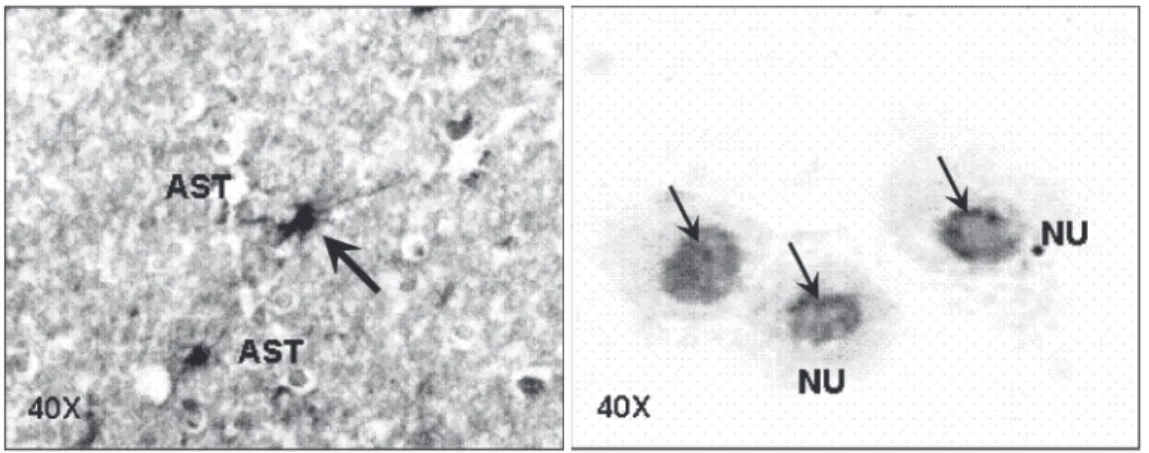
The incubation of different dilutions of NOR-1 with primary human cultured astro-cytes revealed that the cross-reaction previ-ously detected was preferentially localized in the cell nucleus. Astrocytes of the spinal cord and primary cultures showed a peri-nuclear immunostaining that was enhanced by increasing concentrations of the anti-p24gag monoclonal antibody (Figure 3).
Figure 2. Western blot of proteins obtained from cellular components of the human central nervous system using different monoclonal antibodies (mAb) against human T-lymphotro-pic virus type I proteins. NOR-1 (mAb for gag p24). LT-4 (mAb for tax p40). GIN-7 (mAb for gag p19). WAG-15 (mAb for gag p17). A, Proteins from primary cultures of human astro-cytes. B, Proteins from human fetal brain. RC = Reagent control. About 75 µg total protein was loaded per line.
Figure 3. Immunohistochemical reaction of astrocytes with the monoclonal antibody NOR-1 for p24 gag of human T-lymphotro-pic virus type I. A, Histological preparation of the spinal cord of normal young rat. B, Human pri-mary cultures of normal astro-cytes. The arrows indicate a positive immunohistochemistry reaction. AST = astrocytes; NU = nuclear cross-reaction. 98-100 kDa
NOR-1 LT-4 GIN-7 WAG-15 RC NOR-1 LT-4 GIN-7 WAG-15 RC
33-35 kDa
A A A A
Spatial distribution of anti-tax cross-reaction staining in the spinal cord of non-infected rats
In order to assess the location of the anti-tax LT-4 in cells we studied its cross-reactiv-ity with different areas of thoracic and lum-bar spinal cord sections. The immunoreac-tivity with the monoclonal antibody was pre-dominantly observed as an intense nuclear reaction in clusters of neurons of variable sizes located in different laminae of T6 to L2 sections of spinal cord grey matter (Figure 4A). At the level of the intermediate (lamina VII; Figure 4B) and anterior and posterior (lamina V) horns (medial and lateral IX lami-nae; Figure 4C), we observed intense cross-reaction with the monoclonal LT-4 anti-tax antibody. This situation contrasts with the lack of LT-4 cross-reactivity observed in nerve fibers of white matter, in the
ependy-mal channel and in the lamina X (Figure 4A). The negative controls did not exhibit any cross-reaction with the different compo-nents of the spinal cord.
Discussion
An important result of our study was the characterization of the cross-reaction be-tween a perinuclear normal astrocyte protein of 98 to 100 kDa with a monoclonal anti-body against HTLV-I p24 (NOR-1). Our results also showed that this mimicry be-tween the two proteins correlates with the presence of anti-98 to 100-kDa autoantibod-ies in all Colombian TSP/HAM patients tested (11/11). The first report of cross-reaction between sera of TSP/HAM patients and en-dothelial cells of the brain (23) showed that circulation of these autoantibodies in blood is part of the progression not only of TSP/ HAM but also of other demyelinating disor-ders. Recent studies have revisited the mo-lecular mimicry with proteins of HTLV-I as a process that may play some role in the progression of TSP/HAM. Although the molecular mimicry between a peptide of the myelin basic protein and a peptide of HTLV-I with TSP/HAM was not associated with disease progression (24), there is the possi-bility that other CNS targets could mimic HTLV-I antibodies. In this respect, our re-sults agree with data showing that IgG iso-lated from TSP/HAM patients exhibited a specific HTLV-I immunoreactivity with un-infected neurons (13). Neuronal Western blots showed that the IgG reactivity with a single 33- to 35-kDa protein detected in all patients studied is an important process that could play a role in the immunopathogenesis of TSP/HAM (25-27).
Another important result of our study was the characterization of the cross-reac-tion of a perinuclear normal astrocyte pro-tein of 98-100 kDa and a nuclear 33- to 35-kDa human brain protein with a monoclonal antibody against HTLV-I p24 (NOR-1) and
Figure 4. Immunohistochemical reaction of the lumbosacral regions of the spinal cord from a non-human T-lymphotropic virus type I-infected young rat with the monoclonal antibody LT-4 anti-tax. A, General view of motor and sensory areas of the spinal cord that cross-reacted (10X). B, Nuclear cross-reaction observed in some motor neurons of the spinal cord (lamina VII). C, Motor neurons of lamina IX. The arrows indicate a positive immunohis-tochemistry reaction.
Posterior
Anterior 10X
A
A
A
A
A
B
B
B
B
B
C
C
C
C
C
500 µm 500 µm
tax-p40 (LT-4), respectively. Our results also showed that autoantibodies against the as-trocyte 98- to 100-kDa and brain 33- to 35-kDa proteins circulate in the Colombian TSP/ HAM patients included in the present study. The first report of cross-reaction between sera from TSP/HAM patients and endotheli-al cells of the brain (27) showed that circula-tion of autoantibodies in blood is part of the progression not only of TSP/HAM but also of other demyelinating disorders. In this re-spect, our results agree with data showing that IgG isolated from TSP/HAM patients exhibits a specific HTLV-I immunoreactiv-ity with uninfected neurons (28-30).
Monoclonal antibodies that bind HIV-1 gp41 and cross-reacted with astrocytes were recovered from the blood of patients in-fected with HIV-1 (28). Such mimicry by antiviral antibodies interacting with an as-trocyte isoform of alpha actinin (29) could play a role in the dementia of AIDS patients. Since HTLV-I-seropositive patients included in the study were indeed seronegative for HIV-1, it is important to consider that the mimicry between astrocyte proteins and vi-ral proteins may be a common feature of the characteristic neural damage observed not only in a neurological disease associated with HIV-1 infection but also in TSP/HAM. Our results confirmed and extended to human fetal brains and to rat spinal cord the data previously reported by Levin et al. (29) who characterized the molecular mimicry between the HTLV-I tax protein and CNS proteins. In addition to the intense staining of brain neurons, we observed preferential anti-tax IgG staining in the nucleus of T7 to L2 neurons in the rat spinal cord. These results contrast with the absence of staining of glia and dorsal root ganglia (peripheral nervous system) observed with the tax mono-clonal antibody.
Levin et al. (28) have characterized the molecular mimicry between an environmen-tal agent and the CNS. They isolated immu-noglobulin G (IgG) from the serum of
pa-tients with TSP/HAM and tested it for reac-tivity with human tissues. There was intense staining of neurons in the brain and no stain-ing of glia, dorsal root ganglia (peripheral nervous system) or systemic organs. A clonal antibody to HTLV-I-tax (tax mono-clonal antibody) mimicked IgG staining of neurons. To identify the protein, cortical neurons were isolated and subjected to SDS-PAGE and Western blot analysis. The IgG recovered from TSP/HAM patients recog-nized a band of approximately 33 kDa, whereas IgG isolated from negative controls did not. Importantly, the tax monoclonal antibody that stained CNS neurons reacted with the antigen. Consistent with data re-ported by Levin et al. (28), our evidence showed that this cross-reaction can be ex-tended to the proteins of fetal human brains because we characterized a similar cross-reaction between a 33- to 35-kDa neuronal protein and the monoclonal antibody LT-4 anti-tax.
All patients (13/13) with TSP/HAM that
were tested by Levin et al. (28) developed
autoantibodies recognizing the neuronal an-tigen. Nine of ten HTLV-I-seropositive pa-tients without neurological symptoms and 12 HTLV-1-seronegative controls showed no reactivity. All the Colombian TSP/HAM patients studied here reacted against the 33-to 35-kDa brain protein, in contrast 33-to none of the HTLV-I-seropositive asymptomatic and HTLV-I-seronegative subjects.
As shown in previous reports, the main pathological feature of TSP/HAM is a chronic inflammation of the spinal cord character-ized by perivascular cuffing of mononuclear cells accompanied by parenchymal lympho-cyte infiltration (30). Immunological abnor-malities arising from a high HTLV-I proviral load in peripheral blood lymphocytes play an important role in the pathological process of spinal cord lesions in TSP/HAM patients
(30). Concomitant damage to surrounding
HTLV-I-infected lymphocytes, could be involved in the pathological events of TSP/HAM. In this respect, heightened transmigrating activity of HTLV-I-infected CD4+ T lymphocytes to the CNS tissues may play a key role in the development of TSP/HAM.
The cross-reaction between a 98- to 100-kDa astrocytic protein and the HTLV-I p24 gag protein supports the existence of mi-metic targets for viral proteins in this impor-tant cell of the CNS. As previously reported, monoclonal antibodies that bound to HIV-1 gp41 and cross-reacted with astrocytes were recovered from the blood of patients in-fected with HIV-1 (31). Such mimicry by antiviral antibodies interacting with an as-trocyte isoform of alpha actinin (32) could play a role in the dementia of AIDS patients. Since the HTLV-I-seropositive patients in-cluded in the study were indeed HIV-1 sero-negative, it is important to consider that the mimicry between astrocytic proteins and vi-ral proteins may be a common feature of the characteristic neural damage not only in a neurological disease associated with HIV-1 infection but also in TSP/HAM.
Our evidence showed that the molecular mimicry of tax protein is preferentially lo-cated in the large neurons of the spinal cord motor areas of T-6 to L-2. As proposed in previous reports, the main pathological fea-ture of TSP/HAM is a chronic inflammation of the spinal cord characterized by perivas-cular cuffing of mononuclear cells accompa-nied by parenchymal lymphocyte infiltra-tion. Immunological abnormalities arising from a high HTLV-I proviral load in periph-eral blood lymphocytes play an important role in the pathological process of spinal cord lesions in TSP/HAM patients. Although
a concomitant damage to the surrounding
CNS tissues, in which CD8+ I-spe-cific cytotoxic T lymphocytes attack HTLV-I-infected lymphocytes, might be involved in the pathological events of the spinal cords
of TSP/HAM patients as one of the actual pathogenetic mechanisms, heightened trans-migrating activity of HTLV-I-infected CD4+ T lymphocytes to the CNS tissues may play a key role in the development of TSP/HAM. It is not clear how antibodies could enter the CNS and bind to intracellular antigens located in the neurons of motor spinal cord sections. Previous data indicated that IgG has a significant role in the pathogenesis of disease. In TSP/HAM, patients are infected
with HTLV-I, which is tropic for CD4+
T-lymphocytes. Infection of CD4+
T-lympho-cytes results in their activation and may al-low them to cross the blood-brain barrier (32). Interaction of these cells with the CNS results in cytokine expression, adhesion molecule and receptor up-regulation, and metalloproteinase secretion. These events are associated with disruption of the blood-brain barrier. As a consequence, cytotoxic T lymphocytes and antibodies against tax may enter the CNS. Tax-specific antibodies may home to the neurons and disrupt neuronal function, as suggested by previous studies (12).
The results presented here lead us to conclude that the location of the cross-reac-tion with the tax LT-4 monoclonal anti-body in specific sections of the spinal cord may correlate with the pathological damage mediated by molecular mimicry observed in TSP/HAM. This mimicry may involve ax-onal degeneration of the motor areas of the spinal cord, mainly those located in the infe-rior thoracic spinal cord sections. This rea-soning opens the possibility to explain the correlation between molecular mimicry and the pathogenesis of TSP/HAM.
Acknowledgments
References
1. Fujinami R & Oldstone MB (1989). Molecular mimicry as a mechan-ism for virus induced autoimmunity. Immunology Research, 8: 3-15. 2. Fujinami R, Oldstone MB, Wrobleska Z, Frankel ME & Koprowski H (1983). Molecular mimicry in virus infection: cross-reaction of measles virus phosphoprotein of herpes simplex virus protein with intermediate filaments. Proceedings of the National Academy of Sciences, USA, 80: 2346-2350.
3. Dyrberg T, Peterson JS & Oldstone MB (1990). Immunological cross-reactivity between mimicking epitopes on a virus protein and human autoantigen depends on a single amino acid residue. Clinical Immunopathology, 54: 290-297.
4. Fujinami RS & Oldstone MB (1985). Amino acid homology between the encephalitogenic site of myelin basic protein and virus: mechan-ism for autoimmunity. Science, 230: 1043-1045.
5. Srinivasaapa J, Saegusa J & Prabhkar B (1986). Molecular mimicry: frequency of reactivity of monoclonal antiviral antibodies with nor-mal tissues. Journal of Virology, 57: 397-401.
6. Casseb J & Penalva-de-Oliveira AC (2000). The pathogenesis of tropical spastic paraparesis/human T-cell leucemia type I-associ-ated myelopathy. Brazilian Journal of Medical and Biological Re-search, 33: 1395-1401.
7. Romero IA, Prevost MC, Perret E, Adamson P, Greenwood J, Courad PO & Ozden S (2000). Interactions between brain endothelial cells and human T-cell leukemia virus type 1-infected lymphocytes: mechanisms of viral entry into the central nervous system. Journal of Virology, 74: 6021-6030.
8. Lehky TJ, Fox CH, Koenig SM, Levin C, Flerlage N, Izumo S, Sato E, Raine CS, Osame M & Jacobson S (1995). Detection of human T-lymphotropic virus type I (HTLV-I) tax RNA in the central nervous system of HTLV-I-associated myelopathy/tropical spastic parapare-sis patients by in situ hybridization. Annals of Neurology, 37: 167-175.
9. Kubota R, Umehara F, Izumo S, Ijichi S, Matsumuro K, Yashiki S, Fujiyoshi T, Sonoda S & Osame M (1994). HTLV-I proviral DNA amount correlates with infiltrating CD4+ lymphocytes in the spinal cord from patients with HTLV-I-associated myelopathy. Journal of Neuroimmunology, 53: 23-29.
10. Kira J (1994). The presence of HTLV-I proviral DNA in the central nervous system of patients with HTLV-I-associated myelopathy/ tropical spastic paraparesis. Molecular Neurobiology, 8: 139-145. 11. Froes MM, Correia AH, Garcia-Abreu J, Spray D, Campos AC,
Campos de Carvalho AC & Neto VM (1999). Gap-junctional coupling between neurons and astrocytes in primary central nervous system cultures. Proceedings of the National Academy of Sciences, USA, 96: 7541-7546.
12. Giraudon P, Bernard C, Malcus N, Dufay C, Desgranges M & Belin F (1995). Retroviral infection (HTLV-I) induces cytokine-regulated im-munomodulation and cytotoxicity of medulloblastoma cells. Journal of Neuropathology and Experimental Neurology, 54: 165-174. 13. Giraudon P, Dufay N, Hardin H, Reboul A, Tardy M & Belin MF
(1993). Differentiation of a medulloblastoma cell line towards an astrocytic lineage using the human T lymphotropic retrovirus-1.
Neuroscience, 52: 1069-1079.
14. Giraudon P, Vernant JC, Confavreux C, Belin MF & Desgranges C (1998). Matrix metalloproteinase 9 (gelatinase B) in cerebrospinal fluid of HTLV-1 infected patients with tropical spastic paraparesis.
Neurology, 50: 1920.
15. Moritoyo T, Izumo S, Moritoyo H, Tanaka Y, Kiyomatsu Y, Nagai M,
Usuku K, Sorimachi M & Osame M (1999). Detection of human T-lymphotropic virus type I p40tax protein in cerebrospinal fluid cells from patients with human T-lymphotropic virus type I-associated myelopathy/tropical spastic paraparesis. Journal of Neurovirology, 5: 241-248.
16. Umehara F, Okada Y, Fujimoto N, Abe M, Izumo S & Osame M (1998). Expression of matrix metalloproteinases and tissue inhibi-tors of metalloproteinases in HTLV-I-associated myelopathy. Jour-nal of Neuropathology and Experimental Neurology, 57: 839-849. 17. Chen W, Elias RV, Cao W, Lerious V & McGinnis JF (1999).
Anti-recoverin antibodies cause the apoptotic death of mammalian pho-toreceptor cells in vitro. Journal of Neuroscience Research, 57: 706-718.
18. Tanaka Y, Lee B, Inoi T, Tozawa H, Yamamoto N & Hinuma Y (1986). Antigens related to three core proteins of HTLV-I (p24, p19 and p15) and their intracellular localizations, as defined by monoclonal anti-bodies. International Journal of Cancer, 37: 35-42.
19. Tanaka Y, Zeng L, Shiraki H, Shida H & Tozawa H (1991). Identifica-tion of a neutralizaIdentifica-tion epitope on the envelope gp46 antigen of human T cell leukemia virus type I and induction of neutralizing antibody by peptide immunization. Journal of Immunology, 147: 354-360.
20. Lee B, Tanaka Y & Tozawa H (1989). Monoclonal antibody defining tax protein of human T-cell leukemia virus type-I. Tohoku Journal of Experimental Medicine, 157: 1-11.
21. Yavin E & Yavin Z (1974). Attachment and culture of dissociated cells from rat embryo cerebral hemispheres on polylysine-coated surface. Journal of Cell Biology,62: 540-546.
22. Barin F, M’Boup S, Denis F, Kanki P, Allan JS, Lee TH & Essex M (1985). Serological evidence for virus related to simian T-lymphotro-pic retrovirus III in residents of West Africa. Lancet, ii: 1387-1389. 23. Nishizawa H, Kita N, Okimura S, Takao E & Abe Y (1988).
Determi-nation of molecular weight of native proteins by polyacrylamide gradient gel electrophoresis. Electrophoresis, 9: 802-806. 24. Hsu S, Raine L & Fanger H (1981). Use of the
avidin-biotin-peroxi-dase complex (ABC) in immunoperoxiavidin-biotin-peroxi-dase techniques: a compari-son between ABC and unlabeled antibody (PAP) procedures. Jour-nal of Histochemistry and Cytochemistry,29: 577-580.
25. Hökfelt T, Fuxe K & Goldstein N (1975). Application of immunochem-istry to studies of monoamine cell systems with special reference to nervous tissues. Annals of the New York Academy of Sciences, 254: 407-432.
26. Shi SR, Iman A, Young L, Cote R & Taylor CR (1995). Antigen retrieval immunohistochemistry under the influenza of pH using monoclonal antibodies. Journal of Histochemistry and Cytochemis-try,43: 193-201.
27. Tsukada N, Tanaka Y & Yanagisawa N (1989). Autoantibodies to brain endothelial cells in the sera of patients with human T-lympho-tropic virus type I associated myelopathy and other demyelinating disorders. Journal of Neurological Sciences, 90: 33-42.
28. Levin MC, Krichavsky M, Berk J, Foley S, Rosenfeld M, Dalmau J, Chang G, Posner JB & Jacobson S (1998). Neuronal molecular mimicry in immune-mediated neurologic disease. Annals of Neurol-ogy, 44: 87-98.
30. Levin MC, Lee SM, Morcos Y, Brady J & Stuart J (2002). Cross-reactivity between immunodominant human T lymphotropic virus type I tax and neurons: implications for molecular mimicry. Journal of Infectious Diseases, 186: 1514-1517.
31. Eddleston M, de la Torre JC, Xu JY, Dorfman N, Notkins A, Zolla-Pazner S & Oldstone MB (1993). Molecular mimicry accompanying