Peripheral markers of oxidative stress in
chronic mercuric chloride intoxication
1Laboratório de Fisiologia Cardiovascular, Departamento de Fisiologia,
Instituto de Ciências Básicas da Saúde, Universidade Federal do Rio Grande do Sul, Porto Alegre, RS, Brasil
2Cátedra de Química General e Inorgánica, Facultad de Farmacia y Bioquímica,
Universidad de Buenos Aires, Buenos Aires, Argentina L.L.P. Gutierrez1,
N.G. Mazzotti1,
A.S.R. Araújo1, R.B. Klipel1,
T.R.G. Fernandes1,
S.F. Llesuy2
and A. Belló-Klein1
Abstract
The present study was designed to evaluate the time course changes in peripheral markers of oxidative stress in a chronic HgCl2 intoxication model. Twenty male adult Wistar rats were treated subcutaneously daily for 30 days and divided into two groups of 10 animals each: Hg, which received HgCl2 (0.16 mg kg-1 day-1), and control, receiving the same volume of saline solution. Blood was collected at the first, second and fourth weeks of Hg administration to evaluate lipid peroxidation (LPO), total radical trapping antioxidant potential (TRAP), and superoxide dismutase (Cu,Zn-SOD), glutathione peroxidase (GPx), glutathione-S-transferase (GST), and catalase (CAT). HgCl2 adminis-tration induced a rise (by 26%) in LPO compared to control (143 ± 10 cps/mg hemoglobin) in the second week and no difference was found at the end of the treatment. At that time, GST and GPx were higher (14 and 24%, respectively) in the Hg group, and Cu,Zn-SOD was lower (54%) compared to control. At the end of the treatment, Cu,Zn-SOD and CAT were higher (43 and 10%, respectively) in the Hg group compared to control (4.6 ± 0.3 U/mg protein; 37 ± 0.9 pmol/mg protein, respectively). TRAP was lower (69%) in the first week compared to control (43.8 ± 1.9 mM Trolox). These data provide evidence that HgCl2 administration is accompanied by systemic oxi-dative damage in the initial phase of the process, which leads to adaptive changes in the antioxidant reserve, thus decreasing the oxidative injury at the end of 30 days of HgCl2 administration. These results suggest that a preventive treatment with antioxidants would help to avoid oxidative damage in subjects with chronic intoxication. Correspondence
A. Belló-Klein Rua Sarmento Leite, 500 90050-170 Porto Alegre, RS Brasil
Fax: +55-51-3316-3166 E-mail: [email protected] Research supported by CAPES, FINEP, CNPq, and FAPERGS.
Received September 29, 2004 Accepted March 2, 2006
Key words •Heavy metals
•Antioxidant enzymes
•Lipid peroxidation
•Total radical-trapping antioxidant potential
•HgCl2
Mercury is a widespread environmental and industrial pollutant which induces se-vere alterations in the tissues of both animals and humans (1). According to the Brazilian Health Ministry, 974,069 cases of human intoxication were registered in Brazil from 1985-2003 with 6,228 deaths. Mortality due to Hg2+ intoxication observed in Brazil in the
same period was about 2,500, with the agri-cultural use of pesticides promoting greater lethality (2). Various mechanisms have been proposed to explain the biological toxicity of mercuric chloride (HgCl2), including
oxida-tive stress. Hg2+ reacts with thiol groups (-SH),
stress or predisposing cells to it (3). Other antioxidants, including ascorbic acid and vitamin E, have been reported to be depleted in HgCl2-treated rats (4). If animals are
pre-treated with superoxide dismutase (Cu,Zn-SOD) before acute intoxication is induced, histological changes are prevented (5). Many experiments suggest that oxidative stress can be involved in cellular damage and that it can be implicated in the toxicity of many xenobiotics.
The evaluation of the extent of blood oxidative stress by standardized methods can be useful to define the role of oxidative stress in different pathologies and can be used for clinical diagnosis. Kinesiological and electrophysiological parameters, as well as biochemical determinations made in fe-ces, saliva, sweat and urine, are usually con-sidered to be noninvasive peripheral mark-ers. Biochemical procedures carried out in plasma and blood cells are considered to be minimally invasive peripheral markers (6).
Thus, the purpose of the present study was to monitor some peripheral markers of oxidative stress during chronic HgCl2
in-toxication to examine how the antioxidant system of animals responds to HgCl2 with
time.
Twenty male Wistar rats (250 g) were obtained from the Central Animal House of Universidade Federal do Rio Grande do Sul. The animals were housed in plastic cages (4 animals each) and received water and pelleted food ad libitum. They were maintained un-der standard laboratory conditions (controlled temperature of 21ºC, 12-h light/dark cycle). Animals were weighed weekly. The animals were divided into two groups of 10: Hg group, that received daily subcutaneous HgCl2 injections, 0.16 mg kg body weight-1
day-1 (7) dissolved in saline solution, and
control, that received the same volume of saline subcutaneously daily. The animals were injected daily for 30 consecutive days, always at the same time, from 8:00 to 10:00 o’clock, to avoid circadian oscillations.
Blood samples were taken at the end of the 1st, 2nd and 4th weeks of treatment. Blood was collected always at the same time of day and on the same day of the week by retro-orbital venous plexus puncture under ether anesthesia to measure oxidative stress at these three time points. Heparinized venous blood samples were washed in a solution of 9 g/L sodium chloride and centrifuged three times at 3000 g for 10 min at room tempera-ture. Plasma was separated and stored for total radical-trapping antioxidant potential (TRAP) determination. White cells were dis-carded by aspiration and the erythrocytes diluted 1/10 in 1 mMacetic acid and 4 mM magnesium sulfate, placed in an ice bath for 10 min and centrifuged at 4200 g for 20 min at 0ºC. The supernatant was used for enzyme and lipid peroxidation (LPO) assays (6).
At the end of treatment, animals were euthanized by cervical dislocation, and total blood was collected by cardiac puncture to measure Hg2+ concentration. This
determi-nation was done by atomic absorption spec-trophotometry at the Toxilab Clinical Anal-ysis Laboratory (Porto Alegre, RS, Brazil). The lower limit of sensitivity was 0.02 ppb. Chemiluminescence was used to evaluate LPO. Chemiluminescence was measured with a liquid scintillation counter in the out-of-coincidence mode (LKB Rack Beta Liquid Scintillation Spectrometer 1215, LKB -Produkter AB, Bromma, Sweden). Blood samples were placed in low-potassium vials at a protein concentration of 0.5-1.0 mg protein/mL in a reaction medium consisting of 120 mM KCl in 30 mM sodium phosphate buffer, pH 7.4. Measurements were started by the addition of 3 mM tert-butyl hydroper-oxide and the data are reported as counts per second per milligram hemoglobin (cps/mg Hb) (8).
50 µM ABAP, 40 µM luminol, and 50 mM sodium phosphate buffer, pH 7.4, was incu-bated and a steady-state luminescence arose from the free radical-mediated luminol oxi-dation. This emission was almost completely quenched by the addition of Trolox (water-soluble vitamin E), yielding induction times linearly related to the free radical scavenger concentration added. A calibration curve was obtained by using 0.2 to 1 µM Trolox. The addition of plasma samples instead of Trolox elicits an induction time related to the initial amount of sample added (9). Luminescence was measured with a scintillation counter in the out-of-coincidence mode and the results are reported as mmol Trolox L-1 mg protein-1.
Catalase (CAT) activity was determined by following the decrease in 240-nm absorp-tion in a reacabsorp-tion medium containing 50 mM sodium phosphate buffer, pH 7.2, and 10 mM hydrogen peroxide (10). CAT activity is reported as picomol hydrogen peroxide reduced per minute per milligram of protein. Glutathione peroxidase (GPx) oxidizes reduced glutathione in the presence of oxides. Its activity is reported as nmol per-oxide/hydroperoxide reduced min-1 mg
pro-tein-1. Selenium-dependent and
-independ-ent GPx activities were measured by follow-ing NADPH oxidation at 340 nm in a reac-tion medium containing 0.17 mM reduced glutathione, 0.2 U/mL glutathione reduc-tase, and 0.5 mM tert-butyl hydroperoxide (which reacts with both the Se-dependent and non-Se-dependent GPx), as described by Flohé and Gunzler (11).
Cu,Zn-SOD activity was determined based on the inhibition of superoxide radical reaction with pyrogallol and the results are reported as units per milligram of protein (12). Cu,Zn-SOD activity was determined by measuring the rate of pyrogallol oxida-tion. The reaction medium contained 50 mM Tris buffer, pH 8.20, 24 mM pyrogallol, and 30 mM CAT. Absorbance changes were measured at 420 nm for 2 min.
Glutathione-S-transferase (GST)
activ-ity, reported as pmol/mg protein, was meas-ured by the rate of formation of dinitrophenyl-S-glutathione at 340 nm. The reaction medi-um consisted of 19 mM sodimedi-um phosphate buffer, pH 6.5, 1 mM GSH, 1 mM chloride dinitrobenzene, and erythrocytes (13).
The conversion of hemoglobin to cya-nomethemoglobin by the Drabkin reagent was measured against a standard curve (14). Protein was measured by the method of Lowry et al. (15) using bovine serum albu-min as standard.
Tert-butyl hydroperoxide was from Al-drich (Milwaukee, WI, USA) and ABAP was from Polyscience (Warrington, PA, USA). All other chemicals were purchased from Sigma (St. Louis, MO, USA).
Data were analyzed by two-way analysis of variance and are reported as means ± SEM. Values of P < 0.05 were considered to be significant.
Blood Hg2+ concentration was higher in
treated animals (reaching 73.5 µg/dL) com-pared to control (P < 0.01), which showed no detectable concentration of this metal in blood. The body weight curve constructed to follow the animals’ growth demonstrated that the initial body weight of the control (249.0 ± 1.3 g) and Hg (248.1 ± 1.0 g) animals did not differ significantly. Body weight increased by 26% in control from the first and the last day of the experiment (day 30), and was reduced by 19% in the Hg group, during the same period of treatment. There was a significant body weight differ-ence between groups after the first week of treatment (data not shown).
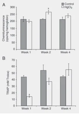
In the Hg group, TRAP was decreased by 69% in the first week of treatment and in-creased progressively in the subsequent weeks, with no difference being observed in relation to control during the last week of treatment. Control animals showed no significant changes in TRAP at the three time points. These data can be observed in Figure 1B.
Table 1 shows the activities of CAT, Cu,Zn-SOD, GPx, and GST in erythrocytes from animals of both experimental groups. There was no difference in antioxidant en-zyme activity between the two groups
dur-ing the first week of treatment. Durdur-ing the second week, GST and GPx activities were significantly increased by 14 and 24%, re-spectively, in the treated group, while Cu,Zn-SOD activity was decreased by 54% com-pared to control. During the last week of treatment, Cu,Zn-SOD was increased by 43% and CAT by 10%, both significantly differ-ent from control animals at the same time point, while the activities of the other en-zymes did not change significantly.
In the present study, chronic administra-tion of HgCl2 to rats caused an imbalance in
oxidative stress as monitored by means of some peripheral markers, providing a ki-netic view of this complex phenomenon. Oxidative stress was measured through very sensitive methods that are detected by chemi-luminescence allowing high precision deter-minations with small amounts of sample.
Chronic HgCl2 intoxication induced a
sig-nificant decrease in the animal’s body weight compared to control. Besides weight loss, the animals showed signs of intoxication like yel-lowish fur and a cachectic appearance.
When peripheral markers of oxidative stress were analyzed in the present study, important HgCl2-induced changes in TRAP
(Figure 1B) and LPO (Figure 1A) were de-tected. The TRAP technique measures the antioxidant capacity of plasma, which is a combination of the effect of all of the chain-breaking antioxidants, including thiol groups (especially glutathione), uric acid and ascor-Figure 1. Effect of HgCl2
admin-istration on lipid peroxidation lev-els and total radical trapping an-tioxidant potential (TRAP) of rats. A, Lipid peroxidation levels, measured by chemilumines-cence and reported as cps/mg hemoglobin. Data are reported as mean ± SEM for 10 animals per group. *P < 0.05 compared to control at the same time point (two-way ANOVA).B, TRAP, re-ported as mM Trolox in plasma. Data are reported as means ± SEM for 10 animals per group. *P < 0.01 compared to all other experimental groups (two-way ANOVA).
Table 1. Antioxidant enzyme activities in erythrocytes of rats treated subcutaneously with HgCl2 daily for 30 days.
Antioxidant enzyme Week 1 Week 2 Week 4
Control Hg Control Hg Control Hg
CAT (pmol/mg protein) 36.7 ± 1.70 41.0 ± 1.40 36.2 ± 1.90 37.0 ± 1.40 37.0 ± 0.96 41.0 ± 0.85*
SOD (U/mg protein) 5.50 ± 0.40 4.10 ± 0.50 4.30 ± 0.30 2.00 ± 0.50* 4.60 ± 0.30 6.60 ± 0.70*
GPx (nmol min-1 mg protein-1) 45.0 ± 2.50 36.5 ± 3.50 46.0 ± 1.30 57.0 ± 0.70* 46.0 ± 4.10 39.6 ± 2.70 GST (pmol min-1 mg protein-1) 0.70 ± 0.01 0.70 ± 0.01 0.70 ± 0.04 0.80 ± 0.03* 0.70 ± 0.04 0.70 ± 0.06
Data are reported as mean ± SEM for 5-7 animals. CAT = catalase; SOD = superoxide dismutase; GPx = glutathione peroxidase; GST = glutathione-S-transferase.
*P < 0.05 compared to control at the same time (two-way ANOVA). Chemiluminescence (cps/mg hemoglobin)
300
Week 1 Week 2 Week 4
Control HgCl2
250
200
150
100
50
0
TRAP (mM Trolox)
70
60
50
40
30
20
0 10
Week 1 Week 2 Week 4
A
B
*
bic acid (16). In the present study, antioxi-dant capacity was decreased in the first week of treatment, probably because of the bind-ing of mercuric ions to thiols, leadbind-ing to intracellular depletion of these groups, espe-cially glutathione. Furthermore, other cellu-lar antioxidants such as vitamins C and E may also have been depleted, as described previously (4), in an attempt to recover the antioxidant status before the occurrence of oxidative damage.
As a consequence, an imbalance in anti-oxidant protective mechanisms takes place, leading to oxidative stress in the cells. In fact, a drastic increase in reactive oxygen species production has been reported during acute treatment with HgCl2, inducing LPO
in rat brain (7). In the present study, we also observed an increase in systemic LPO levels during the second week of treatment (Figure 1A), possibly as a consequence of the deple-tion of antioxidants. During week 2, some changes in antioxidant enzyme activity were observed. Cu,Zn-SOD activity was decreased by 54%, possibly indicating an increased superoxide radical production and a conse-quently higher hydroxyl radical formation. Enhanced levels of the latter may contribute to LPO initiation (8), as observed in the present study. Increased LPO levels are in-dicative of an oxidative stress situation that might lead to the induction of thioredoxin, a member of an evolutionarily conserved fam-ily of redox-active proteins containing a con-served active site dithiol motif. This enzyme could be induced in oxidative stress situa-tions and may play a protective role against specific toxicologic conditions (17). Al-though thioredoxin activity and/or induction were not measured in this experimental mo-del, increased thioredoxin activity may re-place reduced glutathione levels, as reflect-ed by the progressive increase in TRAP observed in the present study.
GST and GPx activities increased during the second week of treatment with HgCl2.
The elevation in GST activity indicates that an oxidative stress condition is occurring, since GST is considered to be an oxidative stress marker (18). GST catalyzes the conju-gation of electrophilic molecules with GSH, being part of the mechanism of detoxifica-tion of xenobiotics (13). GST is also in-volved in the detoxification of peroxidation products (18). Increased GPx activity indi-cates an increase in the amount of organic and non-organic peroxides, such as hydro-gen peroxide, which are substrates for the enzyme (19).
At the end of the experimental protocol, CAT and Cu,Zn-SOD activities were in-creased. This may indicate that hydrogen peroxide and superoxide radical production is enhanced. It has been reported that HgCl2
may disturb mitochondrial inner membrane function, resulting in increased formation of hydrogen peroxide by the mitochondrial elec-tron transport chain (1). The increased CAT activity observed in the present study can be understood as an adaptive response to in-creased hydrogen peroxide production.
Taken together, the present results sug-gest that animals treated with HgCl2 respond
at the cellular level by mobilizing different substances to avoid or compensate for the metal’s toxicity. This adaptive response is reflected on the peripheral markers of oxida-tive stress measured. The novelty of this study is the oxidative stress time course evaluation in the HgCl2 intoxication. This is
a minimally invasive approach and these markers could be determined in patients ex-posed to Hg2+ to monitor the clinical
evolu-tion of antioxidant therapies against cellular oxidative damage.
Acknowledgments
References
1. Lund BO, Miller DM, Woods JS. Mercury-induced H2O2 production and lipid peroxidation in vitro in rat kidney mitochondria. Biochem Pharmacol 1991; 42 Suppl: S181-S187.
2. SINITOX. National system of toxic-pharmacological information. 2002. http://www.fiocruz.br/cict/sinitox. Accessed February, 2006. 3. Gstraunthaler G, Pfaller W, Kotanko P. Glutathione depletion and in
vitro lipid peroxidation in mercury or maleate induced acute renal failure. Biochem Pharmacol 1983; 32: 2969-2972.
4. Fukino H, Hirai M, Hsueh YM, Yamane Y. Effect of zinc pretreatment on mercuric chloride-induced lipid peroxidation in the rat kidney. Toxicol Appl Pharmacol 1984; 73: 395-401.
5. Girardi G, Elias MM. Mercuric chloride effects on rat renal redox enzyme activities: SOD protection. Free Radic Biol Med 1995; 18: 61-66.
6. Repetto MG, Reides CG, Evelson P, Kohan S, de Lustig ES, Llesuy SF. Peripheral markers of oxidative stress in probable Alzheimer patients. Eur J Clin Invest 1999; 29: 643-649.
7. Huang YL, Cheng SL, Lin TH. Lipid peroxidation in rats adminis-trated with mercuric chloride. Biol Trace Elem Res 1996; 52: 193-206.
8. Gonzalez FB, Llesuy S, Boveris A. Hydroperoxide-initiated chemilu-minescence: an assay for oxidative stress in biopsies of heart, liver, and muscle. Free Radic Biol Med 1991; 10: 93-100.
9. Lissi E, Salim-Hanna M, Pascual C, del C. Evaluation of total anti-oxidant potential (TRAP) and total antianti-oxidant reactivity from
lumi-nol-enhanced chemiluminescence measurements. Free Radic Biol Med 1995; 18: 153-158.
10. Aebi H. Catalase in vitro. Methods Enzymol 1984; 105: 121-126. 11. Flohe L, Gunzler WA. Assays of glutathione peroxidase. Methods
Enzymol 1984; 105: 114-121.
12. Marklund S. Handbook of methods for oxygen radical research. Boca Raton: CRC Press; 1985.
13. Mannervik B, Guthenberg C. Glutathione transferase (human pla-centa). Methods Enzymol 1981; 77: 231-235.
14. Drabkin DL, Austin J. Spectrophotometric studies: preparation from washed blood cells, nitric oxide haemoglobin and sulfahaemoglobin. J Biol Chem 1935; 112: 51-57.
15. Lowry OH, Rosebrough AL, Farr AL, Randall R. Protein measure-ment with the Folin phenol reagent. J Biol Chem 1951; 193: 265-275.
16. Ghiselli A, Serafini M, Natella F, Scaccini C. Total antioxidant capac-ity as a tool to assess redox status: critical view and experimental data. Free Radic Biol Med 2000; 29: 1106-1114.
17. Watson WH, Yang X, Choi YE, Jones DP, Kehrer JP. Thioredoxin and its role in toxicology. Toxicol Sci 2004; 78: 3-14.
18. Neefjes VM, Evelo CT, Baars LG, Blanco CE. Erythrocyte glutathi-one S transferase as a marker of oxidative stress at birth. Arch Dis Child Fetal Neonatal Ed 1999; 81: F130-F133.