REVIEW ARTICLE
Rupture of vasa vasorum and intramural hematoma of the
aorta: a changing paradigm
Ruptura dos
vasa vasorum
e hematoma intramural da aorta: um paradigma em
mudança
Adamastor Humberto Pereira*
Introduction
Vasa vasorum (VV) are small arteries which penetra-te the arpenetra-terial wall both through the luminal surface (vasa vasorum internae) and the adventitia (vasa vasorum exter-nae). Adventitial VV ramify into smaller vessels supplying the outer media layers. Venous VV are more numerous and also supply mainly the outer media. In humans, arteries containing less than 29 cellular layers or less than 0.5 mm diameter do not present VV.1,2
hanks to microcomputerized tomography, tridimen-sional study of these vessels became possible,3,4 as well as
microarteriography have enabled a more thorough unders-tanding of their anatomy and distribution5 through the
in-jection of a silicone polymer (Microil®, Flow Tech, U.S.).
VV seem to play a fundamental role in the physio-pathology of the three acute aortic syndrome entities: aortic dissection, intramural hematoma (IMH), and pe-netrating ulcer.
VV reactivity
Anatomically VV are characterized by the presence of endothelial cells, smooth muscle cells and by being ex-ternally covered with connective tissue. hese features are similar to those encountered in small coronary arteries, indicating that VV can regulate their own tone and con-sequently arterial wall perfusion.6-8 Isolated porcine
aor-tic VV responded to endothelium-dependent vasodilator substance P and to bradykinin similarly to the native artery, conirming the afore mentioned autoregulating aspect.9,10
VV hemodynamics
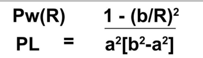
VV’s anatomic localization and ramiication characte-ristics prevent the blood low from reaching the inner me-dia due to the compressive stress inside the arterial wall, as described by the Lamé’s formula (Figure 1). his formula indicates that the compression stress in the arterial wall’s
Abstract
Rupture of vasa varorum has been recognized as one cause of intramural hematoma of the aorta for 90 years. his brief revision presents systematically, the physiology of these vessels and its role in the physiopathology of the alterations in the aortic wall secondary to hypertension, arteriosclerosis and in Acute Aortic Syndrome. he hypothesis is that rupture of vasa vasorum is a secondary phenomenon and not one causal factor in the physiopathology of intramural hematoma.
Keywords: Aortic intramural hematoma, vasa vasorum, aortic dissection.
Resumo
A ruptura dos vasa vasorum tem sido reconhecida como uma das causas do hematoma intramural da aorta há 90 anos. Esta breve revisão apresenta sistematicamente a isiologia desses vasos e o seu papel na isiopatologia das alterações parietais da aorta que ocorrem na hipertensão arterial, na arteriosclerose e na síndrome aórtica aguda. A hipótese defendida aqui é a de que a ruptura dos vasa vasorum ocorre como um fenômeno secundário e não como um dos fatores causais na isiopatologia do hematoma intramural.
Palavras-chave: Hematoma intramural da aorta, vasa vasorum, dissecção da aorta.
* Departamento de Cirurgia, Hospital de Clínicas de Porto Alegre, Faculdade de Medicina da Universidade Federal do Rio Grande do Sul, Porto Alegre, RS, Brazil. No conlicts of interest declared concerning the publication of this article.
VV rupture and aortic IMH - Pereira AH J Vasc Bras 2010, Vol. 9, Nº 2
58
Figure 1 - Lamé’s formula. Compression stress in inner arterial wall equals the luminal pressure in the subendothelial region5
Pw(R)
=
PL
1 - (b/R)
2
a
2
[b
2
-a
2
]
a = arterial radius; b = periadventitial radius; PL = intra-arterial pressure; R = distance between the radii.
interior equals the luminal pressure in subendothelial re-gion, but decreases toward the adventitia.
Consequently, no wall perfusion may occur near the artery’s lumen in the wall’s site where tissue pressure exce-eds vasa vasorum’s pressure (determined mainly by pres-sure drop throughout the vasa vasorum, as described in Poiseulle’s law).11
Flow resistance in the VV is high because its radius is considerably smaller than arterial lumen, especially in large arteries as the aorta. In the inner media the com-pression stresses are higher than VV’s luminal pressure. However, these comments are related to a static condition, without considering the dynamics of systole and diasto-le. Systolic impulsion in the VV progresses throughout its rami and reaches the terminal rami with some delay if compared to the systolic pressure inside the native artery. herefore, perfusion may be slightly higher than expected in a static state.11
Adventitial VV are connected through a plexus, but behave as terminal arteries; VV’s microembolization redu-ces the density of these vessels in that point and increases the number of ramiications in areas of low blood supplian-ce.11 his characteristic may impact on spatial distribution
of perfusion and arterial wall drainage. he fact that the hi-ghest concentration of atheroma plaques in the several arte-rial segments coincides anatomically with lower perfusion by the VV may be a direct evidence of the role these vessels play in atherosclerotic disease.1,2,11
he reason for pulmonary arteries and veins not deve-loping atherosclerosis may be related to the decreased low of solutes toward the wall’s interior due to the lower intralu-minal pressure. In these vessels, the VV are not compressed during the whole cardiac cycle, keeping the media’s perfu-sion stable. On the other hand, VV plexus in large veins is more numerous than in arteries because, contrarily to what happens in the latter, transendothelial difusion from the lumen does not supply the inner media with oxygen and nutrients.11
Interestingly, VV in humans are less numerous in abdo-minal aorta and is probably one reason for a greater chance in developing aneurysms. In other species, like dogs, VV are numerous in the abdominal aorta; characteristically, they do not develop aneurysms.12
Vasa vasorum and aortic dissection
For more than 4 decades it has been known that ad-ventitia excision and, consequently, that of VV, leads to ischemia of the outer media.13 In 2000, Angouras et al.14
published a neat study in swine submitted to adventitia excision associated to intercostal ligature. Ater adventitia excision, the aorta was wrapped in non-porous material (polyvinyl chloride) to prevent periaortic ibrosis and neo-vascularization. Ater 15 days the pigs were sacriiced, and the material was sent to microscopy and in vitro mecha-nical analysis. VV low interruption resulted in ischemic necrosis of the outer media, with complete loss of smooth muscle cells and elastin and collagen ibers’ architecture. In several sections, features of wall dissection were pre-sent. In vitro mechanical analysis evidenced arterial wall stifening, which was, in some specimens, four times hi-gher than in control animals.
Hypertension, the chief condition associated with dis-section, is accompanied by hypertrophy and hyperplasia of smooth muscle cells and increase in oxygen consumption. Chronic hypertension is associated with VV occlusion and neovascularization, increased arterial thickness and acce-lerated atherosclerosis. In hypertensive crisis, ischemia of the media may be aggravated by VV constriction. he in-ner media remains nourished by difusion from the arterial lumen.15-22 hese factors result in two regions with distinct
characteristics in the media: a more elastic inner region and a stifer outer region. Due to the diferences in the two regions’ elastic moduli, an increased shear stress occurs in this interface.
Vasa vasorum and intramural hematoma
IMH is the cause of 5-20% cases of acute aortic syndrome.23-25 The relation between VV rupture and
intramural hematoma was first established in 1920 by Krukenberg et al.;26 since then, IMH has been accepted
VV rupture and aortic IMH - Pereira AH J Vasc Bras 2010, Vol. 9, Nº 2 59
arch and descending aorta. Most publications accept this cause-effect relationship between VV rupture and HI, but evidence is scarce, as observed by Sundt27 in a recent
literature review. It seems difficult to explain how these small vessels with low intraluminal pressure may dissect big portions of the aorta and often lead to arterial wall rupture. This mechanism also does not explain why in penetrating ulcer the hematoma tends to be more res-tricted even when there is a direct communication be-tween the lumen and the media. On the other hand, Park et al.28 have observed a 73% frequency of intimal rupture
in patients with type A IMH (ascending aorta) who had been submitted to surgery with no preoperative defect detected by tomography. In other words, more than 70% of type A IMH patients were erroneously diagnosed, once intimal rupture was present.
In 2008, Grimm et al.29 suggested that small atheroma
plaques was another mechanism for IMH development, afecting the whole thoracic aorta in eight of their treated patients; the authors related the plaques position with the greater or lesser extension of the lesion.
Despite the huge advances in imaging technology, as 128-multidetector computed tomography, IMH diagnosis is still controversial, since it is necessary to demonstrate the absence of intimal rupture to perform this diagnosis. In this context, the quality of the images is crucial. Distinguishing acute dissection with thrombosis of the false lumen from IMH is diicult, some authors prefer to deine the lesion as dissection without reentry oriice.30-32
Discussion on pathophysiological causes of IMH may seem more philosophical than actually helpful for clinical practice, as stated by Reuthebuch,33 since most cases of type
A IMH would be treated by open surgery, and type B IMH, observed with clinical treatment. Knowledge about patho-physiological mechanisms involved in the IMH genesis is notwithstanding desirable and may imply a change in some paradigms in prognosis and treatment in an age of rapid advances of methods and images.
Conclusions
As already established, IMH occurs in older patients, generally hypertensive. Complex structural and metabolic alterations of the aortic wall related with ischemia of the media are a substratum for the division of layers of this tu-nica, as observed above. VV rupture is probably a seconda-ry phenomenon and occurs in other arterial territories, not accompanied by arterial wall lamination. Lower blood sup-ply by the VV is probably involved in the pathophysiology
of the three entities of acute aortic syndrome, but its impor-tance in each of them is yet to be deined.
References
1. Wolinsky H, Glagov S. Nature of species diferences in the me-dial distribution of aortic vasa vasorum in mammals. Cir Res.
1967;20:409-21.
2. Okuyama K, Yagimuna G, Takahashi T, Sasaki H, Mori S. he development of vasa vasorum of the human aorta in various conditions. A morphometric study. Arch Pathol Lab Medicine. 1988;112:721-5.
3. Barger AC, Beeuwkes R 3rd, Lainey LL, Silverman KJ. Hypothesis:
vasa vasorum and neovascularization of human coronary arteries. A possible role in the pathophysiology of atherosclerosis. N Engl J Med. 1984;310:175-7.
4. Gössl M, Malyar NM, Rosol M, Beighley PE, Ritman EL. Impact of coronary vasa vasorum functional structure on coronary ves-sel wall perfusion distribution. Am J Physiol Heart Circ Physiol. 2003;285:H2019-26.
5. Den Hartog JP. Strength of Materials. New York: Dover Publications; 1949. p. 323.
6. Scotland RS, Vallance PJ, Ahluwalia A. Endogenous factors invol-ved in regulation of tone of arterial vasa vasorum: implications for conduit vessel physiology. Cardiovasc Res.2000;46:403-11.
7. Heistad D, Marcus M, Martin J. Efects of neural stimuli on blood low through vasa vasorum in dogs. Circ Res. 1979;45:615-20.
8. Heistad D, Marcus ML. Role of vasa vasorum in nourishment of the aorta. Blood Vessels. 1979;16:225-38.
9. Nikol S, Pelisek J, Engelmann MG, et al. Vascular endothelial growth factor (VEGF165) and its inluence on angiogenesis ver-sus arteriogenesis in diferent vascular beds. J Endovasc her.
2002;9:842-54.
10. Nikol S, Engelmann MG, Pelisek J, et al. Local perivascular applica-tion of low amounts of a plasmid encoding for vascular endothe-lial growth factor (VEGF165) is eicient for therapeutic angiogene-sis in pigs. Acta Physiol Scand. 2002;176:151-9.
11. Erik L, Ritman EL, Lerman A. he Dynamic Vasa Vasorum. Cardiovasc Res. 2007;75:649-58.
12. Gössl M, Rosol M, Malyar NM, et al. Functional anatomy and hemodynamic characteristics of vasa vasorum in the walls of porcine coronary arteries. Anat Rec A Discov Mol Cell Evol Biol. 2003;272:526-37
13. Wilens SL, Malcolm JA, Vazquez JM. Experimental infarction (me-dial necrosis) of the dog’s aorta. Am J Pathol 1965;47:695-711.
14. Angouras D, Sokolis DP, Dosios T, et al. Karayannacosa: Efect of impaired vasa vasorum low on the structure and mechanics of the thoracic aorta: implications for the pathogenesis of aortic dis-section. Eur J Cardiothorac Surg. 2000;17:468-73.
VV rupture and aortic IMH - Pereira AH J Vasc Bras 2010, Vol. 9, Nº 2
60
16. Marcus ML, Heistad DD, Law EG, Armstrong ML, Abboud FM. Efects of chronic hypertension on vasa vasorum in the thoracic aorta. Cardiovasc Res. 1985;19:777-81
17. Ohhira A, Ohhashi T. Efects of aortic pressure and vasoactive agents on the vascular resistance of the vasa vasorum in canine isolated thoracic aorta. J Physiol (Lond). 1992;453:233-45
18. Larson EW, Edwards WD. Risk factors for aortic dissection: a ne-cropsy study of 161 cases. Am J Cardiol. 1984;53:849-55.
19. Roberts WC. Aortic dissection: anatomy, consequences and cau-ses. Am Heart J. 1981;101:195-214.
20. Olivetti G, Melissari M, Marchetti G, Anversa P. Quantitative struc-tural changes of the rat thoracic aorta in early spontaneous hyper-tension. Tissue composition, and hypertrophy and hyperplasia of smooth muscle cells. Circ Res. 1982;51:19-26.
21. Kosan RL, Burton AC. Oxygen consumption of arterial smooth muscle as a function of active tone and passive stretch. Circ Res. 1966;18:79-88.
22. Jones RM. Mechanics of composite materials. Washington: Scripta Book Company; 1975. p. 210-22.
23. Evangelista A, Mukherjee D, Mehta RH, et al. Acute intramu-ral hematoma of the aorta: a mystery in evolution. Circulation. 2005;111:1063-70.
24. Ganaha F, Miller DC, Sugimoto K, et al. Prognosis of aortic intramu-ral hematoma with and without penetrating atherosclerotic ulcer: a clinical and radiological analysis, Circulation. 2002;106:342-8.
25. Nienaber CA, Eagle KA. Aortic dissection: new frontiers in diagno-sis and management: Part I. From etiology to diagnostic strategies. Circulation. 2003;108:628-35.
26. Krukenberg E. Beitrage zur frage des aneurysma dissecans. Beitr Pathol Anat Allg Pathol. 1920;67:329-51.
27. Sundt TM. Intramural hematoma and penetrating atherosclerotic ulcer of the aorta. Ann horac Surg. 2007;83:S835-41.
28. Park KH, Lim C. Choi JO, et al. Prevalence of aortic intimal defect in surgically treated acute type A intramural hematoma. Ann horac Surg. 2008;86:1494-500.
29. Grimm M, Loewe C, Gottardi R, et al. Novel insights into the me-chanisms and treatment of intramural hematoma afecting the entire thoracic aorta. Ann horac Surg. 2008;86:453-56.
30. Matsuo H. he thrombosed type of aortic dissection-its clinical features and diagnosis. Int J Angiol. 1998;7:329-34.
31. Matsuo H. Recognition and management of thrombosed type of aortic dissection with long-term follow-up results. Int J Angiol. 2000;9:27-30.
32. Takagi H, Manabe H, Kawai N, Goto S, Umemoto T. hrombosed-type acute aortic dissection. Ann horac Surg. 2009;88:1389.
33. Reuthebuch O. Invited commentary. Ann horac Surg. 2008;86:1501.
Correspondence: