Orthodontic treatment in patient with idiopathic root
resorption: A case report
Diego Rey1, Rosana Martínez Smit2, Liliana Gamboa3
How to cite this article: Rey D, Smit RM, Gamboa L. Orthodontic treatment in patient with idiopathic root resorption: A case report. Dental Press J Orthod. 2015 Jan-Feb;20(1):108-17. DOI: http://dx.doi.org/10.1590/2176-9451.20.1.108-117.oar
Submitted: September 28, 2013 - Revised and accepted: February 25, 2014
» The authors report no commercial, proprietary or financial interest in the products or companies described in this article.
» Patients displayed in this article previously approved the use of their facial and in-traoral photographs.
1 Assistant Professor and Head, Department of Orthodontics, CES University,
Medellín, Colombia.
2 Assistant Professor, Department of Orthodontics, CES University, Medellín,
Colombia.
3 Specialist in Orthodontics.
Contact address: Rosana Martínez Smit Transversal 27A sur, 42 B - 61, Medellin / Colombia E-mail: [email protected]
DOI: http://dx.doi.org/10.1590/2176-9451.20.1.108-117.oar
Multiple idiopathic external root resorption is a rare pathological condition usually detected as an incidental radiographic finding. External root resorption of permanent teeth is a multifactorial process related to several local and systemic fac-tors. If an etiological factor cannot be identified for root resorption, the term “idiopathic” is applied. This report presents a case of multiple idiopathic apical root resorption. The condition was found in a young female patient seeking orth-odontic treatment due to malocclusion. This kind of resorption starts apically and progresses coronally, causing a gradual shortening and rounding of the remaining root. Patients with this condition are not the ideal candidates for orthodontic treatment; however, the aim of this report is to describe an unusual case of idiopathic root resorption involving the entire dentition, and to present the orthodontic treatment of this patient. It describes the progress and completion of orthodon-tic therapy with satisfactory end results.
Keywords:Root resorption. Orthodontics. Corrective Orthodontics. Tooth resorption.
A reabsorção radicular externa idiopática é uma rara condição patológica, normalmente detectada como um achado fortuito radiológico. Trata-se de um processo multifatorial, relacionado a diversos fatores locais e sistêmicos. Se um fator etiológico não for identificado, a reabsorção radicular é classificada como idiopática. Apresentamos o caso de uma paciente do sexo feminino com reabsorção radicular idiopática, que procurou tratamento ortodôntico para corrigir uma má oclusão. Esse tipo de reabsorção começa apicalmente e evolui coronalmente, provocando o encurtamento gradual e o arredondamento da raiz remanescente. Os pacientes com essa condição não são os candidatos ideais para o tratamento ortodôntico, no entanto, o objetivo desse relato é descrever um caso incomum de reabsorção radicular idiopática que en-volveu toda a dentição. Além disso, pretende-se apresentar o tratamento ortodôntico da paciente. Também são descritos o progresso e a conclusão do tratamento ortodôntico, assim como os satisfatórios resultados finais.
INTRODUCTION
External root resorption in the permanent dentition is usually pathological. Recognized causes of external resorption of primary and permanent teeth include trauma, infection, periodontal disease, endodontic treatment, encroachment from neoplasm, orthodontic treatment, bleaching, Paget’s disease of bone, and trau-ma to the jaws. When none of these causes are present, resorption is termed ‘‘idiopathic resorption of teeth.’’1
Idiopathic external root resorption (IERR) afects ei-ther or both apical and cervical regions of one or several teeth, but most commonly occurs in the apical region. It is relatively rare to ind idiopathic resorption in the cervical areas of a tooth, and even more uncommon for the condition to involve multiple teeth.2
The irst report was published in 1930,3 and
de-scribed a case of progressive cervical root resorption as-sociated with functional hepatic disturbances.
The incidence of IERR seems to be greater in younger women.4,5 Only nine clearly identiied cases
of multiple idiopathic apical root resorption have been reported in the literature.1,3,6-12 All of them were in
rela-tively young individuals aged from 14 to 34 years old, and all except two were in males.11,12
This type of root resorption might have a hereditary familiar component, and can be detected in siblings of a similar age.13 It is also related to other dental anomalies,
as early loss of primary teeth, agenesis, invaginated teeth, conoid teeth, supernumerary teeth, microdontia, taur-odontia and pulp calculus.13-16 Also, it can be associated
with syndromes such as Down and Steven Johnson.17
The clinical shape of IERR does not difer from those of known etiology. Although external root resorption is most commonly diagnosed by evaluation of radiographs,18
the diagnosis of IERR must be an exclusion of local fac-tors and medical conditions and, therefore, the medical history of the patient plays an important role when there is no evidence of an etiological triggering factor.19
IERR presents a common group of characteristics that include involvement of several or all teeth, clinically asymptomatic, which respond to pulp vitality tests and might present mobility, decreased alveolar bone and poor periodontal insertion.10 Radiographic resorption
begins at the cement-enamel junction or in the apical area and there is a loss of more than one third of root length. Histological tests of removed sot tissue of teeth reveal non-speciic chronic inlammation.10
No reports were found in the literature regarding orthodontic management of patients with multiple idiopathic root resorption and which document long term post-treatment stability and prognosis. This ar-ticle describes a case of severe idiopathic apical root resorption in which no cause could be identiied or any reason determined for its occurrence. Also, orth-odontic management aimed at solving the esthetic and functional concerns of the patient. This research also describes the clinical and radiographic indings, as well as the biomechanical management during the evolution of treatment.
DIAGNOSIS AND ETIOLOGY
A 17-year-old female patient whose chief complaint was the presence of diastemas in the maxillary anterior region, an esthetic and psychological concern that she described inhibited and limited her interaction with other people, presented for treatment. She was also con-cerned about the potential risk of losing some of her teeth due to general root resorption which had been previously diagnosed by another orthodontist who had refused to treat her due to the potential risks involved in trying to close the spaces.
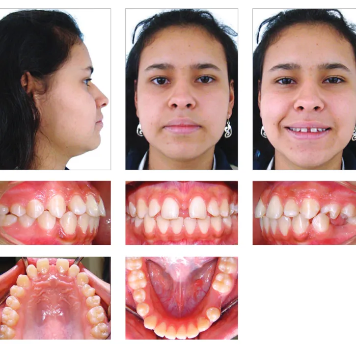
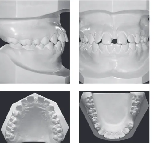
The patient presented a straight proile, good health condition and oral hygiene, normal breathing pattern and atypical swallowing pattern (Fig 1). Intraoral ex-amination revealed Class I malocclusion, 2-mm overjet and 5% overbite, coinciding dental midlines, moderate spacing in both arches and upper and lower labialized and protruded incisors (Figs 1 and 2). Radiographic analysis revealed the presence of all teeth which exhibited altered crown-root proportion, (maxillary right permanent lat-eral incisor, mandibular right irst and second premolars) with thinned and short roots, sclerosis of root canals and complete root resorption of maxillary permanent let lateral incisor. Tooth buds of maxillary and mandibu-lar let third momandibu-lars at Nolla Stage 6 development were observed, as well as the presence of mandibular second primary molar with congenital absence of mandibular let second premolar and mandibular right third molar (Fig 3). The patient presented Class I skeletal pattern with bimaxillary prognathism and macrognathism, pro-clination of maxillary and mandibular incisors and acute nasolabial angle (T0) (Fig 3 and Table 1).
Figure 1 - Initial facial and intraoral photographs.
electrical and heat pulp tests and were negative upon percussion and palpation. Sporadic painful symptom-atology of posterior segments was reported during mastication. All teeth presented normal physiological mobility, except for maxillary left permanent lateral incisor that had grade II mobility. Anatomy and color of crowns were normal. Periodontal examination in-dicated normal probing depths between 2 and 3 mm without bleeding.
TREATMENT OBJECTIVES
The aim of orthodontic treatment was mainly to meet patient’s esthetic expectations, achieve closure of anterior diastemas with light forces and also mainte-nance of crown-root proportion.
TREATMENT ALTERNATIVES
Figure 2 - Initial casts.
Figure 3 - Initial radiographs. A) Cephalometric tracing; B) Panoramic radiograph; C) Periapical radiograph of right upper incisors; D) Periapical radiograph of left upper incisors; E) Periapical radiograph of lower incisors.
A
B
orthodontic treatment was not an option, but the pa-tient was highly concerned about esthetics. Another op-tion was not using Orthodontics to fully close diastemas between maxillary teeth, but distributing those spaces to be restored with composites instead, so as to increase mesiodistal width, and also restore with osseointegrated implants the absent premolar and maxillary perma-nent let lateral incisor. Nevertheless, the patient did not count with the economic resources for this treat-ment option. Thus, it was decided to start orthodontic treatment focused on fully closing diastemas with light forces. The patient agreed and understood the risks.
TREATMENT PROGRESS
Prior to treatment onset, the patient was informed about the characteristics of the progressive pulp pathol-ogy condition she had and the limitations, risks and ob-jectives of treatment. Ater signing an informed consent form, orthodontic therapy was initiated.
Treatment plan required initial consultation with an endodontist in order to evaluate the degree and severity of external root resorption and begin orthodontic treat-ment with minimal risk, while taking into account the existing limitations.
Orthodontic treatment initiated irst in the upper posterior segments between canines and molars with an edgewise-standard technique. During the irst phase of treatment, low caliber NiTi wires were used (Fig 4). Once the posterior segments of the maxillary arch were consolidated, ixed appliances were installed in the upper
Measurement Norm T0 T1 T1-T0
SNA (degrees) 76.2 - 83.8 91.3 89.8 -1.5
SNB (degrees) 75 - 81 88.5 86.8 -1.7
ANB (degrees) 5.1 - 0.5 2.8 3 0.2
Co-A (mm) 90 91.7 92.1 -0.4
Co-Pog (mm) 110 118.1 117.9 -0.2
FMA(degrees) 24.2 18.2 18.8 0.6
Nasolabial angle (degrees) 105 91.2 97.1 5.9
Lower lip to H line (degrees) 0 - 0.5 3 0 -3
U1-FH (degrees) 110 129.9 113.3 -16.6
U1-PP (degrees) 105 - 115 128.5 115.7 -12.8
L1-PM (degrees) 88.5 - 97 106.6 102.8 -3.8
Interincisal angle (degrees) 124 106.5 127.5 -21
Table 1 - Cephalometric measurements.
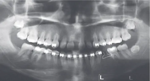
anterior segment where teeth were more afected by re-sorption. Space closure in the lower arch was initiated with a frictional technique using light elastomeric chains. Strict panoramic radiographic control was carried out every eight months based on clinical criteria in order to monitor the progression of pulp pathology (Fig 5). Given the positive response during treatment, the space be-tween mandibular irst premolar and molar was closed by attraction with a closed loop which had a tip back bend on the molar in order to protract and disincline it (Fig 4).
Esthetic contouring of upper anterior crowns was not necessary given the fact that all spaces were closed satisfactorily, thereby achieving an adequate distribution of all spaces. Prosthetic replacement of the maxillary lat-eral incisor was also not necessary due to stability shown during treatment. During the inal phase of treatment, the patient was referred to maxillary labial frenectomy and speech therapy in order to control tongue thrust habit that could afect long-term stability of treatment. Retention was completed with maxillary and mandibu-lar ixed retainers from canine to canine and the use of ESSIX plates. Total treatment time was 2.3 years be-tween 2009 and 2011. Some treatment limitations were encountered during the inal phase of treatment, such as the impossibility of completely aligning midlines due to the initial absence of mandibular let irst molar. The spaces between mandibular second premolar and man-dibular second molar were also not closed completely due to occlusal adjustment in that segment, which re-quired tip-back biomechanical movements that rep-resented high risk of root resorption. Post-treatment periodic radiographic controls were recommended to monitor the progression of root resorption.
TREATMENT RESULTS
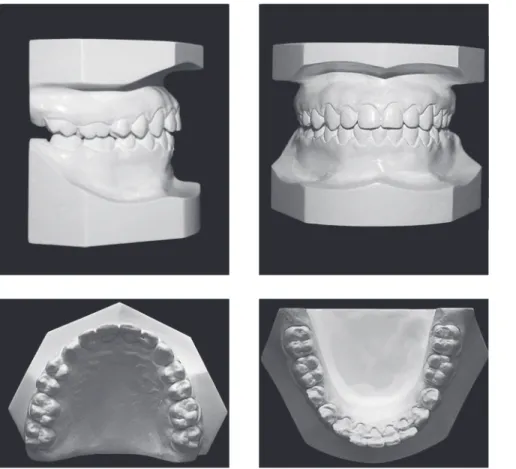
Ater orthodontic treatment with ixed appliances, the shape and contour of both dental arches improved, the rotations were ixed, diastemas were closed, pro-clination of maxillary and mandibular incisors was improved, a better occlusal relationship was achieved, overbite and overjet were corrected, the Curve of Spee was lattened, her nasolabial angle improved (T1), and a harmonic smile was achieved (Figs 6, 7 and 8).
Figure 4 - Control panoramic radiograph.
Figure 5 - Intraoral photographs during orthodontic treatment.
DISCUSSION
Clinical reports of classical idiopathic multiple root resorption are presented for patients whose past medi-cal history did not reveal any associated systemic, dental or familial causes.1,3,6-12 This article presented the
orth-odontic management of a young female patient with severe root resorption whose teeth were preserved es-thetically and functionally. It is important that the clini-cian have an understanding of the incidence, cause and
efects of root resorption in order to ofer patients the best treatment options.
Figure 7 - Post-treatment casts.
Figure 8 - Post-treatment radiographs. A) Cephalometric tracing; B) Panoramic radiograph; C) Periapical radiograph of right upper incisors; D) Periapical radiograph of left upper incisors; E) Periapical radiograph of lower incisors.
A
B
In this report, a patient with apical external root re-sorption with gradual rounding and shortening of roots was orthodontically treated. It was possible to reduce protrusion on both sides by decreasing U1-FH -16o,
U1-PP -12.8o and L1- MP -3.8o (Table 1). The
naso-labial angle reduced in 5.9o while the lower lip
retrud-ed 3 mm (Table 1), thereby improving patient’s pro-ile (Fig 6). The condition remained stable during the course of orthodontic treatment.
Marques et al21 reported a case of a young girl diagnosed
with a condition described as short root anomaly (SRA), a pathology similar to IERR described on this paper; how-ever, SRA is established when family link is established. The authors highlighted the importance of good diagnosis and efectiveness of orthodontic therapy that did not in-volve force applied directly on afected teeth.
The origin of the condition does not seem to be in the pulp and, therefore, interceptive endodontic treat-ment that includes pulp removal and placetreat-ment of cal-cium hydroxide or gutta-percha are not indicated.22
Given that dental and bone resorption is caused by osteoclastic activity,22 it is hypothesized that there is
some triggering factor that activates these cells.
Current management of this condition is conserva-tive, minimally invasive and consists of long-term mon-itoring.23 Orthodontic treatment is a viable alternative
that ofers patients an acceptable esthetic and functional solution. However, there are important considerations that the orthodontist must take into account and follow, such as the prognosis of teeth with a history of severe resorption, progression of the condition, progress and stability of teeth with future restorations.
These cases are best described as idiopathic because no cause or family history could be associated. Man-agement of interceptive therapy of idiopathic root re-sorption depends on the identiication of the speciic cell mechanism and the external factor that cause the disorder. Orthodontic management is an useful alterna-tive that provides these patients with a functional and esthetic option. It is important that the clinician com-pletes a full medical history and detailed initial clinical and radiographic indings and have the patient sign an informed consent document prior to treatment onset. Orthodontic therapy should be focused on solving pa-tient’s esthetic concerns.
Oyana et al,24 using the inite element method,
dem-onstrated that a signiicant amount of stress was concen-trated at the middle of the root in a model of short root. That condition is suicient to increase root resorption in progress on those patients. Orthodontic forces should be applied with caution. In alignment and leveling, the use of intermittent, light and constant forces that do not surpass capillary blood pressure of 20-26 g/cm2 are
rec-ommended. The use of Class II intermaxillary elastics, maxillary expansion appliances anchored on premolars and extraoral forces anchored on irst molars should be avoided, since they have been reported as a potential risk factor for teeth with root resorption.26
Strict radiographic controls during the course of orthodontic therapy in order to monitor the resorptive condition are very important. During retention, ixed retainers in the upper and lower anterior segments are recommended. It is also important to identify the pres-ence of functional habits, such as atypical swallowing or nail biting, both of which could afect treatment results and stability of compromised teeth.
It is also important to emphasize the need to insist on extreme oral hygiene measures in order to maintain patient’s periodontal stability. Post-treatment radio-graphic control is recommended in order monitor the condition and establish a long-term prognosis, in addi-tion to addressing the concerns menaddi-tioned above.
CONCLUSIONS
1) Orthodontic treatment of patients with idiopath-ic multiple root resorption offering them estheti-cal and physiologiestheti-cal solutions is possible consid-ering that the patient understands potential risks and limitations.
2) Orthodontic management is based on simple me-chanical techniques that include light and controlled forces, allowing predictable movements which are physiologically acceptable if pulp and periodontal limitations are considered.
3) A complete history of patient’s medical background allows identiication of any systemic condition that might be associated with the pulp pathology.
1. George DI, Miller RL. Idiopathic resorption of teeth: a report of three cases. Am J Orthod. 1986;89(1):13-20.
2. Liang H, Burkes EJ, Frederiksen NL. Multiple idiopathic cervical root resorption: systematic review and report of four cases. Dentomaxillofac Radiol. 2003;32:150-5.
3. Muller. E. Laboratory studies of an unusual case of resorption. J Am Dent Assoc. 1930;17:326-34.
4. Rivera EM, Walton RE. Extensive idiopathic apical root resorption. A case report. Oral Surg Oral Med Oral Pathol. 1994;78(5):673-7.
5. Postlethwaite KR, Hamilton M. Multiple idiopathic external root resorption. Oral Surg Oral Med Oral Pathol. 1989;68(5):640-3. 6. Carr HG. Multiple idiopathic resorption of teeth. Br Dent J.
1958;105:455-6.
7. Pinska E, Jarzynka W. Spontaneous resorption of the roots of all permanent teeth as a familial disease. Czas Stomatol. 1966;19(2):161-5. 8. Kerr DA, Courtney RM, Burkes EJ. Multiple idiopathic root resorption.
Oral Surg Oral Med Oral Pathol 1970;29:552-65.
9. Hopkins R, Adams D. Multiple idiopathic resorption of the teeth. Br Dent J. 1979;146(10):309-12.
10. Lydiatt DD, Hollins RR, Peterson G. Multiple idiopathic root
resorption: diagnostic considerations. Oral Surg Oral Med Oral Pathol. 1989;67(2):208-10.
11. Moody AB, Speculand B, Smith AJ, Basu MK. Multiple idio-pathic external resorption of teeth. Int J Oral Maxillofac Surg. 1990;19(4):200-2. 12. Moody GH, Muir KF. Multiple idiopathic root resorption: a case report and
discussion of pathogenesis. J Clin Periodontol. 1991;18(8):577-80. 13. Harris EF, Kineret SE, Tolley EA. A heritable component for external
apical root resorption in patients treated orthodontically. Am J Orthod Dentofacial Orthop. 1997;111(3):301-9.
14. Lerman RL, Gold R. Idiopathic short root anomaly. J Pedod. 1977;1(4):327-33.
REFERENCES
15. Brook AH, Holt RD. The relationship of crown length to root length in permanent maxillary central incisors. Proc Br Paedod Soc. 1978;8:17-20. 16. Näsman M, Björk O, Söderhäll S, Ringdén O, Dahllöf G. Disturbances in
the oral cavity in pediatric long-term survivors after diferent forms of antineoplastic therapy. Pediatr Dent. 1994;16(3):217-23.
17. Prahl-Andersen B, Oerlemans J. Characteristics of permanent teeth in persons with trisomy G J Dent Res. 1976;55(4):633-8.
18. Gibilisco J. Oral radiographic diagnosis. 5th ed. Philadelphia: WB Saunders; 1985.
19. Belanger GK, Coke JM. Idiopathic external root resorption of the entire permanent dentition: report of case. ASDC J Dent Child. 1985;52(5):359-63.
20. Schätzle M, Tanner SD, Bosshardt DD. Progressive, generalized, apical idiopathic root resorption and hypercementosis. J Periodontol. 2005;76(11):2002-11.
21. Marques LS, Generoso R, Armond MC, Pazzini CA. Short-root anomaly in an orthodontic patient. Am J Orthod Dentofacial Orthop. 2010;138(3):346-8.
22. Andreasen JO. External root resorption: its implication in dental traumatology, paedodontics, periodontics, orthodontics and endodontics. Int Endod J. 1985;18(2):109-18.
23. Gupta R, Prakash V. Bilateral extensive idiopathic apical root resorption in supraerupted maxillary molars: a case report. Oral Surg Oral Med Oral Pathol Oral Radiol Endod. 2008;106(3):e44-7.
24. Oyama K, Motoyoshi M, Hirabayashi M, Hosoi K, Shimizu N. Efects of root morphology on stress distribution at the root apex. Eur J Orthod. 2007;29(2):113-7.
25. Schwarz. A. Tissue changes incidental to orthodontics tooth movement. Int J Orthod. 1932;18(4):331-52.