The
Plasmodium falciparum
-Specific Human Memory B
Cell Compartment Expands Gradually with Repeated
Malaria Infections
Greta E. Weiss1, Boubacar Traore2, Kassoum Kayentao2, Aissata Ongoiba2, Safiatou Doumbo2, Didier Doumtabe2, Younoussou Kone2, Seydou Dia2, Agnes Guindo2, Abdramane Traore2, Chiung-Yu Huang3, Kazutoyo Miura4, Marko Mircetic1, Shanping Li1, Amy Baughman1, David L. Narum4, Louis H. Miller4, Ogobara K. Doumbo2, Susan K. Pierce1*, Peter D. Crompton1*
1Laboratory of Immunogenetics, National Institute of Allergy and Infectious Diseases, National Institutes of Health, Bethesda, Maryland, United States of America,
2Malaria Research and Training Center, Faculty of Medicine, Pharmacy and Dentistry, University of Bamako, Bamako, Mali,3Biostatistics Research Branch, National Institute of Allergy and Infectious Diseases, National Institutes of Health, Bethesda, Maryland, United States of America,4Malaria Vaccine Development Branch, National Institute of Allergy and Infectious Diseases, National Institutes of Health, Bethesda, Maryland, United States of America
Abstract
Immunity to Plasmodium falciparum (Pf) malaria is only acquired after years of repeated infections and wanes rapidly without ongoing parasite exposure. Antibodies are central to malaria immunity, yet little is known about the B-cell biology that underlies the inefficient acquisition ofPf-specific humoral immunity. This year-long prospective study in Mali of 185 individuals aged 2 to 25 years shows thatPf-specific memory B-cells and antibodies are acquired gradually in a stepwise fashion over years of repeatedPfexposure. BothPf-specific memory B cells and antibody titers increased after acute malaria and then, after six months of decreasedPfexposure, contracted to a point slightly higher than pre-infection levels. This inefficient, stepwise expansion of both thePf-specific memory B-cell and long-lived antibody compartments depends onPf exposure rather than age, based on the comparator response to tetanus vaccination that was efficient and stable. These observations lend new insights into the cellular basis of the delayed acquisition of malaria immunity.
Citation:Weiss GE, Traore B, Kayentao K, Ongoiba A, Doumbo S, et al. (2010) ThePlasmodium falciparum-Specific Human Memory B Cell Compartment Expands Gradually with Repeated Malaria Infections. PLoS Pathog 6(5): e1000912. doi:10.1371/journal.ppat.1000912
Editor:Jean Langhorne, National Institute for Medical Research, United Kingdom
ReceivedDecember 9, 2009;AcceptedApril 19, 2010;PublishedMay 20, 2010
This is an open-access article distributed under the terms of the Creative Commons Public Domain declaration which stipulates that, once placed in the public domain, this work may be freely reproduced, distributed, transmitted, modified, built upon, or otherwise used by anyone for any lawful purpose.
Funding:This work was supported by the Division of Intramural Research of the National Institute of Allergy and Infectious Diseases (NIAID), National Institutes of Health (NIH). The funders had no role in study design, data collection and analysis, decision to publish, or preparation of the manuscript.
Competing Interests:The authors have declared that no competing interests exist.
* E-mail: [email protected] (SKP); [email protected] (PDC)
Introduction
To date, most successful vaccines have targeted pathogens that induce long-lived protective antibodies after a single infection, such as the viruses that cause smallpox, measles and yellow fever [1]. It has proved more difficult to develop highly effective vaccines against pathogens that do not induce sterile immunity such as the human immunodeficiency virus type-1 (HIV-1), Mycobacterium tuberculosis (Mtb), and Plasmodium falciparum malaria [2]. However, unlike HIV-1 and Mtb, clinical immunity to malaria can be acquired, but only after years of repeated Pf infections [3]. Passive transfer studies indicate that antibodies ultimately play a key role in protection from malaria [4], yet several studies show that antibodies toPfantigens are inefficiently generated and rapidly lost in the absence of ongoing exposure to the parasite (reviewed in [5]). Elucidating the cellular basis of the inefficient acquisition of malaria immunity may ultimately prove critical to the design of an effective malaria vaccine.
Despite the key role that antibodies play in protection from a variety of infectious diseases, remarkably little is known about the cellular basis of acquiring humoral immunity in response to natural infections in humans. This gap in our knowledge is due in large part
to the difficulty in studying natural infections in humans when we cannot predict who within a population will be infected with a given pathogen at a given time. Thus, our current understanding of the acquisition of immunity is largely derived from animal models and studies of humans after vaccination. These studies have established that long-lived, antibody-based immunity requires the generation and maintenance of memory B cells (MBCs) and long-lived plasma cells (LLPCs) (reviewed in [6,7]). This process begins when naı¨ve B cells bind antigen near the interface of B and T cell areas of secondary lymphoid organs. Several studies suggest that high-affinity binding drives naı¨ve B cells to differentiate into short-lived, isotyped switched plasma cells (PCs) within the extra-follicular region which contributes to the initial control of infection. In contrast, lower affinity binding selects for entry of naı¨ve B cells into follicles where germinal centers are formed. After a period of 7–10 days, through the CD4+
The mechanisms by which antibody responses are maintained over the human life-span remains an open question. In one model, LLPCs survive indefinitely in the bone marrow and independently maintain steady-state antibody levels [8]. Alter-native models predict that PCs are replenished by MBCs that proliferate and differentiate in response to persistent [9] or intermittent exposure to antigen, and/or through non-specific by-stander activation (e.g. cytokines or TLR ligands) [10]. Unlike PCs, which are terminally-differentiated, MBCs may be maintained through homeostatic proliferation [11], possibly through exposure to polyclonal stimuli [10]. To address fundamental questions related to the generation and mainte-nance of MBCs and Abs specific forPfmalaria in children in malaria endemic areas, we conducted a year-long prospective study in a rural village of Mali that experiences an intense, sharply-demarcated six-month malaria season annually. We determined whetherPfinfection generates MBCs specific forPf blood stage antigens, and if so, whether they accumulate with age and cumulative Pf exposure, and also whether their frequency correlates with protection from malaria. In addition, we determined whether acute, symptomaticPfinfection resulted in an increase in the number ofPf-specific MBCs and the levels of specific antibodies, and if so, whether this increase remained stable over a six-month period of markedly reduced Pf transmission. By taking advantage of the tetanus immunization schedule in Mali in which infants and women of child-bearing age are vaccinated, we compared the relative efficiencies of the acquisition of tetanus toxoid (TT)- and Pf-specific MBCs and Ab, and also tested three hypotheses: 1) that growth of the MBC compartment depends on immunological experience rather than age, 2) that Pf infection induces non-specific activation of bystander B cells [12,13], and 3) that polyclonal activation during heterologous immune responses is a general mechanism for maintaining MBCs and LLPCs [10].
Results
Malaria immunity is acquired gradually despite intense exposure to thePfparasite
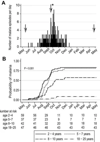
In May 2006 we initiated an observational cohort study in Mali to investigate the mechanisms underlying naturally-acquired malaria immunity. A detailed description of the study site and cohort has been reported elsewhere [14]. The study population was an age-stratified, random sample representing 15% of all individuals living in a small, rural, well-circumscribed, non-migratory community where antimalarial drugs were provided exclusively by the study investigators. During a two-week period one month prior to the abrupt onset of the six-month malaria season, we enrolled 225 individuals in four age groups: 2–4 years (n = 73), 5–7 years (n = 52), 8–10 years (n = 51), and 18–25 years (n = 49). Attendance at scheduled follow-up visits was.99% for children (2–10 years) and 82% for adults (18–25 years) during the one-year study period indicating a high degree of study awareness and participation. For the MBC analysis reported here, a subset of 185 individuals was randomly selected within each of the four age categories. All subsequent data and analysis refer to these 185 individuals. The baseline demographic and clinical characteristics of this subset are shown in Table 1, according to age group. As previously reported [14], only three of the characteristics shown in Table 1 were associated with decreased malaria risk in multivariate analysis—increasing age, sickle cell trait (HbAS), and asymptomaticPfparasitemia at study enrollment. During the one-year study period there were 380 unscheduled clinic visits, during which 219 cases of malaria were diagnosed, five of which met the WHO criteria for severe malaria [15]. Malaria episodes were defined as an axillary temperature $37.5uC, Pf asexual parasitemia$5000 parasites/mL, and a non-focal physical exam by the study physician. As expected in this region of Mali, all malaria cases were confined to a six-month period that began in July, peaked in October, and ended by January (Fig. 1A). The incidence of malaria and the proportion of individuals experienc-ing at least one malaria episode decreased with age, whereas the time to the first malaria episode increased with age (Table 2 and Fig. 1B). Thus, despite intense annualPftransmission at this study site, malaria immunity is acquired slowly.
Analysis ofPf-specific and TT-specific MBCs and Abs inPf -uninfected children and adults before the malaria season
We first established baseline levels of IgG+AMA1-, MSP1- and
TT-specific MBCs and Abs inPf-uninfected, healthy children and adults in May just before the malaria season, a point at which there had been little to noPftransmission for five months. For this analysis we excluded individuals with asymptomaticPfparasitemia (8.7% of total cohort; Table 1), because they showed a decreased risk of malaria and tended toward higher frequencies of AMA1-and MSP1-specific MBCs AMA1-and levels of Ab (data not shown).
We focused our analyses on MBCs and Abs specific for Pf blood-stage antigens because humoral responses are known to be critical to blood-stage immunity [4]. We examined the response to two blood stage proteins, Apical Membrane Antigen 1 (AMA1) and Merozoite Surface Protein 1 (MSP1), because we had previously studied the MBC and Ab responses to these antigens in vaccine trials of Pf-naı¨ve individuals [16]. This afforded the opportunity to compare the acquisition of B cell memory to the same antigens after vaccination versus natural Pfinfection. We express MBC data as ‘MBCs per 106 PBMCs’, where ‘MBCs’ refers to the number of antibody secreting cells derived from MBCs during the six-day culture, and ‘106PBMCs’ refers to the number of PBMCs after culture. In the present study, the mean Author Summary
frequency of AMA1-specific MBCs per 106PBMCs increased with age (Fig. 2A; 2–4 yr: 1.2 [95% CI: 0.45–1.9]; 5–7 yr: 5.0 [95% CI:
20.2–10.1]; 8–10 yr: 8.9 [95% CI: 4.9–12.9]; 18–25 yr: 37.8 [95% CI: 10.4–65.3]; P,0.001), as did the proportion of individuals with detectable AMA1-specific MBCs (2–4 yr: 8.1%; 5–7 yr: 30.8%; 8–10 yr: 50.0%; 18–25 yr: 54.8%; P,0.001). Similarly, AMA1-specific Ab levels and the proportion of individuals seropositive for AMA1-specific Abs increased with age (Fig. 2A; P,0.001 for both comparisons). There was a positive correlation between the frequency of AMA1-specific MBCs and Ab levels (Spearman’s correlation coefficient = 0.35; P = 0.005; Fig. S1).
We observed a similar age-associated increase in the frequency of MSP1-specific MBCs, although the overall frequency was lower than that for AMA1-specific MBCs (Fig. 2B; 2–4 yr: 1.2 [95% CI: 0.55–1.9]; 5–7 yr: 3.2 [95% CI: 1.2–5.2]; 8–10 yr: 5.9 [95% CI: 2.9–9.0]; 18–25 yr: 10.3 [95% CI: 6.3–14.3]; P,0.001). Likewise, the proportion of individuals who had detectable MSP1-specific MBCs (2–4 yr: 9.1%; 5–7 yr: 27.8%; 8–10 yr: 34.3%; 18–25 yr: 47.6%; P = 0.001) was similar to that for AMA1. MSP1-specific Ab levels and the proportion of individuals seropositive for MSP1-specific Abs also increased gradually with age (Fig. 2B; P,0.001 for both comparisons). There was a positive correlation between the frequency of MSP1-specific MBCs and Ab levels (Spearman’s correlation coefficient = 0.34; P = 0.004; Fig. S1). Remarkably, despite exposure to 50–60 infective mosquito bites per month at the peak of each malaria season in this area [17], only approximately half of adults had detectable MBCs specific for AMA1 and MSP1, even though most had detectable AMA1- and MSP1-specific antibodies. Of the 72 individuals without detectable AMA1-specific MBCs before the malaria season, 64 (88.9% [95% CI 79.3–95.1]) did not have detectable MSP1-specific MBCs, suggesting that failure to generate MBCs to one Pf antigen is associated with failure to generate MBCs to otherPfantigens.
To understand if the expansion ofPf-specific MBCs with age was driven by repeated exposure to Pf antigens or simply a function of age, we determined the frequency of MBCs specific for an unrelated antigen, tetanus toxoid (TT), with age. In Mali, a single TT vaccine is administered to infants less than six months of age and a second TT vaccine is administered to females around 15 years of age to prevent neonatal tetanus. Thus, we measured TT-specific antibody and MBC responses at least 18 months after TT vaccination, a point at which the TT-specific response is likely to be at steady state. In contrast to what was observed for AMA1-and MSP1-specific MBCs, the frequency of TT-specific MBCs among males did not change significantly from age 2 to 25 years (Fig. 2C) (2–4 yrs: 10.8 [95% CI27.4–29.0], 5–7 yrs: 7.3 [95% CI 0.7–13.9], 8–10 yrs: 8.0 [95% CI 3.1–12.8], 18–25 yrs: 4.7 [95% CI 1.4–8.1]; P = 0.80). Similarly, the proportion of male adults who were positive for TT-specific MBCs did not differ significantly from male children (2–4 yrs: 25.0%, 5–7 yrs: 33.3%, 8–10 yrs: 40.9%, 18–25 yrs 28.6%; P = 0.80). The slightly higher frequency of TT-specific MBCs in male versus female children was not statistically significant. However, the frequency of TT-specific MBCs was significantly higher in female adults compared to female children (Fig. 2C; mean frequency of TT-specific MBCs per million PBMC by age group (2–4 yrs: 2.9 [95% CI 1.1–4.7], 5–7 yrs: 3.2 [95% CI 0.2–6.1], 8–10 yrs: 3.4 [95% CI 1.1–5.7], 18–25 yrs: 58.7 [95% CI 34.2–83.3]; P,0.001) presumably the result of booster vaccination. Likewise, the proportion of female adults who were positive for TT-specific MBCs was significantly higher as compared to female children (2–4 yrs: 28.1%, 5–7 yrs: 25.0%, 8–10 yrs: 27.3%, 18–25 yrs 88.0%; P,0.001). For both females and males, TT-specific Ab levels mirrored MBC frequencies (Fig. 2C)—clearly increasing from female children to female adults (P,0.001), while not changing significantly by age in males (P = 0.44). Overall, TT-specific Ab levels and MBC frequencies correlated (Spearman’s correlation coefficient = 0.48;
Table 1.Baseline characteristics of the study cohort by age group.
Age group, years All (n = 185)
2–4 (n = 59) 5–7 (n = 37) 8–10 (n = 42) 18–25 (n = 47)
Gender, % female (no.) 66.1 (39) 48.7 (18) 33.3 (14) 61.7 (29) 54.1 (100)
Ethnicity, % (no.)
Bambara 62.7 (37) 51.4 (19) 54.8 (23) 66.0 (31) 59.5 (110)
Sarakole 32.2 (19) 43.2 (16) 35.7 (15) 27.7 (13) 34.1 (63)
Fulani 3.4 (2) 5.4 (2) 7.1 (3) 4.3 (2) 4.9 (9)
Malinke 1.7 (1) 0.0 (0) 2.4 (1) 2.1 (1) 1.6 (3)
Hemoglobin AS, % (no.)a– 13.6 (8) 8.1 (3) 7.1 (3) 10.6 (5) 10.3 (19)
P. falciparumsmear positive at enrollment, % (no.)b– 6.8 (4) 10.8 (4) 11.9 (5) 6.4 (3) 8.7 (16)
Parasitemia if smear positive at enrollment, parasites/microliter [geometric mean (95% CI)]
1438 (159–12973)
3616 (1500–8715)
415 (134–1287)
953 (39–23381)
1137 (579–2232) GI helminth, % positive at enrollment (no.)c– 14.6 (8) 8.3 (3) 11.8 (4) 0 (0) 9.7 (15)
Urine schistosomiasis, % positive at enrollment (no.)d– 0 (0) 0 (0) 5.3 (2) 29.0 (9) 7.4 (11)
Distance lived from clinic, meters (mean6SD) 395 (6116) 408 (6140) 365 (683) 359 (691) 382 (6110) Bed net use, % (no.)–e 27.3 (15) 41.2 (14) 17.1 (7) 39.5 (15) 30.4 (51)
a
–Data available for 177 subjects.
b
–All subjects were asymptomatic at enrollment.
c
–Data available for 154 subjects; GI = gastrointestinal.
d
–Data available for 148 subjects.
e
P,0.001; Fig. S1). The observation that Pf-specific MBCs increased with age while TT-specific MBCs in individuals who received no booster vaccine did not increase and tended to decrease slightly with age indicates that the increase inPf-specific MBCs is driven by repeated antigen exposure and is not simply a function of age. Of note, the size of the total IgG+
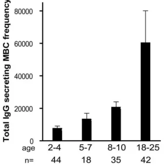
MBC compartment, as reflected in the peripheral blood, increased with age (Fig. 3; P,0.001), consistent with the maturation of the total MBC compartment with immunological experience.
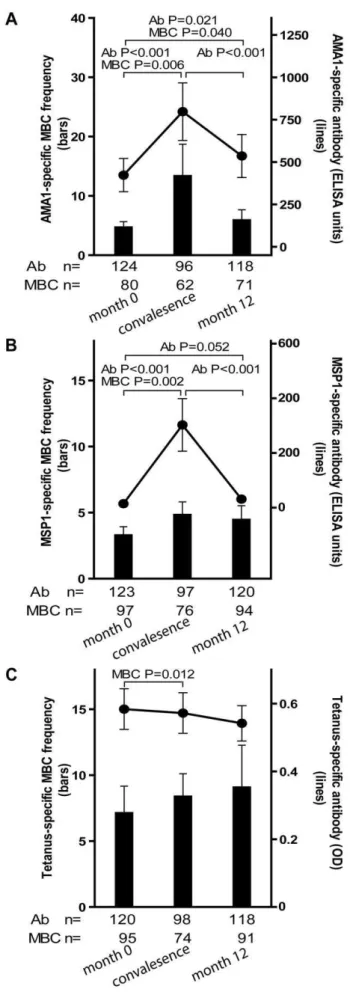
Longitudinal analysis of thePf- and TT-specific MBC and Ab responses two weeks after acute malaria and after a prolonged period of decreasedPfexposure
To assess the Pf-specific MBC and Ab responses to acute malaria, and to determine the stability of this response during a period of little to noPftransmission, we measured the frequencies of MBCs and Ab levels specific for AMA1 and MSP1 14 days after the first episode of malaria (convalescence), and in a cross-sectional survey at the end of the following dry season (month 12), and compared these frequencies to the pre-malaria season baseline (month 0; as detailed above). Malaria episodes were defined as an axillary temperature $37.5uC, Pf asexual parasitemia $5000 parasites/mL, and a non-focal physical exam by the study
physician. Because few adults experienced malaria (Table 2), this analysis only included children aged 2–10 years (see Fig. 4 for sample sizes at each time point). The mean frequency of AMA1-specific MBCs in children aged 2–10 years increased from month 0 to convalescence (Fig. 4A; month 0: 4.7 [95% CI: 2.8–6.6]; convalescence: 13.4 [95% CI: 2.7–24.1; P = 0.006] and then decreased from convalescence to month 12 (Fig. 4A; month 12: 5.9 [95% CI: 2.4–9.4]; P = 0.93 versus convalescence) to a point just above the frequency at month 0 (Fig. 4A; P = 0.021, month 0 vs. month 12). Likewise, the level of AMA1-specific Ab increased from month 0 to convalescence (Fig. 4A; month 0: 422.8 [95% CI: 228.7–617.0]; convalescence: 797.2 [95% CI: 460.0–1134.7; P,0.001], and then decreased from convalescence to month 12 (Fig. 4A; month 12: 535.5 [95% CI: 283.8–787.2]; P,0.001 versus convalescence], to a point just above month 0 levels (Fig. 4A; P = 0.040, month 0 vs. month 12).
The MSP1-specific MBC and Ab responses followed a similar pattern. The mean frequency of MSP1-specific MBCs in children aged 2–10 years increased from month 0 to convalescence (Fig. 4B; month 0: 3.3 [95% CI: 2.0–4.6]; convalescence: 4.8 [95% CI: 2.9– 6.8; P = 0.002] and then decreased from convalescence to month 12 (Fig. 4B; month 12: 4.5 [95% CI: 2.4–6.6]; P = 0.71 versus convalescence) to a point just above the frequency at month 0
Table 2.Malaria clinical outcomes by age group.
Age group, years All (n = 185)
2–4 (n = 59) 5–7 (n = 37) 8–10 (n = 42) 18–25 (n = 47)
Malaria incidence, mean (6SD)–a 1.86 (
61.28) 1.81 (61.17) 0.95 (61.08) 0.09 (60.28) 1.19 (61.27)
Severe malaria incidence, no.b– 4 1 0 0 5
At least one malaria episode, % (no.) 83.1 (49) 81.1 (30) 57.1 (24) 8.5 (4) 57.8 (107)
Time to first malaria episode, days (median)–c 101 121 124 153 118
Parasitemia at first malaria episode, parasites/microliter [geometric mean (95% CI)]
39084 (30579–49954)
26417 (19440–35896)
20561 (15683–26956)
8816 (4082–19037)
28678 (24334 –33799)
a
–Malaria episode defined as T$37.5uC, asexual parasitemia$5000/microliter, and non-focal physical examination.
b
–WHO definition of severe malaria.
c
–Days since study enrollment. doi:10.1371/journal.ppat.1000912.t002
(Fig. 4B; P = 0.156, month 0 vs. month 12). Likewise, the level of MSP1-specific Ab increased from month 0 to convalescence (Fig. 4B; month 0: 14.6 [95% CI: 10.5–18.6]; convalescence: 302.6 [95% CI: 111.7–493.4; P,0.001], and then decreased from convalescence to month 12 (Fig. 4B; month 12: 31.1 [95% CI: 5.5–56.6]; P,0.001 versus convalescence], to a point just above month 0 levels (Fig. 4B; P = 0.052, month 0 vs. month 12). Figure 2. ThePf-specific MBC and long-lived antibody com-partments expand gradually with age. Shown are the MBC frequencies (bars, left axis) and antibody levels (lines, right axis) specific for AMA1 (A) and MSP1 (B) by age category; and TT (C) by age category and gender; before the malaria season inPf-uninfected individuals. The frequency of AMA1- and MSP1-specific MBCs increased with age (P,0.001 for both trends), as did the level of AMA1- and MSP1-specific antibodies (P,0.001 for both trends). There were no significant differences by gender for the AMA1- and MSP1-specific responses (not shown). To determine if the expansion ofPf-specific MBCs with age was driven by exposure to antigen or simply a function of age, we measured the TT-specific MBC and antibody response with age. In Mali, infants are vaccinated with TT, and females receive a TT booster around the age of 15 years to prevent neonatal tetanus. In contrast to AMA1 and MSP1, the frequency of TT-specific MBCs and the level of TT-specific antibodies for males did not change significantly from age 2 to 25 years (P = 0.80 and P = 0.44, respectively). However, the frequency of TT-specific MBCs and the level of TT-TT-specific antibodies was higher in female adults compared to female children (P,0.001 for both comparisons). MBC frequencies were determined by ELISPOT and are expressed per million PBMC. The number of individual samples assayed and the percent of individual samples that exceeded the limit of detection (i.e. those considered positive) is indicated below the graph. The discrepancy in the sample size for ELISA data among 2–4 year olds is due to technical error during the performance of the ELISA. P values were obtained by the Kruskal-Wallis test. Data are shown as mean6 s.e.m.
doi:10.1371/journal.ppat.1000912.g002
Figure 3. The size of total IgG+ MBC compartment expands gradually with age.The frequency of IgG+
MBCs per million PBMCs measured before the malaria season increased with age (P,0.001). The number of individuals in each age category is indicated. ThePvalue was obtained by the Kruskal-Wallis test. Data are shown as mean6 s.e.m.
To determine if malaria induces non-specific activation of ‘bystander’ MBCs, we compared the frequencies of TT-specific MBCs and Ab levels before the malaria season (month 0) to that 14 days after acute malaria (convalescence). We observed a small, but statistically significant increase in the frequency of TT-specific MBCs from month 0 to convalescence (Fig. 4C; month zero: 7.1 [95% CI: 3.1–11.2]; convalescence: 8.4 [95% CI: 5.0– 11.8; P = 0.012) that did not change significantly at month 12 (month 12: 9.1 [95% CI: 3.2–15.4]; P = 0.974 versus convales-cence]. In contrast, TT-specific Ab levels decreased slightly from month 0 to convalescence, and again from convalescence to month 12, although neither decline was statistically significant (Fig. 4C; month 0: 0.58 [95% CI: 0.5–0.7]; convalescence: 0.57 [95% CI: 0.5–0.7; P = 0.063]; month 12: 0.54 [95% CI: 0.4–0.6]; P = 0.525 versus convalescence). Collectively these results indicate that malaria infection results in an increase in the frequencies of both Pf-specific, and bystander MBCs. However, malaria selectively inducesPf-specific Ab production but does not appear to drive the differentiation of bystander naı¨ve and memory B cells into PCs.
B cell subsets inPf-uninfected children and adults before the malaria season
By FACS we determined the proportion of B cell subsets in individuals (2–4 yrs [n = 38], 5–7 yrs [n = 21], 8–10 yrs [n = 23], 18–25 yrs [n = 27]) before the malaria season (Fig. 5A). With increasing age, and as a percentage of total CD19+
B cells we observed a decrease in immature B cells (CD19+
CD10+
; P,0.001) and naı¨ve B cells (CD19+
CD272 CD21+
CD102; P = 0.047) and an increase in resting IgG+
MBCs (CD19+
CD27+
CD21+
; P,0.001) and activated IgG+
MBCs (CD19+
CD27+
CD212 CD20+
CD102; P,0.001). The increase with age of classical MBCs is consistent with the increase in total IgG+
MBCs we observed using the MBC ELISPOT assay (Fig 3).
In a subset of 87 individuals from this same study cohort, we previously reported that Pf exposure is associated with an expanded subset of ‘atypical’ MBCs that express FCRL4 and are hyporesponsive toin vitrostimuli [18], similar to the ‘exhausted’ MBCs described in viremic, HIV-infected individuals [19]. Atypical MBCs are defined as CD19+
CD272 CD212 CD20+
CD102and typically represents,4% of circulating CD19+
B cells in healthy U.S. adults [19]. Here, analyzing a larger number of individuals in the cohort, we confirmed that this subset of MBCs is expanded in Malian children and adults compared to malaria-naı¨ve U.S. adults (U.S. adults: 1.4% [95% CI: 0.9–1.8]; Malian children aged 2–10 years: 10.2% [95% CI: 8.7–11.8], P,0.001 versus U.S. adults; Malian adults aged 18–25 years: 14.8% [95% CI: 11.0–19.1], P,0.001 versus U.S. adults). Thus, in addition to the increase in classical MBCs, an ‘atypical’ MBC subset is expanding with age in this study population.
Figure 4. Longitudinal analysis of thePf- and TT-specific MBC and antibody response. Compared to month zero, the MBC frequencies and antibody levels specific for AMA1 (A) and MSP1 (B) increased two weeks after the first episode of malaria and then contracted to a point slightly higher than pre-infection levels after a six-month period of decreasedPf exposure. Compared to month zero, there was a small but statistically significant increase in TT-specific MBC two weeks after the first episode of malaria (C), whereas the level of TT-specific antibodies did not change. The number of individuals in each age category is indicated. Only statistically significant P values are shown. P values were obtained by the Wilcoxon matched-pairs signed-rank test. Data are shown as mean6s.e.m.
Longitudinal profiling of B cell subsets in children before and after acute malaria
We investigated the impact of acute malaria on the relative proportion of B cells in each subset in children aged 2–10 years. Compared to the pre-malaria season baseline (month 0), there were no significant changes in the percent of lymphocytes that
were CD19+
14 days after acute malaria. Within the CD19+
B cell population there were no significant changes in the percent of immature B cells, naı¨ve B cells, or resting MBCs, after acute malaria. Moreover, there was no change in the proportions of resting and atypical MBCs that were IgG+
. However, we observed a decrease in the percentage of total atypical MBCs (Fig. 5B; month 0: 10.9% [95% CI: 9.4–12.4], convalescence: 8.7% [95% CI: 7.3–10.2]; P = 0.027), and an increase in activated MBCs following acute malaria (month 0: 1.6 [95% CI: 1.2–2.0], convalescence: 1.9 [95% CI: 1.4–2.4]; P = 0.09). Within the activated MBC subset there was a significant increase in the proportion that were IgG+
(month 0: 59.0% [95% CI: 56.0–62.1], month 12: 62.8% [95% CI: 59.2–66.3]; P,0.001). The decrease in the proportion of atypical MBCs in the peripheral blood suggests that this subpopulation may be trafficking out of the circulation into tissues in response to acute malaria.
AMA1- and MSP1-specific MBC frequencies and Ab levels and malaria risk
We determined prospectively whether AMA1- or MSP1-specific Ab levels or MBC frequencies measured just prior to the six month malaria season were associated with the subsequent risk of malaria. For this analysis a malaria episode was defined as an axillary temperature $37.5uC, Pfasexual parasitemia $5000 parasites/
mL, and a non-focal exam by the study physician. Because the
incidence of malaria was very low in adults during the study period (Table 2), they were excluded from this analysis. Three measures of malaria risk were analyzed: 1) whether or not malaria was experienced, 2) the incidence of malaria, and 3) the time to the first malaria episode. In the corresponding multivariate regression models (logistic, Poisson, and Cox regression) which controlled for age, sickle cell trait, and concurrent asymptomaticPfparasitemia, we found no correlation between malaria risk and AMA1- or MSP1- specific Ab levels or MBC frequencies. As discussed below, this finding was not unexpected based on the observation that the malaria vaccine candidates AMA1 and MSP1 did not confer protection against malaria in clinical trials [20,21].
Discussion
In this year-long prospective study of children and adults in an area of intense, annual, sharply demarcatedPftransmission, we show that MBCs specific forPfcan be acquired, but only gradually in a stepwise fashion over years of repeated Pfexposure. MBCs specific for two Pf antigens, AMA1 and MSP1, increased in frequency in response to acutePfinfection, and then contracted during a six-month period of decreasedPf exposure to a point slightly above pre-infection levels. Cross-sectional analysis of individuals aged 2–25 years just before the malaria season indicated that this step-wise, incremental increase in Pf-specific MBCs with each malaria season contributes to the gradual expansion of thePf-specific MBC compartment with cumulativePf exposure. By comparison, the stable frequency of TT-specific MBCs with age after immunization in infancy indicates that growth of antigen-specific MBC compartments does not simply occur with age, but requires repeated antigen exposure. We do not formally know if the gradual gain inPf-specific MBCs is in fact due to an increase in long-lived MBCs, or whether those MBCs requirePf-stimulation and would be lost ifPftransmission did not resume after the six-month dry season. In another setting, namely in an area of Thailand with lowPftransmission, Wipasaet al.[22] recently reported that nearly half of adults studied had acquired long-lived Pf-specific MBCs as a result of infrequent malaria infections. It will be of genuine interest to understand the cellular Figure 5. Profile of B-cell subsets before the malaria season in
children and adults. (A) By flow cytometry the following B cell subsets were quantified from samples collected before the malaria season: immature B cells CD19+
CD10+
, naı¨ve B cells CD19+ CD272 CD21+
CD102, atypical MBCs CD19+
CD272 CD212 CD102, classical MBCs CD19+CD27+CD21+, activated MBCs CD19+CD212CD27+CD20+, and PCs CD19+
CD212CD202CD102. As a percentage of CD19+ B cells, immature B cells (P,0.001) and naı¨ve B cells (P = 0.047) decreased with age, while atypical MBCs (P = 0.002), IgG+
atypical MBCs (P,0.001), IgG+ classical MBCs (P,0.001)), activated MBCs (P = 0.001), IgG+ activated MBCs (P,0.001) and PCs (P = 0.046) increased with age. P values were obtained by the Kruskal-Wallis test. (B) As a percentage of CD19+B cells, atypical MBCs decreased 14 days after acute malaria in children aged 2– 10 years compared to the percentage before the malaria season. The P value was obtained by the Wilcoxon matched-pairs signed-rank test. Data are shown as mean6s.e.m.
and molecular mechanisms at play in the generation of MBCs under these very different conditions of exposure of children versus adults as these could have significance with regard to vaccine development. Moreover, recent studies in mouse models are revealing multiple, phenotypically and functionally distinct populations of MBCs [23,24] and it will be of interest to further characterize Pf-specific MBCs in different malaria endemic settings.
The study described here provides a rare view of the acquisition and maintenance of human B cell memory. Most prospective studies of human B and T cell immunological memory have evaluated responses to vaccination rather than natural infection, in part because of the difficulty of predicting who within a population will be infected with a given pathogen at a given time. In response to a single vaccination, several studies have described an expansion and contraction of vaccine-specific MBCs [25,26] and CD8+
memory T cells [27]. In one of the few longitudinal studies of the MBC response to natural infection, Harriset al. examined antigen-specific MBC responses of patients presenting with acute Vibrio cholerae infection, a pathogen that elicits long-term protective immunity against subsequent disease [28]. In contrast to our results, they observed that the majority of patients acquired IgA and IgG MBCs specific for two Vibrio cholerae antigens and that these persisted up to one year after infection.
Whereas MBCs mediate recall responses to reinfection by rapidly expanding and differentiating into PCs, LLPCs residing in the bone marrow constitutively secrete antibody in the absence of antigen and thus provide a critical first line of defense against reinfection [6]. Logistical constraints precluded the direct measurement of circulating PCs in this study. However, we took advantage of the discrete six-month dry season, a period of little to noPftransmission, to infer the relative contributions of SLPCs and LLPCs to thePf-specific IgG response based on a serum IgG half-life of,21 days [29]. Two weeks after acute malaria, AMA1- and
MSP1-specific Ab levels increased significantly and then decreased over a six-month period to a point just above pre-infection levels, indicating that the majority of PCs generated in response to acute Pfinfection were short-lived. This observation is consistent with previous studies that described rapid declines in Pf-specific Ab within weeks of an acute malaria episode [30,31]. We infer that the small net increase inPf-specific Ab at the end of the six-month dry season represents the acquisition ofPf-specific LLPCs. Because Pftransmission resumes after the six-month dry season, we cannot estimate the long-term decay rate ofPf-specific Ab in the absence of reinfection. It remains to be seen whether long-term decay rates of Pf-specific Ab are comparable to rates of Ab decay after exposure to common viral and vaccine antigens such as mumps and measles, for example, which elicit Ab with half-lives exceeding 200 years [32]. The small incremental gains in AMA1- and MSP1-specific Abs in response to acute malaria mirrors the gradual exposure-related increase inPf-specific MBCs, consistent with the long-lived Abs being the products of LLPCs derived from MBCs. Unlike the response to some other pathogens, such as measles, which induce long-lived protective Abs after a single exposure, it may be that repeated exposure to thePfparasite is necessary to ‘fill’ thePf-specific LLPC compartment to the point where basal levels of circulating Abs to any givenPfantigen reach a protective threshold. In a separate study of this cohort, we observed a similar pattern of transient increases during the malaria season of Abs specific for a large number of Pf antigens using protein microarrays [33] suggesting that malaria induces a relatively high SLPC-to-LLPC ratio that is not exclusively a function of the inherent qualities of any given antigenper se.
that specific parasite products selectively interfere with the regulation of B cell differentiation [45] or with the signals required for sustaining LLPCs in the bone marrow [46]. It is also conceivable that the disproportionately high level of class-switched SLPCs we observed in response toPf infection arises from pre-diversified IgM+
IgD+
CD27+
(marginal zone) B cells—analogous to the rapid protective response against highly virulent encapsulated bacteria that do not elicit classical T-dependent responses [47]. These and other hypotheses could be tested by applying systems biology methods [48] and targeted ex vivo and in vitro assays to rigorously conducted prospective studies of Pf-exposed populations.
We previously reported thatPf exposure is associated with a functionally and phenotypically distinct population of FCRL4+
hypo-responsive atypical MBCs [18], similar to the ‘exhausted’ MBCs described in HIV-infected individuals [19]. In this study, with a larger sample size, we confirmed that Pf exposure is associated with an expansion of FCRL4+
MBCs. The accumula-tion of atypical MBCs could be linked to the slow acquisiaccumula-tion ofPf -specific MBCs, as naı¨ve B cells in response toPfinfection could have a propensity to differentiate into atypical rather than classical MBCs. We also observed that the FCRL4+
MBC population decreased in the peripheral circulation two weeks after acute malaria suggesting that these MBCs are directly involved in the response toPfinfection, possibly trafficking to secondary lymphoid tissues. Although the function of FCRL4+MBCs is not established,
Moir et al. [19] suggested that FCRL4+
‘exhausted MBCs’ contribute to the B cell deficiencies observed in HIV-infected individuals. In contrast, Ehrhardt et al.[49], who first described FCRL4+ ‘tissue-like MBCs’ in lymphoid tissues associated with
epithelium, suggested that these cells may play a protective role during infections. At present, the factors that underlie the expansion of atypical MBCs in this study population are not known. Genetic or environmental factors that are associated with Pftransmission but not accounted for in this study could explain this observation. It will be of interest to understand the origin, antigen-specificity, and function of FCRL4+
MBCs in the context of Pf infection and the potential impact of these MBCs on the ability of children to respond to malaria vaccines.
In multivariate analysis we found no correlation between the frequency of MBCs and levels of Abs specific for AMA1 or MSP1 and malaria risk. This is not necessarily unexpected in light of recent clinical trials that showed that vaccination with either AMA1 or MSP1 did not confer protection [20,21]. Furthermore, we suspect that the frequency of MBCs per se may not reliably predict clinical immunity to malaria regardless of antigen specificity. Malaria symptoms only occur during the blood stages ofPfinfection and can begin as early as three days after the blood stage infection begins [50].Because the differentiation of MBCs into PCs peaks,6–8 days after re-exposure to antigen [10], there
may not be sufficient time for MBCs specific for Pfblood stage antigens to differentiate into the antibody-secreting cells that would prevent the onset of malaria symptoms. In contrast, the longer incubation period of other pathogens allows MBCs to differentiate into protective antibody-secreting cells before symp-toms develop. For example, follow-up studies of hepatitis B vaccinees have shown that protection can persist despite the decline of hepatitis B-specific antibodies to undetectable levels [51], presumably due to the recall response of persistent MBCs. Thus, protection against the blood stages of malaria may depend on achieving and maintaining a critical level of circulating antibody that can rapidly neutralize the parasite. MBCs may contribute to the gradual acquisition of protective immunity by differentiating into LLPCs with eachPfinfection.
Here we also provide evidence concerning the mechanism by which MBCs and LLPCs are maintained. We observed a modest but statistically significant increase in TT-specific MBCs two weeks after acute malaria, in support of the hypothesis that MBCs are renewed by polyclonal or ‘bystander’ activation [10]. The stable frequency of TT-specific MBCs with age suggests that the small increases associated withPf-induced polyclonal activation are matched by the rate of loss of senescent TT-specific MBCs. It has also been proposed that non-specific polyclonal stimulation maintains long-lived Ab responses by driving MBCs to differentiate into SLPCs or LLPCs [10]. Similarly, it has been hypothesized thatPlasmodium infection generates large amounts of non-specific Ig [52] through polyclonal B cell activation [12,13]. However, despite the presence of TT-specific MBCs and their expansion following Pf infection, we did not observe a concomitant increase in TT-specific IgG. This finding is consistent with recent human studies that demonstrate a lack of bystander IgG production after heterologous vaccination or viral infection [32,53]; as well as studies in mice that demonstrate PC persistence after MBC depletion [54], and the failure of MBCs to differentiate into PCs in vivo upon TLR4 and 9 activation [55]. This finding does not represent an overt inability of TT-specific MBCs to differentiate into PCs, since adult females in this study had a sharp increase in tetanus IgG after a single tetanus booster. It is possible that bystander MBCs specific for antigens other than TT differentiate into PCs afterPfinfection, but based on the results of this study we hypothesize that the preponderance of IgG produced in response to malaria is specific for the,2400 Pfproteins expressed during the
blood-stage of infection [56], and that increases in ‘non-specific’ IgG reflect boosting of cross-reactive B cells [57,58]. From a basic immunology perspective, these data support a model in which non-specific stimuli contribute to MBC self-renewal, but not to the maintenance of LLPCs. Studies of other Ab specificities and isotypes before and after malaria and other infections would test this hypothesis further. Although a recent mouse study showed that MBCs do not proliferatein vivoafter immunization with an irrelevant antigen [59], this may reflect the difference in requirements for MBC maintenance in mammals with relatively short life spans.
It is of general interest to determine which parasite products are responsible for the polyclonal activation of MBCs observed here. Studiesin vitrosuggest thatPfdrives polyclonal MBC activation by the cysteine-rich interdomain regions 1a (CIDR1a) of the Pf erythrocyte membrane protein 1 (PfEMP1) [13,60], but it is conceivable thatPf-derived TLR agonists [61,62] or bystander T cell help [63,64,65] also contribute to MBC proliferation in the absence of BCR triggering [66].
Materials and Methods
Ethics statement
The ethics committee of the Faculty of Medicine, Pharmacy, and Odonto-Stomatology, and the institutional review board at the National Institute of Allergy and Infectious Diseases, National Institutes of Health approved this study (NIAID protocol number 06-I-N147). Written, informed consent was obtained from adult participants and from the parents or guardians of participating children.
Study site
This study was carried out in Kambila, a small (,1 km2) rural village with a population of 1500, located 20 km north of Bamako, the capital of Mali.Pftransmission is seasonal and intense at this site from July through December. The entomological inoculation rate measured in a nearby village was approximately 50–60 infective bites per person per month in October 2000 and fell to near zero during the dry season [17]. A detailed description of this site and the design of the cohort study has been published elsewhere [14].
Sampling strategy, study participants, and malaria case definition
In May 2006, during a two-week period just prior to the malaria season, 225 individuals aged 2–10 years and 18–25 years were enrolled after random selection from an age-stratified census of the entire village population. Enrollment exclusion criteria were hemoglobin,7 g/dL, fever$37.5uC, acute systemic illness, use of anti-malarial or immunosuppressive medications in the past 30 days, or pregnancy. All analysis in the present study pertains to an age-stratified subset of individuals (n = 185) randomly selected from those who had complete sets of PBMC samples over the entire study period. From May 2006 through May 2007, participants were instructed to report symptoms of malaria at the village health center, staffed 24 hours per day by a study physician. For individuals with signs or symptoms of malaria, blood smears were examined for the presence ofPf. Patients with positive smear results (i.e. any level of parasitemia) were treated with a standard 3-day course of artesunate plus amodiaquine, following the guidelines of the Mali National Malaria Control Program. Anti-malarial drugs were provided exclusively by the study investigators. Children with severe malaria were referred to Kati District Hospital after an initial parenteral dose of quinine. For research purposes, a malaria episode was defined as an axillary temperature $37.5uC, Pfasexual parasitemia $5000 parasites/
mL, and a nonfocal physical examination by the study physician. Severe malaria, as defined by the WHO [15], was included in this definition. Three clinical endpoints were used to evaluate the relationship between Pf-specific immune responses and malaria risk: 1) whether or not malaria was experienced, 2) the incidence of malaria, and 3) the time to the first malaria episode. Blood smears were prepared and venous blood samples collected during the two-week enrollment period (month 0), 14 days after the first episode of malaria (convalescence), and during a two-week period at the end of the six-month dry season (month 12). Hemoglobin was typed from venous blood samples. Stool and urine were examined at enrollment for the presence of helminth infections. Venous blood samples from ten healthy U.S. adult blood bank donors were analyzed as controls. Travel histories for these U.S. adults were not available, but prior exposure toPfis unlikely.
PBMC and plasma collection
Blood samples (8 ml for children and 16 ml for adults) were drawn by venipuncture into sodium citrate-containing cell
preparation tubes (BD, Vacutainer CPT Tubes) and transported 20 km to the laboratory where they were processed within three hours of collection. Plasma and PBMCs were isolated according to the manufacturer’s instructions. Plasma was stored at 280uC. PBMCs were frozen in fetal bovine serum (FBS) (Gibco, Grand Island, NY) containing 7.5% dimethyl sulfoxide (DMSO; Sigma-Aldrich, St. Louis, MO), kept at280uC for 24 hours, and then stored at2196uC in liquid nitrogen. For each individual, PBMC and plasma samples from all time points were thawed and assayed simultaneously.
Measurement of peripheral bloodPfparasitemia
Thick blood smears were stained with Giemsa and counted against 300 leukocytes.Pfdensities were recorded as the number of asexual parasites/ml of whole blood, based on an average leukocyte count of 7500/ml. Each smear was evaluated separately by two expert microscopists blinded to the clinical status of study participants. Any discrepancies were resolved by a third expert microscopist.
Hemoglobin typing
Hemoglobin was typed by high performance liquid chromatog-raphy (HPLC; D-10 instrument; Bio-Rad, Hercules, CA) as previously described [14].
Stool and urine exam for helminth infection
At enrollment, duplicate stool samples were examined for Schistosoma mansonieggs and other intestinal helminths using the semi-quantitative Kato-Katz method. To detect Schistosoma haematobium eggs, 10 ml of urine were poured over Whatman filter paper. One or two drops of ninhydrine were placed on the filter and left to air dry. After drying, the filter was dampened with tap water and helminths were eggs detected by microscopy.
Geographic information system data collection
Latitude and longitude coordinates of study subjects’ households were measured by a handheld global positioning system receiver (GeoXM; Trimble) and reported earlier [14].
Antibody detection by ELISA
ELISAs were performed by a standardized method as described previously [70]. For both AMA1 and MSP1, a 1:1 mixture of FVO and 3D7 AMA1 and MSP1 isotypes was used to coat the ELISA plates. The limit of detection for the AMA1 and MSP1 ELISA is based on the range of values that gives reproducible results at the Malaria Vaccine and Development Branch at NIAID where the assay is routinely performed. More specifically, the limit of detection is the ELISA unit value at the lowest point on the standard curve, multiplied by the dilution factor at which samples are tested. The minimal detection levels for the MSP1 and AMA1 ELISA assays were 11 and 33 ELISA units, respectively. For analysis, all data below the minimum detection level were assigned a value of one half the limit of detection (i.e. 6 units for MSP1, 17 units for AMA1). The limit of detection for the TT ELISA was not determined because we did not have access to TT-naı¨ve serum.
Memory B cell analysis
alone (RPMI 1640 with L-Glutamine, Penicillin/Streptomycin 100 IU/ml, 10% heat-inactivated FBS, 50mM b-Mercaptoetha-nol) or media plus a cocktail of polyclonal activators: 2.5mg/ml of
CpG oligonucleotide ODN-2006 (Eurofins MWG/Operon, Huntsville, AL), Protein A from Staphylococcus aureus Cowan (SAC) at a 1/10,000 dilution (Sigma-Aldrich, St. Louis, MO), pokeweed mitogen at a 1/100,000 dilution (Sigma-Aldrich), and IL-10 at 25 ng/ml (BD Biosciences). Cells were washed and distributed on 96-well ELISPOT plates (Millipore Multiscreen HTS IP Sterile plate 0.45 um, hydrophobic, high-protein binding) to detect antibody-secreting cells (ASCs). ELISPOT plates were prepared by coating with either: a 10mg/ml solution of polyclonal
goat antibodies specific for human IgG (Caltag) to detect all IgG-secreting cells; a 1% solution of bovine serum albumin (BSA) as a non-specific protein control; or 5mg/ml solutions of either tetanus
toxoid (TT), AMA1, or MSP1 to detect antigen-specific ASCs. For AMA1 and MSP1, a 1:1 mixture of FVO and 3D7 isotypes was used to coat the ELISPOT plates. Plates were blocked by incubation with a solution of 1% BSA. For the detection of antigen-specific ASCs, cells were plated in duplicate in eight serial dilutions beginning with 56105cells/well. For detection of total IgG ASCs cells were plated at six serial dilutions beginning at 46104cells/well. After a five hour incubation of the cells in the
ELISPOT plates, plates were washed four times each in PBS and PBS-Tween 20 0.05% (PBST), and incubated overnight with a 1:1000 dilution of alkaline phosphatase-conjugated goat antibodies specific for human IgG (Zymed) in PBST/1% FCS. Plates were washed four times each in PBST, PBS, and ddH2O; developed using 100ml/well BCIP/NBT for 10 minutes; washed thoroughly
with ddH2O and dried in the dark. ELISPOTS were quantified using Cellular Technologies LTD plate-reader and results analyzed using Cellspot software. Results are reported as frequencies of MBCs per 106 PBMCs after the six-day culture. The limit of detection of the MBC ELISPOT assay for this analysis was five ASCs per 106 PBMC based on the average number of ASCs on the BSA control. Assay failure was defined as fewer than 1000 IgG+
ASCs per 106 PBMCs after the six-day culture which resulted in the exclusion of 15% of individuals at month 0, 13.2% 14 days after the first malaria episode, and 7.3% at month 12. For individuals with a limited number of PBMCs, priority was given to performing the ELISPOT assay for MSP1, then TT, and then AMA1.
Phenotypic analysis of B cell subsets
All phenotypic analyses were performed using mouse mAbs specific for human B cell markers conjugated to fluorophores as previously reported [18]. Fluorophore-conjugated mAbs specific for the following markers were used: PECy7-CD19, PE-CD20, APC-CD10, APC-CD27, PE-IgG (BD Biosciences, San Jose, CA) and FITC-CD21 (Beckman Coulter, Fullerton, CA). A four-color, two-stain strategy was used to identify B cell subsets as follows: plasma cells/blasts (CD19+CD212CD202), naive B cells (CD19+
CD272CD102), immature B cells (CD19+
CD10+
), classical MBCs (CD19+
CD27+
CD21+
), atypical MBCs (CD19+
CD212 CD272 CD102) and activated MBCs (CD19+ CD212 CD27+
CD20+
). FACS analyses were performed on a FACSCa-libur flow cytometer (BD Biosciences) using FlowJo software (Tree Star, Ashland, OR).
Statistical analysis
Data were analyzed using STATA (StataCorp LP, Release 10.0) and GraphPad Prism for Windows (GraphPad Software, version 5.01).The Kruskal-Wallis test was used to compare continuous variables between groups, and the Fisher’s exact test was used to compare categorical variables. The Wilcoxon matched-pairs signed-rank test was used to compare measurements of the same parameter at two time points for the same individual. The correlation between different continuous measures was determined by using the Spearman correlation coefficient. The malaria-free probability over the twelve-month study period was estimated by the Kaplan-Meier curve, and the time to the first malaria episode was compared by the log rank test. Cox’s proportional hazards model was used to assess the effect of the following factors on the hazard of malaria: age, gender, weight, ethnicity, distance lived from study clinic, self-reported bednet use, hemoglobin type, antigen-specific MBC frequencies and Ab levels. The same list of variables was included in logistic and Poisson regression models to determine their impact on the odds and incidence of malaria episodes, respectively. For all tests, two-tailed p values were considered significant if#0.05.
Supporting Information
Figure S1 Correlative analysis of antibody levels and memory B cell frequencies specific for AMA1, MSP1, and tetanus toxoid. Shown are scatterplots of antibody levels versus memory B cell frequencies specific for (A) AMA1 (n = 64), (B) MSP1 (n = 67) and (C) tetanus toxoid (n = 128). Data are derived from venous blood samples drawn before the malaria season. Only individuals with both antibody and memory B cell data are included. For AMA1 and MSP1 the plots include individuals with antibody levels at or above the limit of detection of the ELISA. Individuals with ‘failed’ ELISPOT assays are not included. As described in ‘Materials and Methods’, assay failure was defined as fewer than 1000 IgG+ASCs
per 106 PBMCs after the six-day culture. The Spearman’s correlation coefficient is given for each plot.
Found at: doi:10.1371/journal.ppat.1000912.s001 (3.84 MB TIF)
Acknowledgments
We sincerely thank the residents of Kambila, Mali for their ongoing support of this study. We also thank Tonkoro Diarra for assisting at the study clinic, Bakary Coulibaly and Daouda Kane for helping to prepare the study site, and Julie Kim, Dr. Richard Sakai and the Mali Service Center for logistic support. We also thank the staff at Biologic Laboratories, University of Massachusetts Medical School at Jamaica Plains, MA for generously providing purified tetanus toxoid.
Author Contributions
Conceived and designed the experiments: G. Weiss, B. Traore, K. Kayentao, A. Ongoiba, L. Miller, O. Doumbo, S. Pierce, P. Crompton. Performed the experiments: G. Weiss, S. Doumbo, D. Doumtabe, Y. Kone, S. Dia, A. Guindo, A. Ttraore, M. Mircetic, S. Li, A. Baughman, P. Crompton. Analyzed the data: G. Weiss, P. Crompton. Contributed reagents/materials/analysis tools: C. Huang, K. Miura, D. Narum. Wrote the paper: G. Weiss, S. Pierce, P. Crompton. Provided medical care for study participants and collected clinical data at the field site: K. Kayentao, A. Ongoiba, Y. Kone, S. Dia. Processed biological specimens and performed clinical assays: S. Doumbo, D. Doumtabe, Y. Kone, A. Traore.
References
1. Plotkin SA (2008) Vaccines: correlates of vaccine-induced immunity. Clin Infect Dis 47: 401–409.
2. Vekemans J, Ballou WR (2008) Plasmodium falciparum malaria vaccines in development. Expert Rev Vaccines 7: 223–240.
3. Marsh K, Kinyanjui S (2006) Immune effector mechanisms in malaria. Parasite Immunol 28: 51–60.
5. Langhorne J, Ndungu FM, Sponaas AM, Marsh K (2008) Immunity to malaria: more questions than answers. Nat Immunol 9: 725–732.
6. Gourley TS, Wherry EJ, Masopust D, Ahmed R (2004) Generation and maintenance of immunological memory. Semin Immunol 16: 323–333. 7. Tarlinton DM (2008) Evolution in miniature: selection, survival and distribution
of antigen reactive cells in the germinal centre. Immunol Cell Biol 86: 133–138. 8. Manz RA, Thiel A, Radbruch A (1997) Lifetime of plasma cells in the bone
marrow. Nature 388: 133–134.
9. Zinkernagel RM, Bachmann MF, Kundig TM, Oehen S, Pirchet H, et al. (1996) On immunological memory. Annu Rev Immunol 14: 333–367.
10. Bernasconi NL, Traggiai E, Lanzavecchia A (2002) Maintenance of serological memory by polyclonal activation of human memory B cells. Science 298: 2199–2202.
11. Macallan DC, Wallace DL, Zhang Y, Ghattas H, Asquith B, et al. (2005) B-cell kinetics in humans: rapid turnover of peripheral blood memory cells. Blood 105: 3633–3640.
12. Greenwood BM, Vick RM (1975) Evidence for a malaria mitogen in human malaria. Nature 257: 592–594.
13. Donati D, Zhang LP, Chene A, Chen Q, Flick K, et al. (2004) Identification of a polyclonal B-cell activator in Plasmodium falciparum. Infect Immun 72: 5412–5418.
14. Crompton PD, Traore B, Kayentao K, Doumbo S, Ongoiba A, et al. (2008) Sickle Cell Trait Is Associated with a Delayed Onset of Malaria: Implications for Time-to-Event Analysis in Clinical Studies of Malaria. J Infect Dis 198: 1265–1275.
15. (2000) Severe falciparum malaria. World Health Organization, Communicable Diseases Cluster. Trans R Soc Trop Med Hyg 94 Suppl 1: S1–90.
16. Crompton PD, Mircetic M, Weiss G, Baughman A, Huang CY, et al. (2009) The TLR9 ligand CpG promotes the acquisition of Plasmodium falciparum-specific memory B cells in malaria-naive individuals. J Immunol 182: 3318–3326.
17. Dicko A, Klion AD, Thera MA, Sagara I, Yalcouye D, et al. (2004) The etiology of severe anemia in a village and a periurban area in Mali. Blood 104: 1198–1200.
18. Weiss GE, Crompton PD, Li S, Walsh LA, Moir S, et al. (2009) Atypical memory B cells are greatly expanded in individuals living in a malaria-endemic area. J Immunol 183: 2176–2182.
19. Moir S, Ho J, Malaspina A, Wang W, DiPoto AC, et al. (2008) Evidence for HIV-associated B cell exhaustion in a dysfunctional memory B cell compartment in HIV-infected viremic individuals. J Exp Med 205: 1797–1805.
20. Sagara I, Dicko A, Ellis RD, Fay MP, Diawara SI, et al. (2009) A randomized controlled phase 2 trial of the blood stage AMA1-C1/Alhydrogel malaria vaccine in children in Mali. Vaccine 27: 3090–3098.
21. Ogutu BR, Apollo OJ, McKinney D, Okoth W, Siangla J, et al. (2009) Blood stage malaria vaccine eliciting high antigen-specific antibody concentrations confers no protection to young children in Western Kenya. PLoS One 4: e4708. 22. Wipasa J, Suphavilai C, Okell LC, Cook J, Corran PH, et al. Long-lived antibody and B Cell memory responses to the human malaria parasites, Plasmodium falciparum and Plasmodium vivax. PLoS Pathog 6: e1000770. 23. Dogan I, Bertocci B, Vilmont V, Delbos F, Megret J, et al. (2009) Multiple layers
of B cell memory with different effector functions. Nat Immunol 10: 1292–1299. 24. Anderson SM, Tomayko MM, Ahuja A, Haberman AM, Shlomchik MJ (2007) New markers for murine memory B cells that define mutated and unmutated subsets. J Exp Med 204: 2103–2114.
25. Baer J, Santiago F, Yang H, Wu H, Holden-Wiltse J, et al. B cell responses to H5 influenza HA in human subjects vaccinated with a drifted variant. Vaccine 28: 907–915.
26. Wrammert J, Smith K, Miller J, Langley WA, Kokko K, et al. (2008) Rapid cloning of high-affinity human monoclonal antibodies against influenza virus. Nature 453: 667–671.
27. Miller JD, van der Most RG, Akondy RS, Glidewell JT, Albott S, et al. (2008) Human effector and memory CD8+T cell responses to smallpox and yellow fever vaccines. Immunity 28: 710–722.
28. Harris AM, Bhuiyan MS, Chowdhury F, Khan AI, Hossain A, et al. (2009) Antigen-specific memory B-cell responses to Vibrio cholerae O1 infection in Bangladesh. Infect Immun 77: 3850–3856.
29. Morell A, Terry WD, Waldmann TA (1970) Metabolic properties of IgG subclasses in man. J Clin Invest 49: 673–680.
30. Cavanagh DR, Elhassan IM, Roper C, Robinson VJ, Giha H, et al. (1998) A longitudinal study of type-specific antibody responses to Plasmodium falciparum merozoite surface protein-1 in an area of unstable malaria in Sudan. J Immunol 161: 347–359.
31. Kinyanjui SM, Conway DJ, Lanar DE, Marsh K (2007) IgG antibody responses to Plasmodium falciparum merozoite antigens in Kenyan children have a short half-life. Malar J 6: 82.
32. Amanna IJ, Carlson NE, Slifka MK (2007) Duration of humoral immunity to common viral and vaccine antigens. N Engl J Med 357: 1903–1915. 33. Crompton PD, Kayala MA, Traore B, Kayentao K, Ongoiba A, et al. A
prospective analysis of the antibody response to Plasmodium falciparum before and after a malaria season by protein microarray. Proc Natl Acad Sci U S A. In press.
34. Crotty S, Felgner P, Davies H, Glidewell J, Villarreal L, et al. (2003) Cutting edge: long-term B cell memory in humans after smallpox vaccination. J Immunol 171: 4969–4973.
35. Dorfman JR, Bejon P, Ndungu FM, Langhorne J, Kortok MM, et al. (2005) B cell memory to 3 Plasmodium falciparum blood-stage antigens in a malaria-endemic area. J Infect Dis 191: 1623–1630.
36. Scherf A, Lopez-Rubio JJ, Riviere L (2008) Antigenic variation in Plasmodium falciparum. Annu Rev Microbiol 62: 445–470.
37. Takala SL, Plowe CV (2009) Genetic diversity and malaria vaccine design, testing and efficacy: preventing and overcoming ‘vaccine resistant malaria’. Parasite Immunol 31: 560–573.
38. Ruben FL, Smith EA, Foster SO, Casey HL, Pifer JM, et al. (1973) Simultaneous administration of smallpox, measles, yellow fever, and diphthe-ria-pertussis-tetanus antigens to Nigerian children. Bull World Health Organ 48: 175–181.
39. Delgado MF, Coviello S, Monsalvo AC, Melendi GA, Hernandez JZ, et al. (2009) Lack of antibody affinity maturation due to poor Toll-like receptor stimulation leads to enhanced respiratory syncytial virus disease. Nat Med 15: 34–41.
40. Traore B, Kone Y, Doumbo S, Doumtabe D, Traore A, et al. (2009) The TLR9 agonist CpG fails to enhance the acquisition of Plasmodium falciparum-specific memory B cells in semi-immune adults in Mali. Vaccine.
41. Wykes MN, Zhou YH, Liu XQ, Good MF (2005) Plasmodium yoelii can ablate vaccine-induced long-term protection in mice. J Immunol 175: 2510–2516. 42. Zellweger RM, Hangartner L, Weber J, Zinkernagel RM, Hengartner H (2006)
Parameters governing exhaustion of rare T cell-independent neutralizing IgM-producing B cells after LCMV infection. Eur J Immunol 36: 3175–3185. 43. Carvalho LJ, Ferreira-da-Cruz MF, Daniel-Ribeiro CT, Pelajo-Machado M,
Lenzi HL (2007) Germinal center architecture disturbance during Plasmodium berghei ANKA infection in CBA mice. Malar J 6: 59.
44. Achtman AH, Khan M, MacLennan IC, Langhorne J (2003) Plasmodium chabaudi chabaudi infection in mice induces strong B cell responses and striking but temporary changes in splenic cell distribution. J Immunol 171: 317–324. 45. Schmidlin H, Diehl SA, Blom B (2009) New insights into the regulation of
human B-cell differentiation. Trends Immunol 30: 277–285.
46. O’Connor BP, Raman VS, Erickson LD, Cook WJ, Weaver LK, et al. (2004) BCMA is essential for the survival of long-lived bone marrow plasma cells. J Exp Med 199: 91–98.
47. Weill JC, Weller S, Reynaud CA (2009) Human marginal zone B cells. Annu Rev Immunol 27: 267–285.
48. Querec TD, Akondy RS, Lee EK, Cao W, Nakaya HI, et al. (2009) Systems biology approach predicts immunogenicity of the yellow fever vaccine in humans. Nat Immunol 10: 116–125.
49. Ehrhardt GR, Hsu JT, Gartland L, Leu CM, Zhang S, et al. (2005) Expression of the immunoregulatory molecule FcRH4 defines a distinctive tissue-based population of memory B cells. J Exp Med 202: 783–791.
50. Simpson JA, Aarons L, Collins WE, Jeffery GM, White NJ (2002) Population dynamics of untreated Plasmodium falciparum malaria within the adult human host during the expansion phase of the infection. Parasitology 124: 247–263. 51. West DJ, Calandra GB (1996) Vaccine induced immunologic memory for
hepatitis B surface antigen: implications for policy on booster vaccination. Vaccine 14: 1019–1027.
52. Curtain CC, Kidson C, Champness DL, Gorman JG (1964) Malaria Antibody Content of Gamma 2-7s Globulin in Tropical Populations. Nature 203: 1366–1367.
53. Di Genova G, Roddick J, McNicholl F, Stevenson FK (2006) Vaccination of human subjects expands both specific and bystander memory T cells but antibody production remains vaccine specific. Blood 107: 2806–2813. 54. Ahuja A, Anderson SM, Khalil A, Shlomchik MJ (2008) Maintenance of the
plasma cell pool is independent of memory B cells. Proc Natl Acad Sci U S A 105: 4802–4807.
55. Richard K, Pierce SK, Song W (2008) The agonists of TLR4 and 9 are sufficient to activate memory B cells to differentiate into plasma cells in vitro but not in vivo. J Immunol 181: 1746–1752.
56. Florens L, Washburn MP, Raine JD, Anthony RM, Grainger M, et al. (2002) A proteomic view of the Plasmodium falciparum life cycle. Nature 419: 520–526. 57. Daniel-Ribeiro CT, Zanini G (2000) Autoimmunity and malaria: what are they
doing together? Acta Trop 76: 205–221.
58. Zanini GM, De Moura Carvalho LJ, Brahimi K, De Souza-Passos LF, Guimaraes SJ, et al. (2009) Sera of patients with systemic lupus erythematosus react with plasmodial antigens and can inhibit the in vitro growth of Plasmodium falciparum. Autoimmunity 42: 545–552.
59. Benson MJ, Elgueta R, Schpero W, Molloy M, Zhang W, et al. (2009) Distinction of the memory B cell response to cognate antigen versus bystander inflammatory signals. J Exp Med 206: 2013–2025.
60. Donati D, Mok B, Chene A, Xu H, Thangarajh M, et al. (2006) Increased B cell survival and preferential activation of the memory compartment by a malaria polyclonal B cell activator. J Immunol 177: 3035–3044.
61. Krishnegowda G, Hajjar AM, Zhu J, Douglass EJ, Uematsu S, et al. (2005) Induction of proinflammatory responses in macrophages by the glycosylpho-sphatidylinositols of Plasmodium falciparum: cell signaling receptors, glycosyl-phosphatidylinositol (GPI) structural requirement, and regulation of GPI activity. J Biol Chem 280: 8606–8616.
63. Jones KR, Hickling JK, Targett GA, Playfair JH (1990) Polyclonal in vitro proliferative responses from nonimmune donors to Plasmodium falciparum malaria antigens require UCHL1+ (memory) T cells. Eur J Immunol 20: 307–315.
64. Ho M, Tongtawe P, Kriangkum J, Wimonwattrawatee T, Pattanapanyasat K, et al. (1994) Polyclonal expansion of peripheral gamma delta T cells in human Plasmodium falciparum malaria. Infect Immun 62: 855–862.
65. Hviid L, Kurtzhals JA, Adabayeri V, Loizon S, Kemp K, et al. (2001) Perturbation and proinflammatory type activation of V delta 1(+) gamma delta T cells in African children with Plasmodium falciparum malaria. Infect Immun 69: 3190–3196.
66. Good KL, Avery DT, Tangye SG (2009) Resting human memory B cells are intrinsically programmed for enhanced survival and responsiveness to diverse stimuli compared to naive B cells. J Immunol 182: 890–901.
67. Wykes MN, Good MF (2009) What have we learnt from mouse models for the study of malaria? Eur J Immunol 39: 2004–2007.
68. Mestas J, Hughes CC (2004) Of mice and not men: differences between mouse and human immunology. J Immunol 172: 2731–2738.
69. Davis MM (2008) A prescription for human immunology. Immunity 29: 835–838.
70. Miura K OA, Muratova OV, Miller LH, Saul A, Long CA (2008) Development and characterization of a standardized ELISA including a reference serum on each plate to detect antibodies induced by experimental malaria vaccines. Vaccine 26: 193–200.