Roles and Regulation of Ketogenesis in
Cultured Astroglia and Neurons Under
Hypoxia and Hypoglycemia
Shinichi Takahashi
1, Takuya Iizumi
1, Kyoko Mashima
1, Takato Abe
1,
and Norihiro Suzuki
1Abstract
Exogenous ketone bodies (KBs), acetoacetate (AA), andb-hydroxybutyrate (BHB) act as alternative energy substrates in neural cells under starvation. The present study examined the endogenous ketogenic capacity of astroglia under hypoxia with/without glucose and the possible roles of KBs in neuronal energy metabolism. Cultured neurons and astroglia were prepared from Sprague-Dawley rats. Palmitic acid (PAL) andL-carnitine (LC) were added to the assay medium. The 4- to 24-hr production of AA and BHB was measured using the cyclic thio-NADH method.14C-labeled acid-soluble products (KBs) and 14CO2 produced from [1-14C]PAL were also measured. L-[U-14C]lactic acid ([14C]LAC), [1-14C]pyruvic acid
([14C]PYR), or b-[1-14C]hydroxybutyric acid ([14C]BHB) was used to compare the oxidative metabolism of the glycolysis end products with that of the KBs. Some cells were placed in a hypoxic chamber (1% O2). PAL and LC induced a higher
production of KBs in astroglia than in neurons, while the CO2production from PAL was less than 5% of the KB production in
both astroglia and neurons. KB production in astroglia was augmented by the AMP-activated protein kinase activators, AICAR and metformin, as well as hypoxia with/without glucose. Neuronal KB production increased under hypoxia in the absence of PAL and LC. In neurons, [14C]LAC and [14C]PYR oxidation decreased after 24 hr of hypoxia, while [14C]BHB oxidation was preserved. Astroglia responds to ischemiain vitroby enhancing KB production, and astroglia-produced KBs derived from fatty acid might serve as a neuronal energy substrate for the tricarboxylic acid cycle instead of lactate, as pyruvate dehydrogenase is susceptible to ischemia.
Keywords
acetoacetate, AMP-activated protein kinase,b-hydroxybutyrate, ketone bodies, long-chain fatty acids, palmitic acid
Introduction
Adult brain function is exclusively dependent on the oxi-dative metabolism of glucose under normal physiological conditions (Clarke and Sokoloff, 1999; Dienel, 2009), while ketone bodies (KBs), which are mainly synthesized and supplied by the liver (i.e., exogenous KBs; Robinson and Williamson, 1980; Guzma´n and Geelen, 1993), can fuel the infantile brain (Edmond et al., 1985; Vannucci and Simpson, 2003). Even the adult brain can utilize exogenous KBs when glucose availability is limited (Clarke and Sokoloff, 1999; Dienel, 2009), such as during starvation, prolonged hypoglycemia, or because of insulin resistance associated with diabetes mellitus (Nehlig, 2004; Veech, 2004). Moreover, infusions of
[2,4-13C2]-b-hydroxybutyrate and 1H–13C polarization
transfer spectroscopy in normal human subjects revealed the entry and metabolism in the nonfasted brain (Pan et al., 2002). Furthermore, experimental evidence suggests that exogenous KBs exert neuroprotective roles under several circumstances, including cerebral ischemia (Prins, 2008; Puchowicz et al., 2008). In contrast to
1Department of Neurology, Keio University School of Medicine, Tokyo,
Japan
Corresponding Author:
Shinichi Takahashi, Department of Neurology, Keio University School of Medicine, 35 Shinanomachi, Shinjuku-ku 160-8582, Tokyo, Japan. Email: takashin@tka.att.ne.jp
ASN Neuro
July-September 2014: 1–14
!The Author(s) 2014 Reprints and permissions:
sagepub.co.uk/journalsPermissions.nav DOI: 10.1177/1759091414550997 asn.sagepub.com
Creative Commons CC-BY: This article is distributed under the terms of the Creative Commons Attribution 3.0 License
exogenous KBs, however, the roles and regulation of endogenous KBs remain to be determined.
Fatty acids serve as a main energy substrate of cardiac myocytes, and skeletal muscle displays substantial meta-bolic flexibility, with the capacity to switch from predom-inantly lipid oxidation and high rates of fatty acid uptake during fasting conditions to the suppression of lipid oxi-dation and increased glucose uptake, oxioxi-dation, and stor-age under insulin-stimulated conditions (Andres et al., 1956; Kelley et al., 1990). Although acetyl-CoA, an end product of theb-oxidation of fatty acids, enters the tri-carboxylic acid (TCA) cycle in the mitochondria of these cells, neural cells prefer glucose-derived acetyl-CoA to fatty derived acetyl-CoA. Therefore, fatty acid-derived acetyl-CoA produced in neural cells by b -oxida-tion, the amount of which is minimal (if any), leads to ketogenesis (acetoacetate [AA] and b-hydroxybutyrate [BHB]).
Fatty acids are bound to albumin in the blood, and only free fatty acids can cross the blood–brain barrier (BBB; Dhopeshwarkar and Mead, 1973; Smith and Nagura, 2001). Although the brain levels of soluble fatty acids have not been reported, fatty acids have long been known to be BBB permeable. Neural cells also reportedly express fatty acid-binding proteins that take up free fatty acids (Mitchell et al., 2011). Long-chain fatty acids (>12 carbons) such as palmitic acid
(PAL, 16 carbons), a predominant fatty acid in the body, are then metabolized to produce long-chain fatty acid acyl-CoA. Fatty acid acyl-CoA is then transported into mitochondria by carnitine palmitoyltransferase I (CPT-I), which exists in the outer membrane of mito-chondria and undergoesb-oxidation. Because astrocytes envelop microvessels in the brain (Mathiisen et al., 2010), they are likely to be the main site of fatty acid metabolism in the brain.
Although only astrocytes are thought to be able to utilize fatty acids for CO2 production (Edmond et al.,
1987; Auestad et al., 1991), not only astrocytes but other kinds of brain cells also potentially utilize KBs (Edmond et al., 1987; Auestad et al., 1991; McKenna et al., 1993; McKenna, 2012). A limited number of studies performed by essentially a single laboratory have revealed that endogenous KBs produced from fatty acid by astro-cytes may support neuronal energy metabolism in place of glucose or lactate (Bla´zquez et al., 1998, 1999; Guzma´n and Bla´zquez, 2001, 2004). To date, however, whether hypoxia or hypoglycemia enhances astroglial KB synthe-sis has not been extensively studied (Taı¨b et al., 2013). Furthermore, neuronal utilization of astrocyte-derived KBs after hypoxia or hypoglycemia remains to be determined.
Guzma´n and Bla´zquez (2001) reported that AMP-activated protein kinase (AMPK) regulates astroglial ketogenesis by phosphorylating acetyl-CoA carboxylase
(ACC), thereby inhibiting ACC activity and reducing cytosolic malonyl-CoA—a major physiological inhibi-tor of CPT-I (a rate-limiting enzyme of fatty acid metabolism). In fact, 5-amino-1 -b-D -ribofuranosyl-imidazole-4-carboxamide (AICAR), a cell-permeable analog of AMP that activates AMPK (Corton et al.,
1995), enhances ketogenesis in astroglia. Because
AMPK is a sensor of AMP/ATP, an indicator of the energy reserve in cells, Guzma´n and Bla´zquez speculated that hypoxia/ischemia may stimulate astroglial ketogen-esis and proved that chemical hypoxia induced by 1 mmol/L of NaN3, which inhibits cytochrome oxidase
and thus the mitochondrial respiratory chain (Leary et al., 2002), for 1 hr did indeed enhance ketogenesis in cultured astroglia (Bla´zquez et al., 1999).
Hypoxia also activates glycolytic activity in both astroglia and neurons, leading to enhanced lactate pro-duction. After reoxygenation, lactate can fuel neuronal oxidative metabolism if the function of the mitochondrial
pyruvate dehydrogenase (PDH) enzyme complex
(PDHC) is preserved. However, PDH is known to be susceptible to hypoxia or reactive oxygen species (ROS) induced by reoxygenation (Cardell et al., 1989; Fukuchi et al., 1998). Therefore, lactate may not be a suitable substrate for neuronal metabolism after ischemia–reper-fusion. In contrast, KBs, especially BHB, can enter the TCA cycle directly in the absence of PDHC. The present study examined the effects of hypoxia/hypoglycemia and the pharmacological activation of AMPK by AICAR as well as 1,1-dimethylbiguanide hydrochloride (metformin; Fulgencio et al., 2001; Zang et al., 2008) on ketogenesis in cultured neurons and astroglia and compared the neuronal oxidation of lactate/pyruvate with that of BHB.
Materials and Methods
Animals
Timed-pregnant Sprague-Dawley rats were purchased from Japan SLC, Inc. (Hamamatsu, Japan). All the animal procedures were performed in accordance with ‘‘The Animal Experimentation Guidelines’’ of Keio University School of Medicine and were approved by the Committee on Animal Care and Use, Keio University.
Chemicals
(Boston, MA, USA); b-[1-14C]hydroxybutyric acid
([14C]BHB; specific activity, 2.035 GBq/mmol) was
obtained from American Radiolabeled Chemicals, Inc. (St. Louis, MO, USA); Dulbecco’s modified Eagle media (DMEM) with or without glucose, penicillin, and streptomycin were obtained from Life Technologies (Grand Island, NY, USA); defined fetal bovine serum
(FBS) was obtained from HyClone Laboratories
(Logan, UT, USA); and all other chemicals were obtained from Sigma (St. Louis, MO, USA).
Preparation of Cells
The primary astroglial cultures were prepared from the cerebral cortex of rat pups 24 to 48 hr after birth (Takahashi et al., 2012a). The mechanically dissociated cells from the cortex (2.5105cells/ml) were plated
(15 ml/flask) in uncoated 75-cm2 culture flasks
(Sumitomo Bakelite, Tokyo, Japan) and cultured in a
glucose-containing medium (final concentration,
12 mmol/L of D-glucose) comprising DMEM with 10%
(v/v) FBS, penicillin (100 U/ml), and streptomycin (100mg/ml) at 37C in humidified air containing 21%
O2and 7% CO2(Day 0). The culture medium was
chan-ged every 2 days until the cultures reached confluence. On Day 11, the adherent cells were treated with trypsin-EDTA solution, suspended in fresh high (12 mmol/L)-glucose medium, and placed in uncoated 12-well culture plates (0.8 ml/well, respectively; Nalge Nunc., Rochester, NY, USA) or in 25-cm2culture flasks (5 ml/flask; Nalge Nunc). From the day after subculturing, the cells were cultured in DMEM, FBS, penicillin, and streptomycin and a final D-glucose concentration of 12 mmol/L for 10 days. The culture medium was changed twice a week, and the cells (secondary astroglial cultures) were used for assays once they reached confluence (on Day 21 in vitro). Because the rates of astroglial glucose con-sumption are very high, the nutrient medium had to be replaced with fresh medium containing 12 mmol/L of
D-glucose every 3 or 4 days. In fact, the glucose concen-tration in the medium fell to 5 to 7 mmol/L 4 days after medium change (data not shown). If the nutrient medium remained untouched for more than 5 days, the glucose concentration fell to zero (data not shown).
The primary neuronal cultures were prepared from the cortex and striatum of fetal rats on embryonic Day 16, as described previously (Takahashi et al., 2012a). The mech-anically dissociated cells were placed in 12-well culture plates (0.8 ml/well, respectively) or 25-cm2culture flasks (5 ml/flask) coated with poly-L-lysine (5mg/ml). For the
neuronal cultures, viable cells (1.5106cells per ml) that excluded Trypan blue were placed in the cultures, and cytosine arabinoside (10mmol/L) was added 72 hr later
to induce the mitotic arrest of the astroglia. The cells were cultured in a glucose medium (final concentration,
12 mmol/LD-glucose) at 37
C in humidified air contain-ing 21% O2and 7% CO2.In vitroassays were performed
using cultures that were 7 or 8 days old. Because neurons do not tolerate medium change and replacement by fresh nutrient medium severely damages them (Driscoll et al., 1993), the nutrient medium remained untouched until the experiments were initiated. At the time of experiment (usually on Day 7), the glucose concentration was approximately 5 to 7 mmol/L (data not shown), which is close to astroglial glucose condition.
Preparation of the Assay Solution for PAL Oxidation
and KB Synthesis
For the measurement of [14C]PAL oxidation to 14CO2
and KB synthesis, the nutrient medium was replaced with assay solution containing PAL (100mmol/L) acid
and L-carnitine (LC; 1 mmol/L) with the appropriate drugs.
On the day of use, PAL stock solutions of 200 mmol/L were prepared in 100% ethanol (EtOH). Working water-soluble solutions of 5 mmol/L of PAL with or without 0.04 mmol/L [14C]PAL were then generated by incubating the PAL in phosphate-buffered saline (PBS) containing 10% endotoxin-free and fatty acid-free bovine serum
albumin (BSA) at 37
C for 30 to 60 min with occasional vortexing. This solution was then added to the assay solu-tions (20mL/ml of Dulbecco’s balanced salt solution
[DBSS] containing 110 mmol/L NaCl, 5.4 mmol/L KCl,
1.8 mmol/L CaCl2, 0.8 mmol/L MgSO4, 0.9 mmol/L
NaH2PO4, and 44 mmol/L NaHCO3containing 2 mmol/
L ofD-glucose for the14C method or 20mL/ml of nutrient
medium, DBSS, or DMEM for the cyclic thio-NADH method). Equal volumes of the PBS /EtOH /10% fatty acid-free BSA vehicle were applied to the control cells.
When 24-hr hypoxia experiment was performed, nutri-ent medium was used. For neuronal cells, nutrinutri-ent medium in which cells had been grown was collected and used for the assay by adding PAL and LC because replacement by fresh medium severely damages neurons after 24 hr (Driscoll et al., 1993). For astroglia, fresh nutrient medium that was used for medium change every 3 or 4 days during cultivation was used for the assay by adding PAL and LC. When combination study of hypoxia and hypoglycemia was performed, we used DBSS or DMEM without glucose for shorter period of time (4 or 12 hr) that both neuronal and astroglial cells can tolerate.
Measurement of AA and BHB Using the Cyclic
Thio-NADH Method
PAL and 1 mmol/L LC, 300mL of the supernatant was
sampled and used. First, 150mL of 300mL supernatant
was used to determine total KBs (BHB and AA) content. By the addition of BHB dehydrogenase andb
-nicotina-mide adenine dinucleotide disodium (NADH þ Hþ
, reduced form), AA was converted to BHB. Then,
thio-NADþ
, the oxidized form ofb-thionlactinamide adenine dinucleotide, was added to convert thio-NADþ
to thio-NADH. The produced thio-NADH was quantified using spectrophotometry (404 nm) to determine total BHB (i.e., total KBs) content in 150mL of supernatant. Second, the
remaining 150mL of 300mL supernatant was used to
determine BHB content. AA was removed by converting
AA to acetone and CO2 through the addition of AA
dehydrogenase. Then, BHB was quantified by converting
to AA by adding thio-NADþ, as described earlier.
Finally, the AA content was calculated based on the dif-ference between the total KB (the first sample) and the BHB (the second sample). Net production (negative values indicate consumption) of KBs after treatment was calculated by subtracting the baseline value of KB contents and standardized by cellular protein (mg) and
expressed by per assay period (min or hr).
Measurement of the Rate of [
14C]PAL Oxidation
to
14CO
2The rate of PAL oxidation (i.e., the oxidative metabolism of PAL in the TCA cycle) was measured using a modifi-cation of the method described by Guzma´n and Bla´zquez (2001). After cells cultured in 25-cm2culture flasks were washed twice with PBS without Ca2þand Mg2þ contain-ing no glucose, DBSS containcontain-ing 2 mmol/L ofD-glucose,
100mmol/L PAL, 0.2% BSA, and 1 mmol/L LC was
added. These solutions were labeled using [14C]PAL
(ori-ginal concentrations: 3.7 MBq/ml, 1.67 mmol/L) to
obtain a final concentration of 0.84mmol/L. The flasks
were capped with rubber stoppers containing a center well and were incubated at 37
C for 60 min. The14CO2
produced was trapped using a cotton ball placed in the center well containing 100mL of hyamine hydroxide
10-X. The reactions were terminated by the injection of 250mL of HClO4(2 mol/L) through the rubber stopper,
and the flasks were kept at 4
C overnight to trap the
14
CO2. The center wells were then transferred to 20-ml
glass scintillation counter vials, and 500mL of EtOH and
10 ml of Insta-Fluor Plus were added. The14C contents of the vials were evaluated using a liquid scintillation coun-ter. Waniewski and Martin (2004) reported that substan-tial14C counts were obtained from a flask without cells. Therefore, the 14C count obtained for a flask without cells, in which the reaction had been stopped at 60 min, was regarded as the background value and was sub-tracted from the values of the flasks containing cells in the present study.
Measurement of the Rate of KB Synthesis
From [
14C]PAL
The [14C]PAL assay was initiated by the addition of
0.84mmol/L (final concentration) of albumin-bound
[14C]PAL, PAL (100mmol/L), 0.2% BSA, LC (1 mmol/
L), and 2 mmol/L of D-glucose to cultured cells, as described earlier. The reaction was stopped with 250mL
of HClO4(2 mol/L) after 60 min (PAL oxidation assay).
After the overnight collection of 14CO2, KBs were
extracted and quantified exactly as described by
Guzma´n and Bla´zquez (2001). The assay solution was
collected and spun down (1,500g for 5 min), and
50mL of supernatant was transferred to a 20-ml glass
scintillation counter vial containing 1 ml of H2O; 10 ml
of Insta-Gel Plus was then added. The14C contents of the vials were evaluated using a liquid scintillation counter. As for KB production, background values were deter-mined using blank flask without cells, as described earlier. The cell carpets remaining in the incubation flasks after the removal of the reaction mixtures were then digested with 5 ml of 0.1 mol/L NaOH, and their protein contents were determined. The rates of total PAL metab-olism (pmol PAL/mg protein/60 min) based on the
con-version from [14C]PAL to 14CO2 or the acid-soluble
fraction over 60 min were measured.
Measurement of the Rates of [
14C]LAC, [
14C]PYR,
and [
14C]BHB Oxidation to
14CO
2The rates of [14C]LAC, [14C]PYR, and [14C]BHB oxida-tion to 14CO2were measured using a modification of a
previously described method (Abe et al., 2006). The assay procedure was basically the same as the one used for the measurement of the rate of [14C]PAL oxidation to14CO2
described earlier. The assay solutions that contained no glucose consisted of 2.5 ml of DBSS containing 2 mmol/L of LAC, 2 mmol/L of PYR, or 4 mmol/L of D-/L-BHB with the appropriate 14C-labeled tracers. These solutions were labeled by adding 1.0mL/ml of [14C]LAC (original
concentrations: 3.7 MBq/ml, 0.60 mmol/L), [14C]PYR (original concentrations: 3.7 MBq/ml, 10.51 mmol/L), or
[14C]BHB (original concentrations: 3.7 MBq/ml,
1.8 mmol/L), respectively. For each substrate, 14C count as the background value was obtained from a flask with-out cells. Cellular oxidative capacity of the LAC, PYR, or BHB was calculated by subtracting the background value of each substrate and standardized by protein contents.
Experimental Protocol
To assess the effect of hypoxia on ketogenesis, cells were placed in a hypoxic chamber (Multi-gas incubator APM-30DR; ASTEC, Fukuoka-ken, Japan) at 37
with or without glucose. When pharmacological activators of AMPK (AICAR or metformin), cilostazol, rotenone, or glutamate were applied, the appropriate drugs in a suitable vehicles, that is, 0.1% (v/v) DMSO for cilostazol, 0.1% (v/ v) EtOH for rotenone, and water 0.1% (v/v) for glutamate, were added to the medium, and the cells were incubated for 24 hr. For the neuronal cultures, as changes in the medium per se can damage the cells through glutamate toxicity (Driscoll et al., 1993), the drugs were added to the culture media in which the cells had been grown to achieve the final concentrations. For the assays carried out with glucose-free condition, DBSS or DMEM without glucose was used instead of the nutrient medium for both neurons and astroglia by eliminating FBS. After 4 to 24 hr of exposure, 0.3 ml of the culture medium supernatant was sampled and used for the AA and BHB measurements. The cell carpet was lysed using 0.1 mol/L of NaOH, and the protein contents were measured using a bicinchoninic acid (BCA) assay (Smith et al., 1985).
For the glucose-deprivation assays and the [14C]assays, the nutrient medium was removed, and the cells were washed twice with PBS containing no glucose. Then, the cells were incubated with DBSS and tracer doses of [14C]PAL (with 2 mM glucose), or with [14C]LAC, [14C]PYR, or [14C]BHB (all in the absence of glucose).
Statistical Analyses
Statistical comparisons of the values obtained for each group were performed using grouped t tests or a one
-way analysis of variance (ANOVA) followed by the Dunnett test for multiple comparisons. A p value of
<.05 was considered statistically significant.
Results
Fatty Acid Metabolism in Neurons and Astroglia
as Assessed Using [
14C]PAL
The rates of fatty acid (PAL) oxidation in the presence of glucose in both neurons and astroglia (Figure 1(a)) were similar and much smaller (approximately 1%–5%) than those of KB production, which were predominant in both cell types (Figure 1(b)), and of glucose oxidation (not shown). According to our previous report, the rate of neuronal [U-14C]glucose oxidation is approximately 12 pmol/mg protein/60 min and that of astroglial
oxida-tion is 2 to 6 pmol/mg protein/60 min (Abe et al., 2006).
Interestingly, in contrast to glucose oxidation, which is much higher in neurons than in astroglia, the rates of PAL oxidation were similar in both cell types. The rates
of CO2 production from [14C]PAL were 0.17 and
0.22 pmol/mg protein/60 min in neurons and astroglia
(Figure 1(a);p¼.054), respectively. In contrast, KB pro-duction from PAL was about 5-fold higher in astroglia
than in neurons (Figure 1(b); p<.001). AICAR
(an AMPK activator) did not alter PAL oxidation by neurons and astroglia, whereas it enhanced astroglial KB production (p<.05, grouped t test) but did not
alter neuronal KB production.
KB Production by Neurons and Astroglia in the
Presence or Absence of PAL and
LC
The levels of KBs (AA and BHB) derived from fatty acid (PAL) were 5 to 10 times higher in astroglia than in neurons (Figure 1(c) and (d)). In both types of cells, KB production was not completely PAL-dependent, as astroglia and neurons both produced KBs in the absence of PAL. In astroglia, AA accounted for the major KB fraction (Figure 1(c)), compared with BHB (Figure 1(d)). The astroglial productions of both AA and BHB were augmented by AICAR, while the neuronal KB produc-tion was not affected by AICAR (Figure 1(c) and (d)).
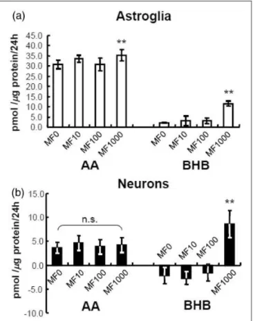
Effects of Metformin and Cilostazol on Ketogenesis
in Astroglia and Neurons
We also examined the pharmacological activation of
AMPK by metformin (1–1,000mmol/L) on ketogenesis
in cultured neurons and astroglia. Metformin is another AMPK activator that is used clinically for the treatment of diabetes mellitus. Lower concentrations of metformin (1–100mmol/L) did not alter KB production by astroglia
or neurons, whereas a higher concentration (1,000mmol/
L) did, indeed, induce KB (BHB) production in astroglia (Figure 2(a)) and neurons (Figure 2(b)). Neuron con-sumed small amounts of BHB during the experimental interval (negative values, Figure 2(b)). Notably, AICAR is a stronger inducer of ketogenesis in astroglia than met-formin (compare Figure 1(c) and (d) with Figure 2(a)). Although the metformin concentration used for the treat-ment of diabetic patients is approximately 10mmol/L, this
concentration did not affect KB production in both cell types. In contrast, metformin at a concentration of 1,000mmol/L reportedly activates AMPK in vitro (Zang
et al., 2008; Figure 2); thus, the clinical relevance of this concentration remains to be established.
Hypoxia-Enhanced Astroglial Ketogenesis and the
Enhancement of Neuronal BHB Production Were
Not Completely PAL Dependent
Hypoxia (1% O2for 24 hr) augmented the astroglial
pro-duction of both AA and BHB, as expected (Figure 4(a)). Unexpectedly, however, neuronal BHB production, but not AA production, also increased under hypoxia (Figure 4(a)). The enhancement of astroglial KB produc-tion under hypoxia disappeared with the eliminaproduc-tion of PAL (Figure 4(b)), indicating that hypoxia does not enhance the influx of glycolytic products available for KB generation. In contrast, the enhanced production of BHB by neurons under hypoxia was not affected in the absence of PAL (Figure 4(b)). Moreover, an increase in
the mitochondrial redox state (NADH/NADþ
) might
facilitate the conversion of AA to BHB as well as AA productionper se.
Combination of Hypoxia and Hypoglycemia
Enhances Astroglial KB Synthesis
To mimic clinical ischemic stroke, we examined the effect of combined insult of hypoxia without glucose (i.e., oxygen–glucose deprivation, OGD). For 24-hr hypoxia experiment, we used nutrient medium that contained 10% FBS to examine the effect of hypoxia per se. For OGD experiment, we removed FBS that contained low amount of glucose. We performed OGD experiment using two different conditions: DBSS without glucose (Figure 5(a) and (b)) or DMEM without glucose (Figure 6(a) and (b)). Because neither neurons nor
Figure 1. Fatty acid oxidation and ketogenesis by neurons and astroglia.
(a) Fatty acid oxidation in neurons (filled bars) and astroglia (blank bars) as determined by quantifying the amount of14CO2derived from
[1-14C]palmitic acid ([14C]PAL). 5-Amino-1 -b-D-ribofuranosyl-imidazole-4-carboxamide (AICAR), an AMP-activated protein kinase
astroglia tolerate OGD for 24 hr, we examined the effect of OGD for 4 and 12 hr. As shown in Figures 5 and 6, glucose deprivation alone induced marked ele-vation of KB synthesis in astroglia but not in neurons. Either 4 - or 12-hr incubation with glucose under hyp-oxia did not induce significant increases in KB
produc-tion in astroglia. The different neuronal BHB
production rates during hypoxia (Figures 4 and 5) might be ascribed to the assay solution (DBSS) that contains more restricted components (i.e., no amino acids or FBS, Figure 5) compared with assay media used for 24-hr hypoxia (i.e., nutrient media that may have provided supplemental substrates, Figure 4).
In summary, the hypoglycemic component of OGD is the major factor involved in induction of a significant elevation in KB synthesis in astroglia after 4 and 12 hr, whereas KB synthesis is not altered by OGD.
Neither Chemical Hypoxia Nor Glutamate
Enhanced Total Ketogenesis in Neurons
and Astroglia
Chemical hypoxia (rotenone, 1mmol/L for 24 hr) in
astro-glia reduced AA production in the presence of PAL plus glucose but raised BHB formation and caused a 25% fall in total KB (i.e., sum of AA and BHB; see, cyclic thio-NADH method in the ‘‘Materials and Methods’’ section) production (Figure 7). BHB production was also enhanced in neurons. Thus, the BHB/AA ratio was
mark-edly elevated in both types of cells, indicating
that NADH/NADþ
increased as a result of the inhibition
of the mitochondrial electron chain. Notably,
astroglial ketogenesis in the presence of fatty acid plus substrates was lower in the presence of chemical hypoxia caused by rotenone (Figure 7) compared with oxygen deprivation (1% O2), that is, it was similar to that of
the 4-hr interval of oxygen deprivation (Figures 5(a) and 6(a)) but about half that of the 12-hr interval (Figures 5(b) and 6(b)).
Furthermore, Guzma´n and Bla´zquez (2001) men-tioned the glutamate-induced enhancement of astroglial KB production as an unpublished observation. However, detailed data have not been presented. Hypoxic insults cause neuronal glutamate release (Nishizawa, 2001). Thus, whether glutamate per se induces astroglial KB production seems to be an important issue. We and others previously demonstrated that glutamate uptake
through a Naþ
-dependent glutamate transporter
enhances astroglial glucose utilization and lactate pro-duction (Pellerin and Magistretti, 1994; Takahashi et al., 1995). However, glutamate did not affect the astro-glial production of AA or BHB (Figure 8) when assayed in the presence of glucose.
Neuronal Oxidation of Lactate and BHB
In neurons, LAC oxidation was reduced by the addition of BHB (Figure 9(a), right), and BHB oxidation was aug-mented by the addition of LAC (Figure 9(b), right). Neither LAC nor BHB oxidation was affected by the addition of BHB or LAC in astroglia (Figures 9(a) and (b)). Notably, BHB oxidation may supplement lactate as fuel, but its overall contribution is relatively small. The role of lactate and BHB as energy substrates for astroglial oxidative metabolism is another important issue to be solved (McKenna, 2012).
Figure 2. Effects of metformin (MF) on ketogenesis from palmitic
acid by neurons and astroglia.
Metformin (1,000mmol/L) activated the production of acetoacetate
(AA) andb-hydroxybutyrate (BHB) in astroglia (blank bars, a) and neurons (filled bars, b) as determined using the cyclic thio-NADH method during 24 hr in the presence of palmitic acid (PAL) andL -carnitine (LC). Glucose (2 mmol/L) was included in all assays. Note that the neuronal nutrient medium that had been present for 5 to 6 days was collected and reused for the neuronal KB assays, and negative values indicate net consumption of KBs that were present in the 5- to 6-day medium during the subsequent assay interval. Neurons consumed small amounts of BHB during the experimental interval. Astrocytes are not sensitive to medium change, and fresh medium was used for the astrocyte assays. Values are the meanSD (n¼6). n.s.¼not significant. **p<.01 (Dunnett test
Effects of 1% Hypoxia (24 hr) on Oxidative
Metabolism of Lactate, Pyruvate, or BHB in
Neurons
The neuronal utilization (oxidative metabolism) of LAC (Figure 10(a)) and PYR (Figure 10(b)) was signifi-cantly reduced when assayed under normoxic conditions after hypoxia (24 hr), while BHB oxidation (Figure 10(c)) was preserved (no statistically significant decrease, grouped t test). In control neuronal cultures, pyruvate was oxidized 3.8-fold faster than lactate and 8.8 times better than BHB (Figure 10), indicating that pyruvate is the best neuronal substrate and that shuttling of NADH equivalents derived from lactate into mitochon-dria may limit lactate oxidation compared with that of pyruvate.
Discussion
The present study demonstrated that both cultured astro-glia and neurons produce KBs from long-chain fatty acids, that is, PAL, while fatty acid oxidation through the TCA cycle was negligible. Regarding glucose oxida-tion, neurons exhibit a much higher rate of glucose oxi-dation, whereas astroglia possess a lower capacity for glucose oxidation irrespective of the higher glucose util-ization, resulting in a large amount of lactate production (Izawa et al., 2009). Both neurons and astroglia produce lactate under hypoxia, and the present study demon-strated that neurons produced BHB probably from endogenous lipids or other compounds in the medium. In contrast, astroglial KB production was exogenous fatty acid derived but not glucose derived. AMPK acti-vation enhanced astroglial KB production through ACC phosphorylation, and hypoxia may also enhance KB pro-duction through the same mechanism, as reported by Guzma´n and Bla´zquez (2001).
Although the function of the adult brain is completely dependent on glucose oxidation for ATP production, suckling baby mainly utilizes KBs instead of glucose. Even the adult brain shifts its energy source from glucose to KBs under conditions of starvation or insulin resist-ance associated with diabetes mellitus, but increased KB utilization is associated with upregulation of monocar-boxylate transporters in the BBB to facilitate greater KB uptake into brain (Pan et al., 2000, 2001, 2002).
An important finding of the present study is that both astroglia and neurons utilize fatty acid to produce KBs instead of utilizing it as a TCA cycle substrate. Astroglia produced more KBs than neurons, as reported by Guzma´n and Bla´zquez (2001). They showed that AICAR (an AMPK activator) stimulates astroglial KB production by phosphorylating ACC. Because phosphor-ylation inhibits ACC activity, the level of malonyl-CoA, which is a main physiological inhibitor of CPT-I (a rate-limiting enzyme of fatty acid metabolism), is reduced.
The present study only partly confirmed their observa-tion, as cilostazol, which also cause phosphorylation of ACC, failed to stimulate KB production, suggesting that other mechanisms may be involved. We previously reported that a clinically relevant concentration of cilos-tazol (3mmol/L) did, indeed, alter glucose metabolism in
cultured astroglia through PKA (Takahashi et al., 2011) Therefore, cilostazol at this concentration should have activated PKA, leading to the augmentation of KB pro-duction. The lack of an effect of cilostazol on fatty acid metabolism in cultured astroglia remains to be explored. Another activator of AMPK, metformin, is clinically used for the treatment of diabetes mellitus. The lower concentration (up to 100mmol/L) of metformin used in
the present study was selected based on the estimated concentration in the brain of patients as determined by calculating the peak blood concentration of metformin assuming a BBB permeability of approximately 10% of
Figure 3. Effects of cilostazol (CZL) on ketogenesis from palmitic acid by neurons and astroglia.
Effects of 3mmol/L of CZL on production of acetoacetate (AA) andb-hydroxybutyrate (BHB) in neurons (filled bars) and astroglia (blank
the concentration in the blood. At this concentration, metformin did not induce KB production in astroglia. In contrast, the higher concentration (1,000mmol/L) did
indeed induce KB production. Interestingly, however,
Fulgencio et al. (2001) reported that metformin
(50–500mmol/L) suppressed ketogenesis and ketogenic
gene expression in freshly isolated hepatocytes. The reason for the contradictory results between hepatocytes and cultured astroglia remains to be determined.
A suggestion made a decade ago that metformin
reduces glucose synthesis through the activation
of AMPK has recently been challenged by genetic
loss-of-function experiments (Foretz et al., 2010). According to Miller et al. (2013), metformin antagonizes the action of glucagon, thus reducing the fasting glucose levels. In mouse hepatocytes, metformin leads to the accu-mulation of AMP and related nucleotides, which inhibit adenylate cyclase, reduce the levels of cAMP and PKA activity, abrogate the phosphorylation of critical protein targets of PKA, and block glucagon-dependent glucose output from hepatocytes. In the present study, cilostazol activated PKA but did not alter KB production, as shown earlier. Whether the mechanism by which a higher concen-tration of metformin induces astroglial ketogenesis involves this novel pathway remains uncertain.
A novel finding of the present study is that neurons under hypoxia produce substantial amounts of KBs.
Figure 4. Effects of hypoxia on ketogenesis in the presence and
absence of palmitic acid by neurons and astroglia.
(a) Production of acetoacetate (AA) andb-hydroxybutyrate (BHB) in neurons (filled bars) and astroglia (blank bars) as determined using the cyclic thio-NADH method during 24 hr under normoxia (21% O2) or hypoxia (1% O2) in the presence of palmitic acid (PAL)
andL-carnitine (LC). Hypoxia increased AA and BHB production in astroglia but only increased BHB production in neurons. (b) Production of AA and BHB in neurons and astroglia under hypoxia in the absence of PAL and LC. Glucose (2 mmol/L) was included in all assays. Astroglial AA and BHB production were markedly reduced by the elimination of PAL and LC, and the hypoxia-induced augmentation disappeared. In contrast, the hypoxia-induced enhancement of BHB production was preserved in neurons even in the absence of PAL and LC. Values are the meanSD (n¼6 for a and b). n.s. ¼ not significant. **p<.01; ***p<.001 (grouped t
test).
Figure 5. Effects of hypoxia with or without glucose-deprivation
on ketogenesis from exogenous palmitic acid by neurons and astroglia.
The effects of hypoxia (1% O2) with glucose (12.5 mmol/L) or
with-out glucose for 4 hr (a) and 12 hr (b) on neuronal or astroglial total ketone body (KB) production were examined using Dulbecco’s balanced salt solution (DBSS) containing 110 mmol/L NaCl, 5.4 mmol/L KCl, 1.8 mmol/L CaCl2, 0.8 mmol/L MgSO4, 0.9 mmol/
L NaH2PO4, and 44 mmol/L NaHCO3. Neither neurons nor
Moreover, this hypoxia-induced KB production by neu-rons was not exogenous PAL dependent. These results suggest that acetyl-CoA from other sources, which nor-mally enters the TCA cycle, is instead used for KB pro-duction under hypoxic conditions. However, astroglia KB production under hypoxia disappeared with the elim-ination of PAL. As KBs are known to play neuroprotec-tive roles under ischemia, KBs derived from both neurons and astrocytes might have important roles in the brain in vivo. In particular, the roles of astroglia seem to be important, as they produce KBs directly from exogenous fatty acids.
Guzma´n and Bla´zquez (2001) reported that chemical hypoxia also enhanced neuronal KB production. In the present study, however, somewhat different results were obtained. The total KB (AA and BHB) production in the neurons did not increase with the addition of rotenone, a mitochondrial complex I inhibitor. Furthermore,
Figure 6. Effects of hypoxia with or without glucose-deprivation
on ketogenesis from exogenous palmitic acid by neurons and astroglia.
The effects of hypoxia (1% O2) with glucose (12.5 mmol/L) or
with-out glucose for 4 hr (a) and 12 hr (b) on neuronal or astroglial total ketone body (KB) production were examined using DMEM contain-ing no fetal bovine serum. Neither neurons nor astroglia tolerate hypoxia without glucose (i.e., oxygen-glucose deprivation, OGD) for 24 hr. Neuronal KB productions during 4 or 12 hr were very small and negligible. Hypoxia with glucose for 4 or12 hr did not elicit statistically significant increases in KB production by astroglia. Values are the meanSD (n¼6). n.s.¼not significant. ***p<.001
(groupedttest).
Figure 7. Effects of chemical hypoxia on ketogenesis by neurons
and astroglia in the presence of exogenous palmitic acid and glucose.
Production of acetoacetate (AA), b-hydroxybutyrate (BHB), and total ketone bodies (tKBs) in neurons (filled bars) and astroglia (blank bars), as determined using the cyclic thio-NADH method during 24 hr of exposure to rotenone or a vehicle with palmitic acid (PAL) andL-carnitine (LC). Glucose (2 mmol/L) was included in all assays. Rotenone reduced tKB production in astroglia compared with ethanol (EtOH)-treated control cells, but it did not affect neuronal tKB production. The BHB/AA ratio in both neurons and astroglia increased markedly in the presence of rotenone. Values are the meanSD (n¼6 for a and b). n.s. ¼ not significant. *p<.05 (groupedttest).
Figure 8. Effects of glutamate (GLU) on astroglial ketogenesis.
Effects of GLU at different concentrations (100 and 500mmol/L) on
production of acetoacetate (AA) andb-hydroxybutyrate (BHB) in astroglia in the presence of glucose (2 mmol/L) were examined using the cyclic thio-NADH method during 24 hr of exposure to GLU (100 or 500mmol/L). Values are the meanSD (n¼4). n.s.¼
not significant (AVOVA). **p<.01 (Dunnett test for multiple
rotenone actually reduced the total KB production in astroglia. Both types of cells showed an increase in the
ratio of BHB/AA, indicating that NADH/NADþ
asso-ciated with rotenone-induced mitochondrial dysfunction. Moreover, glucose deprivation seemed to be more a
potent activator of astroglial ketogenesis. Astroglial energy production seems to be more dependent on glyco-lytic metabolism rather than oxidative metabolism of glu-cose (Abe et al., 2006; Takahashi et al., 2012b). The results of the present study are in accordance with
Figure 9. Neuronal oxidation of lactate (LAC) andb-hydroxybutyrate (BHB).
(a) Effect of addition of BHB on LAC oxidation as determined using [U-14C]LAC in neurons (filled bars) and astroglia (blank bars). (b) Effect of addition of LAC on BHB oxidation as determined using [1-14C]BHB in neurons (filled bars) and astroglia (blank bars). Glucose was not
included in the medium in these assays. Values are the meanSD (n¼4). n.s.¼not significant. *p<.05; **p<.01 (groupedttest).
Figure 10. Hypoxia/reoxygenation-induced alteration of neuronal oxidation of lactate (LAC), pyruvate (PYR), andb-hydroxybutyrate
(BHB).
(a) Effects of hypoxia (1% O2for 24 hr) on [U-14C]LAC in neurons. (b) Effects of hypoxia (1% O2for 24 hr) on [1-14C] PYR in neurons. (c)
Effects of hypoxia (1% O2for 24 hr) on [1-14C]BHB in neurons. Glucose was not included in the medium in these assays, which were
recent findings by Taı¨b et al. (2013), who found that hypothalamic astroglia responds to hypoglycemia by gen-erating KBsin vivo.
The fate of KBs produced by either neurons or astro-glia seems to be important because reoxygenation after hypoxia facilitates ROS production from damaged mito-chondria, leading to PDHC impairment. In vivo studies showed that PDHC activity is enhanced during transient ischemia and then decreases after reperfusion (Cardell et al., 1989; Fukuchi et al., 1998). Thus, even though lactate accumulates during hypoxia, it may not be readily available as an energy source after reoxygenation. The present study clearly demonstrated that lactate and pyruvate oxidation decreased in neurons after hypoxia/ reoxygenation, while BHB oxidation was preserved. Therefore, it is reasonable to speculate that KBs pro-duced by astroglia or neurons, or both cell types, can fuel the neuronal energy state after hypoxia/reoxygena-tion. Whether KBs produced during ischemia support astroglial oxidative metabolism (McKenna, 2012) after reoxygenation is subject of the future study.
Conclusion
Astroglia and neurons responded to hypoxia by enhan-cing KB production, and KBs produced by astroglia or neurons or both might be used as a neuronal energy sub-strate. The activation of astroglial ketogenesis through activated AMPK might reduce ischemic cell damage.
Author contributions
S. T. designed experiments. S. T., T. I., and K. M. performed experiments. S. T., T. A., and N. S. analyzed data and wrote the manuscript.
Funding
The authors disclosed receipt of the following financial support for the research, authorship, and/or publication of this article: This work was supported by Grant-in-Aid for Scientific Research (C) from the Ministry of Education, Culture, Sports, Science, and Technology of Japan: 24591276 (to S. T.).
Declaration of Conflicting Interests
The authors declared no potential conflicts of interest with respect to the research, authorship, and/or publication of this article.
References
Abe T., Takahashi S., & Suzuki N. (2006) Oxidative metabolism in cultured rat astroglia: Effects of reducing the glucose concen-tration in the culture medium and of D-aspartate or potassium stimulation.Journal of Cerebral Blood Flow and Metabolism: Official Journal of the International Society of Cerebral Blood Flow and Metabolism26: 153–160.
Andres R., Cader G., & Zierler K. L. (1956) The quantitatively minor role of carbohydrate in oxidative metabolism by skeletal
muscle in intact man in the basal state; measurements of oxygen and glucose uptake and carbon dioxide and lactate production in the forearm.The Journal of Clinical Investigation35: 671–682. Auestad N., Korsak R. A., Morrow J. W., & Edmond J. (1991) Fatty acid oxidation and ketogenesis by astrocytes in primary culture.Journal of Neurochemistry56: 1376–1386.
Bla´zquez C., Sanchez C., Velasco G., & Guzma´n M. (1998) Role of carnitine palmitoyltransferase I in the control of ketogenesis in primary cultures of rat astrocytes. Journal of Neurochemistry
71: 1597–1606.
Bla´zquez C., Woods A., de Ceballos M. L., Carling D., & Guzma´n M. (1999) The AMP-activated protein kinase is involved in the regulation of ketone body production by astrocytes.Journal of Neurochemistry73: 1674–1682.
Cardell M., Koide T., & Wieloch T. (1989) Pyruvate dehydrogen-ase activity in the rat cerebral cortex following cerebral ische-mia.Journal of Cerebral Blood Flow and Metabolism: Official Journal of the International Society of Cerebral Blood Flow and Metabolism9: 350–357.
Clarke D. D., & Sokoloff L (1999) Circulation and energy metab-olism of the brain. In: Siegel G., Agranoff B., Albers R. W., & Fisher S. (eds)Basic neurochemistry: Molecular, cellular, and medical aspects, 6th ed. Philadelphia, PA: Lippincott-Raven, pp. 637–669.
Corton J. M., Gillespie J. G., Hawley S. A., & Hardie D. G. (1995) 5-aminoimidazole-4-carboxamide ribonucleoside A specific method for activating AMP-activated protein kinase in intact cells?European Journal of Biochemistry/FEBS229: 558–565. Dhopeshwarkar G. A., & Mead J. F. (1973) Uptake and transport of fatty acids into the brain and the role of the blood-brain barrier system.Advances in Lipid Research11: 109–142.
Dienel G. A (2009) Energy metabolism in the brain. In: Byrne J. H., & Roberts J. L. (eds)From molecules to networks: An introduc-tion to cellular and molecular neuroscience, 2nd ed. London, England: Academic Press, pp. 49–110.
Driscoll B. F., Deibler G. E., Law M. J., & Crane A. M. (1993) Damage to neurons in culture following medium change: Role of glutamine and extracellular generation of glutamate.Journal of Neurochemistry61: 1795–1800.
Edmond J., Auestad N., Robbins R. A., & Bergstrom J. D. (1985) Ketone body metabolism in the neonate: Development and the effect of diet.Federation Proceedings44: 2359–2364. Edmond J., Robbins R. A., Bergstrom J. D., Cole R. A., & de Vellis
J. (1987) Capacity for substrate utilization in oxidative metab-olism by neurons, astrocytes, and oligodendrocytes from developing brain in primary culture. Journal of Neuroscience Research18: 551–561.
Foretz M., Hebrard S., Leclerc J., Zarrinpashneh E., Soty M., Mithieux G.,...Viollet B. (2010) Metformin inhibits hepatic
gluconeogenesis in mice independently of the LKB1/AMPK pathway via a decrease in hepatic energy state.The Journal of Clinical Investigation120: 2355–2369.
Fukuchi T., Katayama Y., Kamiya T., McKee A., Kashiwagi F., Terashi A. (1998) The effect of duration of cerebral ischemia on brain pyruvate dehydrogenase activity, energy metabolites, and blood flow during reperfusion in gerbil brain. Brain Research
792: 59–65.
isolated hepatocytes and on specific gene expression in cultured hepatocytes.Biochemical Pharmacology62: 439–446. Guzma´n M., & Bla´zquez C. (2001) Is there an astrocyte-neuron
ketone body shuttle?Trends in Endocrinology and Metabolism
12: 169–173.
Guzma´n M., & Bla´zquez C. (2004) Ketone body synthesis in the brain: Possible neuroprotective effects. Prostaglandins, Leukotrienes, and Essential Fatty Acids70: 287–292.
Guzma´n M., & Geelen M. J. (1993) Regulation of fatty acid oxi-dation in mammalian liver. Biochimica et Biophysica Acta
1167: 227–241.
Izawa Y., Takahashi S., & Suzuki N. (2009) Pioglitazone enhances pyruvate and lactate oxidation in cultured neurons but not in cultured astroglia.Brain Research1305: 64–73.
Kelley D. E., Reilly J. P., Veneman T., & Mandarino L. J. (1990) Effects of insulin on skeletal muscle glucose storage, oxidation, and glycolysis in humans.The American Journal of Physiology
258: E923–E929.
Laun R. A., Rapsch B., Abel W., Schroder O., Roher H. D., Ekkernkamp A.,...Schulte K. M. (2001) The determination
of ketone bodies: Preanalytical, analytical and physiological considerations. Clinical and Experimental Medicine 1: 201–209.
Leary S. C., Hill B. C., Lyons C. N., Carlson C. G., Michaud D., Kraft C. S.,...Moyes C. D. (2002) Chronic treatment with
azide in situ leads to an irreversible loss of cytochrome c oxi-dase activity via holoenzyme dissociation. The Journal of Biological Chemistry277: 11321–11328.
Mathiisen T. M., Lehre K. P., Danbolt N. C., & Ottersen O. P. (2010) The perivascular astroglial sheath provides a complete covering of the brain microvessels: An electron microscopic 3D reconstruction.Glia58: 1094–1103.
McKenna M. C. (2012) Substrate competition studies demonstrate oxidative metabolism of glucose, glutamate, glutamine, lactate and 3-hydroxybutyrate in cortical astrocytes from rat brain.
Neurochemical Research37: 2613–2626.
McKenna M. C., Tildon J. T., Stevenson J. H., Boatright R., & Huang S. (1993) Regulation of energy metabolism in synaptic terminals and cultured rat brain astrocytes: Differences revealed using aminooxyacetate. Developmental Neuroscience 15: 320–329.
Miller R. A., Chu Q., Xie J., Foretz M., Viollet B., Birnbaum M. J. (2013) Biguanides suppress hepatic glucagon signalling by decreasing production of cyclic AMP.Nature494: 256–260. Mitchell R. W., On N. H., Del Bigio M. R., Miller D. W., &
Hatch G. M. (2011) Fatty acid transport protein expression in human brain and potential role in fatty acid transport across human brain microvessel endothelial cells. Journal of Neurochemistry 117: 735–746.
Nehlig A. (2004) Brain uptake and metabolism of ketone bodies in animal models. Prostaglandins, Leukotrienes, and Essential Fatty Acids70: 265–275.
Nishizawa Y. (2001) Glutamate release and neuronal damage in ischemia.Life Sciences69: 369–381.
Pan J. W., de Graaf R. A., Petersen K. F., Shulman G. I., Hetherington H. P., Rothman D. L. (2002) [2,4-13C
2
]-beta-hydroxybutyrate metabolism in human brain. Journal of Cerebral Blood Flow and Metabolism: Official Journal of the International Society of Cerebral Blood Flow and Metabolism
22: 890–898.
Pan J. W., Rothman T. L., Behar K. L., Stein D. T., & Hetherington H. P. (2000) Human brain beta-hydroxybutyrate and lactate increase in fasting-induced ketosis. Journal of Cerebral Blood Flow and Metabolism: Official Journal of the International
Society of Cerebral Blood Flow and Metabolism 20:
1502–1507.
Pan J. W., Telang F. W., Lee J. H., de Graaf R. A., Rothman D. L., Stein D. T.,...Hetherington H. P. (2001) Measurement of
beta-hydroxybutyrate in acute hyperketonemia in human brain.
Journal of Neurochemistry79: 539–544.
Pellerin L., & Magistretti P. J. (1994) Glutamate uptake into astro-cytes stimulates aerobic glycolysis: A mechanism coupling neuronal activity to glucose utilization. Proceedings of the National Academy of Sciences of the United States of America
91: 10625–10629.
Prins M. L. (2008) Cerebral metabolic adaptation and ketone metabolism after brain injury. Journal of Cerebral Blood Flow and Metabolism: Official Journal of the International Society of Cerebral Blood Flow and Metabolism28: 1–16. Puchowicz M. A., Zechel J. L., Valerio J., Emancipator D. S., Xu
K., Pundik S.,...Lust W. D. (2008) Neuroprotection in
diet-induced ketotic rat brain after focal ischemia. Journal of Cerebral Blood Flow and Metabolism: Official Journal of the International Society of Cerebral Blood Flow and Metabolism
28: 1907–1916.
Robinson A. M., & Williamson D. H. (1980) Physiological roles of ketone bodies as substrates and signals in mammalian tissues.
Physiological Reviews60: 143–187.
Smith P. K., Krohn R. I., Hermanson G. T., Mallia A. K., Gartner F. H., Provenzano M. D.,...Klenk D. C. (1985) Measurement of
protein using bicinchoninic acid.Analytical Biochemistry150: 76–85.
Smith Q. R., & Nagura H. (2001) Fatty acid uptake and incorpor-ation in brain: Studies with the perfusion model. Journal of Molecular Neuroscience16: 167–172.
Taı¨b B., Bouyakdan K., Hryhorczuk C., Rodaros D., Fulton S., Alquier T. (2013) Glucose regulates hypothalamic long-chain fatty acid metabolism via AMP-activated kinase (AMPK) in neurons and astrocytes. The Journal of Biological Chemistry
288: 37216–37229.
Takahashi S., Driscoll B. F., Law M. J., & Sokoloff L. (1995) Role of sodium and potassium ions in regulation of glucose metab-olism in cultured astroglia. Proceedings of the National
Academy of Sciences of the United States of America 92:
4616–4620.
Takahashi S., Izawa Y., & Suzuki N. (2011) Cilostazol enhances oxidative glucose metabolism in both neurons and astroglia without increasing ROS production. Pharmacology &
Pharmacy2: 315–321.
Takahashi S., Izawa Y., & Suzuki N. (2012a) Astroglial pentose phosphate pathway rates in response to high-glucose environ-ments.ASN Neuro4(2): e00078 doi:10.1042/AN20120002 Takahashi S., Izawa Y., & Suzuki N. (2012b) Astrogliopathy as a
loss of astroglial protective function against glycoxidative stress under hyperglycemia.Clinical Neurology52: 41–51.
Vannucci S. J., & Simpson I. A. (2003) Developmental switch in brain nutrient transporter expression in the rat. American
Journal of Physiology Endocrinology and Metabolism 285:
E1127–E1134.
Veech R. L. (2004) The therapeutic implications of ketone bodies: The effects of ketone bodies in pathological conditions: Ketosis, ketogenic diet, redox states, insulin resistance, and mitochon-drial metabolism.Prostaglandins, Leukotrienes, and Essential Fatty Acids70: 309–319.
Waniewski R. A., & Martin D. L. (2004) Astrocytes and synapto-somes transport and metabolize lactate and acetate differently.
Neurochemical Research29: 209–217.
Zang Y., Yu L. F., Pang T., Fang L. P., Feng X., Wen T. Q.,...Li J.