online | memorias.ioc.fiocruz.br
Retention of a recombinant GFP protein expressed by the yellow fever
17D virus in the E/NS1 intergenic region in the endoplasmic reticulum
Gisela Freitas Trindade1, Marlon Gilsepp Veloso de Santana1, Juliana Ribeiro dos Santos1, Ricardo Galler2, Myrna Cristina Bonaldo1/+
1Laboratório de Biologia Molecular de Flavivírus, Instituto Oswaldo Cruz
2Instituto de Tecnologia em Imunobiológicos-Fiocruz, Av. Brasil 4365, 21045-900 Rio de Janeiro, RJ, Brasil
The flaviviral envelope proteins, E protein and precursor membrane protein, are mainly associated with the endoplasmic reticulum (ER) through two transmembrane (TM) domains that are exposed to the luminal face of this compartment. Their retention is associated with the viral assembly process. ER-retrieval motifs were mapped at the carboxy terminus of these envelope proteins. A recombinant yellow fever (YF) 17D virus expressing the reporter green fluorescent protein (GFP) with the stem-anchor (SA) region of E protein fused to its carboxy terminus was subjected to distinct genetic mutations in the SA sequence to investigate their effect on ER retention. Initially, we introduced progressive deletions of the stem elements (H1, CS and H2). In a second set of mutants, the effect of a length increase for the first TM anchor region was evaluated either by replacing it with the longer TM of human LAMP-1 or by the insertion of the VALLLVA sequence into its carboxy terminus. We did not detect any effect on the GFP localisation in the cell, which remained associated with the ER. Further studies should be undertaken to eluci-date the causes of the ER retention of recombinant proteins expressed at the intergenic E/NS1 region of the YF 17D virus polyprotein.
Key words: recombinant yellow fever 17D virus - GFP expression - ER retention - deletion and insertion mutants
Yellow fever (YF) virus is the prototype member of the genus Flavivirus of the Flaviviridae family. The YF genome consists of a 10,832-nucleotide (nt) positive, single-stranded RNA and contains a single open reading frame that is translated into a 3410-amino acid polypro-tein precursor. The specific proteolytic cleavage of this precursor by cellular proteases and the viral NS2B/NS3 proteolytic complex leads to the generation of structural proteins [the C, precursor membrane (PrM), M and E constituents of the viral particle] and the nonstructural proteins (the NS1, NS2A, NS2B, NS3, NS4A, NS4B and NS5 components of the viral replicative complex). During flavivirus cell propagation, there is an extensive rearrangement of endoplasmic reticulum (ER)-derived membranes that raises specialised and distinct sites of viral replication and assembly (Mackenzie 2005, Wel-sch et al. 2009, Gillespie et al. 2010). The E and prM proteins, which are the envelope proteins, are translo-cated to the ER and exposed to the luminal face of this compartment. Both proteins remain associated with the ER membrane through their carboxy-terminal regions, which consist of a pair of antiparallel helices traversing the membrane (Zhang et al. 2003).
Fianancial support: PDTIS/FIOCRUZ (RVR03), CNPq, INCTV-CNPq/MCT
MCB and RG were recipients of fellowships from CNPq. +Corresponding author: [email protected] Received 11 August 2011
Accepted 23 November 2011
The E protein is the major constituent of the flaviviral envelope. In the infectious viral particle, E protein exists as a slightly bent, elongated head-to-tail dimer that is ori-entated parallel to the viral membrane. The E monomer has three domains in its ectodomain portion: the struc-turally central, N-terminal domain I, followed by the di-merisation domain II, and the carboxy-terminal, Ig-like domain III. The carboxy terminus of approximately 100 amino acid residues is denominated of stem-anchor (SA) region. The stem is composed of two α-helixes buried in the outer leaflet of the viral membrane and the anchor is composed of two antiparallel, coiled-coil transmem-brane (TM) domains. Similarly, alpha-helix near the N terminus of prM/M is partially submerged in the outer lipid bilayer and the carboxy-terminal region of M con-sists of a pair of antiparallel helices traversing the mem-brane (Zhang et al. 2003).
2008). The heterodimer formation and the virus particle assembly likely depend on the accumulation of these proteins in the ER (Lorenz et al. 2003, Op De Beeck et al. 2004). Several ER-retrieval motifs were mapped in the TM regions of the E and prM proteins, predomi-nantly through chimerisation experiments adding differ-ent parts of the flaviviral E-TM to cell surface proteins, such as CD4 (Hsieh et al. 2010). Hence, substitutions of non-hydrophobic residues in the first and second E pro-tein TM regions and the increase in the length of these alpha-helices led to the export of the chimeric proteins from the ER. Interestingly, the TM domains of the en-velope proteins are relatively shorter than those found in viruses, which are assembled at the cell membrane (Hsieh et al. 2010). The same organelle-specific prop-erties were determined in previous studies comparing the TM domains of early and late compartment proteins and indicated a difference in TM length and thus bilayer thickness (Bretscher & Munro 1993, Levine et al. 2000, Sharpe et al. 2010). These findings support a TM-depen-dent sorting mechanism for viral envelope proteins.
We have developed a method to express foreign an-tigens between the E and NS1 regions of the YF 17D genome (Bonaldo et al. 2007, 2010). The recombinant protein is engineered to contain in its amino and car-boxy termini functional motifs present in conserved se-quences flanking the E and NS1 intergenic region. One of these regions is the SA region of E protein, which was fused to the carboxy terminus of the heterologous green fluorescent protein (GFP). Accordingly, the presence of the retention signal in these domains, which indicates that the recombinant GFP protein is expressed by the YF 17D viruses, was associated exclusively with the ER and not with other organelles or even secreted to the infected cell culture supernatant. In this work, we investigated whether modifications in the SA region fused to GFP could induce the export of the recombinant protein from the ER. For this purpose, we have introduced several mutations in the SA region of the recombinant GFP ex-pressed by the YF 17D viruses. This study represents the first time that these changes were evaluated in a live flavivirus to analyse whether the recombinant protein was retained in the ER.
MaTERialS aND METhoDS
Cell culture and virus multiplication - Vero cells (ATCC) were grown in Earle’s 199 medium supplement-ed with 5% foetal bovine serum (FBS). Viral stocks were obtained after Vero cells monolayer infection at a multi-plicity (MOI) of 0.1. The supernatant was collected and titred by plaque assay on Vero cells at a density of 50,000/ cm2. Growth curves were determined as described
previ-ously (Bonaldo et al. 2005). Briefly, Vero cell monolayers at a density of 62,500/cm2 were infected at a MOI of 0.02.
After 24 h intervals post-infection (p.i.), the supernatant was collected and the viral replication rate was estimated by plaque titration. The results were expressed by plaque forming units (PFU/mL). The viral growth peaks were compared using the one way analysis of variance (ANO-VA) test. Dunnett’s post-test was selected to compare the growth of parental YF17DE200T3 with the other recom-binants 72 h p.i. (GraphPad Prism 5.03 Program).
Construction of cDNA templates and virus recovery - The recombinant YF infectious clones were obtained as described previously (Bonaldo et al. 2007). The YF17D/ EGFP/ΔH1CS insert construct was obtained by two rounds of polymerase chain reaction (PCR) amplification. In the first round, the amplification of the GFP fragment (pEGFP-C2) (Clontech) with forward primer RG328 (5’-CTAGGAGTTGGCGCCGATCAAGGAGCGCCA-TCAACTTTGGCGTGAGCAAGGGCGAGGAGCT-3’) and reverse primer RG332 (5’-CCGTATGAATTC- CTTTCCCAACCGAAGTCTTGTACAGCTCGTCCA-TGCCG-3’) generated a product of 768 bp containing 15 nt of the 3’E gene, 27 nt of 5’NS1, the GFP gene and 28 nt of the 5’E-H2 gene. A second fragment was also cre-ated via the amplification of 22 nt of the 3’GFP gene, E-H2, E-TM1, E-TM2 and 27 nt of the 5’NS1 coding sequence from pT3 plasmid (15) with forward primer RG333 (5’-CGGCATGGACGAGCTGTACAAGACT-TCGGTTGGGAAAGGAATTCATACGG-3’) and re-verse primer RG331 (5’-GCCAAAGTTGATGGCG-CATCCTTGATCGGCGCCAACTCCTAGAGAC-3’), which created a DNA fragment of 244 bp. These two DNA fragments were used to obtain a 981 bp recombi-nant cassette that contained 15 nt of the 3’E gene, 27 nt of 5’NS1, the GFP gene, E-H2, E-TM1, E-TM2 and 27 nt of 5’NS1 in the presence of RG328 and RG331, which was then cloned into the pT3 plasmid and used to obtain the cDNA template. A similar approach was employed to obtain the other GFP derivatives. The YF17D/EGFP/ ΔH1CSH2 virus construct was generated by means of a first round amplicon of 793 bp, which was derived from pEGFP-C2 amplification using the RG328 and RG412 primers (5’-CCTTTGTTATCCAGTTCAAGCCGCCA- AATAGAGACCCCTTGTACAGCTCGTCCATGC-CGAGAG-3’) containing 15 nt of the 3’E gene, 27 nt of 5’NS1, the GFP gene and 36 nt of 5’E-TM1 and a 205 bp fragment containing 24 nt of 3’GFP, E-TM1, E-TM2 and 27 nt of the 5’NS1 gene from the pT3 plasmid in the presence of primers RG413 (5’-CTCTCGGCATGGAC- GAGCTGTACAAGGGGTCTCTATTTGGCGGCTT-GAACTGGATAACAAAGG-3’) and RG331. The 936 bp GFP resulting DNA fragment containing 15 nt of the 3’E gene, 27 nt of 5’NS1, the GFP gene, E-TM1, E-TM2 and 27 nt of 5’NS1 was obtained using the RG328 and RG331 oligonucleotides. To generate the construct YF17D/GFP/ TM1-TM LAMP, where the E-TM1 of the YF 17D virus was replaced by the human LAMP-1 protein TM region, we first produced four fragments derived from the pT3 plasmid and the pEGFP-C2 plasmid. Fragment 1 was 785 bp long and was amplified from pEGFP with for-ward primer RG328 and reverse primer RG452 (5’-GT- GAACAACTTTCCTATTGAGCTTCCCTCCTTGTA-CAGCTCGTCCATGCCGAGAG-3). This fragment had 15 nt of the 3’E gene, 27 nt of the 5’NS1 gene, the entire GFP gene and 31 nt of the 5’ E-TM1 gene. Fragment 2 was 230 bp long and was obtained from the amplification of pT3 with forward primer RG453 (5’-CTCTCGGCAT- GGACGAGCTGTACAAGGAGGGAAGCTCAATAG-GAAAGTTGTTCAC-3’) and reverse primer RG458
contained the 24 nt of the 3’GFP gene, H1, CS, H2, 6 nt of E-TM1 and 36 nt of the 5’TM LAMP-1 gene. Frag-ment 3 was 247 bp long and was amplified using pT3 as a template with forward primer RG459 (5’-CTTGATT- GCTTATCTGGTGGGAACAAGAAACATGACAAT-GTCCATGAGCATGATC-3’) and reverse primer RG460 (5´-CACAGGATCTTCTGGATAGTATGAGTACTTGT TCAGCCAGTCATCAGAGTCTC-3’) and consisted of 21 nt of the 3’TM LAMP-1 gene, E-TM2 and 149 nt of the 5’NS1 gene. Fragment 4 was 266 bp long and was obtained with fragment 2 as an amplification template with forward primer RG453 and reverse primer RG461 (5’-TCCCACCAGATAAGCAATCAAGACAATAA- GAACTAGCCCAGCCAGGGCACCCCCGACGGC-TATAGGGAT-3). Fragment 4 contained 24 nt of the 3’GFP gene, H1, CS, H2, 6 nt of E-TM1 and the entire sequence of TM LAMP-1. The second round of ampli-fication consisted of the creation of the larger fragment 5 (492 bp) by PCR fusion of fragments 3 and 4 using the primers RG453 and RG460. Fragment 5 contained 24 nt of the 3’GFP gene, H1, CS, H2, 6 nt of E-TM1, TM LAMP-1, E-TM2 and 149 nt of 5’NS1. The entire GFP expression cassette was yielded by fusion PCR us-ing the 1st and 5th fragments with the oligonucleotides RG328 and RG460. This fragment of 1,101 bp consist-ed of 15 nt of the 3’E gene, 27 nt of 5’NS1, the entire GFP gene, H1, CS, H2, 6 nt of E-TM1, TM LAMP-1, E-TM2 and 149 nt of 5’NS1. The recombinant GFP cas-sette carrying E-TM1 sequence with an insert coding from VALLLVA was obtained from GeneScript and was cloned into pT3 to generate the virus 17D/EGFP/TM1 + VALLLVA. All PCR amplifications were performed with Pfx DNA polymerase (Invitrogen) and the products were purified with a silica-based column QIAquick PCR Purification Kit (QIAGEN) and cloned into the pGEM-T plasmid (Promega) using electrocompetent Escherichia coli Sure (Stratagene). The recombinant pT3 plasmids were obtained by cloning Nar I GFP inserts and were se-quenced to confirm the insert orientation and integrity. All of the YF cDNA templates were obtained using the two-plasmid method as previously described (Rice et al. 1989, Bonaldo et al. 2002). The other viruses included in our study were YF17D/EGFP/SA, which contained the entire YF SA and the parental control virus YF17D/ E200T3 (Bonaldo et al. 2007). All of the cDNA tem-plates were linearised by XhoI digestion and subjected to in vitro transcription by SP6 RNA polymerase (Am-pliScribe SP6 kit, Epicentre Technologies). The RNA samples were transfected into Vero cells as previously reported (Bonaldo et al. 2002). The cultures were ob-served daily until a cytopathic effect was evident. The viral supernatants resulting from transfection were used to prepare viral stocks.
Western blot analysis - The cellular extracts were obtained after two washes with phosphate buffered saline (PBS) and resuspension in lysis buffer (50 mM Tris-HCl, pH 7.5; 150 mM NaCl; 1 mM EDTA; 0.25% Triton X-100; 25 mg/mL PMSF, 50 µM leupeptin) at ap-proximately 5 x 10⁶ cells/mL. Lysates were vortexed for
1 min and clarified by spinning for 1 min at top speed on a benchtop microfuge. The solutions were mixed with
4 x Laemmli buffer (125 mM Tris-HCl, pH 6.8; 20% glycerol; 4% SDS; 0.1% bromophenol blue dye; 10% beta-mercaptoethanol) and heated for 5 min at 95ºC. Proteins in the cellular extracts were resolved by sodium dodecyl sulfate (SDS-PAGE) and subsequently transferred to ni-trocellulose membranes. The membranes were blocked with 5% milk in PBS, washed with PBS and incubated overnight at 4ºC with rabbit polyclonal NS3 antibody (Carpp et al. 2011) or rabbit polyclonal GFP antibody (Invitrogen). The blots were then treated with horserad-ish peroxidase-conjugated rabbit (GE Healthcare) for 1 h at room temperature (RT), washed and detected by a chemiluminescence reagent (ECL, GE Healthcare).
Metabolic labelling and immunoprecipitation - Vero cells were infected at a MOI of 0.5 PFU/cell in 60 mm dishes at 62,500 cells/cm2. After 72 h of incubation, the
cells were starved in methionine-free Dulbecco’s modi-fied Eagle medium and labelled with 50 μCi L-[35S]
me-thionine (Easy Tag, Perkin Elmer) for 1 h. The cells were washed and incubated with 2 mL of Earle’s 199 medium with 2% FBS for 6 h at 37ºC in a CO2 incubator. The supernatants were removed and centrifuged for 2 min at 1,500 g. Phenylmethanesulfonyl fluoride (PMSF) was added to 0.1 mM and the adherent cells were scraped off and lysed under non-denaturing conditions (50 mM Tris-HCl, pH 7.5; 150 mM NaCl; 1 mM EDTA; 0.25% Triton X-100; 25 mg/mL PMSF, 50 µM leupeptin). The cell extracts (250 μL) and culture supernatants (800 μL) were immunoprecipitated with mouse polyclonal hyper-immune ascitic fluid to the YF 17D (NIAID) and rabbit anti GFP IgG fractions (Invitrogen). Immunoprecipitates were fractionated with protein A-agarose (Invitrogen) and analysed by 10% SDS-PAGE. The gels were dried and fluorographed using Amersham Amplify fluoro-graphic reagent (GE Healthcare) at -80ºC.
Confocal immunofluorescence microscopy - Vero cells at 30,000 cell/cm2 were grown on 8-well Lab-Tek
RESulTS
Construction of recombinant viruses - The insertion of foreign proteins in the intergenic E/NS1 region must not interfere with the correct topology of the YF viral proteins and precursor polyprotein processing in the ER membrane. Consequently, the insertion takes into ac-count the fusion to the heterologous protein of nine no acid residues of the N-terminal of NS1 and of 96 ami-no acid residues of the E protein SA region at the amiami-no and carboxy termini, respectively (Bonaldo et al. 2007). The SA region is relatively conserved among flavivi-ral E proteins and has four alpha-helixes that comprise two TM domains (Fig. 1). As previously described, the recombinant protein expressed by the YF17D/GFP/SA carrying the two TM segments at its carboxy terminus is associated with the luminal side of the ER membrane (Bonaldo et al. 2007). To study the involvement of the SA motifs in the retention of recombinant GFP in the ER, we initially introduced several deletions in this region and created recombinant YF viruses expressing GFP fused to truncated SA sequences (Fig. 2), which consisted of the SA region, with deletions of H1 and CS of the stem-domain (virus YF17D/GFP/∆H1CS) and only the anchor region, with the removal of the two alpha-helixes of the stem-domain (virus YF17D/GFP/∆H1CSH2). Addition -ally, we modified the SA region fused to GFP by either replacing the TM1 domain (YF E protein amino acid se-quence from 449-467) with the TM domain of the lyso-somal membrane glycoprotein-1 (amino acid sequence from 383-405) (GenBank AAA60382) or inserting the motif VALLLVA at its end (Fig. 2). These modifica-tions promptly promoted an increase in the hydrophobic lengths of these TM helices from 16 residues, as previ-ously established (Zhang et al. 2003), to 23 residues in both cases. After transfection of the synthetic viral RNA samples of each construct into Vero cells, we could de-tect cytopathic effects and the autofluorescence of GFP in all of the transfected cell monolayers by fluorescence microscopy (results not shown).
Replication in Vero cells - We determined the growth ability of the recombinant viruses in Vero cells. Most of the recombinant viruses peaked at 72 h p.i. and exhibited lower rates of replication compared to the parental virus YF17D/E200T3 (Fig. 3). The exception was that the YF/ GFP/TM1 + VALLLVA virus that displayed a peak titre
of 7.24 ± 0.44 log10 PFU/mL that is similar to peak titre of the parental virus YF17D/E200T3 (7.31 ± 0.35 log10 PFU/mL). At 72 h, the peak titres were significantly dif-ferent compared to the parental virus YF17D/E200T3 (One Way ANOVA/Dunnett’s multiple comparison test; p < 0.05). However, there was no significant difference between the YF17D/E200T3 and YF17D/GFP/TM1 + VALLLVA viruses (p > 0.05).
Expression of GFP by recombinant YF 17D virus-es - The exprvirus-ession of GFP in virus-infected cells was verified by western blotting analysis of Vero cell lysates using a GFP-specific antibody (Fig. 4). Each viral
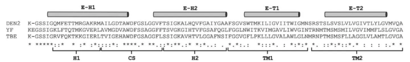
con-Fig. 1: amino acid sequences of flaviviral protein E stem-anchor (SA) regions. Alignment of yellow fever (YF) 17D virus (amino acid posi-tion from 397-493, GenBank P03314), dengue 2 virus (DEN2) (amino acid posiposi-tion from 459-495, GenBank P29990), tick-borne encephalitis virus (TBE) (amino acid position from 460-496, Genbank Q88474). The domains of SA regions are indicated as H1, CS, H2, TM1 and TM2 accordingly to Allison et al. (1999). The alpha-helices of SA (E-H1 and E-H2) and the stem-region (E-T1 and E-T2) determined by cryoelectron microscopic studies are indicated above the alignment (Zhang et al. 2003). The following symbols represent the degree of conservation observed at each amino acid position: identical in all sequences (*), conserved substitutions (:) and semi-conserved substitutions (.).
Fig. 2: genomic structure of recombinant yellow fever (YF) 17D vi-ruses at the E/NS1 intergenic region. A: schematic drawing of part of the polyprotein precursor [C, precursor membrane (PrM), E and NS1] of YF virus. The E protein stem-anchor (SA) region is shown, as well as the signal peptidase cleavage site (black arrow head); B: schematic diagram representing this region in the recombinant YF 17D/green fluorescent protein (GFP) viruses in which the heterologous protein consists of duplicated portions of NS1 (9 amino acid residues of the N-terminus) and the E protein (SA domains) fused to it. The prote-olytic sites of signal peptidase flanking the recombinant cassette are indicated (black arrow head). Below this illustration the amino acid sequences of the SA elements of each recombinant virus are shown;
C: exchange performed in YF17D/GFP/TM1→TM LAMP-1 virus; D:
struct containing a reporter GFP protein with different SA compositions was capable of expressing the recombi-nant proteins. The recombirecombi-nant proteins were processed by the cell signal peptidase, generating GFP proteins with distinct molecular weight bands as shown by SDS-PAGE: 37.9 kDa corresponds to YF17D/GFP/SA, 34.4 kDa corresponds to YF17D/GFP/ ΔH1CS, 33.1 kDa cor-responds to YF17D/GFP/ΔH2, 38.5 kDa corcor-responds to YF17D/GFP/TM1-TM LAMP and 38.8 kDa corresponds to YF17D/EGFP/TM1 + VALLLVA. These values are consistent with the MWs predicted by in silico analysis of the lower bands. The upper bands are not suggestive of GFP dimers because the increase in the molecular weight was only of 10-14 kDa. This difference was likely due to covalent binding to some cellular protein because all of the cell lysates were boiled at 95ºC for 10 min in Laemmli buffer containing beta-mercaptoethanol
Fig. 3: replication profile of recombinant viruses in Vero cells. Cells were infected at multiplicity of infection of 0.02. Each time point constitutes the average titre obtained from at least three independent experiments with the corresponding standard deviation. GFP: green fluorescent protein; PFU: plaque forming units; p.i.: post-infection; SA: stem-anchor; YF: yellow fever.
Fig. 4: detection of green fluorescent protein (GFP) expression af-ter infection of Vero cells with recombinant yellow fever (YF) 17D viruses. Cell lysates were subjected to western blot analysis using a rabbit anti NS3 serum and a rabbit anti-GFP polyclonal antibody. At panel A the Lanes correspond to the following samples: Mock (1),
YF17D/E200T3 (2), YF17D/GFP/SA (3), YF17D/GFP/ΔH1CS (4), YF17D/GFP/ΔH1CSH2 (5), YF17D/GFP/TM1-TM LAMP (6) and B
to Mock (1), YF17D/E200T3 (2), YF17D/GFP/SA (3), YF17D/GFP/ TM1 + VALLLVA (4). On the left side of the A and B are indicated the molecular weight markers and on the right side the NS3 protein and the region containing the GFP cassette variants. SA: stem-anchor.
Fig. 5: analysis of recombinant yellow fever (YF) 17D infected cells expressing green fluorescent protein (GFP) protein. Immunopre-cipitation profiles obtained from protein extracts of mock-infected Vero cells (Lanes Mock), YF17D/GFP/stem-anchor (SA), YF17D/
GFP/ΔH1CS (ΔH1CS), YF17D/GFP/ΔH1CSH2 (ΔH1CSH2), YF17D/
and SDS prior to SDS-PAGE analysis. The control vi-rus YF17D/E200T3 did not produce GFP and no protein could be detected with the GFP antiserum, as expected.
We also analysed GFP expression from the different recombinant YF 17D viruses in Vero cells by means of cell metabolic labelling and immunoprecipitation with GFP and YF polyclonal antisera. We could detect YF virus structural proteins C (11 kDa), E (53 kDa) and prM (26 kDa) and non-structural proteins NS5 (103 kDa), NS3 (70 kDa) and NS1 (46 kDa) in the immunoprecipi-tates from all of the preparations (Fig. 5). Similar to the western blot analysis results, molecular weight variants of GFP contained different domains of the SA, which displayed comparable molecular weights (Fig. 5). In the cell culture supernatant, only viral E and NS1 proteins were detectable, suggesting that there is no secretion of GFP to the extracellular medium during viral infection.
Intracellular localisation of recombinant GFP ex-pressed by YF 17D viruses - The original GFP heterolo-gous cassette expressed by the YF17D/GFP/SA virus is
fused to the entire SA, which contains motifs involved in ER retention, such as E-TM1. We investigated wheth-er the deletions and inswheth-ertions introduced into the SA domains could modify the ER-retention profile of the recombinant protein, as previously demonstrated (Bon-aldo et al. 2007). We examined the intracellular distri-bution of GFP fused to different variations of the YF E protein SA domains in Vero cell monolayers infected by the parental and recombinant YF17D/GFP viruses for up to 120 h. We subsequently established that the maxi-mum autofluorescence of GFP was reached at 72 h p.i. for each recombinant virus.
The association of GFP with the ER was accessed by staining Vero cells infected by the recombinant GFP viruses with an ER-specific labelling marker (Fig. 6). The recombinant proteins of all of the variant viruses were apparently dispersed at the perinuclear region, which is suggestive of ER retention. Merge images con-firmed this finding for all of the recombinant viruses. It was suggested that recombinant GFP proteins may
be confined to a subcompartment of the ER (Welsch et al. 2009). To further investigate whether the recombi-nant protein was directed to the secretory pathway, we also labelled the Golgi system. The perinuclear staining pattern of GFP did not overlap with the Golgi appara-tus (Fig. 7). Because one of the constructs contained a swap of the E-TM1 domain for the human LAMP-1 TM domain, we also investigated the association of GFP to the lysosome. Again, it was not possible to detect any GFP fluorescence associated with this organelle (Fig. 8). Rhodamine-conjugated WGA was used to label the sur-face of fixed Vero cells that were not permeabilised with Triton X-100.Fig. 9 shows that there was no co-localisa-tion of the GFP expressed by the different recombinant viruses with WGA cell surface labelling. These data in-dicate that the modifications at the SA region fused to GFP did not promote its exodus from the ER. Therefore, the location of heterologous GFP for each of the viral constructs was restricted to the interior of the cell and not detected at the infected cell surface.
DiSCuSSioN
Given the outstanding properties of the YF 17D vac-cine virus, its use as a vector for the expression of het-erologous antigens toward the development of new live attenuated vaccines has been pursued by several labo-ratories (McAllister et al. 2000, Franco et al. 2010, Guy et al. 2010). We previously developed a methodology for the expression of heterologous proteins by the YF vi-rus based on the insertion of foreign sequences between the genes that code for the E and NS1 proteins of the 17D virus (Bonaldo et al. 2007). This approach involves cleavage between E and NS1 by a cellular enzyme signal peptidase (Stadler et al. 1997) present in the ER lumen (Bonaldo et al. 2007). To guarantee the correct cleavage of the precursor polyprotein and its membrane topology, we duplicated and fused the first 27 nt of the NS1 gene and the last 288 nt of the E gene to the 5’ and 3’ ends, respectively, of the heterologous gene. Initially, we per-formed the insertion of the GFP gene into the YF ge-nome due to the ease of viral viability and heterologous
Fig. 8: lysosome stained at infected cells. Vero cells 72 h infected with recombinant viruses expressing the green fluorescent protein (GFP) (green) were labelled with LysoTracker (red). Nuclei were stained with 4’-6-diamidino-2-phenylindole (DAPI) (blue). SA: stem-anchor. Bar = 20 µm.
protein expression monitoring. To date, several other genes have been expressed, such as the simian immuno deficiency virus Gag 45-269 fragment (Bonaldo et al. 2010), the 261-380 fragment of the amastigote surface protein-2 of Trypanosoma cruzi (Nogueira et al. 2011) and the 19 kDa domain of the Plasmodium falciparum merozoite surface protein-1 (MSP-1) (unpublished data). These different recombinant proteins expressed at the E/NS1 region with the described genomic structure are generally retained in the ER (unpublished observations). This finding was expected because all recombinant vi-ruses have fused to the carboxy terminus of the foreign gene, a duplication of the E protein SA domain, which is well known to contain molecular determinants that promote E protein retention in the ER as required for viral assembly (Lorenz et al. 2003, Ciczora et al. 2010, Hsieh et al. 2010). Because the development of YF 17D virus as a multipurpose vector may require the synthe-sis and trafficking of foreign proteins to other organelles to improve their immunogenicity, we have introduced
modifications in the SA portion of the heterologous GFP expression cassette and checked whether the modifi-cations could support the exit of the cassette from the ER compartment. The exit from the ER might also be advantageous in viral assembly kinetics with a conse-quent increase in viral titres and yields because the ER retention of the recombinant protein could disturb the heterodimerisation of the E and prM proteins and, con-sequently, the viral assembly.
Lin et al. 2011). CS is a conserved and flexible region connecting the two alpha-helices of the stem. Peptides containing part of this region are capable of binding to late-stage fusion intermediates and inhibit viral entry in the cell (Schmidt et al. 2010a, b). The E-H2 element has been associated with the stabilisation of prM-E heterodi-mer interactions. Hence, mutations in E-H2 disturb the membrane binding ability of the stem, cause deficiencies in viral assembly and reduce infectiousness (Allison et al. 1999, Lin et al. 2011). Because the different deletions introduced in the carboxy terminus of the recombinant GFP allowed the regeneration of three different YF vi-ruses with similar growth properties, they likely have the same effect on viral fitness. The other group of re-combinant YF 17D viruses expressed GFP rere-combinant proteins with alterations in E-TM1. In this case, we in-vestigated whether a longer TM domain with 23 amino acids, instead of 16 amino acids, could promote the ex-port of recombinant GFP from the ER. This assumption was based on the findings that the TM domain lengths
of early and late compartment proteins display organ-elle-specific properties, which are related to the lipidic bilayer thickness (Bretscher & Munro 1993, Levine et al. 2000, Sharpe et al. 2010). The first modification con-sisted of the swap of E-TM1 for the single TM domain of the lysosomal membrane glycoprotein-1 or LAMP-1. LAMP-1 possesses a highly N-linked glycosylated lumi-nal domain, a single-TM domain and a short cytoplasmic tail carrying a lysosome-targeting signal at the COOH terminus (Chen et al. 1988). We did not expect this LAMP-1 TM domain to promote the delivery of GFP to the lysosomes because only its TM domain was used in the viral construction and not the LAMP-1 cytoplasmic tail motif. However, we intended to investigate whether the presence of a 23 amino acid-long TM domain in the recombinant GFP cassette could support its departure from the ER to another organelle. The same rationale was employed in designing other recombinant viruses, in which the E-TM1 of recombinant GFP was modified by the insertion of the VALLLVA sequence. The
tion of this or a similar motif (ALALAL) in the TM1 of chimeric CD4 proteins containing the flaviviral E pro-tein anchor caused the departure of ER and membrane surface association, which corroborated the evidence in favour of the TM length as a ER retention determinant (Ciczora et al. 2010, Hsieh et al. 2010). Given the role of TM2 in translocation of NS1 and signalase cleavage, this SA segment was not modified to not compromise the polyprotein processing and recovery of the viruses. However, both modifications that resulted in increasing the E-TM1 length in the GFP cassette expressed by re-combinant YF 17D viruses did not lead to the export of GFP from the ER. Interestingly, this type of mutation did not considerably affect the ER retention phenotype when the prM-E interactions were assayed in silico in addi-tion to the levels of virus-like particle (VLP) producaddi-tion (Ciczora et al. 2010, Hsieh et al. 2010). Because the as-sembly process requires several E proteins with their re-spective TM regions in close proximity in a defined area of the replication vesicle membrane (Welsch et al. 2009), other ER retention motifs could keep envelope proteins inside the ER until the budding of the VLP. Notably, a number of signals of different strengths may contribute to the ER retention, such as the presence and interac-tion of non-hydrophobic residues in both E-TM domains, the length of the alpha-helices of the prM and E protein TM domains and a retention determinant also present in the TM1 of prM (Op De Beeck et al. 2004, Hsieh et al. 2008, 2010, Ciczora et al. 2010). It was observed that GFP was concentrated in distinct areas of the ER and this result could indicate that recombinant protein was segregated into sub-compartments of the ER. Nonethe-less, it is clear that these retention signals are related to the flavivirus-derived anchor portion of the recombinant GFP expression cassette because the GFP portion does not contain any specific ER retrieval or retention signals in its sequence (Tsien 1998). A careful mutational analy-sis of non-hydrophobic residues of the recombinant GFP TM segments is underway to investigate their effect on the organelle location. However, it is possible that in the context of a live virus infection, a number of factors will be involved rather than the expression of a single protein or VLP assembly. Further studies should be undertaken to elucidate the causes of ER retention of the recombi-nant proteins expressed at the intergenic E/NS1 region of the YF 17D virus polyprotein, given the usefulness of this site for the expression of foreign genes for new live attenuated viral vaccine development.
aCkNowlEDGEMENTS
To Regiane Burger, for the anti-NS3 antibodies, to Heloísa Diniz, for image editing, and to PDTIS-Fiocruz, for support-ing the sequencsupport-ing and confocal microscopy studies through the Genomics and Confocal Microscopy Facilities.
REFERENCES
Allison SL, Stiasny K, Stadler K, Mandl CW, Heinz FX 1999. Mapping of functional elements in the stem-anchor region of tick-borne en-cephalitis virus envelope protein E. J Virol73: 5605-5612.
Bonaldo MC, Garratt RC, Caufour PS, Freire MS, Rodrigues MM, Nussenzweig RS, Galler R 2002. Surface expression of an
immu-nodominant malaria protein B cell epitope by yellow fever virus.
J Mol Biol315: 873-885.
Bonaldo MC, Garratt RC, Marchevsky RS, Coutinho ES, Jabor AV, Almeida LF, Yamamura AM, Duarte A, Oliveira PJ, Lizeu JO, Camacho A, Freire MS, Galler R 2005. Attenuation of recombi-nant yellow fever 17D viruses expressing foreign protein epitopes at the surface. J Virol79: 8602-8613.
Bonaldo MC, Martins MA, Rudersdorf R, Mudd PA, Sacha JB, Piaskowski SM, Costa Neves PC, Veloso de Santana MG, Vojnov L, Capuano S 3rd, Rakasz EG, Wilson NA, Fulkerson J, Sadoff JC, Watkins DI, Galler R 2010. Recombinant yellow fever vac-cine virus 17D expressing simian immunodeficiency virus SIV-mac239 gag induces SIV-specific CD8+ T-cell responses in
rhe-sus macaques. J Virol84: 3699-3706.
Bonaldo MC, Mello SM, Trindade GF, Rangel AA, Duarte AS, Ol-iveira PJ, Freire MS, Kubelka CF, Galler R 2007. Construction and characterization of recombinant flaviviruses bearing inser-tions between E and NS1 genes. Virol J4: 115.
Bretscher MS, Munro S 1993. Cholesterol and the Golgi apparatus.
Science261: 1280-1281.
Carpp LN, Galler R, Bonaldo MC 2011. Interaction between the yel-low fever virus nonstructural protein NS3 and the host protein Alix contributes to the release of infectious particles. Microbes Infect.13: 85-95.
Chen JW, Cha Y, Yuksel KU, Gracy RW, August JT 1988. Isolation and sequencing of a cDNA clone encoding lysosomal membrane gly-coprotein mouse LAMP-1. Sequence similarity to proteins bearing onco-differentiation antigens. J Biol Chem263: 8754-8758.
Ciczora Y, Callens N, Seron K, Rouille Y, Dubuisson J 2010. Identifica-tion of a dominant endoplasmic reticulum-retenIdentifica-tion signal in yel-low fever virus pre-membrane protein. J Gen Virol91: 404-414.
Franco D, Li W, Qing F, Stoyanov CT, Moran T, Rice CM, Ho DD 2010. Evaluation of yellow fever virus 17D strain as a new vector for HIV-1 vaccine development. Vaccine28: 5676-5685.
Gillespie LK, Hoenen A, Morgan G, Mackenzie JM 2010. The endo-plasmic reticulum provides the membrane platform for biogenesis of the flavivirus replication complex. J Virol84: 10438-10447.
Guy B, Guirakhoo F, Barban V, Higgs S, Monath TP, Lang J 2010. Preclinical and clinical development of YFV 17D-based chime-ric vaccines against dengue, West Nile and Japanese encephalitis viruses. Vaccine28: 632-649.
Hsieh SC, Liu IJ, King CC, Chang GJ, Wang WK 2008. A strong en-doplasmic reticulum retention signal in the stem-anchor region of envelope glycoprotein of dengue virus type 2 affects the produc-tion of virus-like particles. Virology374: 338-350.
Hsieh SC, Tsai WY, Wang WK 2010. The length of and nonhydro-phobic residues in the transmembrane domain of dengue virus envelope protein are critical for its retention and assembly in the endoplasmic reticulum. J Virol84: 4782-4797.
Levine TP, Wiggins CA, Munro S 2000. Inositol phosphorylceramide synthase is located in the Golgi apparatus of Saccharomyces cer-evisiae. Mol Biol Cell11: 2267-2281.
Lin SR, Zou G, Hsieh SC, Qing M, Tsai WY, Shi PY, Wang WK 2011. The helical domains of the stem region of dengue virus envelope protein are involved in both virus assembly and entry.
J Virol85: 5159-5171.
Lorenz IC, Allison SL, Heinz FX, Helenius A 2002. Folding and dimerization of tick-borne encephalitis virus envelope proteins prM and E in the endoplasmic reticulum. J Virol76: 5480-5491.
subviral particles from tick-borne encephalitis virus. J Virol77: 4370-4382.
Mackenzie J 2005. Wrapping things up about virus RNA replication.
Traffic6: 967-977.
McAllister A, Arbetman AE, Mandl S, Pena-Rossi C, Andino R 2000. Recombinant yellow fever viruses are effective therapeutic vac-cines for treatment of murine experimental solid tumors and pul-monary metastases. J Virol74: 9197-9205.
Nogueira RT, Nogueira AR, Pereira MC, Rodrigues MM, Galler R, Bonaldo MC 2011. Biological and immunological characteriza-tion of recombinant yellow fever 17D viruses expressing a Try-panosoma cruzi amastigote surface protein-2 CD8+ T cell epitope
at two distinct regions of the genome. Virol J8: 127.
Op De Beeck A, Rouille Y, Caron M, Duvet S, Dubuisson J 2004. The transmembrane domains of the prM and E proteins of yellow fe-ver virus are endoplasmic reticulum localization signals. J Virol 78: 12591-12602.
Rice CM, Grakoui A, Galler R, Chambers TJ 1989. Transcription of infectious yellow fever RNA from full-length cDNA templates produced by in vitro ligation. New Biol1: 285-296.
Schmidt AG, Yang PL, Harrison SC 2010a. Peptide inhibitors of dengue-virus entry target a late-stage fusion intermediate. PLoS Pathogens6: e1000851.
Schmidt AG, Yang PL, Harrison SC 2010b. Peptide inhibitors of flavivirus entry derived from the E protein stem. J Virol84: 12549-12554.
Sharpe HJ, Stevens TJ, Munro S 2010. A comprehensive comparison of transmembrane domains reveals organelle-specific properties.
Cell142: 158-169.
Stadler K, Allison SL, Schalich J, Heinz FX 1997. Proteolytic activation of tick-borne encephalitis virus by furin. J Virol71: 8475-8481.
Tsien RY 1998. The green fluorescent protein. Annu Rev Biochem 67: 509-544.
Welsch S, Miller S, Romero-Brey I, Merz A, Bleck CK, Walther P, Fuller SD, Antony C, Krijnse-Locker J, Bartenschlager R 2009. Composition and three-dimensional architecture of the dengue vi-rus replication and assembly sites. Cell Host Microbe5: 365-375.
Yu IM, Zhang W, Holdaway HA, Li L, Kostyuchenko VA, Chipman PR, Kuhn RJ, Rossmann MG, Chen J 2008. Structure of the im-mature dengue virus at low pH primes proteolytic maturation.
Science319: 1834-1837.