Mechanical, antibacterial and bond strength
properties of nano-titanium-enriched glass
ionomer cement
Rene GARCIA-CONTRERAS1, Rogelio Jose SCOUGALL-VILCHIS2, Rosalía CONTRERAS-BULNES3, Hiroshi
SAKAGAMI4, Raul Alberto MORALES-LUCKIE5, Hiroshi NAKAJIMA6
1- Laboratorio de Investigación Interdisciplinaria, Área de Biomateriales Dentales, Escuela Nacional de Estudios Superiores (ENES) Unidad León, Universidad Nacional Autónoma de México (UNAM), Guanajuato, México.
2- Chairman of Dental Research Center “Dr. Keisaburo Miyata”. Department of Orthodontics, Autonomous University State of Mexico (UAEM), Toluca, México. 3- Department of Pediatric Dentistry, Dental and Advanced Studies Research Center (CIEAO), Faculty of Dentistry, Autonomous University State of Mexico (UAEM), Toluca, México.
4- Division of Pharmacology, Meikai University School of Dentistry, Sakado, Saitama, Japan.
5- Sustainable Chemistry Research Center, Autonomous University State of Mexico (UAEM), Toluca, México. 6- Division of Dental Biomaterials Science, Meikai University School of Dentistry, Sakado, Saitama, Japan.
Corresponding address: Rogelio J. Scougall-Vilchis - Chairman of Dental Research Center “Dr. Keisaburo Miyata”. Department of Orthodontics, Dental and Advanced Studies Research Center (CIEAO) - Faculty of Dentistry, Autonomous University State of Mexico (UAEM) - Tollocan-Jesus Carranza - Colonia Universidad - Toluca - México - e-mail:[email protected]/[email protected]/[email protected]
Submitted: December 11, 2014 - Modiication: April 15, 2015 - Accepted: May 27, 2015
ABSTRACT
http://dx.doi.org/10.1590/1678-775720140496
T
he use of nanoparticles (NPs) has become a signiicant area of research in Dentistry. Objective: The aim of this study was to investigate the physical, antibacterial activity and bond strength properties of conventional base, core build and restorative of glass ionomer cement (GIC) compared to GIC supplemented with titanium dioxide (TiO2) nanopowder at 3% and 5% (w/w). Material and Methods: Vickers microhardness was estimated with diamond indenter. Compressive and lexural strengths were analyzed in a universal testing machine. Specimens were bonded to enamel and dentine, and tested for shear bond strength in a universal testing machine. Specimens were incubated with S. mutans suspension for evaluating antibacterial activity. Surface analysis of restorative conventional and modiied GIC was performed with SEM and EDS. The analyses were carried out with Kolmogorov-Smirnov, ANOVA (post-hoc), Tukey test, Kruskal-Wallis, and Mann Whitney. Results: Conventional GIC and GIC modiied with TiO2 nanopowder for the base/liner cement and core build showed no differences for mechanical, antibacterial, and shear bond properties (p>0.05). In contrast, the supplementation of TiO2 NPs to restorative GIC signiicantly improved Vickers microhardness (p<0.05), lexural and compressive strength (p<0.05), and antibacterial activity (p<0.001), without interfering with adhesion to enamel and dentin. Conclusion: GIC supplemented with TiO2 NPs (FX-II) is a promising material for restoration because of its potential antibacterial activity and durable restoration to withstand the mastication force.
Keywords: Glass ionomer cements. TiO2 nanoparticles. Antibacterial activity. Physical
properties. Shear bond strength.
INTRODUCTION
Glass ionomer cement (GIC) possesses certain properties of adhesive23, biocompatibility2, and
luoride releasing3, which have led to worldwide
use as luting, base, liners and restorative materials. However, the major disadvantages are fracture
toughness, low wear-resistance and in the past high dissolution in a water sorption23 resulting in a base,
properties for compressive strength, hardness, higher modulus of elasticity, higher resistance to solubility and resistance to bacterial adhesion14.
Signiicant perfections have been developed since the invention of GIC, numerous iller components
have been added including; silver-amalgam particles1, spherical silica26, zirconia12, glass
iber13, hydroxyapatite20, bioactive glass particles
as pre-reacted glass ionomer particles (PRG), giomer restorative material15. The incorporation of
the iller particles above to GIC has signiicantly modiied the mechanical properties of cements;
however, fillers can interfere with metabolic activities for bacterial adhesion and inhibit the antibacterial activity of GIC4. In contrast, the use of
nanoparticles (NPs) has become a signiicant area
of research in Dentistry, the main use have been focused in increasing the mechanical properties and antibacterial effect; altering the hydrogen bonding, respiratory process, DNA unwinding, cell wall synthesis and division by making “pits” in the wall and increasing the permeability resulting in a bacterial death11. Recently, incorporation of
hydroxyapatite and luoroapatite nanobioceramics
into conventional GIC improved their mechanical properties and bond strength to dentine22. Titanium
dioxide (TiO2) as an inorganic additive has many promising properties as it is chemically stable, biocompatible and antibacterial28. NPs have been
proposed as reinforcing fillers to dental resin composites and epoxy30. It has recently been
reported that (i) the incorporation of TiO2 NPs to
GIC at 3% and 5% (w/w) signiicantly enhanced the fracture toughness, compressive strength, lexural
strength and hardness, and (ii) GIC supplemented with TiO2 NPs showed antibacterial activity against
Streptococcus mutans without interference with
luoride release; nevertheless, (iii) the incorporation
of 7% of TiO2 NPs compromised the mechanical
properties and adhesion6. We recently reported
that, for TiO2 nanoparticles in culture with human
gingival ibroblast (HGF)9 and oral squamous cell
carcinoma cells (HSC-2)7, some particles were
incorporated into the cells, exclusively in the vacuoles and showed no cytotoxic nor hormetic growth stimulation at lower concentrations. However, TiO2 NPs exert pro-inlammatory action by
Interleukin-1β (IL-1β) and stimulated the secretion
of prostaglandin E2 (PGE2), Cyclooxygenase (COX) 1 and 2, and induced drastic metabolic changes10
to the culture medium by HGF cells and TiO2 NPs also induced PGE2 production, in synergy with IL-1β, the enhanced production of PGE2 was not simply due to LPS contamination9. Also, the incorporation
of TiO2 NPs to GIC exhibits acceptable to moderate biocompatibility in culture with human oral normal
cells [pulp cells (HPC), gingival ibroblast (HGF), periodontal ligament ibroblast (HPLF)] and human
cancer cells [oral squamous cell carcinoma (OSCC): HSC-2, HSC-3, HSC-4 and gingival carcinoma (Ca9-22)]8.
Based in the previously reports, we expected that the supplementation of TiO2 NPs to GIC enhance its mechanical and antibacterial properties, the objective of this research is to investigate the
physical properties (microhardness, lexural and
compressive strength), the antibacterial activity and the bond strength of base, core build up and
restorative GIC compared to GIC modiied with TiO2
nanopowder at 3% and 5% (w/w).
MATERIAL AND METHODS
Powder of each GIC was blended with TiO2 nanopowder, anatase phase, particle size <25 nm
(Sigma-Aldrich, St. Louis, MO, USA) at 3% and 5%
(w/w). GIC powder and TiO2 NPs were mixed in a vortex for one minute.
Vickers microhardness test
GIC cylinders (9.5x1 mm) (n=5) were made
in a Telon mold according to ADA speciication 27
after being prepared following the manufacturer´s instruction. The recommended powder/liquid (P/L) ratio of 2.6/1 g was mixed for cements. Cylinders
were tested in ISO 9001:2008 certiied diamond
indenter (DongGuan Sinowon precision instruments, Nancheng, China) with 10 N and a dwell time of 10 s were employed for 10 indentations across the specimens of each group resulting in 50 indentations of each group. Since Vickers microhardness test is more sensitive to measurement errors than Knoop test and best for small rounded areas, we decided to use the method based on the ISO 9917-1:200716.
Flexural and compressive strength
Twenty cylinder specimens were prepared as mentioned above. Cylinders were subjected to three points bending in a universal testing machine (AGS-X, Shimadzu, Kyoto, Japan) at cross speed of 1 mm/min (MPa). Flexural strength (MPa) was calculated using the following formula:
O´=3Pl/2bd2
where O´ is the lexural strength, P (N) is the
load at fracture, l is the distance between the two supports (mm), b is the width of the specimen (mm), and d is the thickness (mm). On the other hand, compressive strength of specimens was performed by the universal testing machine at cross speed of 1 mm/min (MPa), and calculated using the following equation:
CS=2P/πdh
to ISO 9917-1:200716 and ISO 9917-2:201017.
Shear bond strength to enamel and dentine
A total of 180 freshly extracted anterior bovine teeth were stored in 0.1 thymol solution. Teeth were randomly divided into the nine groups
(n=20/group). Samples were ixed in acrylic resin
(NicTone 62, MDC Dental, Guadalajara, Mexico) with a label bearing the number of each sample. A mounting jig was used to align each tooth’s labial surface. Standardized GIC blocks (4x4x1 mm) were preformed in a metal mold following the manufacturer’s instructions. Before adhering the block to the dental surfaces with fresh cement, the sample surfaces were finished with #400 waterproof abrasive paper (Fuji Star, Sankyo, Rikagaku, Okegawa, Japan). In the case of enamel bond strength, vestibular surface was sandblasted (Micro Cab, Danville, San Ramon, CA, USA) with 50 µm of aluminum dioxide (Danville, San Ramon, CA, USA) for one minute. Then, teeth underwent ultrasonic cleaning for one minute (Quantrex, Kearny, NJ, USA). Consequently, for testing the bond strength in dentin, the vestibular surfaces of the teeth were reduced approximately 1.5 mm with a high speed diamond bur (SS White Burs Inc, Lakewood, NJ, USA). At that point, dentinal surface was sandblasted and underwent ultrasonic cleaning as mentioned above. Immediately after direct bonding the GIC block with appropriate powder/ liquid proportion, samples were stored in water at 37°C during 24 h. Shear bond strength to enamel and dentine was carried out in a universal testing machine at cross speed of 1 mm/min (MPa). Force was applied at the interface of the GIC block and dental surface.
Antibacterial activity
Suspension of approximately 105 Streptococcus
mutans (S. mutans, ATCC 35668) was cultivated in brain heart infusion broth (Becton Dickinson, NJ, USA) for 18 hours. Bacteria solution was sub-cultivated in brain heart agar (Becton Dickinson, NJ, USA). Immediately, blocks (4x4x1 mm) of the
different conventional GIC and GIC modiied with
TiO2 NPs at 3% and 5% (w/w) were set in direct
contact over the agar containing the bacteria, after 24 hours of incubation at 37°C, inhibit halos were measured with electronic digital caliper (NSK, Tochigi, Japan). Three blocks were set on each 100 mm plate containing the brain heart agar. Experiment was performed in triplicate to obtain reproducible data.
SEM and EDS analysis
Standardized GIC blocks (4x4x1 mm) of FX-II
conventional, FX-II 3% (w/w) TiO2 NPs, and FX-II
5% (w/w) TiO2 NPs were prepared in the metallic
mold and covered with microslide glass. Samples
were gently polished and inished with #400, 1000
and 1500 waterproof abrasive paper (Fuji Star, Sankyo, Rikagaku, Okegawa, Japan). Subsequently,
blocks were ultrasonically cleaned for ive minutes
in distilled water (Quantrex, Kearny, NJ, USA). All samples were adhered to aluminum stubs with conductive tape, coated with carbon and observed under SEM (PHILIPS XL-30, North Billerica, MA, USA) with secondary electrons at ×100, ×500, and
×3,000 magniication by 20 kV. Energy-dispersive
X-ray (EDS) analysis was developed at the same time of SEM micrographs. An area of approximately 20×15 µm was selected for analysis; relative values were obtained after 300 s of measurement.
Statistical analysis
Mean values and standard deviations were estimated. Vickers microhardness data were subjected to Kolmogorov-Smirnov normality test and ANOVA (post-hoc) Tukey test. In order
to examine compressive and lexural strength,
shear bond strength to enamel and dentine, and antibacterial activity data were analyzed with non-parametric Kruskal-Wallis and multiple comparisons of Mann-Whitney, the analyses were carried out with SPSS 18.0 (SPSS Inc., Chicago, Ill, USA). A value of
0.05 was considered statistically signiicant.
RESULTS
Vickers microhardness
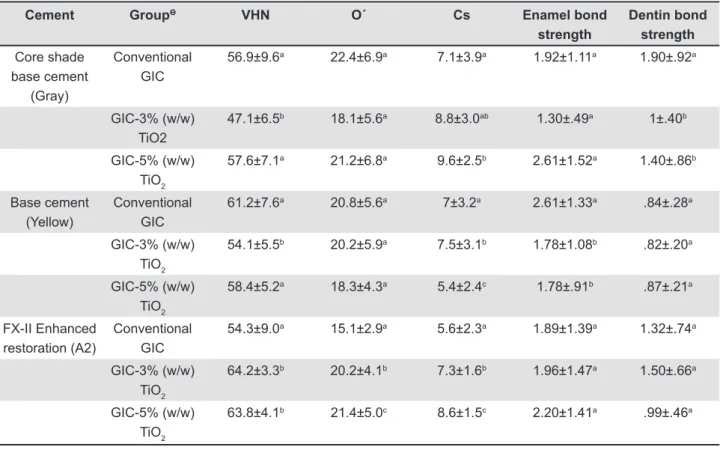
Vickers microhardness data indicated normality and ANOVA test showed statistical differences (p<.0001) between groups and post-hoc Tukey test results are enlisted in Table 1. It must be mentioned that, in all cases, the size of the indentations was
larger than the iller particles, based on the size of illers reported by the manufacturer. Data showed a signiicant increase in microhardness for the FX-II
containing 3% and 5% (w/w) TiO2 NPs compared
to the conventional cement. Nevertheless, core shade and base cement did not present increased microhardness values; actually, the inclusion of nanopowder at both concentrations decreased the microhardness.
Flexural and compressive strength
The supplementation of 3% and 5% (w/w)
TiO2 NPs into FX-II enhanced lexural strength
(p<0.05) and compressive strength (p<0.0001), compared to the conventional cement. The minimal
supplementation at 3% improved the properties of deinitive restoration cement. Core shade build up
cement improved only compressive strength when
5% (w/w) (p<0.05) TiO2 NPs were incorporated,
Cement GroupƟ VHN O´ Cs Enamel bond strength
Dentin bond strength
Core shade base cement
(Gray)
Conventional GIC
56.9±9.6a 22.4±6.9a 7.1±3.9a 1.92±1.11a 1.90±.92a
GIC-3% (w/w) TiO2
47.1±6.5b 18.1±5.6a 8.8±3.0ab 1.30±.49a 1±.40b
GIC-5% (w/w) TiO2
57.6±7.1a 21.2±6.8a 9.6±2.5b 2.61±1.52a 1.40±.86b
Base cement (Yellow)
Conventional GIC
61.2±7.6a 20.8±5.6a 7±3.2a 2.61±1.33a .84±.28a
GIC-3% (w/w) TiO2
54.1±5.5b 20.2±5.9a 7.5±3.1b 1.78±1.08b .82±.20a
GIC-5% (w/w) TiO2
58.4±5.2a 18.3±4.3a 5.4±2.4c 1.78±.91b .87±.21a
FX-II Enhanced restoration (A2)
Conventional GIC
54.3±9.0a 15.1±2.9a 5.6±2.3a 1.89±1.39a 1.32±.74a
GIC-3% (w/w) TiO2
64.2±3.3b 20.2±4.1b 7.3±1.6b 1.96±1.47a 1.50±.66a
GIC-5% (w/w) TiO2
63.8±4.1b 21.4±5.0c 8.6±1.5c 2.20±1.41a .99±.46a Table 1- Mean (standard deviation) of Vickers microhardness (VHN) (n=50), lexural (O´) and compressive strength (Cs)
and shear bond strength to enamel and dentin (n=20) of GIC and GIC incorporated with 3% and 5% (w/w) TiO2 nanopowder
* GIC: Glass ionomer cement. Ɵ TiO2: Titanium dioxide nanopowder.
Mean values for each cement group with the same superscript letter (column) are not signiicantly different (p>0.05), while mean values with different letters are signiicantly different (p<0.05). Vickers microhardness was analyzed with ANOVA (post-hoc) Tukey test, while lexural and compressive strength, shear bond strength to enamel and dentin were analyzed by Mann Whitney test.
Cement GroupƟ n Inhibit halos (mm)
Core shade base cement (Gray)
Conventional GIC* 18 None
GIC-3% (w/w) TiO2 18 None
GIC-5% (w/w) TiO2 18 None
Base cement (Yellow) Conventional GIC 18 None
GIC-3% (w/w) TiO2 18 None
GIC-5% (w/w) TiO2 18 None
FX-II Enhanced restoration (A2)
Conventional GIC 18 0.92±0.22a
GIC-3% (w/w) TiO2 18 2.11±0.82b
GIC-5% (w/w) TiO2 18 1.53±0.79
b
Table 2- Antibacterial activity of GIC and GIC incorporated with 3% and 5% (w/w) TiO2 nanopowder against Streptococcus
mutans (ATCC 35668)
* GIC: Glass ionomer cement. Ɵ TiO
2: Titanium dioxide nanopowder.
addition of TiO2 NPs compared to conventional GIC. The results are summarized in Table 1.
Shear bond strength
Data for shear bond strength (MPa) to enamel and dentine showed no statistical differences
between the conventional GIC and that modiied
with TiO2 NPs (neither at 3% nor at 5%). There was
a slight but insigniicant increase in the shear bond
strength to enamel in the case of the core shade
with 5% (w/w) TiO2 NPs, and FX-II with 3% and
5% (w/w) TiO2 NPs (Table 1).
Antibacterial activity
Bacterial growth activity (Table 2) was reduced
on direct contact to FX-II conventional, FX-II 3%
and 5% (w/w) TiO2 NPs. Inhibit halos values (n=18)
obtained corresponded to 0.92±.22 mm, 2.11±0.82 mm, and 1.53±0.79 mm, respectively. When the antibacterial activity of FX-II 3% and 5% (w/w)
TiO2 NPs was compared with conventional FX-II,
signiicant differences were observed (p<0.001)
in both groups, and no difference was observed
between FX-II 3% and 5% (w/w) TiO2 NPs. The
minimum supplementation of 3% or 5% (w/w)
Element FX-II FX-II-3% (w/w) TiO2 FX-II-5% (w/w) TiO2
C 78.1 59.6 60.3
O 11.2 30.56 30.63
F 2.4 5.72 5.7
Al 3.3 1.68 1.33
Si 2.93 1.34 1.08
P 0.85 0.37 0.28
S 0.006 0.001 0
Ti 0 0.11 0.17
Sr 1.16 0.57 0.44
Total 100% 100% 100%
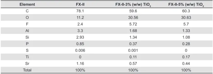
Table 3- Energy-dispersive X-ray (EDS) analysis of conventional FX-II, FX-II with 3% and 5% (w/w) TiO2 nanopowder. Values represent atomic percentage (a%)
FX-II: Enhanced restorative cement TiO2: Titanium dioxide nanopowder
Figure 1- Blocks (4x4x1 mm) of (a) conventional FX-II, (b) FX-II 3% (w/w) TiO2, and (c) FX-II 5% (w/w) TiO2. Samples
were gently polished and inished with #400, #1,000, and #1,500 waterproof abrasive paper and ultrasonically cleaned. Topographically, there are no differences between specimens. Nevertheless, hybrid particles are observed, microparticles (1c, black circle and arrow) are uniformly lay between (matrix) macroparticles, and such particles seem to be grouped of TiO2 nanoparticles due to their angular and semispherical shape conirmed by the 1d micrograph and EDS of this area, the
TiO2 NPs to GIC showed higher antibacterial activity against S. mutans than conventional FX-II. Nevertheless, core shade and base cement
conventional GIC with or without modiication with
TiO2 nanopowder showed no antibacterial properties in any specimens.
SEM and EDS analysis
Representative SEM micrographs are shown in Figure 1. Topographically, there are no apparent
differences in the inish surfaces for FX-II, FX-II
3% and 5% (w/w) TiO2 NPs. In Figure 1C, hybrid
particles are observed, microparticles uniformly lay between (matrix) macroparticles, and such particles seem to be grouped of TiO2 NPs due to
their angular and semispherical shape conirmed by
the 1D micrograph. The composition of conventional
GIC FX-II, FX-II 3% and 5% (w/w) TiO2 NPs are
shown in Table 3. Based on EDS data, all materials showed dominant portions of carbon and oxygen.
Titanium was detected in FX-II containing 3%
and 5% (w/w) TiO2 NPs, while the concentration
of oxygen increased and strontium decreased, by incorporating the TiO2 NPs.
DISCUSSION
Flexural and compressive strength
Compressive and flexural tests are used in Dentistry for laboratory simulation of the stress that may result from forces applied clinically to a restorative, base/liner or core build material24. Most
mastication forces are compressive in nature, but exact critical value is unknown28. Therefore, it is
important to investigate whether compressive force contributes to fracture failure during mastication process. The minimum value necessary to resist the masticatory forces in the posterior teeth would be 125 MPa, while 100 MPa for primary dentition29. Flexural forces are generated under
clinical situations, and the dental materials need
to withstand the repeated lexing, bending, and
twisting forces. Microhardness test is a parameter frequently used to evaluate the material surfaces resistance to plastic deformation by penetration28.
The powder/liquid ratio of GIC has an inluence on
the mechanical properties and bond strength31.
Improvement in lexural strength of the GIC FX-II was signiicantly higher at concentrations of 3% and
5% (w/w) TiO2 NPs than conventional. Therefore,
Ketac-Molar (3M ESPE, Seefeld, Germany) and Fuji IX (GC Corporation, Tokyo, Japan) have
showed higher values of lexural strength than the
cements in this study when performed specimens
of 25x2x2 mm. The reported lexural strength
values of the cements above represent 33.3, 34.5 MPa, respectively19. Supplementation of 3% and
5% (w/w) TiO2 NPs to FX-II have results similar
to those reported by Elsaka, et al.6 (2011) when
restorative GIC (Kavitan Plus, SpofaDental, Czech
Republic) was modiied with TiO2 NPs.
On the other hand, the test procedures for compressive strength are not complicated. Although the compression specimen has a convenient cylindrical geometry, perfection of the ends (which is essential to produce uniform contact between the specimen and the testing device) is difficult to achieve. Compressive strengths
for GIC FX-II containing 3% and 5% (w/w) TiO2
NPs were higher than that of conventional GIC. Compressive strengths of different conventional GIC such as Ketac Molar (3M ESPE), Fuji IX (GC),
and Ketac-il plus (3M ESPE) (146.28 to 152.41
MPa) were higher than that of GIC studied here with or without supplementation of TiO2 NPs; the difference in values can be explained by the size of specimens (4 mm diameter and 6 mm high)19.
Flexural and compressive strength improvement
of FX-II containing 3% and 5% (w/w) TiO2 NPs
can be attributed to the small sizes of the TiO2
particles supplemented into the glass powder and the presence of the NPs can occupy the empty spaces between the larger GIC glass particles and act as additional bonding sites for the polyacrylic polymer; this means that the base cement did not incorporate particles because of the small size
particles and greater surface of TiO2 NPs compared
to those of the glass.
Vickers microhardness
The GIC FX-II enhanced restoration containing
3% and 5% (w/w) TiO2 NPs exhibit signiicantly
higher Vickers microhardness compared to
conventional GIC, while GIC with 5% (w/w) TiO2
NPs for base and core build showed no statistical differences in relation to conventional cement.
The 3% (w/w) TiO2 NPs rather decreased the
Vickers microhardness; the supplementation of TiO2 NPs to FX-II powder possibly is related to the fewer glass particles on the surface of GIC, which result in greater amount of acid to react
with the NPs. Different studies have focused on
determining the hardness of conventional and
modiied GIC. Thus, conventional GIC as Ketac-il (3M, ESPE), Fuji IX (GC), and Ionoil Molar (VOCO,
Cuxhaven, Germany) have values of 90, 69.7, and 57.4 VHN, respectively18. Conventional FX-II
enhanced restoration showed 54.3 VHN, lower values than the other GICs. On the other hand, the
supplementation of 3% and 5% (w/w) TiO2 NPs to
conventional FX-II showed higher values of 64.2 and 63.8 VHN, respectively. Meanwhile, microhardness values of metal reinforced cements like Fuji IX GP (GC) (from 54.44 to 61.77 VHN)21 showed lower
containing 3% (w/w) TiO2 NPs represents 48.34
VHN6. Microhardness values of RMGIC such as
Photac Fil (3M, ESPE), Vitremer (3M, St. Paul, MN, USA), and Fuji II LC (GC) showed values of 46.2, 51.4, 69.2 VHN21, respectively.
Shear bond strength
The chemical adhesion of GIC to enamel and dentin is achieved by reaction of phosphate ions in the dental tissue with carboxylate groups in the
polyacrylic acid. Several factors can inluence the
bond strength, one of which is the type of dental substrate. Theoretical considerations and results of experiments show that enamel is much more susceptible to adhesion than dentin21. Enamel
has a surface that is essentially homogeneous, dense, and mainly composed of hydroxyapatite, which possesses high surface energy. Dentin has a heterogeneous surface, containing dental tubules that contain odonto-plastic processes, consists of
approximately 30% volume organic matter, and
consequently has low surface energy19. The enamel
bond strength of different GIC modiied with 3%
and 5% (w/w) TiO2 NPs studied here showed similar
values in relation to conventional cements except
for Core shade containing 5% (w/w) TiO2 NPs,which
showed signiicantly higher values when bonding to
enamel surface. Data reported here have similar or lower values of enamel shear bond strength than different studies carried out with conventional
GIC such as Ketac-il plus (3M ESPE), Ketac-Molar
(3M ESPE), and Fuji IX (GC). These cements have reported values as follows: 4.9, 5.31, and 5 MPa when debonding 3 mm in diameter of GIC adhered to enamel surface5,25. These low values were
observed due to the sensitivity of GIC to moisture during setting. In our study, the comparison of scores recorded among the conventional GIC and
GIC supplemented with 3% and 5% (w/w) TiO2 NPs
demonstrated that there is no difference between groups of cements, except for the core shade cement, which conventionally has higher adherence
to dentinal surface than GIC modiied with TiO2 NPs.
Results can be explained by the incorporation of TiO2 NPs to powder of GIC, which does not interfere with the shear bond strength to dentin. In addition, some studies recorded shear bond strength values of 2.05, 308, and 3.79 MPa, respectively, for GIC
Ketac-il plus (3M ESPE), Ketac-Molar (3M ESPE),
and Fuji IX (GC)5,25. Therefore, when both enamel
and dentinal surfaces were sandblasted, the values of shear bond strength increase twice when debonding GIC specimens.
Consequently, GIC containing 3% and 5% (w/w)
TiO2 NPs seem to be much more susceptible to dissolution in contact to water than conventional cement; it can be explained by the low ionic
attraction between iller particles and TiO2 NPs and
the heterogeneous distribution of NPs into the iller
particles when mixed at the recommended powder/ liquid ratio.
Antibacterial activity
On the other hand, the minimum supplementation
of 3% or 5% (w/w) TiO2 NPs to the FX-II
showed better antibacterial activity against S. mutans (ATCC 35668) than conventional FX-II. Similar antibacterial activity results are obtained for specimens of restorative GIC Kavitan Plus
(SpofaDental) added with 3%, 5%, and 7% (w/w)
TiO2 nanopowder on direct contact to S. mutans
(ATCC 27351) reported by Elsaka, et al.6 (2011).
The base cement and core shade cement showed no antibacterial activity, possibly explained by the agglomeration of TiO2 NPs forming a conjugated particle that was not perfectly incorporated between
the iller particles and matrixes of GIC as well as that
particle attraction was positioned near the center of the cement without reactive surfaces in direct contact to bacteria, leading to ineffective bacterial growth inhibition. The antibacterial mechanism suggested that TiO2 NPs to produced reactive
oxygen species (ROS), speciically, hydroxyl free
radicals and peroxide, as previously reported27.
SEM and EDS analysis
Due to the unique properties detected in the FX-II supplemented with TiO2 NPs at 3% and 5% (w/w), SEM observation and EDS analysis were performed to identify the topographical aspect and chemical interaction and composition of supplemented GIC; however, it is necessary to investigate the chemical interaction between TiO2 NPs and GIC composition
by speciic analyses, such as transmission electron
microscopy (TEM) and sophisticated spectroscopies. Findings of EDS analysis showed as follows: between higher TiO2 amounts, lesser carbon composition and higher quantity of oxygen. On the
other hand, the luor composition when TiO2 NPs
is added to conventional FX-II GIC powder at 3% and 5% increases, probably, due to the suitable
interaction of glass particles and NPs showed better antibacterial effect28.
Among the limitations of study, further in-depth antibacterial activity tests are necessary to be performed in future research to obtain reliable results using not only S. mutans but also aerobic, anaerobic and facultative bacterial. Further research
is necessary to understand the luor releasing from
the GIC modiied with TiO2 nanopowder.
CONCLUSIONS
in high-tension restoration considering the force of mastication.
REFERENCES
1- Bala O, Arisu HD, Yikilgan I, Arslan S, Gullu A. Evaluation of surface roughness and hardness of different glass ionomer cements. Eur J Dent. 2012;6:79-86.
2- Brentegani LG, Bombonato KF, Carvalho TL. Histological evaluation of the biocompatibility of a glass-ionomer cement in rat alveolus. Biomaterials. 1997;18:137-40.
3- Coffey JP, Robertello FJ, Lynde TA, King P. Fluoride release of glass ionomer-based luting cements in vitro. J Prosthet Dent. 1999;82:172-6.
4- Dhull KS, Nandlal B. Comparative evaluation of luoride release from PGR-composites and compomer on application of topical luoride: an in vitro study. J Indian Soc Pedod Prevent Dent. 2009;27:27-32.
5- El-Askary FS, Nassif MS, Fawzy AS. Shear bond strength of glass-ionomer adhesive to dentin: effect to smear layer thickness and different dentin conditioners. J Adhes Dent. 2008;10:471-9. 6- Elsaka SE, Hamouda IM, Swain MV. Titanium dioxide nanoparticles addition to a conventional glass-ionomer restorative: inluence on physical and bacterial properties. J Dent. 2011;39:589-98.
7- Garcia-Contreras R, Scougall-Vilchis RJ, Contreras-Bulnes R, Ando Y, Kanda Y, Hibino Y, et al. Effect of TiO2 nanoparticles on
cytotoxic action of chemotherapeutic drugs against a human oral squamous cell carcinoma cell line. In Vivo. 2014;28:209-15. 8- Garcia-Contreras R, Scougall-Vilchis RJ, Contreras-Bulnes R, Kanda Y, Nakajima H, Sakagami H. Effect of TiO2 nano glass
ionomer cements against normal and cancer oral cells. In Vivo. 2014;28:895-907.
9- Garcia-Contreras R, Scougall-Vilchis RJ, Contreras-Bulnes R, Kanda Y, Nakajima H, Sakagami H. Induction of prostaglandin E2 production by TiO2 nanoparticles in human gingival ibroblast. In
Vivo. 2014;28:217-22.
10- Garcia-Contreras R, Susigmoto M, Umemura N, Kaneko M, Hatakeyama Y, Soga T, et al. Alteration of metabolomic proiles by titanium dioxide nanoparticles in human gingivitis model. Biomaterials. 2015;57:33-40
11- García-Contreras R, Argueta-Figueroa, Mejía-Rubalcava C, Jiménez Martínez R, Cuevas-Guajardo S, Sánchez-Reyna PA, et al. Perspectives for the use of silver nanoparticles in dental practice. Int Dent J. 2011;61:297-301.
12- Gu YW, Yap AU, Cheang P, Koh YL, Khor KA. Development of zirconia-glass ionomer cement composites. J Non Cryst Solids. 2005;351:508-14.
13- Hammouda IMN. Addition of glass ibers to conventional glass ionomer and composite resin restorative materials. Int J Mat Sci. 2007;2:123-36.
14- Hibino Y, Kuramochi K, Harashima A, Honda M, Yamazaki A, Nagasawa Y, et al. Correlation between the strength of glass ionomer cements and their bond strength to bovine teeth. Dent Mater J. 2004;23:656-60.
15- Ikemura K, Tay FR, Endo T, Phashley DH. A review of chemical-approach and ultramorphological studies in the development of luoride-releasing dental adhesives comprising new pre-reacted glass ionomer (PGR) illers. Dent Mater J. 2008;27:315-39. 16- International Organization for Standardization. ISO 9917:2007: Dentistry-water-based cements-part 1: powder/ liquid acid-base cements. Geneva: International Organization for Standardization; 2007.
17- International Organization for Standardization. ISO 9917-2:2010: Water-based cements - Part 2: Resin-modiied cements.. Geneva: International Organization for Standardization; 2010. 18- Khouw-Liu VH, Anstice HM, Pearson GJ. An in vitro investigation of a poly (vinyl phosphonic acid) based cement with four conventional glass-ionomer cement Part 2: Maturation in relation to surface hardness. J Dent. 1999;27:359-65.
19- Lohbauer U. Dental glass ionomer cements as permanent illing material? Properties, limitations and future trends. Materials. 2010;3:76-96.
20- Lucas ME, Arita K, Nishino M. Toughness, bonding and luoride-release properties of hydroxyapatite-added glass ionomer cement. Biomaterials. 2003;24:3787-94.
21- Magni E, Ferrari M, Hickel R, Ilie N. Evaluation of the mechanical properties of dental adhesives and glass-ionomer cements. Clin Oral Invest. 2010;14:79-87.
22- Moshaverinia A, Ansari S, Moshaverinia M, Roohpour N, Darr JA, Rehman I. Effect of incorporation of hidroxyapatite and luoroapatite nanobioceramics into conventional glass ionomer cements (GIC). Acta Biomater. 2008;4:432-40.
23- Pereira LC, Nunes MC, Dibb RG, Powers JM, Roulet JF, Navarro MF. Mechanical properties and bond strength of glass-ionomer cements. J Adhes Dent. 2002;4:73-80.
24- Peutzfeldt A. Restorative materials for the direct technique. In: Roulet JF, DeGrange M. Adhesion: the silent revolution in dentistry. Chicago: Quintessence Publishing; 2000. p. 61-80.
25- Souza-Zaroni WC, Nhani VT, Ciccone-Nogueira JC, Chinalatti MA, Palma-Dibb RG, Corona SA. Shear bond strength of glass-ionomer cements to air-abraded dentin. J Adhes Dent. 2006;8:233-7.
26- Tjandrawinata R, Irie M, Susuki K. Effect of 10wt% spherical silica iller addition on the various properties of conventional and resin-modiied glass-ionomer cements. Acta Odntol Scand. 2005;63:371-5.
27- Wang H, Tang B, Li X, Ma Y. Antibacterial properties and corrosion resistance of nitrogen-doped TiO2 coatings on stainless
steel. J Mat Sci Technol. 2011;27:309-16.
28- Wang L, D´Alpino PH, Lopes LG, Pereira JC. Mechanical properties of dental restorative material: relative contribution of laboratory test. J Appl Oral Sci. 2003;11:162-7.
29- Williams JA, Billington RW. Increase in compressive strength of glass ionomer restorative materials with respect to time: a guide to their suitability for use in posterior primary dentition. J Oral Rehab. 1989;16:475-9.
30- Xia Y, Zhang F, Xie H, Gu N. Nanoparticle-reinforced resin-based dental composites. J Dent. 2008;36:450-5.