Sixty Hertz Neurostimulation Amplifies
Subthalamic Neural Synchrony in Parkinson
’
s
Disease
Zack Blumenfeld1, Anca Velisar1, Mandy Miller Koop1, Bruce C. Hill1, Lauren A. Shreve1, Emma J. Quinn1, Camilla Kilbane1,2, Hong Yu2¤, Jaimie M. Henderson1,2,
Helen Brontë-Stewart1,2*
1Department of Neurology and Neurological Sciences, Stanford University, Stanford, California, United States of America,2Department of Neurosurgery, Stanford University, Stanford, California, United States of America
¤ Current address: Department of Neurological Surgery, Vanderbilt University Medical Center, Nashville, Tennessee, United States of America
*hbs@stanford.edu
Abstract
High frequency subthalamic nucleus (STN) deep brain stimulation (DBS) improves the car-dinal motor signs of Parkinson’s disease (PD) and attenuates STN alpha/beta band neural synchrony in a voltage-dependent manner. While there is a growing interest in the behavior-al effects of lower frequency (60 Hz) DBS, little is known about its effect on STN neurbehavior-al syn-chrony. Here we demonstrate for the first time that during intra-operative 60 Hz STN DBS, one or more bands of resting state neural synchrony were amplified in the STN in PD. We recorded intra-operative STN resting state local field potentials (LFPs) from twenty-eight STNs in seventeen PD subjects after placement of the DBS lead (model 3389, Medtronic, Inc.) before and during three randomized neurostimulation sets (130 Hz/1.35V, 130 Hz/2V, 60 Hz/2V). During 130 Hz/2V DBS, baseline (no DBS) STN alpha (8–12 Hz) and beta (13–35 Hz) band power decreased (N=14, P<0.001 for both), whereas during 60 Hz/2V
DBS, alpha band and peak frequency power increased (P = 0.012, P = 0.007, respectively). The effect of 60 Hz/2V DBS opposed that of power-equivalent (130 Hz/1.35V) DBS (alpha: P<0.001, beta: P = 0.006). These results show that intra-operative 60 Hz STN DBS
ampli-fied whereas 130 Hz STN DBS attenuated resting state neural synchrony in PD; the effects were frequency-specific. We demonstrate that neurostimulation may be useful as a tool to selectively modulate resting state resonant bands of neural synchrony and to investigate its influence on motor and non-motor behaviors in PD and other neuropsychiatric diseases.
Introduction
High frequency subthalamic nucleus (STN) deep brain stimulation (DBS) provides consistent
long-term improvement of the cardinal motor signs of Parkinson’s disease (PD) [1–6]. STN
a11111
OPEN ACCESS
Citation:Blumenfeld Z, Velisar A, Miller Koop M, Hill BC, Shreve LA, Quinn EJ, et al. (2015) Sixty Hertz Neurostimulation Amplifies Subthalamic Neural
Synchrony in Parkinson’s Disease. PLoS ONE 10(3):
e0121067. doi:10.1371/journal.pone.0121067
Academic Editor:Robert Chen, University of Toronto, CANADA
Received:October 15, 2014
Accepted:January 27, 2015
Published:March 25, 2015
Copyright:© 2015 Blumenfeld et al. This is an open access article distributed under the terms of the
Creative Commons Attribution License, which permits unrestricted use, distribution, and reproduction in any medium, provided the original author and source are credited.
Data Availability Statement:All relevant data are within the paper.
Funding:Support from the John A. Blume Foundation (HBS), Robert and Ruth Halperin Foundation (HBS), and Cahill Family Foundation (HBS). The funders had no role in the study design, data collection and analysis, decision to publish, or preparation of the manuscript.
DBS and dopaminergic medication attenuate pathological neuronal oscillations and neural
synchrony in the alpha/beta band (8–30 Hz) in the Parkinsonian STN, and the degree of
atten-uation by either therapy has been correlated with the degree of improvement in bradykinesia
and rigidity [7–20]. This has led to the suggestion that neural synchrony in the alpha/beta band
is related to Parkinsonian limb motor disability, and that successful high frequency DBS over-rides pathological neural oscillations and synchrony in the sensorimotor network [21,22].
The mechanism of high frequency DBS is not clearly understood. Experimental and
theoret-ical evidence have suggested a dual effect of cell soma inhibition and axonal excitation [22–26],
which may lead to several changes in the local and downstream neural network. Another theo-ry is that high frequency DBS attenuates pathological resting state neural synchrony in the alpha/beta band in the STN region, either by affecting local neuronal oscillations or by sup-pressing cortical beta band synchrony that was transmitted to the STN via the striatum or
di-rectly via the hyperdirect pathway (HDP) [7–9,18,20,27–32]. Until recently, it was difficult to
measure the real-time effect of high frequency STN DBS on STN neural synchrony due to the eclipsing of the underlying neural activity by the stimulation artifact. Using an analog filter, we and others have successfully recorded STN local field potentials (LFPs) during high frequency STN DBS and have confirmed that an effect of high frequency STN DBS is to attenuate resting
state beta band neural synchrony in a voltage-dependent manner in the STN region [10,18,33–
35]. No such investigation has been performed yet on the effect of different frequencies of DBS
on STN neural synchrony, even though low (60–80 Hz) DBS may have different effects on
motor behaviors than high frequency DBS does [36–41].
In this study, we investigated whether the effect of low frequency STN DBS on resting state neural synchrony in the STN in PD was the same or different from that seen at high frequency and, if different, whether this was due to a different total power delivered or due to frequency itself. We used the lowest frequency of DBS (60 Hz) during which we could reliably distinguish
the 8–35 Hz range from the stimulation artifact. This is the first study to determine the effect of
intra-operative 60 Hz DBS on neural synchrony in awake human subjects with PD.
Methods
Patient assessment
Intra-operative LFP recordings were collected in the operating room immediately after DBS lead (model 3389, Medtronic, Inc., Minneapolis, MN, USA) implantation in twenty-eight sub-thalamic nuclei from seventeen subjects with PD, off medication. All subjects signed a written consent for the study, and the study as well as the patient consent form were approved by the Stanford Institutional Review Board. The pre-operative selection criteria and assessment of subjects have been previously described [7,28,42]. Long-acting dopaminergic medication was withdrawn over twenty-four hours before and short-acting medication was withdrawn over twelve hours prior to DBS lead implantation. LFP recordings were taken after the therapeutic effectiveness of the DBS lead placement was verified by the absence of adverse effects and an improvement in motor signs [7,43]. Subjects were awake and not on any
benzodiazepine medication.
Intra-operative LFP recordings and experimental protocol
The DBS lead was implanted in the sensorimotor region of the STN using frameless stereotactic technique and multi-pass microelectrode recording (MER) [44]. The base of electrode 0 was placed at the ventral border of the STN, which had been determined during MER. LFPs were recorded differentially from the DBS lead electrodes 0 and 2 before and during constant voltage stimulation was delivered through electrode 1 using an external stimulator (model 3625
screener, Medtronic, Inc., Minneapolis, MN, USA) with the current return through the guide tube dorsal to the electrode [18]. Limb and head movements were monitored using angular ve-locity sensors on the limbs (Motus Bioengineering, Inc., Benicia, CA, USA), an accelerometer placed on the forehead, surface electromyography over the forearm flexor and extensor mus-cles, continuous synchronized full-body videography, and intra-operative notes. Patients were instructed to lie still without speaking while keeping their eyes open; the neurologist (HBS, CK) monitored them continuously. After thirty seconds of baseline recording without stimula-tion, twenty-second epochs of stimulation were applied in randomized order, separated by ad-ditional twenty- to thirty-second periods without stimulation. The DBS parameter sets used were: 130 Hz/1.35V, 130 Hz/2V, and 60 Hz/2V. DBS at 130Hz/1.35V had equivalent power to 60 Hz/2V DBS. This was calculated according to the total equivalent energy delivered (TEED)
formula: TEED = [voltage2frequencypulse width]/impedance [45]. We will refer to this as
power, not energy. Stimulation pulse width was sixty microseconds.
Data acquisition and analysis
LFP signals were pre-amplified with a gain of sixteen by an isolated amplifier (BioAmp 100, Axon Instruments, Inc.) and then passed through a Brownlee Precision 440 Amplifier/Filter (8-pole Bessel low-pass/high-pass filter) followed by an Axon Cyberamp Amplifier/Filter (4-pole Bessel low-pass/high-pass filter), providing a total gain of 50,000. The corner frequen-cies of the filters were 1 Hz and 30 Hz. The 30 Hz low-pass filter corner frequency was chosen to attenuate the stimulation artifacts produced by 60 Hz stimulation and thereby improve the dynamic range of the digitization process. The kinematic signals (from the accelerometer and angular velocity sensors, all sampled at 1 kHz), the electromyographic signals (sampled at 2 kHz), and the video recording (thirty frames/second) were acquired concurrently with the LFP signals (sampled at 1 kHz) using a data acquisition interface (Power1401) and Spike software (version 2.7) (Cambridge Electronic Design, Ltd., Cambridge, England). Signal analysis was performed in MATLAB (version 8.2, The Mathworks, Inc., Natick, MA, USA). Prior to analy-sis, the data were parsed into epochs using Spike software for each side corresponding to a baseline state and the various stimulation states, with care being taken to exclude any sections containing signal or movement artifacts from further analysis. Transient voluntary movements and involuntary movements, such as tremor and dyskinesias, may attenuate LFP power and
therefore make subsequent power calculations inaccurate [7,32,46–48]. If an inter-stimulation
interval was used as the baseline, the power analysis was performed on the interval beginning at least ten seconds after stimulation had ended to avoid including persistent DBS-induced LFP power attenuation [7]. Each epoch contained at least ten seconds of data. Spectrograms of LFP epochs were generated using a short-time Fourier transform of a one second Hanning fil-tered sliding window and 50% overlap. The power spectral density (PSD) estimate was
calculat-ed using Welch’s method with the aforementioned windows and overlap parameters [49]. The
frequency response of the filters was measured using a continuous train of rectangular pulses as input signal. Both input and output signals were recorded and the PSD estimate of each was computed. For each 1 Hz frequency component up to 35 Hz, a multiplicative compensation factor was computed to make the output spectrum match the input spectrum. These compen-sation factors were then applied to our recorded physiological signal PSD estimates to produce a flat frequency response up to 35 Hz.
Alpha/beta band analysis
(8–12 Hz) and beta (13–35 Hz). For a given side, the absolute power values in the bands of in-terest for all epochs were divided by the corresponding absolute power values of the bands of interest from baseline.
Peak detection and analysis
The frequency resolution of the PSD estimate was 1 Hz. Spectral peaks were detected using a conservative algorithm we have described previously [28]. Peaks were defined as local eleva-tions of spectral power and detected using a 12 Hz sliding window. Within the band of interest
(8–35 Hz), the power of the maximum peak during DBS was normalized to the power of the
maximum peak at baseline.
Statistics
Normalized alpha/beta band power was analyzed using a one-way repeated measures ANOVA and Bonferroni correction to determine significant differences in LFP power among the peri-ods of interest across all hemispheres. A paired t-test was used to compare normalized peak power during 60 Hz DBS to baseline across subjects.
Results
Fourteen out of twenty-eight sides were excluded from all analyses due to the presence of either artifact or movement that corrupted any of the four main epochs of interest (baseline, 130 Hz/ 2V, 60 Hz/2V, and 130 Hz/1.35V) such that less than ten seconds of usable data for any of them remained. The age (mean ± standard deviation) of the remaining thirteen patients was 64.0 ± 7.9 years at the time of surgery, disease duration from the onset of symptoms was 9.3 ± 5.4 years, and disease duration from the time of diagnosis was 8.0 ± 4.5 years. The
UPDRS III scores in the practically defined“off”and“challenged-on”medication states were
38.5 ± 9.7 and 23.6 ± 12.0, respectively.
Alpha/beta band power is attenuated during 130 Hz DBS but is not
attenuated during 60 Hz DBS
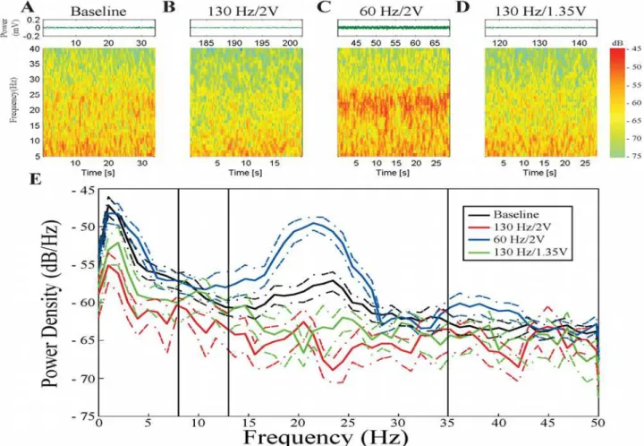
Fig. 1A-Ddisplays the raw data and time frequency spectrograms from a representative STN,
when the subject was in the resting state without DBS (Fig. 1A) and during different DBS ep-ochs (Fig. 1B-D). Visual inspection demonstrated that baseline alpha/beta band spectral power was attenuated during 130 Hz DBS but amplified during 60 Hz/2V DBS.
The PSD in this example (Fig. 1E) confirmed this observation. In this subject during rest,
there was increased LFP power at baseline in a sub-band (~17–26 Hz) within the beta band.
During 130 Hz DBS at 1.35V, the LFP power in this frequency sub-band was attenuated, whereas during 130 Hz DBS at 2V, baseline LFP power was attenuated across a wider
frequen-cy range, from 8–35 Hz. In contrast, LFP power was not attenuated during 60 Hz/2V DBS.
In-stead, there was amplification of LFP power within the sub-band (~17–26 Hz). The effect of 60
Hz/2V DBS opposed that of the power-equivalent DBS set (130 Hz/1.35V).
The group analysis revealed significant attenuation of baseline LFP power in both the alpha
and beta bands during 130 Hz/2V DBS (both P<0.001) (Table 1). There was significantly
Fig 1. Example STN during the different stimulation sets. A,B,C, andDare raw waveforms and spectrograms from the baseline epoch, 130 Hz/2V DBS epoch, 60 Hz/2V DBS epoch, and 130 Hz/1.35V DBS epoch, respectively. Spectrograms are displayed with 99% window overlap.E: Relative PSD traces of the four epochs. Black = baseline; red = 130 Hz/2V; blue = 60 Hz/2V; green = 130 Hz/1.35V. Colored dashed lines represent 95% confidence intervals for each spectrum.
doi:10.1371/journal.pone.0121067.g001
Table 1. Change (Δ) in Mean Power with Respect to Baseline.
DBS Set (number of sides) Alpha (8–12 Hz) Beta (13–35 Hz)
130 Hz/2V Δ= -0.265 Δ= -0.431
(N = 14) #P<0.001 #P<0.001
130 Hz/1.35V Δ= -0.0685 Δ= -0.155
(N = 14) #P = 1 #P = 0.193
60 Hz/2V Δ= + 0.182 Δ= + 0.0932
(N = 14) "P<0.05 "P = 1
The total power delivered during 60 Hz/2V DBS was equivalent to that delivered during 130 Hz/1.35V DBS. However, their effects on baseline spectral power were significantly different from each other in both the alpha (P<0.001) and beta (P = 0.006) bands.
Patient-specific sub-bands of baseline LFP power are amplified during
60 Hz DBS
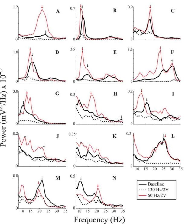
Fig. 2demonstrates the resting state PSDs of LFP power from the DBS lead (across electrodes
0–2) from all fourteen sides at baseline, during 130 Hz/2V DBS, and during 60 Hz/2V DBS.
Baseline LFP power was attenuated in at least one sub-band of the alpha/beta band during 130 Hz/2V DBS in twelve out of the fourteen cases (Fig. 2). This was determined based on
Fig 2. Peak detection for all fourteen sides. A—Nare absolute PSDs from all fourteen recordings. Black = baseline; red = 60 Hz/2V; dashed = 130 Hz/2V. Arrows indicate where peaks were detected by the algorithm, and the color of an arrow corresponds to its PSD spectrum. Black asterisks onJ(one for 60 Hz/ 2V) andK(two, one for 60 Hz/2V and one for baseline) indicate peaks chosen visually.
separation of the confidence intervals of the respective spectra, for which the upper confidence limit of the 130 Hz/2V DBS power spectrum was lower than the lower confidence limit of the baseline spectrum. In contrast, ten out of fourteen sides showed amplification of one or more sub-bands of the baseline alpha/beta band based on a similar analysis of separation of the lower confidence limit of the 60 Hz/2V DBS power spectrum and the upper confidence limit of the baseline spectrum. The effect of 60 Hz DBS varied among subjects: in three cases (Fig. 2F, J, and K), 60 Hz/2V DBS appeared to amplify certain sub-bands that were barely appreciated at baseline; and in three other cases (Fig. 2F, M, and N), similar sub-bands of LFP power were evi-dent at baseline and during 60 Hz/2V DBS; however, the relative power switched between base-line and 60 Hz/2V DBS such that a different sub-band had the greatest power. In certain cases (Fig. 2F, I, and J), there appeared to be a downward shift of LFP power during 60 Hz/2V DBS that may have contributed to the significant amplification of LFP power within the alpha band demonstrated in the group band analyses (Table 1).
To quantify whether the peaks of the sub-bands of neural synchrony were greater during 60 Hz/2V DBS than at baseline, we focused on the peak with the maximum power in either state for each side. A conservative peak detection algorithm detected a peak during baseline and
dur-ing 60 Hz/2V for twelve out of fourteen sides (arrows,Fig. 2). For the other two sides
(Fig. 2J-K), maximum power peaks were chosen visually (black asterisks). Including these sides in the analysis, the maximum peak power was significantly greater during 60 Hz/2V DBS than at baseline (P = 0.007).
Discussion
Low frequency DBS increases STN neural synchrony
To our knowledge, this is the first study to investigate the effects of 60 Hz DBS on resting state
neural synchrony in the STN in people with Parkinson’s disease and to compare such effects to
that of high frequency (130 Hz) DBS in the same STNs.
During high frequency (130 Hz) DBS at 2 Volts, there was significant attenuation of both alpha and beta band LFP power, consistent with previous reports [7,10,18,34,35]. The effect of 130 Hz DBS at 1.35V was significantly different from that at 2V. We have previously shown
that randomized presentations of different voltages of high frequency DBS produced a“dose”
dependent attenuation of baseline resting state neural synchrony in the STN [18].
In contrast, lower frequency (60 Hz) DBS at 2 Volts did not attenuate STN alpha/beta band
synchrony. In fact, there was a significant increase in alpha band (8–12 Hz) LFP power in the
STN region during 60 Hz/2V DBS. The effect of 60 Hz DBS on STN neural synchrony did not appear to be due to lower total power delivered, as its effect opposed that of the power-equivalent DBS setting at 130 Hz (130 Hz/1.35V). This demonstrated that the effects of 60 Hz/ 2V DBS were most likely frequency-specific. These observed differences between 60 Hz DBS and 130 Hz DBS support the theory that these two frequencies have different effects on under-lying neural circuitry.
do not believe that the heterogeneic effects of 60 Hz DBS on the STNs were due to different DBS lead locations; there was similarity among the effects on each STN within an individual (Fig. 2M-N) that were different from the effect on the two STNs of a different individual (Fig. 2A and L). We have previously shown that the baseline resting state LFP spectral peaks are similar and coherent between bilateral STNs of an individual but vary among PD subjects, and it was interesting that the effects of 60 Hz DBS also appeared to be patient-specific [28]. All the PD subjects have shown clinical improvement from STN DBS (data not shown), sug-gesting that the DBS lead was placed in or close to the sensorimotor region in the STN. The phenotype of PD subjects also varies so it is possible that this may contribute to different baseline LFP spectral profiles as well as the different effects of STN DBS. We have addressed this in a larger cohort of PD subjects but were not statistically powered to address this in the current group [32].
Mechanisms of high frequency DBS
The STN is a critical node within the cortico-basal ganglia-thalamo-cortical network: it is
inter-connected with sensorimotor, associative, and limbic sub-/cortical structures [50–53]. This
net-work consists of many nested loops whose oscillatory periods depend on the length of time it takes a signal to propagate around any given loop [54,55].
One of the proposed mechanisms of high frequency STN DBS is that the inter-pulse interval of the pulse train is similar to the period of certain nested loops within the network [21]. Thus, high frequency STN DBS may result in resonance within the sensorimotor network that over-rides the pathological lower frequency synchrony in PD [21,22,24,56,57].
Another theory proposes that high frequency STN DBS directly reduces the imposition of pathological alpha/beta band synchrony on the STN by its action on the cortex. Pathological neural synchrony in the alpha/beta band in the sensorimotor region of the STN in PD may be locally derived, or it may be imposed from cortical regions either via striatal-pallidal-STN afferents due to the direct effect of dopamine depletion in the striatum or via the
cortico-STN hyperdirect pathway (HDP) [18,19,58–62]. Experimental evidence using optogenetics
demonstrated that activation of the cortico-STN HDP, either at its efferent projection site or via the HDP itself, replicated the behavioral effects of electrical STN DBS [63]. Recent theoreti-cal models have predicted that STN DBS antidromic activation of the cortico-STN HDP would modulate cortical firing, leading to a disabling of excessive beta synchrony imposed by the cor-tex on the STN [64]. High frequency STN DBS thus may attenuate STN alpha/beta synchrony by antidromically inhibiting the cortical source(s) of these pathological rhythms.
Proposed mechanism of sixty hertz DBS
It has been suggested that low frequency neurostimulation might have an inter-pulse interval that is too long to suppress intrinsic pathological and bursting activity within the STN. The stimulating spike train may combine with intrinsic pathological firing patterns to cause addi-tional disruption of neural activity [21,24].
Our results suggest an alternative mechanism for 60 Hz DBS. We observed that in most cases, the effect of 60 Hz DBS was to amplify the already identified sub-bands of resting state
neural synchrony, rather than to increase LFP power overall across the 8–35 Hz range. In some
Neurostimulation as an investigative tool for individuals and for individual
behaviors in PD
Clinical evidence suggests that 60 Hz DBS may have a beneficial effect on axial motor
behav-iors, such as freezing of gait and speech, but not on tremor [36–41]. In contrast, high frequency
DBS appears to be therapeutic for tremor but not always for freezing of gait or speech. Howev-er, results have varied among studies and among individuals. The results of this investigation demonstrated heterogeneity among STNs regarding the effect of 60 Hz DBS on baseline neural synchrony, and it is possible that this may contribute to the heterogenic clinical effects of 60 Hz DBS in PD reported so far. We have shown previously that the resting state STN spectral profile is stationary but varies among subjects [7,28,32]. We propose that one explanation for whether high or low frequency DBS is or is not therapeutic for different motor behaviors may lie in the resonant frequency of the nested loops pertinent to the networks mediating such be-haviors. These may vary across the population, and may also be affected by amplitude- or frequency-modulation of interconnecting circuits. We demonstrate here that neurostimulation
itself can be a powerful tool, whereby it is possible to“knock-in”and“knock-out”
patient-specific bands of neural synchrony to determine their causal influences on human behaviors.
Conclusion
This is the first study to investigate the effects of 60 Hz DBS on neural synchrony in the STN in
people with Parkinson’s disease. Intra-operative 60 Hz DBS amplified baseline neural
synchro-ny in contrast to the attenuation seen during high frequency (130 Hz) DBS. The effect on neu-ral synchrony of 60 Hz/2V DBS opposed that of the total power-equivalent stimulation (130 Hz/1.35V DBS), suggesting that the effect is frequency- (not power-) dependent, and that high and low frequencies of DBS may resonate with different nested loops within the cortico-subcortical network. It may now be possible to selectively modulate individualized pathological
“brain arrhythmias”in PD and other neuropsychiatric diseases to investigate their influence on
specific motor and non-motor behaviors.
Acknowledgments
We would like to thank Megan H. Trager for helpful comments on the manuscript.
Author Contributions
Conceived and designed the experiments: AV MMK BCH HBS. Performed the experiments: ZB AV BCH LAS EQ CK HY JMH HBS. Analyzed the data: ZB AV MMK BCH LAS EQ HBS. Contributed reagents/materials/analysis tools: ZB AV MMK BCH CK HY JMH HBS. Wrote the paper: ZB HBS.
References
1. The Deep Brain Stimulation for Parkinson’s Disease Study Group. Deep-brain stimulation of the sub-thalamic nucleus or the pars interna of the globus pallidus in Parkinson’s disease, 2001. New Engl J Med. 2001; 345: 956–963. PMID:11575287
2. Deuschl G, Schade-Brittinger C, Krack P, Volkmann J, Schäfer H, Bötzel K, et al. A randomized trial of deep-brain stimulation for Parkinson's disease. New Engl J Med. 2006; 355: 896–908. PMID: 16943402
3. Krack P, Batir A, Van Blercom N, Chabardes S, Fraix V, Ardouin C, et al. Five-year follow-up of bilateral stimulation of the subthalamic nucleus in advanced Parkinson’s disease. New Engl J Med. 2003; 349: 1925–1934. PMID:14614167
5. Limousin P, Krack P, Pollak P, Benazzouz A, Ardouin C, Hoffmann D, et al. Electrical stimulation of the subthalamic nucleus in advanced Parkinson’s disease. New Engl J Med. 1998; 339: 1105–1111. PMID:9770557
6. Rodriguez-Oroz MC, Moro E, Krack P. Long-term outcomes of surgical therapies for Parkinson’s dis-ease. Mov Disord. 2012; 27: 1718–1728. doi:10.1002/mds.25214PMID:23208668
7. Brontë-Stewart H, Barberini C, Koop MM, Hill BC, Henderson JM, Wingeier B. The STN beta-band pro-file in Parkinson's disease is stationary and shows prolonged attenuation after deep brain stimulation. Exp Neurol. 2009; 215: 20–28. doi:10.1016/j.expneurol.2008.09.008PMID:18929561
8. Brown P, Oliviero A, Mazzone P, Insola A, Tonali P, Di Lazzaro V. Dopamine dependency of oscilla-tions between subthalamic nucleus and pallidum in Parkinson’s disease. J Neurosci. 2001; 21: 1033–1038. PMID:11157088
9. Cassidy M, Mazzone P, Oliviero A, Insola A, Tonali P, et al. Movement-related changes in synchroniza-tion in the human basal ganglia. Brain. 2002; 125: 1235–1246. PMID:12023312
10. Eusebio A, Thevathasan W, Doyle Gaynor L, Pogosyan A, Bye E, et al. Deep brain stimulation can sup-press pathological synchronisation in parkinsonian patients. J Neurol Neurosurg Psychiatry. 2011; 82: 569–573. doi:10.1136/jnnp.2010.217489PMID:20935326
11. Kühn AA, Kempf F, Brücke C, Gaynor Doyle L, Martinez-Torres I, Foltynie T, et al. High-frequency stim-ulation of the subthalamic nucleus suppresses oscillatory beta activity in patients with Parkinson's dis-ease in parallel with improvement in motor performance. J Neurosci. 2008; 28: 6165–6173. doi:10. 1523/JNEUROSCI.0282-08.2008PMID:18550758
12. Kühn AA, Tsui A, Aziz T, Ray N, Brücke C, Kupsch A, et al. Pathological synchronisation in the subtha-lamic nucleus of patients with Parkinson's disease relates to both bradykinesia and rigidity. Exp Neurol. 2009; 215: 380–387. doi:10.1016/j.expneurol.2008.11.008PMID:19070616
13. Lalo E, Thobois S, Sharott A, Polo G, Mertens P, Pogosyan A, et al. Patterns of bidirectional communi-cation between cortex and basal ganglia during movement in patients with Parkinson disease. J Neu-rosci. 2008; 28: 3008–3016. doi:10.1523/JNEUROSCI.5295-07.2008PMID:18354004
14. Levy R, Ashby P, Hutchison WD, Lang AE, Lozano AM, Dostrovsky JO. Dependence of subthalamic nucleus oscillations on movement and dopamine in Parkinson's disease. Brain. 2002; 125: 1196–1209. PMID:12023310
15. Priori A, Foffani G, Pesenti A, Tamma F, Bianchi AM, Pellegrini M, et al. Rhythm-specific pharmacologi-cal modulation of subthalamic activity in Parkinson's disease. Exp Neurol. 2004; 189: 369–379. PMID: 15380487
16. Ray NJ, Jenkinson N, Wang S, Holland P, Brittain JS, Joint C, et al. Local field potential beta activity in the subthalamic nucleus of patients with Parkinson's disease is associated with improvements in brady-kinesia after dopamine and deep brain stimulation. Exp Neurol. 2008; 213: 108–113. doi:10.1016/j. expneurol.2008.05.008PMID:18619592
17. Weinberger M, Mahant N, Hutchison WD, Lozano AM, Moro E, Hodaie M, et al. Beta oscillatory activity in the subthalamic nucleus and its relation to dopaminergic response in Parkinson's disease. J Neuro-physiol. 2006; 96: 3248–3256. PMID:17005611
18. Whitmer D, de Solages C, Hill B, Yu H, Henderson JM, Bronte-Stewart H. High frequency deep brain stimulation attenuates subthalamic and cortical rhythms in Parkinson’s disease. Front Hum Neurosci. 2012; 6: 1–18. doi:10.3389/fnhum.2012.00001PMID:22279433
19. Williams D, Tijssen M, van Bruggen G, Bosch A, Insola A, Di Lazzaro V, et al. Dopamine-dependent changes in the functional connectivity between basal ganglia and cerebral cortex in humans. Brain. 2002; 125: 1558–1569. PMID:12077005
20. Wingeier B, Tcheng T, Koop MM, Hill BC, Heit G, Bronte-Stewart H. Intra-operative STN DBS attenu-ates the prominent beta rhythm in the STN in Parkinson’s disease. Exp Neurol. 2006; 197: 244–251. PMID:16289053
21. Birdno MJ, Grill WM. Mechanisms of deep brain stimulation in movement disorders as revealed by changes in stimulus frequency. Neurother. 2008; 5: 14–25.
22. McIntyre CC, Hahn PJ. Network perspectives on the mechanisms of deep brain stimulation. Neurobiol Dis. 2010; 38: 329–337. doi:10.1016/j.nbd.2009.09.022PMID:19804831
23. Anderson M, Postupna N, Ruffo M. Effects of high-frequency stimulation in the internal globus pallidus on the activity of thalamic neurons in the awake monkey. J Neurophysiol. 2003; 89: 1150–1160. PMID: 12574488
24. Grill WM, Snyder AN, Miocinovic S. Deep brain stimulation creates an informational lesion of the stimu-lated nucleus. Neuroreport. 2004; 15: 1137–1140. PMID:15129161
26. McIntyre CC, Grill WM, Sherman DL, Thakor NV. Cellular effects of deep brain stimulation: model-based analysis of activation and inhibition. J Neurophysiol. 2004; 91: 1457–1469. PMID:14668299 27. Alonso-Frech F, Zamarbide I, Alegre M, Rodríguez-Oroz MC, Guridi J, Manrique M, et al. Slow
oscil-latory activity and levodopa-induced dyskinesias in Parkinson's disease. Brain. 2006; 129: 1748–1757. PMID:16684788
28. de Solages C, Hill BC, Koop MM, Henderson JM, Brontë-Stewart H. Bilateral symmetry and coherence of subthalamic nuclei beta band activity in Parkinson's disease. Exp Neurol. 2010; 221: 260–266. doi: 10.1016/j.expneurol.2009.11.012PMID:19944098
29. Little S, Pogosyan A, Kuhn AA, Brown P. Beta band stability over time correlates with Parkinsonian ri-gidity and bradykinesia. Exp Neurol. 2012; 236: 383–388. doi:10.1016/j.expneurol.2012.04.024PMID: 22572590
30. Marsden JF, Limousin-Dowsey P, Ashby P, Pollak P, Brown P. Subthalamic nucleus, sensorimotor cor-tex and muscle interrelationships in Parkinson’s disease. Brain. 2001; 124: 378–388. PMID:11157565 31. Moshel S, Shamir RR, Raz A, de Noriega FR, Eitan R, Bergman H, et al. Subthalamic nucleus
long-range synchronization—an independent hallmark of human Parkinson’s disease. Front Hum Neurosci. 2013; 7: 1–14. doi:10.3389/fnhum.2013.00001PMID:23355817
32. Shreve LA, Velisar A, Shanidze NM, Hill BC, Kilbane C, Henderson JM, et al. Incidence and modulation of resting state subthalamic nucleus beta rhythm in Parkinson’s disease. Soc Neurosci Abstr 2013. 33. Marceglia S, Rossi L, Foffani G, Bianchi A, Cerutti S, Priori A. Basal ganglia local field potentials:
appli-cations in the development of new deep brain stimulation devices for movement disorders. Expert Rev Med Devices. 2007; 4: 605–614. PMID:17850195
34. Rosa M, Giannicola G, Servello D, Marceglia S, Pacchetti C, Porta M, et al. Subthalamic local field beta oscillations during ongoing deep brain stimulation in Parkinson's disease in hyperacute and chronic phases. Neurosignals. 2011; 19: 151–162. doi:10.1159/000328508PMID:21757872
35. Giannicola G, Rosa M, Servello D, Menghetti C, Carrabba G, Pacchetti C, et al. Subthalamic local field potentials after seven-year deep brain stimulation in Parkinson’s disease. Exp Neurol. 2012; 237: 312–317. doi:10.1016/j.expneurol.2012.06.012PMID:22735488
36. Brozova H, Barnaure I, Alterman RL, Tagliati M. STN-DBS frequency effects on freezing of gait in advanced Parkinson disease. Neurol. 2009; 72: 770–771. doi:10.1016/j.surneu.2009.04.011PMID:19604547 37. Khoo HM, Kishima H, Hosomi K, Maruo T, Tani N, Oshino S, et al. Low-frequency subthalamic nucleus
stimulation in Parkinson’s disease: a randomized clinical trial. Mov Disord. 2014; 29: 270–274. doi:10. 1002/mds.25810PMID:24449169
38. Moreau C, Defebvre L, Destée A, Bleuse S, Clement F, Blatt JL, et al. STN-DBS frequency effects on freezing of gait in advanced Parkinson disease. Neurol. 2008; 71: 81–84.
39. Moreau C, Pennel-Ployart O, Pinto S, Plachez A, Annic A, Viallet F, et al. Modulation of dysarthropneu-mophonia by low-frequency STN DBS in advanced Parkinson’s disease. Mov Disord. 2011; 26: 659–663. doi:10.1002/mds.23538PMID:21506146
40. Sidiropoulos C, Walsh R, Meaney C, Poon YY, Fallis M, Moro E. Low-frequency subthalamic nucleus deep brain stimulation for axial symptoms in advanced Parkinson’s disease. J Neurol. 2013; 260: 2306–2311. doi:10.1007/s00415-013-6983-2PMID:23749331
41. Stegemöller EL, Vallabhajosula S, Haq I, Hwynn N, Hass CJ, Okun MS. Selective use of low frequency stimulation in Parkinson’s disease based on absence of tremor. NeuroRehab. 2013; 33: 305–312. 42. Taylor Tavares AL, Jefferis GSXE, Koop M, Hill BC, Hastie T, Heit G, et al. Quantitative measurements
of alternating finger tapping in Parkinson’s disease correlate with UPDRS motor disability and reveal the improvement in fine motor control from medication and deep brain stimulation. Mov Disord. 2005; 20: 1286–1298. PMID:16001401
43. Koop MM, Andrzejewski A, Hill BC, Heit G, Brontë-Stewart HM. Improvement in a quantitative measure of bradykinesia after microelectrode recording in patients with Parkinson’s disease during deep brain stimulation surgery. Mov Disord. 2006; 21: 673–678. PMID:16440333
44. Brontë-Stewart H, Louie S, Batya S, Henderson JM. Clinical motor outcome of bilateral subthalamic nu-cleus deep-brain stimulation for Parkinson’s disease using image-guided frameless stereotaxy. Neuro-surg. 2010; 67: 1088–1093.
45. Koss AM, Alterman RL, Tagliati M, Shils JL. Calculating total electrical energy delivered by deep brain stimulation systems. Ann Neurol. 2005; 58: 168. PMID:15984018
46. Brontë-Stewart H, Shreve LA, Hill BC, Yu H, Henderson JM, Velisar A. Sixty hertz deep brain stimulation does not attenuate subthalamic nucleus beta rhythm in Parkinson’s disease. Soc Neurosci Abstr 2013. 47. Miller Koop M, Brontë-Stewart H. Spectral distributions of STN local field potential in the beta band are
48. Velisar A, Shreve LA, Hill BC, Yu H, Henderson JM. Resolution of rest tremor reveals underlying sub-thalamic nucleus beta band synchrony in Parkinson’s disease. Soc Neurosci Abstr 2013.
49. Welch PD. The use of fast fourier transform for the estimation of power spectra: a method based on time averaging over short, modified periodograms. IEEE Trans Audio Electroacoust. 1967; 15: 70–73. 50. Haber SN, Lynd-Balta E, Mitchell SJ. The organization of the descending ventral pallidal projections in
the monkey. J Comp Neurol. 1993; 329: 111–128. PMID:8454722
51. Haynes WIA, Haber SN. The organization of prefrontal-subthalamic inputs in primates provides an ana-tomical substrate for both functional specificity and integration: implications for basal ganglia models and deep brain stimulation. J Neurosci. 2013; 33: 4804–4814. doi:10.1523/JNEUROSCI.4674-12. 2013PMID:23486951
52. Mallet L, Schüpbach M, N'Diaye K, Remy P, Bardinet E, Czernecki V, et al. Stimulation of subterritories of the subthalamic nucleus reveals its role in the integration of the emotional and motor aspects of be-havior. Proc Natl Acad Sci USA. 2007; 104: 10661–10666. PMID:17556546
53. Nambu A, Takada M, Inase M, Tokuno H. Dual somatotopical representations in the primate subthala-mic nucleus: evidence for ordered but reversed body-map transformations from the primary motor cor-tex and the supplementary motor area. J Neurosci. 1996; 16: 2671–2683. PMID:8786443
54. Alexander GE, Crutcher MD. Functional architecture of basal ganglia circuits: neural substrates of par-allel processing. Trends Neurosci. 1990; 13: 266–271. PMID:1695401
55. Parent A, Hazrati LN. Functional anatomy of the basal ganglia. I. The cortico-basal ganglia-thalamo-cortical loop. Brain Res Rev. 1995; 20: 91–127. PMID:7711769
56. Montgomery EB. Basal ganglia physiology and pathophysiology: a reappraisal. Parkinsonism Relat Disord. 2007; 13: 455–465. PMID:17977052
57. Montgomery EB, Gale JT. Mechanisms of action of deep brain stimulation (DBS). Neurosci Biobehav Rev. 2008; 32: 388–407. PMID:17706780
58. Brown P. Abnormal oscillatory synchronisation in the motor system leads to impaired movement. Curr Opin Neurobiol. 2007; 17: 656–664. doi:10.1016/j.conb.2007.12.001PMID:18221864
59. Costa RM, Lin SC, Sotnikova TD, Cyr M, Gainetdinov RR, Caron MG, et al. Rapid alterations in corti-costriatal ensemble coordination during acute dopamine-dependent motor dysfunction. Neuron. 2006; 52: 359–369. PMID:17046697
60. Courtemanche R, Fujii N, Graybiel AM. Synchronous, focally modulated beta-band oscillations charac-terize local field potential activity in the striatum of awake behaving monkeys. J Neurosci. 2003; 23: 11741–11752. PMID:14684876
61. Gatev P, Wichmann T. Interactions between cortical rhythms and spiking activity of single basal ganglia neurons in the normal and parkinsonian state. Cereb Cortex. 2009; 19: 1330–1344. doi:10.1093/ cercor/bhn171PMID:18842667
62. Magill PJ, Bolam JP, Bevan MD. Dopamine regulates the impact of the cerebral cortex on the subthala-mic nucleus-globus pallidus network. Neuroscience. 2001; 106: 313–330. PMID:11566503
63. Gradinaru V, Mogri M, Thompson KR, Henderson JM, Deisseroth K. Optical deconstruction of parkin-sonian neural circuitry. Science. 2009; 324: 354–359. doi:10.1126/science.1167093PMID:19299587 64. Kang G, Lowery MM. Effects of antidromic and orthodromic activation of STN afferent axons during