Early Priming Minimizes the Age-Related Immune
Compromise of CD8
+
T Cell Diversity and Function
Sophie A. Valkenburg1, Vanessa Venturi2, Thurston H. Y. Dang2, Nicola L. Bird1, Peter C. Doherty1,3, Stephen J. Turner1, Miles P. Davenport4, Katherine Kedzierska1*
1Department of Microbiology and Immunology, University of Melbourne, Parkville, Melbourne, Australia,2Computational Biology Group St Jude Children’s Research Hospital, Memphis, Tennessee, United States of America,3Department of Immunology, St Jude Children’s Research Hospital, Memphis, Tennessee, United States of America,4Complex Systems in Biology Group, Centre for Vascular Research, University of New South Wales, Kensington, Australia
Abstract
The elderly are particularly susceptible to influenza A virus infections, with increased occurrence, disease severity and reduced vaccine efficacy attributed to declining immunity. Experimentally, the age-dependent decline in influenza-specific CD8+T cell responsiveness reflects both functional compromise and the emergence of ‘repertoire holes’ arising from the
loss of low frequency clonotypes. In this study, we asked whether early priming limits the time-related attrition of immune competence. Though primary responses in aged mice were compromised, animals vaccinated at 6 weeks then challenged .20 months later had T-cell responses that were normal in magnitude. Both functional quality and the persistence of ‘preferred’ TCR clonotypes that expand in a characteristic immunodominance hierarchy were maintained following early priming. Similar to the early priming, vaccination at 22 months followed by challenge retained a response magnitude equivalent to young mice. However, late priming resulted in reduced TCRbdiversity in comparison with vaccination earlier in life. Thus, early priming was critical to maintaining individual and population-wide TCRb diversity. In summary, early exposure leads to the long-term maintenance of memory T cells and thus preserves optimal, influenza-specific CD8+T-cell
responsiveness and protects against the age-related attrition of naı¨ve T-cell precursors. Our study supports development of vaccines that prime CD8+T-cells early in life to elicit the broadest possible spectrum of CD8+T-cell memory and preserve
the magnitude, functionality and TCR usage of responding populations. In addition, our study provides the most comprehensive analysis of the aged (primary, secondary primed-early and secondary primed-late) TCR repertoires published to date.
Citation:Valkenburg SA, Venturi V, Dang THY, Bird NL, Doherty PC, et al. (2012) Early Priming Minimizes the Age-Related Immune Compromise of CD8+
T Cell Diversity and Function. PLoS Pathog 8(2): e1002544. doi:10.1371/journal.ppat.1002544
Editor:E. John Wherry, University of Pennsylvania, United States of America
ReceivedAugust 7, 2011;AcceptedJanuary 7, 2012;PublishedFebruary 23, 2012
Copyright:ß2012 Valkenburg et al. This is an open-access article distributed under the terms of the Creative Commons Attribution License, which permits unrestricted use, distribution, and reproduction in any medium, provided the original author and source are credited.
Funding:This work was supported by Australian National Health and Medical Research Council (NHMRC) Project Grants to KK (AI454312; AI1008854), an NHMRC Program Grant (APP567122) to PCD and SJT, an ARC Project Grant to MPD, SJT, and VV (DP0771340) and NIH grant (AI170251) to PCD. SAV is a recipient of the Australian Postgraduate Award, KK is an NHMRC RD Wright Fellow, SJT is a Pfizer Australia Senior Research Fellow, VV is an ARC Future Fellow and MPD is an NHMRC Senior Research Fellow. The funders had no role in study design, data collection and analysis, decision to publish, or preparation of the manuscript.
Competing Interests:The authors have declared that no competing interests exist. * E-mail: [email protected]
Introduction
The elderly population is particularly susceptible to novel infections, especially the annual, seasonal epidemics caused by influenza A viruses [1,2], with increased occurrence, severity of infection and reduced vaccine efficacy being attributed to age-related decline in immune capacity [3–6]. The ageing effect on the immune system is considered to be multifactorial, arising from the diminished thymic export of naı¨ve precursors due to thymic involution [7,8], the impaired recruitment [9,10] of naı¨ve CD8+
T cell precursors and the replicative senescence of memory cells [11–14]. Ageing can also be associated with abnormal cellular functions such as distorted cytokine secretion (IL-2, IL-4 and IFN-c) profiles [15–17], decreased granzyme B production [18,19] and reduced proliferative capacity due to the loss of CD28 expression [20]. Perturbations in the naı¨ve TCR repertoire have also been reported, with abnormal TCR spectratype (CDR3b length) patterns in aged mice reflecting the massive, antigen-independent expansion, of a few clonotypes [21]. Naı¨ve T cell attrition has also been inferred from observed reductions in
the diversity of antigen-specific TCR repertoires in aged mice [5,22].
Previous mouse studies have established that ageing can be associated with diminished CD8+
T cell efficacy and delayed influenza virus clearance [23–25]. Recent evidence has further shown that the selective loss of primary, influenza-specific CD8+T
cell responsiveness in older mice is characterized by a narrowing in the spectrum of TCR usage and is seen predominantly for low frequency populations, with this effect being best characterized for the prominent DbNP366+CD8+ T cell set [5,26]. Overall, the
findings so far suggest that the capacity to respond effectively to new influenza infections in aged mice requires the maintenance of a diverse pool of functional peripheral T cells.
As CD8+
T cells tend to be specific for peptides derived from more conserved proteins that are internal to the virus, priming effective CD8+T cell memory has obvious potential for countering
newly emerged seasonal or pandemic influenza strains. The importance of long-lived, antigen-specific memory CD8+
humans [29,30]. Such long-term maintenance of memory T cells leading to enhanced secondary response forms the basis for vaccination strategies based on priming CD8+
T cell memory to promote early virus clearance and decreased morbidity. The question is though, whether such CD8+ T cell memory can be
effectively recalled in the elderly.
A recent study [6] suggested that infecting mice with LCMV or influenza at an extreme age (18–20 months) leads to defective CD8+T cell memory and diminished recall responses following
virus challenge. What happens, though, if CD8+
T cell memory is established when the mice are young? The analysis reported here compares the CD8+
T cell response profiles for young (,3 months) and aged (22 month) mice, with the latter cohort being first exposed to immunogenic influenza epitopes early or late in life. The results suggest that designing influenza vaccines which promote as broad as possible spectrum of CD8+T cell memory in
adolescence could be beneficial, even if such benefit emerges long after the subject has first been given the protective immunogen.
Results
To validate the previous studies [5,31] and determine the ageing effect on primary immune responsiveness (Figure 1A) for immunodominant DbNP366+ and DbPA224+ CD8+ pools, we
infected young (,3 month) and old (.22 month) mice intranasally (i.n.) with 16104pfu of an infectious (H3N2, HK) influenza A virus. More importantly, as a main question of the present study, we asked whether any age-related compromise of CD8+
T cell function and diversity might be modified by priming early (at 2 months) or late (at 22 months) i.p. with 1.56107pfu of the
serologically distinct PR8 (H1N1) virus that has the same immunogenic CD8+
T cell peptides as HK.
We used the i.p. priming route with the influenza virus as it does not lead to a productive viral replication (similarly to the current i.m. human influenza vaccines), but gives one-stop growth cycle with full protein production. Such non-productive immunisation with the whole virus results in priming of antigen-specific effector T cells and establishment of long-term T cell memory for subsequent challenge (Figure S1), comparable to those observed after the natural (i.n.) influenza infection [32–34]. Importantly, the i.p. priming does not elicit the whole cascade of detrimental inflammatory responses in the virally-infected lung [35] and thus avoids double pathology at the site of infection. The i.p. route of influenza priming is equivalent to the
current i.m. vaccination approaches used in humans with respect to the non-productive viral immunisation.
CD8+T cell responsiveness following early versus late infection of aged mice
The comparison of the HK-induced CD8+
T cell responses utilized young or old mice that were either immunologically naı¨ve (primary, 10; Figure 1A) or had been primed at 2 months of age with the PR8 virus and challenged 20 months later (secondary, 20; Figure 1B). Immunodominant and subdominant CD8+
T cell responses were measured in the spleen (Figure 1CD) by theex vivo
IFN-c ICS assay. Following 10 challenge, the size of the low precursor frequency DbNP366+CD8+set in the spleen (Figure 1C)
was markedly diminished in the aged animals relative to the young controls as previously observed [5,31].
Conversely, any age-related effects on CD8+ T cell numbers
were not significant for DbPA224 (Figure 1C). The unaffected
DbPA224+CD8+T cell responses are intriguing, as the naı¨ve CD8+
T cell frequencies [36] found for DbPA224–specific T cells in young
mice are significantly higher than those detected for DbNP366
($72 versus ,40 per individual, respectively), suggesting that a larger naı¨ve CD8+T cell pool size minimizes the extent of
age-related attrition and, as a consequence, the effect on primary CD8+
T cell response magnitude (Figure 1C).
Reduced magnitude of the immunodominant DbNP366-specific
CD8+
T cell response that was detected for the primary, influenza-specific CD8+T cell response in older mice (Figure 1CD), was not
sustained following secondary HK challenge of mice that had been primed early with the PR8 virus (Figure 1D). The numbers of DbPA224CD8+ T cells were significantly diminished across
combined experiments but, otherwise, the recall responses for memory T cell pools in young or old mice primed at,2 months (at least 20 months previously) were not obviously different, emphasizing the durability of virus-specific CD8+
T cell memory [37]. In particular, the overdominance of the DbNP366-specific set
that is characteristic of the secondary response to these viruses [38] was still apparent in the aged mice (Figure 1D).
The beneficial effect of the early CD8+ priming on the
immunodominant low-precursor responses like the DbNP366
-specific population following influenza infection at the extreme age was most striking when the relative contributions of particular antigen-specific CD8+T cells were analysed based on total cell
numbers (Figure 2, calculations based on Figure 1 for immuno-dominant DbNP366+CD8+ and DbPA224+CD8+ pools, and data
not shown for subdominant DbPB1703+CD8+ and K b
PB1-F262+CD8+populations). In the aged mice, the primary CD8+T
cell responses showed a shift in the typical immunodominance hierarchy (Figure 2B), with the contribution of the immunodomi-nant DbNP366+CD8+population being significantly lower in the
aged mice (9.463.6%) in comparison to young animals (43.4615%; p,0.01; Figure 2A). The differential immunodomi-nance hierarchy resulted mainly from significantly increased contribution of KbPB1703+CD8+ T cells (Figure 2). This led to
major modifications in response hierarchy following primary influenza virus infection of aged mice KbPB1703.DbPA224.
DbPB1-F262.DbNP366, with the comparable profile for young
mice being DbNP366.DbPA224= KbPB1703&DbPB1-F262.
Conversely, recall of CD8+
T cells primed at a young age preserved the overall contribution of T cell specificities and retained the immunodominance hierarchy in aged mice primed early at 6 weeks (Figure 2D), reflecting the characteristic immunodominance hierarchy in young controls (Figure 2C). These findings show clearly that priming the CD8+
T cell compartment at an early age leads to subsequent preservation of
Author Summary
CD8+
T cell numbers and immunodominance hierarchies for influenza infection in the elderly.
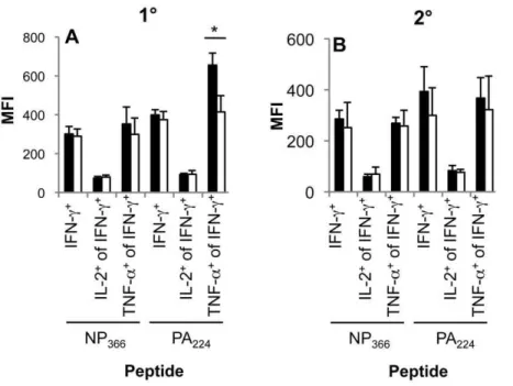
Age-related effects on cytokine polyfunctionality and activation status
One measure of CD8+T cell function is the capacity to produce
multiple cytokines simultaneously [39] followingin vitrostimulation with peptide in the standard, 5 h ICS assay. For the primary DbPA224+CD8+ T cell population that remained relatively
constant in numbers with age (Figure 1C), the frequencies of
double (IFN-c/TNF-a) and triple-producers (IFN-c/TNF-a/IL-2) were significantly lower in comparison with the young mice (Figure 3AB). Furthermore, taking mean fluorescence intensity (MFI), which represents the intensity and therefore amount of cytokine production, it also seems that the DbPA224+CD8+
population tended to produce less TNF-a, though this diminution effect was not apparent for either IFN-c or IL-2 (Figure 4A). Taking the prevalence and MFI data together (Figure 3 and Figure 4), there appears to be a general decrease in cytokine polyfunctionality for the primary DbPA224+CD8+response.
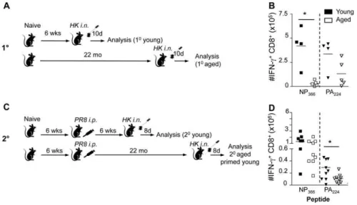
Figure 1. Effect of age and early priming on 10and 20CD8+T cell responses.(A) For the primary responses, naı¨ve mice were infected i.n.
with 16104pfu of the HK (H3N2) influenza A virus either at a young (,3 months; mo) or extreme (22 mo) age. Analysis of CD8+
T cell responses was performed on d10 after the primary infection. (B) For the secondary responses of the early-primed mice, animals were primed at,2 mo i.p. with 1.56107pfu the PR8 (H1N1) influenza A virus, then challenged 6 weeks (young) or.22 mo (aged) later i.n. with 16104pfu of the HK virus. Analysis of
CD8+
T cell responses was performed on d8 after the secondary infection. (C, D) Numbers of epitope-specific CD8+
T cells in the spleens recovered from young (filled symbols) or aged (.22 month, open symbols) B6 mice on d10 (10, C) or d8 (2o, D) following primary (10) or secondary (primed
young) (20) i.n. infection with the HK (H3N2) influenza A virus. Memory mice had been injected i.p. with the PR8 (H1N1) influenza A virus at ,2 mo and were challenged. Lymphocyte populations were stimulated with the NP366or PA224peptides in the presence of Brefeldin A for 5 hrs, then stained
with the anti-CD8PerCPCy5.5 mAb, fixed/permeabilised and stained with anti-IFN-c-FITC mAb. Cytokine (IFN-c) production was calculated by subtracting background fluorescence for the no-peptide controls, and the numbers of IFN-c+CD8+DbNP
366- and DbPA224-specifc CD8+T cells were
determined from the % cells staining and the total cell counts. Data represent individual mice (symbols) and the mean (line). Experiments were performed at least twice. * = p,0.05.
doi:10.1371/journal.ppat.1002544.g001
Figure 2. Immunodominance hierarchies in aged mice after 10infection or 20challenge of primed-early mice.The relative prevalence
of the immunodominant DbNP
366+CD8+and DbPA224+CD8+T cell population over the subdominant DbPB1703+CD8+and KbPB1-F262+CD8+sets.
Results are shown for (A, B) 10and (C, D) 20HK infection in (A, C) young and (B, D) aged mice. The relative contributions of particular antigen-specific
CD8+T cells were analysed based on total cell responses (Figure 1 for DbNP
366+CD8+and DbPA224+CD8+and data not shown for DbPB1703+CD8+and
KbPB1-F262+CD8+). Data represent the mean proportion of a particular peptide-specific CD8+population. * = p,0.01 shows a difference between
young and aged animals. Experimental outline as in Figure 1AB. doi:10.1371/journal.ppat.1002544.g002
Conversely, analysis of aged mice primed early showed that functional characteristics appear to be locked-in early and maintained in the long-term for memory T cell populations (Figure 3CDEF).
Can we detect other evidence of enduring functional change? Given that the influenza-specific CD8+T cells generated following
primary infection of aged mice were either of suboptimal functional quality (DbPA224+CD8+; Figure 3, Figure 4) or reduced
in number (DbNP366+CD8+; Figure 1), the further question was
whether there was any effect on cell surface activation phenotype [34,40–42]. Comparison of phenotypic markers associated with activation, trafficking and memory potential: CD62L vs. IL-7Ra
(CD127), CD27 vs. CD43, and IL-7Ra vs. KLRG-1 for the DbPA224+CD8+and DbNP366+CD8+sets (Figure S2) showed that,
with the exception of a decrease in the relative prevalence of the less activated CD27loCD43loCD8+ Db
PA224+ cells in the older
mice (Figure S2AD), there were no significant differences in phenotype with age.
Aged memory T cells have a young-type TCRbusage profile
Previous studies have found a significant skewing in TCR Vb
usage (mAb staining) and CDR3blength (spectratyping) for CD8+
T cell responses developed from naı¨ve and memory populations by
Figure 3. Cytokine polyfunctionality following 10or 20challenge.Epitope-specific CD8+T cells generated following 10(A, B) or 20(C, D) i.n
HK challenge (see legend to Figure 1) of young (black bar) and aged (white bar) mice were assessed for the simultaneous production of IFN-c, TNF-a
(A, C) and IL-2 (B, D) using the ICS assay. The % values (A–F) were compared for spleens from groups of 3–5 mice and representative dot plots are shown (E, F). * = p,0.05. Experimental outline as in Figure 1AB.
the infection of aged versus young mice [13,21,43]. Thus, we looked more closely at the expansion and maintenance of responding T cell clonotypes [44,45]. As our earlier analysis of influenza-specific CD8+
TCR clonotype diversity has focused on the prominent Vb8.3+
DbNP366+[44,46] and Vb7+DbPA224+sets
[47], we first assessed the VbmAb-staining profiles to determine whether these characteristic TCRs were also selected following primary or secondary challenge of aged mice. Indeed for both DbNP366+ CD8+ and D
b
PA224+CD8+ T cell responses, the
characteristic Vb8.3 and Vb7 usage was observed (Figure 5), though additional Vb6, Vb7 and Vb9 biases were variously detected in individual, older mice for the primary DbNP366+CD8+
population (Figure 5C), possibly due to the recruitment of low frequency alternate DbNP366-specific CD8+T cells. Despite the
presence of a prominent Vb8.1/8.2+Db
NP366+set in one of the
early-primed, secondarily-challenged at 22 month mice, the bias was generally to Vb8.3 suggesting that the characteristic DbNP366+CD8+TCRb usage profile is retained in the persistent
memory population. The DbPA224+set was characterised across
groups by Vb7 TCR usage (Figure 5BDF), which was more consistent than the DbNP366+Vb8.3 usage, possibly reflecting the
higher number of precursors with Vb7 surviving within the 22 month old mice.
Priming at an extreme age does not impair the recall response magnitude
Since priming at a young age led to the typical magnitude and quality of influenza-specific CD8+
T cell responses following viral infection in the aged mice, we asked whether priming the mice via a non-replicative route (i.p. priming with 1.56107pfu of PR8) at extreme age (22 months) would be also beneficial for the subsequent influenza virus infection. Since the reduced primary DbNP366+CD8+T cell responses in aged mice has been attributed
to the lower naı¨ve precursors in young mice [5], this experiment would determine whether old naive mice could be primed at an
extreme age (at 22 months) and subsequently challenged i.n. with 16104pfu of the HK influenza strain (at ,24 months; Figure 6A) to mount an effective recall response after the attrition had occurred. Surprisingly, despite the reduced primary DbNP366+CD8+T cell responses (Figure 1C) and lower magnitude
of secondary DbPA224+CD8+sets (Figure 1E) in the spleens of aged
animals, the recall of influenza-specific CD8+
T cells was robust and equivalent in magnitude to the young controls (Figure 6). The numbers of both immunodominant DbNP366+CD8+ and
DbPA224+CD8+ populations were normal (Figure 6B). This
resulted in the maintained contribution of each of the T cell specificities to influenza-specific responses (Figure 6E). Conversely, the polyfunctionality of those secondary CD8+
T cell populations in mice primed at the extreme age did not always resemble effectiveness of influenza-specific CD8+T cells recruited in young
individuals (Figure 6C). Perturbations in the TCR usage with extreme age were evident macroscopically in the TCR Vbusage for DbPA224+CD8+(Figure 6G) and especially the DbNP366+CD8+
(Figure 6F) responses, with the usage of alternate Vb8.1/8.2 for DbPA224+CD8+, and Vb7 and Vb8.1/8.2 for DbNP366+CD8+
populations. The characteristic Vb8.3 usage for DbNP366+CD8+
was only dominant in 1 of 4 mice (Figure 6F), reflecting narrowing of the naı¨ve DbNP366+CD8+set with extreme age that initially
limited the primary response (Figure 1C) and/or the clonal expansions characteristic for the aged animals as previously reported [13,14].
Early but not late priming preserves TCRb usage of ‘preferred’ clonotypes in the aged mice
A substantial body of work from previous studies has defined the young B6 CDR3bTCR usage at high resolution [44,47], therefore using these data sets from young mice we were able to compare the spectrum of clonotype prevalence in aged mice using single-cell RT-PCR and sequencing of the CDR3bregion to determine the spectrum of TCRb diversity. Analysis of 1489 CDR3b
Figure 4. Impaired polyfunctionality of DbPA224-specific CD8+T cells in the aged mice during primary but not secondary influenza infection.(A) Primary or (B) secondary (primed young) influenza-specific CD8+
T cell responses were assessed for simultaneous production of IFN-c, TNF-aand IL-2 in the spleen of aged (22 months old) and young (6–8 weeks) mice. Compiled data (n = 3–5, mean6SD) are shown for the mean fluorescence intensity (MFI) of IFN-c, IFN-cand TNF-aas well as IFN-cand IL-2 staining. * = p,0.05. Experimental outline as in Figure 1AB. doi:10.1371/journal.ppat.1002544.g004
sequences for primary and secondary (young and primed-old) responses from the 22 month old mice (Tables 1 and 2) showed that the dominant Jbregions and CDR3bloop lengths in the aged animals (Tables S1, S2, S3, S4, S5, S6) were comparable to those found early in life (Figures 7 and 8 for comparison with young animals). However, more inter-individual variation in the primary responses was observed in the older group (Figure S3). While .83% of each of the TCRb repertoires involved in the primary responses to DbNP366in young mice utilized Jb2.2 and a
CDR3 length of 9 amino acids (aa), this profile was substantially diminished to,57% of the TCRbrepertoire for 2/7 aged mice. Similarly, Jb1.1, Jb1.5, and Jb2.6 collectively dominated the primary DbPA224+CD8+ responses for 7/7 young mice, while
Jb2.1 and Jb2.3 emerged strongly (.55% each) for 2 of the older mice. While the primary DbPA224+CD8+repertoires in individual
young mice mostly featured diverse CDR3 lengths of 5, 6, and 7 aa,.94% of the primary DbPA224+CD8+T cell repertoires in two
of the aged mice could be attributed to one particular CDR3 length (i.e. 6 aa in one mouse and 7 aa in the other mouse).
Age-associated changes in TCRb repertoire usage were investigated for the DbNP366+CD8+and DbPA224+CD8+
popula-tions by sequencing individual CDR3bTCR signatures (Tables 1 and 2, Tables S1, S2, S3, S4, S5, S6) and the extent of TCRb
repertoire diversity was then assessed using both the number of different aa-defined clonotypes and Simpson’s diversity index, which accounts for the clonal dominance hierarchy. These measures of diversity were estimated for a standard 22 TCRb
sequences per epitope per mouse to adjust for differences in total number of sequences obtained per mouse [48]. The primary DbPA224+CD8+TCRaˆ repertoires were found to be significantly
less diverse in aged versus young mice, with a lower number of clonotypes per individual (median: 8 vs. 14, p = 0.005; Figure 7C) and a decreased Simpson’s diversity index (median: 0.72 vs 0.94, p = 0.007; Figure 7G), despite there being no significant change in the DbPA224-specific CD8+T cell response magnitude (Figure 1A).
Some age-related contraction in the number of different DbPA224+CD8+ TCRaˆ clonotypes was also found following
secondary infection (early priming) (median: 10 vs. 12, p = 0.007; Figure 7D), though the difference was not as large as in the primary response, largely due to the increased median diversity for the recall response in older mice. Interestingly, when mice were primed at 22 months of age and then challenged (primed old), similar results were obtained as early priming, however there appeared to be substantial increase in the similarity between some pairs of mice (Figure 7P). Surprisingly, the reduced diversity seen in the DbPA224+ CD8+ primary response (Figure 7CG), from
which the late priming response is derived, was not carried over to the primed-old recall TCRb repertoire (Figure 7DH). This suggests that priming plays a positive role in preserving a broader spectrum of clonotype availability within the inherently diverse DbPA224+CD8+ T cell repertoire, due to enhanced response
magnitude.
In contrast, despite the greatly diminished magnitude of the primary DbNP366+CD8+T cell response in older mice (Figure 1A),
the extent of TCRb repertoire diversity analysed at the aa level was not significantly different for young and old mice (Figure 7AE, Table 1, Table S1). The public DbNP366+Vb8.3 clonotypes can
be encoded by up to 10 different nucleotide (n.t.) sequences each, with as many as 4 distinct n.t.-defined variants being present in an individual young mouse [44]. Following primary exposure
Figure 5. Profiles of Vbusage for tetramer+CD8+T cells.
Profiles of TCR Vbusage are shown for d10 (10, A–D) or d8 (20, early priming EF) CD8+
T cells from young (AB) or aged (C–F) mice. The splenocytes were stained with DbNP
366(ACE) and DbPA224(BDF) PE tetramers, anti-CD8-APC and a
of aged animals or when mice were primed late, the three main public Vb8.3+DbNP
366+CD8+clonotypes: SGGANTGQL,
SGGGNTGQL, SGGSNTGQL [44] were encoded by a total of 10 and 9 distinct n.t. sequences respectively (Tables S1 and S5), in contrast to the 16 different clonotypes detected in the
secondary-infected (primed early), aged mice (Table S3). As a consequence, priming early or late prior to challenge preserved a mean of 2.961.1 and 3.060.7 n.t.-defined public clonotypes in comparison to the 1.761.1 public n.t. sequences detected following infection of old, naı¨ve mice. Such reduced availability of n.t.-defined public
Figure 6. Priming at an extreme age leads to normal secondary influenza-specific CD8+ T cell responses.(A) For the secondary
responses of the old-primed mice, naı¨ve B6 mice were i.p. primed with 1.56107pfu of the PR8 virus either at 6 weeks of age (young mice) or at 22
months (primed late aged mice), followed by a secondary i.n. challenge with 16104pfu of the HK influenza strain 6 weeks later. (B) The magnitude of
CD8+
T cell responses in the spleen at the peak (d8) of secondary phase following influenza virus infection are shown for young (6–8 weeks) and aged (22 months old) B6 mice. Immunodominant DbNP
366+and DbPA224+influenza-specific CD8+T cell responses were assessed by IFN-cproduction in an
ex vivoICS assay. (C, D) Polyfunctionality of influenza-specific CD8+
T cell responses was assessed by simultaneous production of IFN-c, TNF-aand IL-2 in the spleen and of young and aged mice. (E) The contribution of immunodominant DbNP
366+CD8+and DbPA224+CD8+ T cell responses in
comparison to subdominant DbPB1
703+CD8+and KbPB1-F262+CD8+sets was calculated based on the proportions of IFN-c+CD8+populations depicted
in (B for DbNP
366+CD8+and DbPA224+CD8+and data not shown for DbPB1703+CD8+and KbPB1-F262+CD8+). TCR Vbusage for the (F) DbNP366and (G)
DbPA
224CD8+sets in the spleen of recall responses of mice primed late. TCR Vbresults represent individual mice of 3 per group. * = p,0.05.
doi:10.1371/journal.ppat.1002544.g006
Table 1.CDR3bTCR repertoire of DbNP
366+Vb8.3+CD8+T cells at the acute phase of primary and secondary influenza virus
infection of young and aged mice.
Primary Secondary
DbNP366+Vb8.3+CD8+ Young Aged Young Aged primed young Aged primed old
Mice analysed 5 7 6 7 5
TCRs sequenced 287 284 383 358 153
Different clonotypes (aa) 24 29 22 15 9
Different clonotypes (nt) 37 37 41 29 16
Clonotypes per mouse (aa) 7.065.1 5.163.5 6.562.3 3.661.8 3.261.5 Clonotypes per mouse (nt) 8.665.6 5.663.8 8.864.2 4.761.4 4.461.1
aPredominant when found in more than 15% of mice. doi:10.1371/journal.ppat.1002544.t001
sequences in the primary aged mice resulted in a loss of one of the major public clonotypes SGGGNTGQL (Figure 8A) in all 7 animals tested following primary virus challenge (Table S1). This was associated in turn with a markedly greater contribution of the SGGANTGQL clonotypes (57% versus 23%) in primarily-infected aged animals in comparison to those that were secondary challenged (Figure 8B). It is interesting to note that previously the SGGANTGQL clonotype has been associated with low pMHC avidity [49]. Thus, although the DbNP366+CD8+ repertoire is
dominated by public TCRs encoded by multiple distinct n.t. sequences, due to codon redundancy the selective, age-related exclusion of one n.t.-defined clonotype does not necessarily equate to the disappearance of any given aa clonotype from the naı¨ve pool. However, it is still possible that the prominent TCR signatures (like SGGGNTGQL) can be lost or significantly decreased with ageing.
Significantly higher inter-individual similarity of DbNP366TCRb
repertoires was seen in the recall response of aged mice that were primed old compared with aged mice primed young (Figure 7N). The proportion of individual mouse TCRbrepertoires comprised of shared clonotypes was consistently high across age and priming groups (Figure 7IJ). Furthermore, there was higher inter-individual similarity during the secondary DbNP366+CD8+responses in aged
mice primed old (Figure 7N) was largely due to the SGGSNTGQL clonotype that was dominant in 4/5 mice, and therefore dominated the primed aged secondary response (Figure 8B, Table S5). The lesser prevalence and dominance of this SGGSNTGQL clonotype in the aged primary response (Figure 8A, Table S1) could be related to the avidity of individual clonotypes recruited during recall and preferential homeostatic proliferation, which is reminiscent of the lower avidity SGGANTGQL clonotype dominating the primary aged response above. Overall, there was a trend towards lower TCR diversity in the DbNP366+CD8+ response to secondary
infection in aged mice, regardless of age of priming, compared with young mice. However, due to the extreme dominance of SGGSNTGQL (Figure 8B), and the significantly greater inter-individual similarity (Figure 7N) in aged mice primed late versus early, the timing of priming has a narrowing effect on the population-wide Vb8.3+
DbNP366+CD8+TCRbrepertoire. Thus,
encountering an immunogenic epitope leads to a relative preser-vation of TCRbdiversity at the n.t. level (the ‘actual’ clonotypes), even if repertoire diversity at the aa level appears unchanged. Priming also prevents the attrition of dominant public TCRs with age and mediates their recruitment into the CD8+
T cell effector pool in the elderly.
The results of the present study also confirm our previous longitudinal analysis of DbNP366+CD8+ responses [44] and
differential clonotype hierarchy usage in the primary young and secondary young mice (Figures 8A and 8B). While SGGGNTGQL is a preferential clonotype after the i.p. priming (as well as after the primary i.n. infection), the hierarchy changes after re-challenge, with SGGANTGQL and SGGSNTGQL clonotypes dominating the secondary response.
Discussion
The present analysis establishes the importance of priming the CD8+T cell compartment early in life in order to preserve CD8+
T cell numbers, functional quality and preferential profiles of TCR usage for influenza-specific CD8+
effector T cell responses in the elderly. In contrast, primary CD8+
T cell responses in aged animals tended to show alterations in the typical CD8+
T cell immunodominance hierarchy, with T cell responses to some epitopes being reduced in magnitude, a decrease in the capacity to make multiple cytokines, and changes in the extent of TCRb
repertoire diversity as a consequence of the diminished availability of naı¨ve clonotypes. These effects were minimal for the recall responses generated from memory T cell populations that were generated early, and then recalled by virus challenge more than 18 months later. Overall, the results emphasize both the durability and constancy of immune memory.
The response hierarchy following primary influenza virus infection of aged mice was KbPB1703.DbPA224.Db
PB1-F262.DbNP366, with the comparable profile for young mice being
DbNP366.D b
PA224= K b
PB1703&PB1-F262. Typically
subdomi-nant epitopes accounted for 59% of the response in aged naı¨ve mice challenged with virus compared with a 34% (Figure 2A) contribution in the young. Thus, immunodominance hierarchies may be relative to age, an idea that is clearly more relevant to the situation in long-lived humans than in mice. In contrast, the typical hierarchy [36] was maintained for both young and old mice that were primed early, with a relative contribution by subdominant epitopes of 10% and 12% (Figure 2D) respectively. Whereas when mice were primed at an extreme age subdominant epitopes contributed 26% of the anti-influenza CD8+ T cell
response and, therefore, the immundodominance hierarchy was perturbed (Figure 6E), to a lesser extent than the primary response in aged mice.
The difference in naı¨ve precursor frequency for the DbNP366+CD8+and DbPA224+CD8+T cell sets is only two-fold
(36 vs 79 naı¨ve precursors, respectively) [36], yet any age-related diminution in magnitude for the primary response to DbPA224was
less apparent, suggesting that expanding CD8+
T cell precursors prevalence by an estimate of 2–4 fold may protect immune
Table 2.CDR3bTCR repertoire of DbPA224+Vb7+CD8+T cells at the acute phase of primary and secondary influenza virus infection
of young and aged mice.
Primary Secondary
DbPA
224+Vb7+CD8+ Young Aged Young Aged primed young Aged primed old
Mice analysed 7 6 6 6 6
TCRs sequenced 373 277 347 249 168
Different clonotypes (aa) 115 55 105 66 54
Different clonotypes (nt) 150 58 128 79 55
Clonotypes per mouse (aa) 23.966.8 10.064.8 21.863.8 13.363.7 11.062.4 Clonotypes per mouse (nt) 26.068.1 10.265.0 23.564.0 13.864.2 11.062.4
capacity in the long term. As all the naı¨ve, endogenous and non-transgenic DbNP366+CD8+, and D
b
PA224+CD8+ T cells are
recruited into the primary immune response [36], there would be no naı¨ve precursors left to mount a primary CD8+
T cell responses after re-challenge for these three sets of influenza-specific CD8+
T cell populations, unless new precursors had emerged subsequently from the thymus.
With age, the relative loss in magnitude for the normally prominent DbNP366-specific response can be most likely attributed
to the loss of naı¨ve precursors with time as previously suggested [5]. Despite multiple attempts to repeat the naı¨ve CD8+T cell
analysis for aged (22 mo) B6 mice, we were unable to recover viable tetramer+
CD8+
populations (data not shown) following the application of the rigorous magnetic separation procedure that is required to recover very small numbers of antigen-specific cells from the total, peripheral CD8+
T cell pool [36,50] in the aged mice comparing to normal precursor frequencies in the young controls. This could reflect diminished structural integrity due, for
Figure 7. Comparison of TCRb diversity, and inter-individual sharing and similarity for the DbNP366+Vb8.3+CD8+ and DbPA
224+Vb7+CD8+repertoires.Shown are the relative measures of TCRbrepertoire diversity, (A–D) the number of different clonotypes and
(E–H) Simpson’s diversity index, and (I–L) % of repertoire comprised of shared clonotypes, and (M–P) inter-individual TCR repertoire similarity, as measured by the Morisita-Horn similarity index. The Simpson’s diversity and Morisita-Horn similarity indices account for the clonal dominance hierarchy among the different clonotypes and vary between 0 (minimum diversity/similarity) and 1 (maximum diversity/similarity). Each of the diversity, inter-individual sharing and similarity measures were estimated for a standard sample size of 22 TCR sequences per individual mouse repertoire. The repertoire diversities were calculated for each mouse per age/priming group for primary (A, E) and secondary (B, F) DbNP366+Vb8.3+CD8+TCR repertoires and for primary (C, G) and secondary (D, H) DbPA224+Vb7+CD8+TCR repertoires. The repertoire similarities
were assessed between pairs of primary (M) and secondary (N) DbNP
366+Vb8.3+CD8+TCR repertoires and between pairs of primary (O) and secondary
(P) DbPA224+Vb7+CD8+TCR repertoires within the same age/priming group. To evaluate TCR sharing, clonotypes were first defined as shared or
non-shared across all DbNP
366-specific or DbPA224-specific TCRbrepertoires. The proportions of the 22 TCRbsequences per DbNP366+Vb8.3+CD8+TCR
repertoire (I, J) or DbPA224+Vb7+CD8+TCR repertoire (K, L) that were comprised of shared clonotypes were then estimated. A Mann-Whitney test was
used to compare between young and aged mice for the primary responses and between young mice, aged mice primed young and aged mice primed old for the secondary responses. For the comparison between age/priming groups for the secondary responses, the statistical significance for each pairwise comparison was determined at p,0.0167 (*), using Bonferroni correction for multiple pairwise comparisons.
doi:10.1371/journal.ppat.1002544.g007
instance, to senescence-associated changes in membrane lipids [51]. Thus, at this time we were unable to compare naı¨ve influenza-specific CD8+T cell precursor frequencies of aged mice
to established precursor frequencies in the young controls, but rather infer results from the immunodominance hierarchy of the aged primary responses.
The comparable sizes and immunodominance hierarchies of influenza-specific CD8+ T cell responses in young and elderly
following recall reflects the stability of long term-memory pools, which has also been evidenced by earlier data showing stable memory numbers for both DbNP366+CD8+and D
b
PA224+CD8+T
cells until at least d575 after primary infection [32]. Together with the present analysis, evidence for the preservation of Vaccinia virus-specific memory populations in humans primed more than 20 years previously [52] reinforces the view that early antigen encounter minimizes the attrition of CD8+
T cell responses in the elderly. Furthermore, analysis of the 2009 H1N1 (swine-origin influenza) response in human populations showed that this newly emerged pandemic virus shared immunogenic peptides with the catastrophic 1918 H1N1 strain [53], emphasizing the likely value of establishing effective CD8+
T cell memory to all known influenza epitopes.
Early priming of the CD8+
T cell compartment also preserves CD8+
T cell functionality in the very long term. In contrast to the suboptimal peptide-induced, polyfunctional cytokine profiles expressed by CD8+
T cells generated from naı¨ve CD8+
T cells in aged animals, the recall of influenza-specific CD8+
T cell memory in the elderly is associated with functional profiles comparable to those found in the young. Since polyfunctionality (simultaneous IFN-c, TNF-aand IL-2 production) of CD8+
T cells is thought to correlate with protective efficacy [54,55,56], establishing optimal cytokine profiles early may provide a clear advantage for virus-specific CD8+
T cell responses in the elderly.
Ageing is often associated with the attrition of the peripheral TCR repertoire, reflecting the loss of some T cell clonotypes and the large expansion of others [5,14,21]. Our study provides the most comprehensive analysis of the aged (primary, secondary primed-early and secondary primed-late) TCR repertoire pub-lished to date. The present, unbiased single-cell RT-PCR analysis of CDR3busage in the elderly showed a diminished number of clonotypes during the aged primary DbPA224+CD8+ responses
when compared with the normal profiles for young individuals [44,47,57]. As naı¨ve DbNP366+CD8+ and DbPA224+CD8+ T
precursors are efficiently recruited into the primary immune response [36], this primary repertoire analysis can be considered to reflect the loss of a substantial proportion of naı¨ve TCRs with ageing. Whilst a previous study [42] suggested that age-related clonal TCR attrition is more prevalent for the low precursor frequency DbNP366+CD8+ repertoire, we found a greater
reduction in the numbers of DbPA224+CD8+(down 60.8%) versus
DbNP366+CD8+(down 34.9%)-specific nucleotide clonotypes per
mouse recovered following primary infection of older mice (Tables 1 and 2). This is likely to reflect that there are a greater variety of n.t. types encoding public DbNP366-specific aa
clonotypes across all mice than for DbPA224+-specific aa
clonotypes, which potentially makes DbPA224+CD8+ aa-defined
clonotypes more vulnerable to total clonotype loss and thus reduced diversity.
The public, aa-defined DbNP366+CD8+CDR3bclonotypes can
be encoded by up to 10 different n.t. sequences [44], meaning that the loss of one n.t.-defined public TCR may not necessary result in the elimination of that particular CDR3baa sequence. Thus, it is not surprising that the DbNP366+CD8+CDR3bclonotypes in the
aged mice following primary infection are encoded by a limited number of n.t. sequences (1.761.1 per mouse) inferring a loss of DbNP366-specific CD8+ T cells. This was associated with the
Figure 8. Comparison between aged and young mice of the dominance of shared DbNP366+CD8+Vb8.3+and DbPA224+CD8+Vb7+ TCR clonotypes during primary and secondary (primed-young and primed-old) infections. Shown are the percentages of the DbNP
366+CD8+Vb8.3+(A, B) and DbPA224+CD8+Vb7+(C, D) TCR repertoires per mouse that are comprised of aa clonotypes shared between a particular
number of mice (indicated by colour-coding) during primary (A, C) and secondary (B, D) infections. The number of mice sharing a TCR clonotype was determined across young and aged mice and both primary and secondary challenges. For example, the public DbNP
366+CD8+Vb8.3+TCR clonotype
SGGANTGQL (red) was observed in 23 out of 30 mice (A, B). This clonotype contributed to 95% of the primary DbNP366+CD8+Vb8.3+TCR repertoire of
aged mouse M4 (A). There were five DbNP
366+CD8+Vb8.3+TCR clonotypes that were each observed in a number of mice ranging between 4 and 7
mice (beige). Three of these five clonotypes contributed to the DbNP366+CD8+Vb8.3+TCR repertoire responding to secondary infection in young
mouse M7 (B, as indicated the three beige segments). Multiple unshared clonotypes (light grey), which were observed in only one mouse, contributed to the TCR repertoires of many of the mice (as indicated by multiple light grey segments per column).
decreased contribution of two main public clonotypes (SGGGNTGQL and SGGSNTGQL) and the increased promi-nence of one public clonotype (SGGANTGQL) in aged mice following primary influenza virus challenge. Similar epitope-specific TCRb repertoire homogenisation across a population of aged mice has been recently observed for CD8+
T cell responses to HSV-1 [22]. As SGGANTGQL is of lower pMHC avidity [49], the dominance of this clonotype in the aged repertoire may be one reason for the lower functional quality of DbNP366+CD8+T cell
responses in the elderly.
The real advantage of priming CD8+T cell responses early in
life is reinforced by the demonstration that n.t.-defined clonotype diversity is preserved for the public DbNP366+ CD8+ T cell
response, resulting in more equal contribution of the 3 main public clonotypes (SGGANTGQL, SGGGNTGQL and SGGSNTG-QL), which was not seen when mice were primed later in life (where SGGSNTGQL alone dominated). Similarly, the secondary DbPA224+CD8+response in aged mice is slightly more diverse than
that generated following primary virus challenge. Thus, early priming of the CD8+
T cell compartment induces a more diverse, aged repertoire by promoting the survival of public DbNP366+CD8+clonotypes. This may in turn reflect the selection
of ‘‘best-fit’’ TCRs. Maintaining TCR repertoire diversity can enhance the efficacy of CD8+
T cell-mediated immunity [58], diminish the likelihood that mutated pathogens ‘escape’ immune recognition [59] and lead to more cross-reactive CD8+
T cell responses [53,60]. Preserving a greater breadth of responding TCRs is thus likely to be favorable for the elderly population. Taken together, our study supports the evolution of vaccine strategies to prime CD8+
T cells early in life in order to preserve the magnitude, functionality, TCR repertoire diversity and preferential TCR usage of responding populations.
Materials and Methods
Ethics statement
All animal experimentation was conducted following the Australian National Health and Medical Research Council Code of Practice for the Care and Use of Animals for Scientific Purposes guidelines for housing and care of laboratory animals and performed in accordance with Institutional regulations after pertinent review and approval by the University of Melbourne Animal Ethics Experimentation Committee in Melbourne.
Mice and influenza virus infection
Female C57BL/6J (B6, H2b) mice were bred and housed under specific pathogen free (SPF) conditions at the Department of Microbiology and Immunology, University of Melbourne. Primary responses: For generation of acute primary influenza CD8+T cell
responses, mice were lightly anaesthetised by inhalation of methoxyflurane and infected intranasally (i.n.) with 16104plaque forming units (pfu) of H3N2 (HK) influenza A viruses in 30mml of PBS. Young mice were infected at 6–8 weeks, while aged mice were infected at 22 months of age. Secondary responses: To study the effects ofearlypriming on aged CD8+
T cell responses, mice were first primed intraperitoneally (i.p.) at 6 weeks of age with 1.56107pfu of H1N1 PR8 influenza A virus and subsequently
challenged with the serologically distinct H3N2 HK virus at extreme age of 22 months (6 weeks-.22 months; primed young-.challenged old). To study the effects of late priming on aged CD8+
T cell responses, mice were first primed i.p. with PR8 at 22 months and challenged 6 weeks later with HK (22 months -.23.5 months; primed old-.challenged old). Control young animals were primed at 6 weeks, then challenged at 12 weeks of age (6
weeks-.12 weeks; primed young-.challenged young). The aged cohort of mice were held for up to 24 months in SPF conditions, monitored for signs of infection, weight loss and spontaneous tumor growth.
Tissue sampling and cell preparation
Spleens were recovered from mice at acute phases of the primary and secondary infections (day (d) 10 and d8, respectively). Spleens were depleted of B cells by incubation on aIgG/IgM coated plates (Jackson ImmunoResearch Labs) for 45 mins at 37uC, and unbound cells harvested.
Tetramer and phenotypic staining of CD8+T cells
Enriched lymphocytes from the spleen were stained with DbNP366and D
b
PA224tetramers conjugated to Strepavidin-APC
or -PE (Invitrogen) at optimal staining concentrations for 1 hr at room temperature. Cells were then washed twice in FACS buffer (PBS with 1%BSA/0.02% sodium azide) and stained with 1mg/ ml CD8-PerCP Cy5.5 (all BD Biosciences unless stated) plus either: 5mg/ml CD27-PE and 5mg/ml CD43-FITC (activation associated glycoform: clone 1B11, eBiosciences) or 5mg/ml CD62L-FITC and 5mg/ml CD127-PE (IL-7Rachain), or 5mg/ ml KLRG1-FITC (Abcam) and 5mg/ml CD127-PE. For Vb usage analysis, tetramer-stained cells were incubated a panel of FITC conjugated anti-VbmAbs (2, 3, 4, 5.1/5.2, 6, 7, 8.1/8.2, 8.3, 9, 10, 12, 13, 14 and 17) [61] at 5mg/ml, and 1mg/ml anti-CD8-PerCPCy5.5. Cells were stained for 30 mins on ice, washed twice and analyzed by flow cytometry using a FACS Calibur (BD Biosciences) and Flowjo software (Treestar).
Peptide stimulation and intracellular cytokine staining
Splenocytes were stimulated with 1mM NP366 or PA224
peptides (AusPep) for 5 hrs at 37uC, 5% CO2in the presence of
1mg/ml Golgi-Plug (BD Biosciences) and 10 U/ml recombinant human IL-2 (Roche). Cells were washed twice with FACS buffer, stained with 1mg/ml anti-CD8-PerCP Cy5.5 mAb for 30 mins on ice, fixed, permeabilised using the BD Cytofix/Cytoperm kit and stained with 5mg/ml anti-IFN-c-FITC, 2mg/ml anti-TNF-a -APC, and 2mg/ml anti-IL-2-PE mAbs. Samples were acquired by flow cytometry using a FACS Calibur and analysed by Flowjo. The total cytokine production was calculated by subtracting background fluorescence using no peptide controls.
Isolation of single-cell tetramer-specific CD8+T cells, RT-PCR and CDR3bsequencing
Splenocytes were stained with DbNP366-PE or DbPA224-PE
tetramers in sort buffer (PBS with 0.1% BSA) for 1 hr at room temperature, washed and stained with 1mg/ml anti-CD8-APC and 5mg/ml of either anti-Vb8.3 or anti-Vb7-FITC for 30 mins on ice, washed twice with sort buffer. Single lymphocytes were isolated by sorting with a FACS Aria (BD Immunocytometry) into 80 wells of an empty 96 well twin-tec plate (Eppendorf). mRNA transcripts were reversed transcribed to cDNA, using a Sensiscript kit (Qiagen) according to manufacturer’s instructions, and the CDR3b region amplified by a nested PCR using Vbprimers [44,47,57]. Positive PCR products were purified using QIAGEN PCR purification kit and sequenced.
Statistical analysis
Magnitude, phenotype and function were compared between experimental aged and young groups by an unpaired Student’st
TCRb repertoires between mice was quantified using both the proportion of the TCRb repertoires per mouse comprised of shared clonotypes and the Morisita-Horn similarity index. The Simpson’s diversity and Morisita-Horn similarity indices account for both the variety of distinct clonotypes (defined either at the level of the amino acid or nucleotide sequence) and the clone size (number of copies) of each clonotype involved in the epitope-specific response within each mouse [48,62]. The Simpson’s diversity and Morisita-Horn similarity indices vary between 0 (minimum diversity/similarity) and 1 (maximum diversity/simi-larity). The diversity and similarity measures were calculated in conjunction with a randomization procedure to correct for differences in sample sizes between mice [48,62], and were estimated for a subsample of 22 TCRbsequences. To estimate the proportion of the TCRb repertoires per mouse comprised of shared clonotypes, clonotypes were pre-defined as shared based on their presence in more than one mouse prior to the random subsampling of 22 sequences. A Mann-Whitney test was used to compare, between pairs of groups, the diversity (and similarity) between the aged and young groups of mice in primary responses and between young and aged young) and aged (primed-old) in secondary responses. Bonferroni correction for multiple pairwise comparisons was applied for the comparisons between the three secondary response groups (i.e. each pairwise test was assessed at the significance level of a= 0.05/3 = 0.0167). All statistical analyses were performed using GraphPad Prism version 5.04 (GraphPad Software Inc, San Diego, CA).
Supporting Information
Figure S1 Analysis of acute and memory CD8+ T cell
responses elicited by i.p. priming.Naı¨ve B6 mice were i.p. primed with 1.56107pfu of the PR8 virus. Influenza-specific CD8+
T cell responses were analysed in the spleen at the acute (d10), early memory (d23) and late memory (10 mths) phases of infection. (A) Total numbers of tetramer+
CD8+
T cells are shown for immunodominant DbNP366+CD8+and DbPA224+CD8+T cell
responses. (B) The contribution of immunodominant DbNP366+
CD8+
, DbPA224+CD8+ T cell responses in comparison with
subdominant DbPB1703+CD8+and KbPB1-F262+CD8+sets were
calculated based on the proportions of IFN-c+
CD8+
populations. (C, D) Polyfunctionality of influenza-specific CD8+ T cell
responses was assessed by simultaneous production of IFN-c, TNF-aand IL-2.
(TIF)
Figure S2 The expression of activation markers on
influenza-specific CD8+T cells in young and aged mice.
Phenotypic analysis of (A, D) CD27 vs CD43, (B, E) KLRG1 vs IL-7R, and (C, F) CD62L vs IL7-R was determined at the acute day 8 secondary time-point for (A–C) DbNP366, and (D–F)
DbPA224 splenocytes from aged mice primed at 3 months and
challenged at 22 months in comparison to young animals. Similar phenotypic data were obtained when aged mice were either primed at 22 months (primary response) or primed when young (at 6 weeks) and challenged at 22 months (secondary response). Data represent the mean6SD of 3–5 mice per group. * = p,0.05. (TIF)
Figure S3 Comparison between aged and young mice of
the characteristics of the DbNP366+ CD8+
Vb8.3+ and DbPA224+CD8+ Vb7+ TCR repertoires during primary and secondary (primed-young and primed-old) infec-tions.The distributions of Jbgene usage (A, B, E, F) and CDR3b
length (C, D, G, H) among all DbNP366+CD8+ Vb8.3+ TCR
sequences during primary (A, C) and secondary (B, D) infections and all DbPA224+CD8+Vb7+TCR sequences during primary (E,
G) and secondary (F, H) infections. (TIF)
Table S1 Nucleotide and amino acid CDR3bdiversity profiles for primary DbNP366+Vb8.3+CD8+ T cells in the aged
($22months) mice. (DOC)
Table S2 CDR3b diversity profiles for primary DbPA
224+
Vb7+CD8+T cells in the aged (
$22months) mice. (DOC)
Table S3 Nucleotide and amino acid CDR3bdiversity profiles for secondary DbNP366+Vb8.3+CD8+T cells in the aged (primed
at 2months-.challenged at 24 months) mice. (DOC)
Table S4 CDR3b diversity profiles for secondary DbPA224+
Vb7+
CD8+
T cells in the aged (primed at 2months,-.challenged at 24 months) mice.
(DOC)
Table S5 Nucleotide and amino acid CDR3bdiversity profiles for secondary DbNP366+Vb8.3+CD8+T cells in the aged (primed
at$22months, challenged 6 weeks later) mice. (DOC)
Table S6 Amino acid CDR3b diversity profiles for secondary DbPA224+Vb7+CD8+T cells in the aged (primed at$22months,
challenged 6 weeks later) mice. (DOC)
Author Contributions
Conceived and designed the experiments: KK SAV MPD VV PCD. Performed the experiments: SAV NLB. Analyzed the data: SAV KK VV THYD NLB. Wrote the paper: SAV KK VV THYD NLB MPD SJT PCD.
References
1. Couch RB, Kasel JA, Glezen WP, Cate TR, Six HR, et al. (1986) Influenza: its control in persons and populations. J Infect Dis 153: 431–440.
2. Webster RG (2000) Immunity to influenza in the elderly. Vaccine 18: 1686–1689. 3. LeMaoult J, Messaoudi I, Manavalan JS, Potvin H, Nikolich-Zugich D, et al. (2000) Age-related dysregulation in CD8 T cell homeostasis: kinetics of a diversity loss. J Immunol 165: 2367–2373.
4. Haynes L, Swain SL, Cambier J, Fulder R (2005) Aging and immune function. Summary of a workshop held at Trudeau Institute, Saranac Lake, NY. Mech Ageing Dev 126: 822–825.
5. Yager EJ, Ahmed M, Lanzer K, Randall TD, Woodland DL, et al. (2008) Age-associated decline in T cell repertoire diversity leads to holes in the repertoire and impaired immunity to influenza virus. J Exp Med 205: 711–723. 6. Decman V, Laidlaw BJ, Dimenna LJ, Abdulla S, Mozdzanowska K, et al. (2010)
Cell-intrinsic defects in the proliferative response of antiviral memory CD8 T cells in aged mice upon secondary infection. J Immunol 184: 5151–5159.
7. Yunis EJ, Fernandes G, Smith J, Stutman O, Good RA (1973) Involution of the thymus dependent lymphoid system. Adv Exp Med Biol 29: 301–306. 8. Simpson JG, Gray ES, Beck JS (1975) Age involution in the normal human adult
thymus. Clin Exp Immunol 19: 261–265.
9. Vezys V, Yates A, Casey KA, Lanier G, Ahmed R, et al. (2009) Memory CD8 T-cell compartment grows in size with immunological experience. Nature 457: 196–199.
10. Cicin-Sain L, Messaoudi I, Park B, Currier N, Planer S, et al. (2007) Dramatic increase in naive T cell turnover is linked to loss of naive T cells from old primates. Proc Natl Acad Sci U S A 104: 19960–19965.
11. Effros RB (1996) Insights on immunological aging derived from the T lymphocyte cellular senescence model. Exp Gerontol 31: 21–27.
decreased frequency of their antigen-specific responsiveness. Mech Ageing Dev 124: 477–485.
13. Ely KH, Ahmed M, Kohlmeier JE, Roberts AD, Wittmer ST, et al. (2007) Antigen-specific CD8+T cell clonal expansions develop from memory T cell pools established by acute respiratory virus infections. J Immunol 179: 3535–3542.
14. Kohlmeier JE, Connor LM, Roberts AD, Cookenham T, Martin K, et al. (2010) Nonmalignant clonal expansions of memory CD8+T cells that arise with age vary in their capacity to mount recall responses to infection. J Immunol 185: 3456–3462.
15. Canonica GW, Ciprandi G, Caria M, Dirienzo W, Shums A, et al. (1985) Defect of autologous mixed lymphocyte reaction and interleukin-2 in aged individuals. Mech Ageing Dev 32: 205–212.
16. Wiedmeier SE, Araneo BA, Huang K, Daynes RA (1991) Thymic modulation of IL-2 and IL-4 synthesis by peripheral T cells. Cell Immunol 135: 501–518. 17. Boon AC, Fringuelli E, Graus YM, Fouchier RA, Sintnicolaas K, et al. (2002)
Influenza A virus specific T cell immunity in humans during aging. Virology 299: 100–108.
18. McElhaney JE, Gravenstein S, Upshaw CM, Hooton JW, Krause P, et al. (2001) Granzyme B: a marker of risk for influenza in institutionalized older adults. Vaccine 19: 3744–3751.
19. Globerson A, Effros RB (2000) Ageing of lymphocytes and lymphocytes in the aged. Immunol Today 21: 515–521.
20. Boucher N, Dufeu-Duchesne T, Vicaut E, Farge D, Effros RB, et al. (1998) CD28 expression in T cell aging and human longevity. Exp Gerontol 33: 267–282.
21. Ahmed M, Lanzer KG, Yager EJ, Adams PS, Johnson LL, et al. (2009) Clonal expansions and loss of receptor diversity in the naive CD8 T cell repertoire of aged mice. J Immunol 182: 784–792.
22. Rudd BD, Venturi V, Davenport MP, Nikolich-Zugich J (2011) Evolution of the antigen-specific CD8+TCR repertoire across the life span: evidence for clonal homogenization of the old TCR repertoire. J Immunol 186: 2056–2064. 23. Bender BS, Johnson MP, Small PA (1991) Influenza in senescent mice: impaired
cytotoxic T-lymphocyte activity is correlated with prolonged infection. Immunology 72: 514–519.
24. Bender BS, Taylor SF, Zander DS, Cottey R (1995) Pulmonary immune response of young and aged mice after influenza challenge. J Lab Clin Med 126: 169–177.
25. Dong L, Mori I, Hossain MJ, Kimura Y (2000) The senescence-accelerated mouse shows aging-related defects in cellular but not humoral immunity against influenza virus infection. J Infect Dis 182: 391–396.
26. Toapanta FR, Ross TM (2009) Impaired immune responses in the lungs of aged mice following influenza infection. Respir Res 10: 112.
27. Roberts AD, Ely KH, Woodland DL (2005) Differential contributions of central and effector memory T cells to recall responses. J Exp Med 202: 123–133. 28. Kohlmeier JE, Cookenham T, Roberts AD, Miller SC, Woodland DL (2010)
Type I interferons regulate cytolytic activity of memory CD8(+) T cells in the lung airways during respiratory virus challenge. Immunity 33: 96–105. 29. McMichael AJ, Gotch FM, Noble GR, Beare PA (1983) Cytotoxic T-cell
immunity to influenza. New Eng J Med 309: 13–17.
30. Epstein SL (2006) Prior H1N1 influenza infection and susceptibility of Cleveland Family Study participants during the H2N2 pandemic of 1957: an experiment of nature. J Infect Dis 193: 49–53.
31. Po JL, Gardner EM, Anaraki F, Katsikis PD, Murasko DM (2002) Age-associated decrease in virus-specific CD8+ T lymphocytes during primary influenza infection. Mech Ageing Dev 123: 1167–1181.
32. Kedzierska K, Venturi V, Field K, Davenport MP, Turner SJ, et al. (2006) Early establishment of diverse TCR profiles for influenza-specific CD62Lhi CD8+
memory T cells. Proc Natl Acad Sci U S A 103: 9184–9189.
33. Stambas J, Doherty PC, Turner SJ (2007) An In Vivo Cytotoxicity Threshold for Influenza A Virus-Specific Effector and Memory CD8+T Cells. J Immunol 178: 1285–1292.
34. Croom HA, Denton AE, Valkenburg SA, Swan NG, Olson MR, et al. (2011) Memory precursor phenotype of CD8+ T cells reflects early antigenic experience rather than memory numbers in a model of localised acute influenza infection. Eur J Immunol 41: 682–693.
35. La Gruta NL, Kedzierska K, Stambas J, Doherty PC (2007) A question of self-preservation: immunopathology in influenza virus infection. Immunol Cell Biol 85: 85–92.
36. La Gruta NL, Rothwell WT, Cukalac T, Swan NG, Valkenburg SA, et al. (2010) Primary CTL response magnitude in mice is determined by the extent of naive T cell recruitment and subsequent clonal expansion. J Clin Invest 120: 1885–1894.
37. Kedzierska K, La Gruta NL, Turner SJ, Doherty PC (2006) Establishment and recall of CD8+T cell memory in a model of localized transient infection. Immunol Rev 211: 133–145.
38. Flynn KJ, Belz GT, Altman JD, Ahmed R, Woodland DL, et al. (1998) Virus-specific CD8+T cells in primary and secondary influenza pneumonia. Immunity 8: 683–691.
39. La Gruta NL, Turner SJ, Doherty PC (2004) Hierarchies in cytokine expression profiles for acute and resolving influenza virus-specific CD8+T cell responses: correlation of cytokine profile and TCR avidity. J Immunol 172: 5553–5560. 40. Sallusto F, Lenig D, Forster R, Lipp M, Lanzavecchia A (1999) Two subsets of
memory T lymphocytes with distinct homing potentials and effector functions. Nature 401: 708–712.
41. Kaech SM, Tan JT, Wherry EJ, Konieczny BT, Surh CD, et al. (2003) Selective expression of the interleukin 7 receptor identifies effector CD8 T cells that give rise to long-lived memory cells. Nat Immunol 4: 1191–1198.
42. Hikono H, Kohlmeier JE, Takamura S, Wittmer ST, Roberts AD, et al. (2007) Activation phenotype, rather than central- or effector-memory phenotype, predicts the recall efficacy of memory CD8+ T cells. J Exp Med 204: 1625–1636.
43. Lang A, Brien JD, Messaoudi I, Nikolich-Zugich J (2008) Age-related dysregulation of CD8+ T cell memory specific for a persistent virus is independent of viral replication. J Immunol 180: 4848–4857.
44. Kedzierska K, Turner SJ, Doherty PC (2004) Conserved T cell receptor usage in primary and recall responses to an immunodominant influenza virus nucleoprotein epitope. Proc Natl Acad Sci U S A 101: 4942–4947.
45. Kedzierska K, La Gruta NL, Stambas J, Turner SJ, Doherty PC (2008) Tracking phenotypically and functionally distinct T cell subsets via T cell repertoire diversity. Mol Immunol 45: 607–618.
46. Deckhut AM, Allan W, McMickle A, Eichelberger M, Blackman MA, et al. (1993) Prominent usage of V beta 8.3 T cells in the H-2Db-restricted response to an influenza A virus nucleoprotein epitope. J Immunol 151: 2658–2666. 47. Turner SJ, Kedzierska K, Komodromou H, La Gruta NL, Dunstone MA, et al.
(2005) Lack of prominent peptide-major histocompatibility complex features limits repertoire diversity in virus-specific CD8(+) T cell populations. Nat Immunol 6: 382–389.
48. Venturi V, Kedzierska K, Turner SJ, Doherty PC, Davenport MP (2007) Methods for comparing the diversity of samples of the T cell receptor repertoire. J Immunol Methods 321: 182–195.
49. Kedzierska K, Thomas PG, Venturi V, Davenport MP, Doherty PC, et al. (2008) Terminal deoxynucleotidyltransferase is required for the establishment of private virus-specific CD8+ TCR repertoires and facilitates optimal CTL responses. J Immunol 181: 2556–2562.
50. Moon JJ, Chu HH, Pepper M, McSorley SJ, Jameson SC, et al. (2007) Naive CD4(+) T cell frequency varies for different epitopes and predicts repertoire diversity and response magnitude. Immunity 27: 203–213.
51. Ohno-Iwashita Y, Shimada Y, Hayashi M, Inomata M (2010) Plasma membrane microdomains in aging and disease. Geriatr Gerontol Int 10 Suppl 1: S41–52.
52. Simpson EL, Hercher M, Hammarlund EK, Lewis MW, Slifka MK, et al. (2007) Cutaneous responses to vaccinia in individuals with previous smallpox vaccination. J Am Acad Dermatol 57: 442–444.
53. Gras S, Kedzierski L, Valkenburg SA, Laurie K, Liu YC, et al. (2010) Cross-reactive CD8+T-cell immunity between the pandemic 2009 and H1N1-1918 influenza A viruses. Proc Natl Acad Sci U S A 107: 12599–12604. 54. Betts MR, Nason MC, West SM, De Rosa SC, Migueles SA, et al. (2006) HIV
nonprogressors preferentially maintain highly functional HIV-specific CD8+T cells. Blood 107: 4781–4789.
55. Darrah PA, Patel DT, De Luca PM, Lindsay RW, Davey DF, et al. (2007) Multifunctional TH1 cells define a correlate of vaccine-mediated protection against Leishmania major. Nat Med 13: 843–850.
56. Daucher M, Price DA, Brenchley JM, Lamoreaux L, Metcalf JA, et al. (2008) Virological outcome after structured interruption of antiretroviral therapy for human immunodeficiency virus infection is associated with the functional profile of virus-specific CD8+T cells. J Virol 82: 4102–4114.
57. Turner SJ, Diaz G, Cross R, Doherty PC (2003) Analysis of clonotype distribution and persistence for an influenza virus-specific CD8+T cell response. Immunity 18: 549–559.
58. Messaoudi I, Guevara Patino JA, Dyall R, LeMaoult J, Nikolich-Zugich J (2002) Direct link between mhc polymorphism, T cell avidity, and diversity in immune defense. Science 298: 1797–1800.
59. Price DA, West SM, Betts MR, Ruff LE, Brenchley JM, et al. (2004) T cell receptor recognition motifs govern immune escape patterns in acute SIV infection. Immunity 21: 793–803.
60. Selin L, Welsh R (2004) Plasticity of T cell memory responses to viruses. Immunity 20: 5–16.
61. Arden B, Clark SP, Kabelitz D, Mak TW (1995) Mouse T-cell receptor variable gene segment families. Immunogenetics 42: 501–530.
62. Venturi V, Kedzierska K, Tanaka MM, Turner SJ, Doherty PC, et al. (2008) Method for assessing the similarity between subsets of the T cell receptor repertoire. J Immunol Methods 329: 67–80.