Journal of Evolution of Medical and Dental Sciences/ eISSN- 2278-4802, pISSN- 2278-4748/ Vol. 4/ Issue 94/ Nov. 23, 2015 Page 15994
MULTIMODALITIES OF MANAGEMENT OF PAEDIATRIC ILEOCOLIC INTUSSUSCEPTION
Keshava Murthy M1, Naveen S2, Tulsi3, Sunil B4
1Associate Professor, Department of Paediatrics Surgery, RRMCH. 2Professor, Department of General Surgery, RRMCH.
3Assistant Professor, Department of Anaesthesia, BMCRI.
4Associate Professor, Department of Paediatrics, KIMS.
ABSTRACT: Intussusception is a life-threatening illness and occurs when a portion of the intestine folds like a telescope with one segment slipping inside another segment. Hirschsprung was the first to publish a series of reports on successful hydrostatic reduction in 1876. Intussusception remains one of the most commonly encountered paediatric surgical emergencies. Enema reduction still remains the mainstay of non-operative care today. Barium and more recently air contrast enemas have been the initial diagnostic and therapeutic investigation of choice. Successful reduction rates vary widely from 42 to 95% (eee).
AIMS AND OBJECTIVES: Aims and objectives of this article with a series of cases of intussusception is to review the various clinical presentation and to study the effectiveness of non-surgical intervention using contrast or AIR enema reduction under fluoroscopic guidance.
METHODS AND RESULTS: A prospective study from October 2010 to September 2015 was carried out in hospitals in Bangalore. Complete review of medical records for clinical and demographic information was only performed for those cases fulfilling diagnostic criteria. During the study, A total of 62 patients diagnosed cases were included in this study. Cases identified in the study were similar in presentation and demographics as those observed in other South Asian Countries. These patients were haemodynamically stabilized and were subjected to definitive procedure; enema reduction was tried in all patients. Multimodalities of management included nonoperative reduction that is hydrostatic enema reduction in 2 cases (3.2), barium enema reduction 45 cases (72.5) and air enema recution in 12 cases (19.3), finally 3 cases (4.8) which failed enema reduction were operated. One case (1.61) intussusception - associated death was recorded.
CONCLUSION: Surgical intervention in intussusception can be prevented by nonoperative reduction, especially if presented early and no signs of peritonitis.
HOW TO CITE THIS ARTICLE: Keshava Murthy M, Naveen S, Tulsi, Sunil B. Multimodalities of Management of Paediatric Ileocolic Intussusception. Journal of Evolution of Medical and Dental Sciences 2015; Vol. 4, Issue 94, November 23;
Page: 15994-15998, DOI: 10.14260/jemds/2015/2331.
INTRODUCTION: Intussusception is a life-threatening illness that occurs when a portion of the intestine folds like a telescope with one segment slipping inside another segment. Intussusception is the most common cause of acute intestinal obstruction in infants and young childrenbetween ages three months and three years. An enema reduction procedure is used to treat intussusception, additionally fluoroscopy is required to identify the intussusception to be sure that reduction has been accomplished. If treated early, most can be reduced by enema or surgery, and if untreated most have fatal outcomes.
In India, rotavirus-associated acute gastroenteritis accounts for 5–70% of all hospitalizations and one of the leading causes of intussusception, thus WHO and Global Alliance for Vaccines and Immunization (GAVI) have identified rotavirus vaccines as a priority. Mortality caused by intussusception in infants and children is now uncommon in
Financial or Other, Competing Interest: None. Submission03-11-2015, Peer Review 04-11-2015, Acceptance 13-11-2015, Published 23-11-2015. Corresponding Author:
Dr. Keshava Murthy M,
No. 14/1, 2nd Cross, AT Street, 6th Block,
Yediyur, Jayanagar-560082, Bangalore.
E-mail: drkeshav_2000@yahoo.com DOI:10.14260/jemds/2015/2331.
developed countries due to better access to healthcare facilities. In contrast, intussusception-associated mortality remains high in some developing countries. This study outlines the clinical presentation, diagnosis, management and outcome of intussusception in infants and young children in India.
MATERIALS AND METHODS: This prospective hospital-based
study carried out at teritiary care hospitals in Bangalore, India. A prospective study from October 2010 to September 2015 was spread over 5years’ period. Complete review of medical records for clinical and demographic information was only performed for those cases fulfilling diagnostic criteria. During the study, a total of 62diagnosed cases were included. Cases identified in the study were similar in presentation and demographics as those observed in other South Asian Countries.
The male: female ratio was 2.4:1. Intussusception cases occurred round the year with no distinct seasonality. These patients were haemodynamically stabilized and were subjected to definitive procedure, enema reduction was tried in all patients. Multimodalities of management included nonoperative reduction, that is hydrostatic enema reduction in 2 cases (3.2), barium enema reduction 45 cases (72.5) and air enema reduction in 12 cases (19.3), finally 3 cases (4.8)
Journal of Evolution of Medical and Dental Sciences/ eISSN- 2278-4802, pISSN- 2278-4748/ Vol. 4/ Issue 94/ Nov. 23, 2015 Page 15995
which failed enema reduction were operated. One case (1.61) intussusception-associated death was recorded. Exclusion criteria was patients with clinical and radiological sign and symptoms of peritonitis, duration of symptoms more than 30 hours and massive abdominal distension. Under general anaesthesia in lithotomy position a Foley catheter number 16 was passed per rectum and balloon inflated, enema reduction done under fluoroscopy guidance from height of 3 feet, 3 attempts were made and each attempt was for 3 minutes.
The claw sign was identified and stasis of dye was noted at that point by further active push of barium in case of barium reduction, saline in case of hydrostatic reduction through a 50cc syringe very gently or maintaining air pressure between 90 to 100 mmHg for 3 minutes in case of pneumatic reduction, the intussusception reduction was achieved. We finally confirmed the flow of enema into the distal small bowel and thus indication used to stop further push of enema.
During the procedure, we encountered 2 cases (3.2%) of sudden reflux of stomach content into esophagus which was appropriately managed by the anaesthetist which prevented aspiration risks in the children. All this procedure was carried out with anesthesia. But during this procedure, operation theatre was ready for any surgical intervention in case of perforation or failure of the procedure.
CONCLUSION: Surgical intervention in intussusception can be prevented by nonoperative reduction, especially if presented early and no signs of peritonitis.
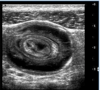
Fig. 1: Ultrasound showing Target Sign
RESULTS: Out of 62cases, cases in the age-group of 0–5 year(s), the median age of subjects was nine months. The majority (75.0%) of these cases were infants with a peak between 6 and 12 months of age. The male:female ratio was 2.4:1.
All the subjects were Indians. Modalities of management included nonoperative reduction that is hydrostatic enema reduction in 2 cases (3.2), barium enema reduction 45 cases
(72.5) and air enema reduction in 12 cases (19.3), finally 3cases (4.8) which failed enema reduction were operated; 1
case (1.61) intussusception-associated death was recorded.
Fig. 2: Multimodalities of Management
DISCUSSION: The present study represents the review of current practice of intussusception reduction in India. The standardization of care has improved with centralization of care to paediatric surgical centres and the use of draft BSPR guidelines. These guidelines were based on the study and review of the best evidence by Rosenfeld and McHugh.(1)
Hirschsprung was the first to publish a series of reports on successful hydrostatic reduction in 1876. Harald
Hirschsprung’s description of a systematic approach to
hydrostatic reduction of intussusception, the reported success rate of this nonoperative intervention has widely varied (<40% to >90%).(2)
Intussusception, a common gastrointestinal (GI) emergency in children develops when a proximal segment of the gastrointestinal tract (Intussusceptum) telescopes into a distal portion (Intussuscipiens), most commonly located near the ileocecal valve. Intussusception occurs in approximately 17.7 children per 100,000 per year in India.(3)
Population-based incidence of intussusception and a case-control study to examine the association of intussusception with natural rotavirus infection among Indian children were conducted.(4) Of these, 60% of cases occur before 1 year of age and 80% before 2years. Its incidence is highest between 5 and 9 months of age, being uncommon in neonates. There is a male predominance of 3:2.(5)
The majority of pediatric cases involve the ileocolic (Orileocecal) portion of the intestine. When the intussusceptum invaginates and pulls along the mesentery, there is compromise of venous return. This is followed by engorgement of the intussusceptum, edema and bleeding from the mucosa which lead to currant jelly stools. Finally, the arterial blood supply of the intestine gets compromised leading to necrosis, perforation and/or shock.
Children less than 3months and greater than 2years old
are most likely to have a lead point or specific identified
cause, such as: Meckel diverticulum, duplications, polyposis
and lymphomas. Hyperplasia of the intestinal Peyer’s patches
have been identified as the leading point in infant cases.
Peyer’s patches hyperplasia can occur secondary to viruses
such as adenovirus, enterovirus, echovirus and human herpes virus.(6)
Journal of Evolution of Medical and Dental Sciences/ eISSN- 2278-4802, pISSN- 2278-4748/ Vol. 4/ Issue 94/ Nov. 23, 2015 Page 15996
The clinical presentation having typical triad of severe intermittent abdominal pain, currant jelly stools and vomiting is seen in less than 20% of cases.9
Bouts of abdominal pain may be evidenced by pulling of knees against the abdomen with an interim asymptomatic healthy child. In ileocolic intussusception palpation of the abdomen may reveal a sausage-shaped mass which is located in the RUQ. A wide spectrum of symptoms may range from painless intussusception to constipation, dehydration, diarrhea, intestinal prolapse, rectal bleeding, sepsis, shock, syncope, vomiting and altered mental status (Lethargy and irritability).10
Lethargy is seen most frequent in infants and young children with or without history of gastrointestinal symptoms. Altered mental status has been hypothesized to be secondary to a combination of factors such as dehydration, electrolyte imbalance, and endorphins or toxic metabolic products released from the ischemic bowel which can affect the brain.11 Recurrent intussusception is present in only 5-8% of children and is most common after hydrostatic versus surgical reduction. Fifty percent of recurrent intussusception cases occur within 48 hours of a prior episode (But have been reported up to 18 months later).12 A recent and extensive review by the World Health Organization (WHO) on intussusception concluded that, in developed countries, the baseline incidence of intussusception is between 0.5 and 4.3 cases per 1,000 live births or 0.7–1.2 cases per 1,000 children aged less than one year. Accurate estimates of the incidence of intussusception are not available for most developing countries. In India, rotavirus-associated acute gastroenteritis accounts for 5–70% of all hospitalizations and one of the leading causes of intusuception, thus WHO and Global Alliance for Vaccines and Immunization (GAVI) have identified rotavirus vaccines as a priority.13
Standard Diagnostic Modalities includes Ultrasound which is a fast, non-invasive and simple reproducible test. Its sensitivity (98-100%) and specificity (88-100%) is high, but is clearly operator dependant. On ultrasound "Target" sign=hypoechoic ring with an echogenic center on transverse US image or a "Pseudokidney" sign=hypoechoicbowell wall extending along a hyperechoic mucosa is seen.14
At some centers, it has displaced radiographs as the initial imaging test of choice. Other indications for its use are: Suspected cases of intussusception where abdominal x-rays are non-conclusive, evaluation for reducibility, presence of a lead point mass, potential incomplete reduction after enema and intussusception limited to small bowel.
Computed tomography (CT) scan is usually not indicated in children, since the diagnosis is generally confirmed by ultrasound or enema. The CT scan involves high costs, radiation exposure and sedation risk which is overall less convenient for this population group.Once the diagnosis of intussusception is established, an intravenous line inserted, and intravenous hydration started.
A nasogastric tube should be inserted and placed to suction. If the patient is markedly distended or has a dilated loop of bowel, an abdominal radiograph should be obtained. Antibiotics should be administered based on clinical suspicion of peritonitis or infection (Sepsis) or in patients with a markedly elevated WBC count.
Preoperatively, intravenous crystalloid resuscitation is begun. Broad-spectrum intravenous antibiotics are administered. Body temperature must be preserved in the operating room. A type and screen of the patient's blood should be obtained. As with any patient with a bowel obstruction, careful induction (i.e., rapid sequence) of anesthesia should take place because of the risk of regurgitation and aspiration.
Therapeutic contrast enemas (Barium, water-soluble and air) are diagnostic and therapeutic techniques with reduction rates of 70 to 90%. This should be considered only after stabilizing the child, adequate hydration, and consultation with a pediatric surgeon. Barium enema was considered to be gold standard until recent introduction of Air
enema’s use, which has subsequently increased due to its lower perforation risk, less radiation exposure, faster and better reduction rate. But the disadvantages of air contrast enema compared to a barium or water-soluble contrast enema is air leaking into the peritoneal cavity because of intestinal perforation may also be difficult to see. Those in favor of using the air contrast enema technique argue that with perforation, the sudden loss of pressure would signal to the radiologist to stop the procedure.
If a tension pneumoperitoneum results, this should be decompressed immediately with an 18-gauge needle. Barium leaking into the peritoneal cavity may cause a chemical peritonitis. Using a water-soluble contrast may decrease this complication. An air contrast enema is advocated as the preferred method by many pediatric surgeons, but since there is no clear consensus among radiologists of the best contrast enema option, this decision is best left to the pediatric surgeons performing the contrast enema procedure.
Intussusception recurrence rates for air versus liquid enema are reported to be similar, approximately 10%.15
Traditionally, an attempt was not considered successful until the reducing agent, whether air, barium, or water-soluble contrast, was observed refluxing back into the terminal ileum, but evidence has shown that this is not entirely necessary. Most intussusceptions that failed to show reflux into the ileum were due to either an edematous or competent ileocecal valve. When these patients were explored, they displayed a completely reduced intussusception. According to this study, a patient who becomes asymptomatic after nonoperative reduction that fails to show reflux of the reducing agent into the ileum can safely be observed.16
Enema reduction should be attempted 3 times at the most before considering it unsuccessful and submitting the patient to surgery. Poor enema reduction rate is determined by (in descending order): symptoms longer than 24 hours, age less than 3 months, dehydration and intussusception of the rectum. Repeat enema is both safe and effective in recurrent intussusception. Spontaneous reduction occurs in approximately 10-17% of patients, being most common in the small bowel.
Journal of Evolution of Medical and Dental Sciences/ eISSN- 2278-4802, pISSN- 2278-4748/ Vol. 4/ Issue 94/ Nov. 23, 2015 Page 15997
If operative reduction is successful, appendectomy is often performed if the blood supply of the appendix is compromised. A cecopexy is not necessary. Risk of recurrence of the intussusception after operative reduction is less than 5%.18
Finally, barium enema is still the gold standard in confirming the diagnosis and nonsurgical reduction of an intussusception. Water-soluble contrast has been used and more recently air enema reduction has been introduced. There are several reasons why radiologists have different preferences for which type of contrast they choose to use for the procedure.
Laparoscopy in the management of intussusception was initially limited to a diagnostic role. It was used to confirm unreduced bowel following an enema with prompt conversion to an open procedure. The laparoscope allowed the surgeons to avoid unnecessary open procedures in cases of spontaneous reduction following enema and enhanced the efficacy of hydrostatic or pneumatic reductions, reducing the need for an open procedure. Continued experience with laparoscopy and improved technology has led some centers to successfully utilize the technique for therapeutic reduction in confirmed cases of pediatric intussusception.
Postoperatively, intravenous fluid resuscitation is continued and calculated, taking into consideration maintenance requirements and third-space losses. Upon resolution of ileus, diet is advanced at the discretion of the surgeon.
At Follow-upin older children and in cases of recurrent intussusception (After 3-4 episodes) successfully reduced with an enema, consider evaluating the patient for a lead point (eg, upper GI series, Meckel scan).
Few complications such as dehydration and aspiration from emesis can occur. Ischemia and bowel necrosis can cause bowel perforation and sepsis. Necrosis of a significant length of intestine can lead to complications associated with short bowel syndrome. Whether treated by operative or radiographic reduction, late stricture (4-8week) may occur within the length of intestine involved.
The overall mortality rate of intussusception is less than 1%. Recurrence rates following nonoperative reduction and surgical reduction are approximately 5% and 1%-4%, respectively.
A decreased rate of operative intussusception management is noted in specialized pediatric hospitals compared with nonpediatric hospitals. This is attributed to the increased experience with and use of the various radiologic reduction techniques.
CONCLUSIONS/SUMMARY: This article highlights the diverse signs and symptoms pediatric intussusception. Only 20% of children will present with the triad (Abdominal pain, currant jelly stools and vomiting). Clinical suspicion is the key factor in making a diagnosis of intussusception even in absence of typical signs and symptoms. Delay in diagnosis increases risk for bowel obstruction, perforation, necrosis and death.
ACKNOWLEDGEMENTS: We thank the tertiary care hospitals in South Bangalore, India, for their valuable contribution.
We also express gratitude with anaesthesia, pediatric team and hospital staff for their coordination and active participation.
REFERENCES:
1. K. Rosenfeld, K. McHugh., Survey of intussusception reduction in England, Scotland and Wales: How and why we could do better. Clinical Radiology
(1999) 54, 452-458. Hirschsprung, H.: 107 Falle von DarminvaginationbeiKindern, behandelt in KoniginLouisen-Kinderhospital in Kopenhagenwahrend der Jahre I87I-I904. Mitteilungenaus den Grenzegebieten den Medezin und Chirurgie,
14: 555, 1905.
2. Glass RI, Bresee JS, Turcios R, Fischer TK, Parashar UD, Steele AD. Rotavirus vaccines: Targeting the developing world. J Infect Dis. 2005;92(Suppl 1):S160–6. [PubMed]. 3. Bahl R1, Saxena M, Bhandari N, Taneja S, Mathur M,
Parashar UD, Gentsch J, Shieh WJ, Zaki SR, Glass R, Bhan MK; Delhi Intussusception Study Hospital Group.2001 4. Sorantin E, Lindbichler F. Management of
intussussception.
EurRadiol. 2004; 14(suppl 4): L146-L154.
5. Behrman RE, Kliegman RM, Jenson HB: Nelson Textbook of Pediatrics, Ed 18: Chapter 330-Intussusception.
2007, 1569-1570.
6. Bines J. Intussusception and rotavirus vaccines. Vaccine. 2006 May 1;24(18):3772-6. Epub 2005 Jul 28.
7. Hyser JM and Estes MK. Rotavirus vaccines and Pathogenesis: 2008. CurrOpinGastroenterol.
2009 Jan;2005(1):36-43.
8. Fleisher G. In: Textbook of Pediatric Emergency Medicine, Fourth Edition: Chapter 118: Abdominal Emergencies;2000:1611-1613.
9. Hickey RW, Sodhi SK, Johnson WR. Two children with lethargy and intussusception. Ann Emerg Med. 1990;19:390-392.
10.Goetting MG, Tiznado-Garcia E, Bakdash TF. Pediatric neurology. 1990 Nov-Dec;6(6):419-421.
11.Difiore JW. Clinical manifestations in intususception. SeminPediatr Surg. 1999 Nov;8(4):214-20.
12.Global initiative to fast-track vaccine for world's leading cause of severe diarrhea among children. Project expected to speed rotavirus vaccine availability to developing countries by 15–20 years.
13.(http://www.globalhealth.org/news/article/2805, accessed on 8 December 2006).
14.RathachaiKaewlai., Intussusception: Ultrasound., M.D(rad)Rit radiology.com. September 21, 2011.
15.Meyer JS, Dangman BC, Buonomo C, et al. Air and liquid contrast agents in the management of intussusception: A controlled, randomized trial.
16.Radiology 1993; 188: 507-511.
17.Shekherdimian S, Lee SL, Sydorak RM, Applebaum H. Contrast enema for pediatric intussusception: Is reflux into the terminal ileum necessary for complete reduction?.
J
Pediatr Surg
. 2009 Jan. 44(1):247-9; discussion 249-50. 18.Waseem M and Rosenberg HK. Pediatric Emergency Care.Journal of Evolution of Medical and Dental Sciences/ eISSN- 2278-4802, pISSN- 2278-4748/ Vol. 4/ Issue 94/ Nov. 23, 2015 Page 15998
19. Niramis R, Watanatittan S, Kruatrachue A, et al. Management of recurrent intussusception: Nonoperative or operative reduction?.J Pediatr Surg. 2010 Nov. 45(11):2175-80.