Barrier in an
In Vitro
Model
Angelika Stammler1,2, Dieter Mu¨ller1, Yoshiaki Tabuchi3, Lutz Konrad2*, Ralf Middendorff1*
1Institute of Anatomy and Cell Biology, Justus-Liebig-University Giessen, Giessen, Germany,2Department of Gynecology and Obstetrics, Justus-Liebig-University Giessen, Giessen, Germany,3Division of Molecular Genetics Research, Life Science Research Center, Graduate School of Medicine Pharmaceutical Sciences, University of Toyama, Toyama, Japan
Abstract
The blood-epididymis barrier (BEB) is formed by epithelial tight junctions mediating selective permeability of the epididymal epithelium. Defective barrier function can disturb the balance of the epididymal milieu, which may result in infertility. The stroma of the epididymis contains high amounts of cytokines of the TGFb family of unknown function. We screened possible effects of all three TGFbisoforms on paracellular tightness in a BEBin vitromodel based on the strongly polarized mouse epididymal epithelial MEPC5 cells in the transwell system. In this model we found a robust transepithelial electrical resistance (TER) of about 840Vx cm2. Effects on the paracellular permeability were evaluated by two methods, TER and
FITC-Dextran-based tracer diffusion assays. Both assays add up to corresponding results indicating a time-dependent disturbance of the BEB differentially for the three TGFbisoforms (TGFb3.TGFb1.TGFb2) in a TGFb-recetor-1 kinase- and Smad-dependent manner. The tight junction protein claudin-1 was found to be reduced by the treatment with TGFbs, whereas occludin was not influenced. Epididymal epithelial cells are predominantly responsive to TGFbs from the basolateral side, suggesting that TGFbmay have an impact on the epididymal epithelium from the stromain vivo. Our data show for the first time that TGFbs decrease paracellular tightness in epididymal epithelial cells, thus establishing a novel mechanism of regulation of BEB permeability, which is elementary for sperm maturation and male fertility.
Citation:Stammler A, Mu¨ller D, Tabuchi Y, Konrad L, Middendorff R (2013) TGFbs Modulate Permeability of the Blood-Epididymis Barrier in anIn VitroModel. PLoS ONE 8(11): e80611. doi:10.1371/journal.pone.0080611
Editor:Michael Koval, Emory University School of Medicine, United States of America
ReceivedSeptember 3, 2013;AcceptedOctober 7, 2013;PublishedNovember 13, 2013
Copyright:ß2013 Stammler et al. This is an open-access article distributed under the terms of the Creative Commons Attribution License, which permits unrestricted use, distribution, and reproduction in any medium, provided the original author and source are credited.
Funding:This project is part of LOEWE Focus Group Male Infertility and Urogenital Infections (MIBIE) funded by state of Hesse, Germany. The funders had no role in study design, data collection and analysis, decision to publish, or preparation of the manuscript.
Competing Interests:The authors have declared that no competing interests exist.
* E-mail: [email protected] (RM); [email protected] (LK)
Introduction
The epididymis mediates maturation, transport and protection of sperm in the luminal fluid [1]. The epididymal duct is a continuous tube of a length of 1 m in mouse, 3 m in rat and 6 m in human [2]. The duct is formed by a pseudostratified columnar epithelium surrounded by a thin layer of muscle cells [1].
The blood-epididymis barrier (BEB) protects sperm during storage and maturation and controls the state of luminal fluid [1]. The barrier is composed of tight junctions between the epithelial cells. In epididymis, tight junction proteins claudin-1, -3, -4, -7 and occludin were found to be expressed [3,4]. Inin vitrocell culture models, the knockdown of one of these claudins (1, -3, -4, or -7) resulted in dramatically decreased transepithelial electrical resis-tance (TER) tested in human epididymal cell lines [5]. Claudin-1 knockout mice die immediately after birth due to dehydration caused by lack of epidermal barrier. Thus, claudin-1 seems to be pivotal for survival and cannot be compensated by other tight junction proteins [6]. The role of occludin for barrier formation is controversial [7] since occludin knockout mice are viable and have an intact barrier in the intestine and bladder [8]. However, male occludin knockout mice are infertile [9].
Quantitative changes of tight junction proteins on the cell surface have been reported to be caused by diverse substances or processes. The decrease of tight junctions on the cell surface connected with disruption of epithelial barrier has been reported
for toxins such as cadmium chloride in the seminiferous epithelium [10] but also for diverse cytokines associated with inflammation and immunoregulation, such as TNFa, IFN-c, interleukins [11,12,13,14] or TGFb. TGFb was reported to increase perme-ability in diverse epithelia, such as trachea epithelium [15], the seminiferous epithelium [16] and ovarian surface epithelium [17] as well as between the cells of the blood-brain barrier [18]. Several TGFb pathways have been reported to mediate quantitative changes of tight junctions. In the murine trachea, the JNK pathway was described to be essential for TGFb response [15], whereas in the seminiferous epithelium Ras/ERK pathway was reported to mediate the signal [16]. In the blood-brain barrier, changes in the permeability were found to be mediated by Smad signaling [18].
perinatally [22]. TGFb3 knockout mice also die perinatally due to developmental defects of the lung [23]. TGFb3 knockout mice also show defective palatogenesis [24].
TGFbs are secreted as non-covalent complexes associated with the latency-associated peptides (LAPs). After activation TGFbs bind to TGFb-receptor-2 (TGFb-R2), which in turn dimerizes with TGFb-R1. In response to the binding of the ligand, the intracellular kinase domain of TGFb-R1 phosphorylates Smad2 or Smad3 that eventually act as transcription factors together with Smad4 [25].
In the epididymis, high amounts of TGFbs were found [26,27,28]. TGFb1 mRNA was described to be present in the stroma of all regions of rat epididymis analyzed by Northern blot [26]. In the same study, TGFb3 mRNA was predominantly found in the corpus region of epididymis, whereas TGFb2 mRNA was not detected [26].
In our investigation we aimed to test the influence of cytokines of the TGFbfamily on the barrier of epididymal epithelium. For this purpose we used anin vitromodel of blood-epididymis barrier based on polarized mouse epididymal cells of the cell line MEPC5 [29] cultured on transwell inserts. Cell line MEPC5 was previously established by us and characterized in detail [29]. In this model we evaluated paracellular permeability by two methods, the mea-surement of transepithelial electrical resistance (TER) and by tracer diffusion. We observed that TGFbs increase paracellular permeability significantly within 4 h depending on (i) the isoform of TGFb, (ii) the side of application (apical vs. basolateral), and (iii) TGFb-R1 kinase activity. Tight junction protein claudin-1 was found to be reduced by the treatment with TGFbs (whereas occludin was not influenced), suggesting a TGFb-R1 kinase-dependent effect on distinct tight junction proteins modulating the BEB.
Methods
Cell culture and viability assay
MEPC5 cell line [29] was cultured in DMEM supplemented with 10% FCS and penicillin/streptavidin at 33uC/5% CO2/96% humidity. After 2–3 days of culture, cells were detached from 75 cm2 flasks using accutase (PAA, Co¨lbe, Germany) and were seeded (36105cells/cm2) in 12-well plates. After 24 h of starvation
with 2% FCS, TGFb-1, -2 or -3 human recombinant protein (Promokine, Heidelberg, Germany) were added combined with fresh medium in a final concentration of 10 ng/ml, as described previously for other cell lines [30]. After 24 h of TGFbtreatment, cells were detached and analyzed concerning viability and number of cells using BioRad Cell Counter System (BioRad, Hercules, CA, USA) combined with Trypan blue (BioRad) staining.
Measurement of transepithelial electrical resistance (TER) For TER measurement, cells were seeded (36105cells/cm2) on ThinCert inserts (Greiner, Frickenhausen, Germany) with 0.4mm pore size in 24-well plates (0.33 cm2 per insert) in culture conditions described above. TER measurements were conducted using Millicell ERS-2 (Merck Millipore, Billerica, MA, USA). TER was calculated according to the instructions of the Millicell ERS-2 manual, subtracting the background resistance of blank filters in the same experiment and multiplying the TER [V] with the area of the monolayer [cm2].
Under these conditions, the TER increases from about 200Vx cm2within one day after seeding to about 840Vx cm2on second day after seeding, correlating with increasing cell number. After day 2 no further increase of cell number could be observed. From this point on further increase of TER slowed down. To
standardize experimental conditions, all experiments were per-formed between day 2 and day 3 despite of ongoing slight increase of TER especially at the end of day 3 (‘‘24 h’’ i.e. 72 h after seeding). In order to allow a more convenient comparison of experiments, values were normalized to the initial value at 0 h (i.e. 48 h after seeding) for each individual insert. This 0 h value was described as 100%.
Between day 1 and day 2 cells were starved for 24 h with 2% FCS. At day 2, after measurement of the initial value (0 h), TGFb -1, -2 or -3 (Promokine) were added in a final concentration of 10 ng/ml combined with fresh medium. For inhibitor experi-ments, the specific inhibitors were added 3 h prior to TGFb addition. After the first 6 h medium supplemented with the specific stimulants was exchanged. Inhibitor Ly364947 (Sigma-Aldrich, St Louis, MO, USA) was dissolved 5 mg/ml in DMSO (Sigma-Aldrich) and stored at 220uC. Stocks were diluted in medium to a final concentration of 5mM/ml. Inhibitor SiS3 (Merck Millipore) was dissolved 15 mg/ml in DMSO and stored at220uC. Stocks were diluted in medium to a final concentration of 2mM/ml. Experiments with vehicle DMSO were conducted with adjusted concentrations.
Tracer diffusion assay
After 24 h of TGFb treatment, fresh medium supplemented with TGFband 5 mg/ml FITC-coupled Dextran, MW 4 (FD4, Sigma-Aldrich) was loaded into the upper compartment of the insert. FD4 has a radius of 14 A˚ and, thus, can pass a cell monolayer only paracellularly. Medium change with TGFb complementation was also applied in the lower compartment. After another 24 h of incubation and careful handling, a 100ml sample was taken from the lower compartment and measured in a dilution series. Fluorescence intensity at wavelength extinction 490 nm/emission 520 nm was measured by ELISA reader (Tecan, Ma¨nnedorf, Switzerland) combined with a black 96-well chimney plate (Greiner). Permeability coefficient (Papp) was calculated using following equation: Papp= (1/A?C0)?(dQ/dt) with
dQ/dtas the solute flux (dQamount of relocated tracer anddttime span),Aas the surface of the insert membrane andC0as the initial concentration.
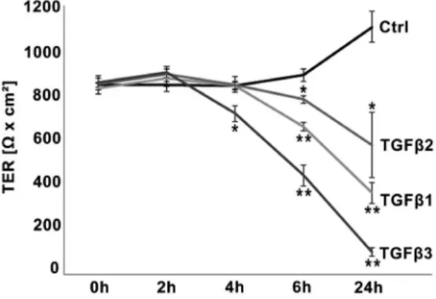
Figure 1. Time-dependent TGFbeffects on TER in the BEBin vitromodel.TGFbs decreased epithelial barrier in a time-dependent manner. TGFb3 decreased absolute values of TER (indicated byVx cm2) significantly within 4 h, 6 h and 24 h compared to untreated control each. TGFb1 and TGFb2 influenced the TER significantly after 6 h and 24 h. Data points represent mean values obtained from n = 6, generated by three independent repetitions performed in duplicate. SEM is indicated, p-values#0.05 (Mann-Whitney-test) were considered significant (*), p#0.005 highly significant (**).
Immunofluorescence
Cells were fixed in 4% saline buffered paraformaldehyde for 10 min and blocked with 2% NGS for one hour. Polyclonal antibodies diluted in saline buffered 0.2% BSA/0.1% NaN3 against claudin-1 (Invitrogen, Camarillo, CA, USA, dilution 1:150) were applied overnight at 4uC. Slides were washed and incubated with secondary antibody (Alexa goat-anti-rabbit Fluor 488, Invitrogen, Camarillo, CA, USA, dilution 1:500) and DAPI
(Roche, Indianapolis, IN, USA, 1:2000) for 2 h at room temperature. After washing and glycerol imbedding, photos were made using Axioskop 2 plus (Carl Zeiss, Go¨ttingen, Germany) supplied with AxioCam MRc (Carl Zeiss) and AxioVision, version 4.8 (Carl Zeiss).
Generation of cell lysates
Cells (36105cells/cm2) were seeded on ThinCert inserts
(Greiner) with 0.4mm pore size in 6-well plates (each insert 4.254 cm2). After 24 h of starvation with 2% FCS, TGFb-1, -2 or -3 (Promokine) were added in a final concentration of 10 ng/ml combined with fresh medium. After 24 h in the presence or absence of TGFbs, filters were rinsed once with ice-cold PBS. The cells were scraped off with a flexible cell scratcher (TPP, Trasadingen, Switzerland) into 1 ml of ice-cold PBS, transferred into tubes and centrifuged for 10 min at 5000 g. Supernatant fractions were removed, and cell pellets were dissolved in 150ml lysis buffer (20 mM Tris-Cl, pH 7.5, 150 mM NaCl, 1 mM EDTA, 1 mM EGTA, 1% Triton X-100), containing 7ml/ml protease inhibitor cocktail (P1860, Sigma). Samples were kept on ice for 30 min and frequently vortexed and centrifugation at 10 000 g for 10 min at 4uC in order to remove cell debris and nuclei. The supernatant fractions, representing the cell lysates, were stored at280uC until usage for Western blot analyses.
Membrane and cytosolic protein fractions from mouse epidid-ymis were prepared as described previously [31]. After homoge-nization by ten strokes in a Potter–Elvehjem homogenizer (Wheaton, Millville, NJ, USA), samples were centrifuged at 3000 g for 8 min at 4uC to remove cell debris and nuclei. The supernatant fractions were ultra-centrifuged for 30 min at 100 000 g at 4uC. The resulting supernatant represents the cytosolic Figure 2. TGFbeffects on tracer diffusion in the BEBin vitro
model.Treatment with TGFb3 increased the permeability coefficient (Papp) highly significant (p = 0.0002) compared to untreated control,
whereas for TGFb1 the level of significance (p = 0.0281) is lower. Each column represents a mean value obtained from n = 6, generated by three independent repetitions performed in duplicate. SEM is indicated, p-values #0.05 (Mann-Whitney-test) were considered significant (*),
p60.005 highly significant (**).
doi:10.1371/journal.pone.0080611.g002
Figure 3. Cell morphology and claudin-1 immunostaining after treatment with TGFbs.MEPC5 monolayers photographed in bright field (A–D), DAPI staining (E–H), and claudin-1 immunostaining (I–L). A: untreated MEPC5; B–D: MEPC5 cells treated with TGFbs for 24 h. No obvious changes in cell morphology are detectable. E: DAPI labeling of the nucleus in untreated MEPC5; F–H: MEPC5 cells treated with TGFbs for 24 h. No obvious changes are detectable. I: Immunostaining of claudin-1 in untreated MEPC5, DAPI labeling of the nucleus. The pattern of claudin-1 localization shows numerous cell clusters characterized by an intense immunofluorescence at the cell borders. J–L: MEPC5 cells treated with TGFbs for 24 h. After treatment with TGFb3 (and to a lesser intent with TGFb1) the staining was generally weaker, the striking immunofluorescence at cell borders was clearly reduced. Photos are examples from at least three independent experiments. Scale bar: 12.5mm in A–D, 25mm in E–L.
fraction. The pellets, representing the membrane fraction, were resuspended in 50 mM Tris-HCl buffer, pH 7.5, and stored at
280uC.
In all samples, protein concentrations were determined by using a kit from Bio-Rad (Munich, Germany) with bovine serum albumin (Sigma) as standard.
Immunoblotting
After separation by SDS-PAGE, proteins were transferred to nitrocellulose membranes (Amersham, Braunschweig, Germany) at 30 V for 12–14 h at 4uC. Blots were stained with Ponceau S (P7170, Sigma), and the positions of co-migrated reference proteins (SDS-6H, Sigma) were marked. Records of the protein images served as additional protein loading control. The blots were then treated for 2 h with blocking solution (Roche, Mannheim, Germany) prior to probing with antibodies directed against claudin-1 (Invitrogen, 1:2000), occludin (Invitrogen, 1:2000), and vinculin (Sigma, 1:6000). Either mouse or anti-rabbit IgG, linked to peroxidase (Pierce, Rockford, IL, USA), were used as secondary antibodies. Signals were detected using enhanced chemiluminescence (Amersham, RPN 2105) on Fuji (13862 C) X-ray films. After stripping of bound antibodies [32], blots were re-used for detection of other proteins, especially
vinculin, which was used for the normalization of densitometric measurements.
Statistics
All data were gained from three independent experiments conducted in duplicate. Mean values and SEM were calculated using MS Excel, Version 2010. Calculation of p-values was done by Students t-test using Excel or non-parametric Mann-Whitney-test using GraphPad Prism 5 (GraphPad Software, La Jolla, CA, USA).
Results
TGFbs mediate disruption of epididymal barrier in vitro In order to investigate the influence of TGFbs on the barrier of the epididymal epithelium, confluent monolayers of MEPC5 cultured on inserts were treated after two days of culture with one of the three TGFbisoforms, which were added in both, the apical and the basolateral compartment of the transwell inserts.
The TER value at day 2 (0 h/initial value) was on average of 837Vx cm2611.2 (Fig. 1). After 24 h (day 3) the TER increased to 1092Vx cm2668.7 in the untreated control (Fig. 1A). All three isoforms of TGFb decreased the TER. Compared to control, treatment with TGFb3 significantly decreased the TER to 710Vx
Figure 4. Western blot analysis of claudin-1 and occludin expression after treatment with TGFbs.A: Western blot with lysates of MEPC5 after combined basolateral and apical stimulation with TGFb1, TGFb2 or TGFb3 for 24 h compared to untreated cells. Lysates from three independent experiments (I–III) were used. TGFb1 and especially TGFb3 decreased the level of claudin-1 compared to the untreated sample. The levels of occludin were unaffected. Membrane (M) and cytosolic (C) protein fractions of mouse epididymis served as controls. B, C: Densitometric analysis of claudin-1 (B) and occludin (C) protein expression. Claudin-1 showed a significant reduction of expression by TGFb3 by TGFb1 treatment (p60.05). All arbitrary units were normalized to the corresponding values of vinculin, serving as loading control. Columns represent mean values of three independent experiments (I–III) with SEM indicated. p-values compared to untreated controls according to Students t-test.
cm2634.1 after 4 h, 437V x cm2647.4 after 6 h and 98 Vx cm2618.7 after 24 h, respectively (Fig. 1A). Treatment with TGFb1 and TFGb2 decreased TER significantly within 6 h (TGFb1: 652Vx cm2618.8; TGFb2: 773 Vx cm2615.8) and 24 h (TGFb1: 360Vx cm2645.9; TGFb2: 571Vx cm26145.5). Regularly, TGFb2 effects were less pronounced than TGFb3 and TGFb1 effects, but have to be considered as significant after 24 h compared to control due to increase of TER in the control. When TGFbs were removed after 24 h of incubation the effect could be partially reversed (data not shown).
Since the initial TER values (0 h) regularly showed slight variations, even between duplicates, in all following experiments the TER values were normalized to the initial value (0 h) of each individual insert (100%). This normalization did not change the level of significance compared to calculation by absolute values (data not shown).
In the tracer diffusion assay, monolayers were pretreated for 24 h with TGFbs before TGFbs together with FD4 were applied for another 24 h. The amount of FD4 diffusing through the monolayer (measured by the permeability coefficient,Papp) treated with TGFb3 was about 10fold higher (highly significant, p = 0.0002) compared to an untreated monolayer (Fig. 2). TGFb1 also showed impressive effects on the capacity of diffusion, whereas TGFb2 effects were negligible (TGFb1, p = 0.0281; TGFb2, p = 0.2072). These results mirror the findings from TER measurements, indicating that the TER actually reflects the paracellular permeability of thisin vitromodel.
TGFbs effects on cell numbers, viability and morphology To exclude that changes of paracellular permeability by TGFbs are the consequence of TGFb-dependent cell number variation, cells were counted using BioRad Cell Counter System. Compared to controls, treatment for 24 h with TGFb1, TGFb2 and TGFb3, respectively, showed only slight effects on cell numbers, which were not significant (all p-values.0.15, Fig. S1). Combination of cell counting with Trypan blue staining did not give any hint for increased cell death after TGFb treatment (data not shown). In addition, the phenotypic shape of the cells did not change under
culture conditions with TGFbs (Fig. 3A–D). According to these results, TGFbs do not affect cell numbers, cell viability or morphology to an extent that is relevant for permeability.
Changes of localization and protein expression of tight junction proteins after TGFbtreatment
Following the axiom, that the paracellular barrier is mediated by tight junctions, the localization of tight junction proteins that were described to be expressed in the epididymis were checked on TGFb-treated monolayers of MEPC5 cells (Fig. 3). In untreated controls (Fig. 3I) the pattern of claudin-1 localization showed numerous cell clusters comprising six or more cells, characterized by an intense immunofluorescence at the cell borders. This pattern is comparable to the well known staining of tight junctions in tangentially cut epithelial cells from tissues sections. We found such a staining pattern for claudin-1 also in murine epididymis sections (data not shown).
After the application of TGFbs, this pattern is disturbed (Fig. 3K–L). Especially treatment with TGFb3 (Fig. 3L) resulted in essential changes of the claudin-1 staining pattern. Larger cell clusters and the striking immunofluorescence of cell borders were barely detectable. These TGFb-dependent changes are not due to reduced cell numbers as shown by (i) cell counting (see above, Fig. S1), (ii) bright field photographs (Fig. 3A–D) and (iii) additional DAPI stainings (Fig. 3E–H).
Western blot analyses of lysates from MEPC5 cells treated in 6-well inserts from the apical and basolateral side (Fig. 4A) revealed a significantly reduced expression of claudin-1 after treatment with TGFb1 and TGFb3 (Fig. 4B), whereas the expression of the tight junction protein occludin was unchanged (Fig. 4C). Control experiments using mouse tissue confirmed expression of claudin-1 and occludin in the mouse epididymis and showed their predominant localization in the membrane fraction (Fig. 4A, right lanes). Vinculin served as standard for normalization of claudin-1 and occludin expression in densitometric measurements (Fig. 4B, 4C).
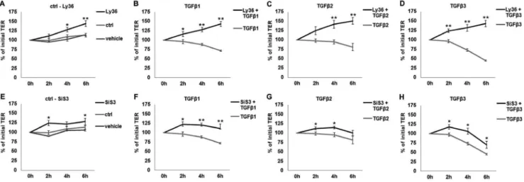
Figure 5. Time-dependent effects of TGFbpathway inhibitors on TER in thein vitromodel.In case of inhibitor application, cells were incubated with inhibitors alone for 3 h and then with TGFbs+inhibitors for 2 h, 4 h and 6 h. Values prior to any treatment were taken as baseline
(100%). A: The inhibition of TGFb-R1 kinase activity by Ly364947 (Ly36) compared to untreated and vehicle-treated cells. B-D: TGFbs-affected TER values in the presence and absence of Ly364947. Effects observed for TGFbs are significantly inhibited by TGFb-R1 inhibition. E: The inhibition of Smad3 by SiS3 compared to untreated and vehicle-treated cells. F–H: TGFbs-affected TER values in the presence and absence of SiS3. Inhibition of Smad3 resulted in attenuation of TGFbeffects. Data points represent mean values obtained from n = 6, generated by three independent repetitions performed in duplicate. SEM is indicated, p-values60.05 (Mann-Whitney-test) were considered significant (*), p60.005 highly significant (**).
TGFb-receptor-1 kinase and Smad3 are involved in TGFb -dependent changes of TER
To make sure that the effects observed are TGFb -R1-dependent, Ly364947, an inhibitor of TGFb-R1 kinase, was applied 3 h prior to TGFbstimulation. The inhibition of TGFb -R1 kinase was able to fully prevent the effects of all three TGFbs (Fig. 5A–D), indicating that all effects of TGFbs are mediated by TGFb-R1 kinase activity.
In the next step, we analyzed how the effects are mediated downstream of the TGFbreceptor complex. Inhibition of Smad3 phosphorylation by the inhibitor SiS3 attenuated the effects of the TGFbisoforms (Fig. 5E–H), indicating that Smad3 is involved in increased TGFb-mediated permeability. Inhibition of non-canon-ical TGFbpathways via JNK did not have any significant effect on TER (data not shown).
Epididymal epithelial cells are responsive to TGFb
predominantly from basolateral side
Since MEPC5 cells are strongly polarized [29], we used the MEPC5 model to analyze whether the epididymal epithelium is predominantly responsive to the TGFb signals from luminal (apical) or stromal (basolateral) side. For this, TGFb was added either to the apical, or to the basolateral or to both compartments (Fig. 6).
Stimulation from basolateral side with TGFb3 resulted in effects similar to those observed for simultaneous stimulation from both sides, decreasing the TER significantly at the data points 4 h, 6 h and 24 h (Fig. 6B and 6C). Effects after apical stimulation (Fig. 6A) were barely detectable. Even after 24 h the TER remained in the range of the initial value. Only due to a slight increase of TER in the untreated control, TER effects became significant after 6 h and 24 h.
According to these data, MEPC5 cells are predominantly responsive to TGFb3 from the basolateral side, suggesting that TGFb3 might affect the permeability of the epididymal epithelium
in vivo predominantly from the interstitial side. Similar but less pronounced results were obtained for TGFb1 (data not shown).
Protein expression of the tight junction proteins claudin-1 and occludin after TGFbtreatment from different sides In order to test if the distinct side-specific TGFbeffects on TER correspond to tight junction protein levels, the expression of claudin-1 was quantified by Western blot. Lysates were gained from cells cultured on 6-well inserts and identical amounts of proteins were analyzed by Western blot (Fig. 7). Whereas combined stimulation from the basolateral and apical side resulted in a strong decrease of claudin-1 levels by TGFb3 and TGFb1 (see Fig. 4A, 4B), stimulation from the apical side alone showed only slight changes of claudin-1 expression (Fig. 7A, 7B), which were not significant (p.0.05). The level of occludin was not affected by apical (Fig. 7A, 7C) or apical/basolateral treatments (see Fig. 4A, 4C). Vinculin served as standard for normalization of claudin-1 and occludin expression in densitometric measurements (Fig. 7B, 7C).
Discussion
Ourin vitrosystem – TER and Tracer diffusion as
benchmarks of paracellular permeability
Based on our preparatory experiments [29] we established a BEBin vitromodel. In this model a highly efficient barrier of 840V x cm2was produced, which is in the range of TER observed in gut epitheliumin vitromodels [33] and is higher than described forin
vitromodels of the blood-testis barrier with less than 250Vx cm2 [34,35]. Inin vitromodels of renal proximal tube up to 240Vx cm2were observed [36].
Furthermore, we were able to modulate the BEBin vitro in a highly reproducible manner, thus we established a robust system that allows screening of substances affecting the BEB in future Figure 6. Dependency of TGFbeffects on TER from the side of stimulation.TGFb3 was applied into the upper compartment (apical) only (A), into the lower compartment (basolateral) only (B), or into both (apical +basolateral), (C). Basolateral application of TGFb3, whether alone or together with apical stimulation decreased TER significantly at 4 h, 6 h and 24 h compared to untreated control, whereas apical stimulation had only slight effects. Due to increase of TER in the untreated control the effect of apical stimulation has to be considered significant after 6 h and 24 h. Data points represent mean values obtained from n = 6, generated by three independent repetitions performed in duplicate. Value prior to treatment was taken as baseline (100%). SEM is indicated, p-values 60.05 (Mann-Whitney-test) were considered significant (*), p60.005 highly significant (**).
studies. Our permeability experiments were based on two entities, electrical resistance and passive diffusion. The measurement of the electrical resistance is a widely accepted method to evaluate epithelial barriers, but TER cannot discriminate between para-cellular and transpara-cellular ion flux [12]. Evaluation of tracer diffusion measures paracellular permeability exclusively, because FD4 has a radius of 14 A˚ and can pass through the monolayer only paracellularly [12]. In our experiments, both methods resulted in comparable conclusions about the distinct effects of the three TGFbisoforms, indicating a high reliability.
Different TGFbisoforms have a distinct influence on the BEB
For the first time, we demonstrate that the permeability of the BEB can be affected by cytokines of the TGFbfamily. We found that the permeability of the BEB can be influenced by the TGFb isoforms differentially. An influence of TGFbs on epithelial paracellular permeability has been shown for other organs, whereas a screening for the differential effect of the distinct isoforms has not been conducted in any other organ before.
Our observations suggest that the three TGFbisoforms differ in their effect on paracellular permeability of the BEB. The strongest effect within a few hours was found for TGFb3. TGFb3 has also been reported to have a strong effect on the blood-testis barrier
[35]. TGFb1 affected the BEB less pronounced. However, the effect of TGFb2 was rather weak. These findings correspond to the differing TGFb-R2 affinity of the three isoforms, with TGFb3 having the highest affinity, the affinity of TGFb1 being less strong and the affinity of TGFb2 being rather weak [37]. All effects on permeability were demonstrated to be TGFb-R1-dependent, which activates the Smad pathway after dimerization with TGFb-R2 after ligand interaction. The differing affinity of the TGFbisoforms to TGFb-R2 might be one of the reasons for the gradual differences of effects.
Regulation of paracellular permeability by tight junctions The tightness of epithelia is a balance between forming of compartments (‘‘barrier’’) and paracellular permeability in order to fulfill specific functions (‘‘fence’’). Paracellular flux is regulated by tight junction proteins on the cell surface. The mechanism of regulation of tight junction quantity is discussed controversially. Most authors suppose a regulation mediated by degradation or endocytosis of tight junction proteins [14]. However, transcrip-tional control of tight junction biosynthesis was also suggested to be involved in the regulation of barrier tightness [38].
The immunofluorescence and Western blot revealed that the level of claudin-1 protein is reduced after treatment with TGFb1 and especially TGFb3, mirroring the ratios found in the Figure 7. Western blot analysis of claudin-1 and occludin expression after apical treatment with TGFbs.A: Western blot with lysates of MEPC5 after apical stimulation with TGFb1, TGFb2 or TGFb3 compared to untreated cells. TGFb1 and TGFb3 decreased the level of claudin-1 slightly compared to the untreated sample. The levels of occludin were unaffected. Membrane (M) and cytosolic (C) protein fractions of mouse epididymis served as controls. B, C: Densitometric analysis of claudin-1 (B) and occludin (C) protein expression. Claudin-1 showed a modest reduction of expression by TGFb1 and TGFb3 treatment which is not significant (p.0.05). All arbitrary units were normalized to the corresponding values of vinculin, serving as loading control. Columns represent mean values of three independent experiments with SEM indicated. p-values compared to untreated controls according to Students t-test.
permeability assessment. In contrast to claudin-1, occludin was not affected significantly, suggesting that the tight junction proteins claudin-1 and occludin are differently influenced by TGFbs. Claudin-1 reduction is associated with the breakdown of the barrier. Occludin cannot rescue this effect.
Of note, claudin-1 knockouts die perinatally because the impaired epithelial barrier results in dehydration [6]. In contrast, occludin knockout mice are viable and show intact epithelial barriers in the intestine and bladder [8], but occludin knockout male mice are infertile [9]. Our data do not provide any evidence that the infertility of occludin knockout mice is due to dysfunction of the epididymal epithelium.
Side-specific effects of TGFbs
In our experiments, we found that the cultured epididymal epithelial cells are predominantly responsive to TGFb3 from the basolateral side. Stimulation from the apical side did not show significant effects neither on TER nor on claudin-1 expression.
Transferring these data into thein vivosituation, the basolateral stimulation would represent TGFb signal from interstitial tissue. The stroma of the epididymis comprises reservoirs of growth factors such as TGFbs in the ECM of the connective tissue [26,27]. TGFb3 was described to be predominantly present in corpus region [26] and has the strongest effect of the three isoforms on paracellular permeability. TGFb1, which was found in all parts of the epididymal stroma [26], has a strong effect on paracellular permeability. However, TGFb2, which seems not to be present in the epididymis [26], had the slightest effect on the permeability of the epididymal epithelium compared to the other two isoforms. Interestingly, Desai and coauthors [26] did not detect the active form of TGFb1 in the epididymis, suggesting activation of stored TGFb1 might be restricted to distinct situations, such as infection or inflammation of the epididymis.
In vivo, the epididymal epithelium could be affected by TGFb activation from the stroma. In contrast, TGFbs, produced in the testis, and presumably present in the luminal fluid of the epididymis, might not have relevant effects on the epididymal epithelium.
The function of the high amounts of inactive TGFb in the stroma has not been elucidated so far. We speculate that activation of TGFbs might provide a mechanism to loosen the BEB in order to allow migration of dendritic cells into the epididymal epithelium. The presence of dendritic cells in the epididymal epithelium has been described, whereas the mechanisms of migration remained unresolved [39].
In conclusion, our experiments show that TGFbs modulate the permeability of the BEB. Comparing the three isoforms, TGFb3 has an outstanding strong effect on the permeability which corresponds to strong reduction of claudin-1 levels. Inhibitor experiments suggest a TGFb-R1 kinase- and Smad-dependent mechanism. Our data provide a mechanism how the tightness of the blood-epididymis barrier is modulated. We developed a robust BEBin vitromodel that can path the route of investigation to the role of BEB in male fertility.
Supporting Information
Figure S1 Influence of TGFbs on the number of MEPC5 cells.No significant difference was observed comparing the cell number after 24 h treatment with TGFbs and control evaluated by automated cell counting. Data points represent mean values obtained from n = 6, generated by three independent repetitions performed in duplicate. SEM is indicated, p-values#0.05 (Mann-Whitney-test) were considered significant (*), p#0.005 highly significant (**).
(TIF)
Acknowledgments
We are grateful to Ingrid Schneider-Hu¨ther, Sabine Tasch and Jo¨rn Lu¨bberstedt for their excellent technical assistance.
Author Contributions
Conceived and designed the experiments: AS DM LK RM. Performed the experiments: AS DM. Analyzed the data: AS DM LK RM. Contributed reagents/materials/analysis tools: YT. Wrote the paper: AS LK RM.
References
1. Cornwall GA (2009) New insights into epididymal biology and function. Hum Reprod Update 15: 213–227.
2. Hinton BT, Galdamez MM, Sutherland A, Bomgardner D, Xu B, et al. (2011) How do you get six meters of epididymis inside a human scrotum? J Androl 32: 558–564.
3. Gregory M, Cyr DG (2006) Identification of multiple claudins in the rat epididymis. Mol Reprod Dev 73: 580–588.
4. Cyr DG, Gregory M, Dube E, Dufresne J, Chan PTK, et al. (2007) Orchestration of occludins, claudins, catenins and cadherins as players involved in maintenance of the blood-epididymal barrier in animals and humans. Asian J Andrology 9: 463–475.
5. Dube E, Dufresne J, Chan PTK, Hermo L, Cyr DG (2010) Assessing the role of claudins in maintaining the integrity of epididymal tight junctions using novel human epididymal cell lines. Biol Reprod 82: 1119–1128.
6. Furuse M, Hata M, Furuse K, Yoshida Y, Haratake A, et al. (2002) Claudin-based tight junctions are crucial for the mammalian epidermal barrier: a lesson from claudin-1-deficient mice. J Cell Biol 156: 1099–1111.
7. Schulzke J-D, Gu¨nzel D, John LJ, Fromm M (2012) Perspectives on tight junction research. Ann N Y Acad Sci 1257: 1–19.
8. Schulzke J-D, Gitter AH, Mankertz J, Spiegel S, Seidler U, et al. (2005) Epithelial transport and barrier function in occludin-deficient mice. Biochim Biophys Acta 1669: 34–42.
9. Saitou M, Furuse M, Sasaki H, Schulzke J-D, Fromm M, et al. (2000) Complex phenotype of mice lacking occludin, a component of tight junction strands. Mol Biol Cell 11: 4131–4142.
10. Siu ER, Wong EWP, Mruk DD, Sze KL, Porto CS, et al. (2009) An occludin-focal adhesion kinase protein complex at the blood-testis barrier: a study using the cadmium model. Endocrinology 150: 3336–3344.
11. Capaldo CT, Nusrat A (2009) Cytokine regulation of tight junctions. Biochim Biophys Acta 1788: 864–871.
12. Shen L, Weber CR, Raleigh DR, Yu D, Turner JR (2011) Tight junction pore and leak pathways: a dynamic duo. Annu Rev Physiol 73: 283–309. 13. Wang F, Graham WV, Wang Y, Witkowski ED, Schwarz BT, et al. (2005)
Interferon-gamma and tumor necrosis factor-alpha synergize to induce intestinal epithelial barrier dysfunction by up-regulating myosin light chain kinase expression. Am J Pathol 166: 409–419.
14. Chalmers AD, Whitley P (2012) Continuous endocytic recycling of tight junction proteins: how and why? Essays Biochem 53: 41–54.
15. Alcorn JF, Guala AS, van der Velden J, McElhinney B, Irvin CG, et al. (2008) Jun N-terminal kinase 1 regulates epithelial-to-mesenchymal transition induced by TGF-beta1. J Cell Sci 121: 1036–1045.
16. Xia W, Wong EWP, Mruk DD, Cheng CY (2009) TGF-beta3 and TNFalpha perturb blood-testis barrier (BTB) dynamics by accelerating the clathrin-mediated endocytosis of integral membrane proteins: a new concept of BTB regulation during spermatogenesis. Dev Biol 327: 48–61.
17. Zhu Y, Nilsson M, Sundfeldt K (2010) Phenotypic plasticity of the ovarian surface epithelium: TGF-beta 1 induction of epithelial to mesenchymal transition (EMT) in vitro. Endocrinology 151: 5497–5505.
18. Ronaldson PT, Demarco KM, Sanchez-Covarrubias L, Solinsky CM, Davis TP (2009) Transforming growth factor-beta signaling alters substrate permeability and tight junction protein expression at the blood-brain barrier during inflammatory pain. J Cereb Blood Flow Metab 29: 1084–1098.
19. Shull MM, Doetschman T (1994) Transforming growth factor-beta 1 in reproduction and development. Mol Reprod Dev 39: 239–246.
22. Sanford LP, Ormsby I, Gittenberger-de Groot AC, Sariola H, Friedman R, et al. (1997) TGFbeta2 knockout mice have multiple developmental defects that are non-overlapping with other TGFbeta knockout phenotypes. Development 124: 2659–2670.
23. Kaartinen V, Voncken JW, Shuler C, Warburton D, Bu D, et al. (1995) Abnormal lung development and cleft palate in mice lacking TGF-beta 3 indicates defects of epithelial-mesenchymal interaction. Nat Genet 11: 415–421. 24. Proetzel G, Pawlowski SA, Wiles MV, Yin M, Boivin GP, et al. (1995) Transforming growth factor-beta 3 is required for secondary palate fusion. Nat Genet 11: 409–414.
25. Derynck R, Zhang YE (2003) Smad-dependent and Smad-independent pathways in TGF-beta family signalling. Nature 425: 577–584.
26. Desai KV, Flanders KC, Kondaiah P (1998) Expression of transforming growth factor-beta isoforms in the rat male accessory sex organs and epididymis. Cell Tissue Res 294: 271–277.
27. Bomgardner D, Wehrenberg U, Rune GM (1999) TGF-beta could be involved in paracrine actions in the epididymis of the marmoset monkey (Callithrix jacchus). J Androl 20: 375–383.
28. Henderson NA, Cooke GM, Robaire B (2006) Region-specific expression of androgen and growth factor pathway genes in the rat epididymis and the effects of dual 5alpha-reductase inhibition. J Endocrinol 190: 779–791.
29. Tabuchi Y, Toyama Y, Toshimori K, Komiyama M, Mori C, et al. (2005) Functional characterization of a conditionally immortalized mouse epididymis caput epithelial cell line MEPC5 using temperature-sensitive simian virus 40 large T-antigen. Biochem Biophys Res Commun 329: 812–823.
30. Konrad L, Scheiber JA, Schwarz L, Schrader AJ, Hofmann R (2009) TGF-beta1 and TGF-beta2 strongly enhance the secretion of plasminogen activator inhibitor-1 and matrix metalloproteinase-9 of the human prostate cancer cell line PC-3. Regul Pept 155: 28–32.
31. Middendorff R, Mu¨ller D, Mewe M, Mukhopadhyay AK, Holstein AF, et al. (2002) The tunica albuginea of the human testis is characterized by complex
contraction and relaxation activities regulated by cyclic GMP. J Clin Endocrinol Metab 87: 3486–3499.
32. Mu¨ller D, Greenland KJ, Speth RC, Middendorff R (2010) Neuronal differentiation of NG108-15 cells has impact on nitric oxide- and membrane (natriuretic peptide receptor-A) cyclic GMP-generating proteins. Mol Cell Endocrinol 320: 118–127.
33. Mochizuki T, Satsu H, Totsuka M, Shimizu M (2009) Transepithelial transport of macromolecular substances in IL-4 treated human intestinal T84 cell monolayers. Biosci Biotechnol Biochem 73: 2422–2426.
34. Kaitu’u-Lino TuJ, Sluka P, Foo CFH, Stanton PG (2007) Claudin-11 expression and localisation is regulated by androgens in rat Sertoli cells in vitro. Reproduction 133: 1169–1179.
35. Lui W-Y, Lee WM, Cheng CY (2001) Transforming growth factor-beta3 perturbs the inter-Sertoli tight junction permeability barrier in vitro possibly mediated via its effects on occludin, zonula occludens-1, and claudin-11. Endocrinology 142: 1865–1877.
36. Prozialeck WC, Edwards JR, Lamar PC, Smith CS (2006) Epithelial barrier characteristics and expression of cell adhesion molecules in proximal tubule-derived cell lines commonly used for in vitro toxicity studies. Toxicol In Vitro 20: 942–953.
37. Cheifetz S, Hernandez H, Laiho M, ten Dijke P, Iwata KK, et al. (1990) Distinct transforming growth factor-beta (TGF-beta) receptor subsets as determinants of cellular responsiveness to three TGF-beta isoforms. J Biol Chem 265: 20533– 20538.
38. Runkle EA, Rice SJ, Qi J, Masser D, Antonetti DA, et al. (2012) Occludin is a direct target of thyroid transcription factor-1 (TTF-1/NKX2-1). J Biol Chem 287: 28790–28801.