1
CD81 promotes a migratory phenotype in neuronal-like cells
1 2
Soraia A. Martins1,2,3,*, Patrícia D. Correia1,*, Roberto A. Dias1,2, Odete A. B. da Cruz e 3
Silva2,4,# and Sandra I. Vieira1,4,#,@ 4
1Cell Differentiation and Regeneration Laboratory, Institute of Biomedicine (iBiMED), 5
Department of Medical Sciences, Universidade de Aveiro, Campus de Santiago, 3810-193 6
Aveiro, Portugal. 7
2Neurosciences and Signalling Laboratory, Institute of Biomedicine (iBiMED), Department 8
of Medical Sciences, Universidade de Aveiro, Campus de Santiago, 3810-193 Aveiro, 9
Portugal; 10
3 Current address: Institute for Stem Cell Research and Regenerative Medicine, Heinrich 11
Heine University Duesseldorf, Moorenstr. 5, 40225, Duesseldorf, Germany. 12
4The Discovery CTR, University of Aveiro Campus, 3810-193 Aveiro, Portugal. 13
*,#Equally contributing authors 14
@Email: [email protected], phone number 00351 234247256 15
16
Brief title: CD81 in neuronal migration 17
2
Abstract
19 20
Tetraspanins, such as CD81, can form lateral associations with each other and with other 21
transmembrane proteins. These interactions may underlie CD81 functions in multiple cellular 22
processes, such as adhesion, morphology, migration and differentiation. Since CD81 role in 23
neuronal cells’ migration has not been established, we here evaluated CD81 effects in the 24
migratory phenotype of SH-SY5Y neuroblastoma cells. CD81 was enriched at SHSY-5Y 25
cell’s membrane, co-localizing with its interactor F-actin in migratory-relevant structures of 26
the leading edge (filopodia, stress fibres and adhesion sites). CD81 overexpression increased 27
the number of cells with a migratory phenotype, in a potentially PI3K-AKT mediated 28
manner. Indeed, CD81 also co-localized with AKT, a CD81-interactor and actin remodel 29
agent, at the inner leaflet of the plasma membrane. Pharmacologic inhibition of PI3K, the 30
canonical AKT activator, led both to a decrease in the acquisition of a migratory phenotype 31
and to a redistribution of intracellular CD81 and F-actin into cytoplasmic agglomerates. 32
These findings suggest that in neuronal-like cells CD81 bridges active AKT and actin, 33
promoting the actin remodelling that leads to a motile cell morphology. Further studies on 34
this CD81-mediated mechanism will improve our knowledge on important physiological and 35
pathological processes such as cell migration and differentiation, and tumour metastasis. 36
37
Keywords: CD81 tetraspanin; actin remodelling; Neuronal migration; PI3K-AKT signalling;
38
SH-SY5Y neuroblastoma cells. 39
3
Introduction
41
CD81 is a 26 kDa integral membrane protein member of the tetraspanin family, an 42
evolutionarily conserved family of membrane proteins containing 4 transmembrane domains 43
and expressed in most human tissues (Hemler 2005). Tetraspanins have the striking ability to 44
form lateral associations with each other and with other tissue type-specific proteins, at the 45
cell membrane. These associations take the form of tetraspanin-enriched microdomains 46
(TEM) that form a dynamic membrane network known as the ‘tetraspanin web’ (Shoham et 47
al. 2006). Tetraspanins regulate a wide range of proteins such as integrins, cell surface 48
receptors, and signalling molecules (Jiang et al. 2015). A major difficulty in the study of 49
tetraspanins is to identify functions that are specific for a given tetraspanin, and to determine 50
how this function relates to specific tetraspanin-associated proteins (Boucheix et al. 2001). 51
CD81 has been emerging as a regulator of a multitude of cellular processes, including 52
adhesion, proliferation, differentiation, cell migration, and microvillus formation (Bari et al. 53
2011; Levy et al. 1998). Cell migration is usually driven by extracellular signals and involves 54
an assemblage of protein-protein interactions. The proteins that play a role in this process 55
include cadherins (involved in cell-cell adhesion), integrins (involved in cell-extracellular 56
matrix (ECM) adhesion), Rac/Rho (for actin cytoskeletal remodelling, 57
protrusion/contraction), and matrix metalloproteinases (for pericellular proteolysis/proteolytic 58
ECM remodelling) (Jiang et al. 2015). Increasing evidence indicates that the CD81 59
tetraspanin may help to coordinate the cell migration process by regulating the function of 60
key proteins involved in all aspects of this process. For example, CD81 supports maturation 61
and surface expression of EWI-2, which modulates integrin-dependent cell motility and 62
spreading. CD81 is also closely associated with the α4β1 integrin, regulating α4β1 adhesion 63
under shear flow conditions. The association of CD81 with type II phosphatidylinositol 4-64
4 kinase (PI4K) is also believed to play a role in cell migration and tumour cell proliferation 65
(Boucheix & Rubinstein 2001; Hemler 2005; Jiang et al. 2015). In immune cells, CD81 66
regulates the dynamics and membrane localization of the small GTPase Rac1 during 67
membrane protrusion, and promotes the formation of adhesion complexes (Tejera et al. 68
2013). 69
CD81 is mainly studied in the immune system, but a few studies have started to implicate 70
CD81 in the nervous system physiology (Boucheix et al. 2001; Hemler et al. 2005). As 71
various mechanisms of cell migration are similar in cells of various origins, a role for CD81 72
in neuronal migration is plausible. Neuronal migration is, along with axon guidance, a 73
fundamental and critical mechanism underlying the development of the brain architecture, 74
and also in neuroregeneration. Migrating neurons are highly polarized in the direction of their 75
movement and undergo the extension of a leading process, the translocation of the nucleus 76
into the leading process, and the elimination of the migrating neuron’s trailing process, 77
leading to the net movement of the cell (Marín et al. 2010). As all these involve actin 78
remodelling and cell adhesion, we here evaluated, for the first time, if CD81 regulates actin 79
remodelling and motility-related cell morphology in neuronal-like cells. 80
5
Materials and Methods
82
SH-SY5Y cell culture, transfection and pharmacologic modulation
83
The SH-SY5Y human neuroblastoma cell line is one of the best well-established in vitro 84
neuronal model (da Rocha et al. 2015). Undifferentiated SH-SY5Y cells are morphologically 85
described as neuroblast-like, non-polarized cell bodies with few truncated processes 86
(Kovalevich & Langford 2013). These characteristics make them a suitable model to study 87
the effects of CD81 in the migratory phenotype of neuronal-like cells. SH-SY5Y cells were 88
maintained in Minimum Essential Media (MEM):F12 (1:1) supplemented with 10% Fetal 89
Bovine Serum (FBS; Gibco, Thermo Fisher) and 1% Antibiotic-Antimycotic solution (AA; 90
Gibco, Thermo Fisher), in a 5% CO2 humidified incubator at 37oC. Cells were sub-cultured 91
when a cell density of 90% was achieved, and plated on sterilized coverslips inside six-well 92
plates. When indicated, SH-SY5Y cells were transfected for 24 h with 1 µg of pCDM8 93
hCD81 cDNA (Addgene plasmid # 11588; a kind gift from Dr. Shoshana Levy) using the 94
TurboFect™ reagent (Fermentas Life Science). For the inhibition of PI3K, CD81 transfected 95
cells were treated with 10 µM of the phosphatidylinositol 3 kinase (PI3K) inhibitor 96
LY294002 (Selleck Chemicals), for 18 hours before fixation. 97
98
Immunocytochemistry (ICC) and microscopy
99
Non-transfected and CD81 transfected SH-SY5Y cells were fixed with 4% paraformaldehyde 100
and permeabilized with 0.2% Triton X-100 for 10 min. For immunocytochemistry (ICC) 101
procedures, the following primary and secondary antibodies were incubated for 1-2h, 102
following the manufacturer’s instructions: mouse monoclonal anti-CD81 M38 (Abcam, Cat. 103
No. ab79559); rabbit monoclonal anti-AKT (Cell Signalling Cat. No. 9272) and Alexa 488- 104
and 594-conjugated IgGs (Molecular Probes). To stain F-actin, Alexa Fluor Phalloidin in 1% 105
6 bovine serum albumin (BSA) phosphate-buffered saline (PBS) was added 30 min in the dark. 106
After three washes with PBS and a last one with distilled water, preparations were mounted 107
with Vectashield® media (Vector Laboratories) containing or not the DAPI nuclear staining 108
probe. Fixed cells were visualized by confocal microscopy [LSM 510 Meta confocal 109
microscope (Carl Zeiss), with a 63x oil objective]. 110
111
Cells morphological analysis
112
The morphology of the transfected and non-transfected cells was monitored to score the 113
typical migratory phenotype of spatial asymmetry with a clear distinction between cell front 114
and rear, stress fibres along the cell, lamellipodia and filopodia at the cell front, and actin 115
filament rearrangements (Qian et al. 2005). The migratory phenotype was monitored in ~65 116
transfected and ~100 non-transfected cells, in five independent biological replicas (n=5). Cell 117
count was performed using the ‘cell counter’ plugin of the ImageJ software (U.S. National 118
Institute of Health), and data expressed as the percentage of cells with a migratory phenotype 119
as a function of the total number of scored cells. In the PI3K inhibition assays, the decrease in 120
the percentage of CD81 transfected migratory cells, resulting from the presence of the PI3K 121
LY294002 inhibitor, was quantified in three independent biological replicas (n=3), where the 122
migratory phenotype was monitored in 100-135 transfected cells per condition. 123
124
Data analysis and statistics
125
Data was expressed as mean values of at least triplicates, standard error of the mean. 126
Statistical analysis was performed using the Statistical Package for the Social Sciences 127
(SPSS) software (version 19). The normality was tested with the Shapiro-Wilk test and all p 128
values were > 0.05, so the null hypotheses that these data were sampled from normally 129
distributed populations, were not rejected. Nevertheless, due to the small sample size, both 130
7 parametric and non-parametric tests were used for post hoc evaluations of differences among 131
groups. The independent sample t-test and the Mann-Whitney U test were used for statistical 132
significance analysis of the migratory phenotype data of Figure 1. The one-tail Student’s t-133
test and the Wilcoxon test were used for statistical significance analysis of PI3K inhibition 134
data (Figure 3). p < 0.05 in at least one of the tests was considered as statistically significant. 135
8
Results
137 138
CD81 co-localizes with F-actin in motile-related structures
139
To be able to migrate, cells have to acquire a polarized migratory phenotype that involves the 140
remodelling of its cytoskeleton, with particular emphasis on the actin cytoskeleton (Qian et 141
al. 2005; Xue & Hemmings 2013). CD81 is known to physically interact with actin, and to 142
promote cytoskeleton remodelling in other cell types (Perez-Hernandez et al. 2013; Tejera et 143
al. 2013). In order to better understand the influence of CD81 in neuronal cytoskeleton 144
remodelling, and to access its co-localization with actin in SH-SY5Y cells, an ICC analysis 145
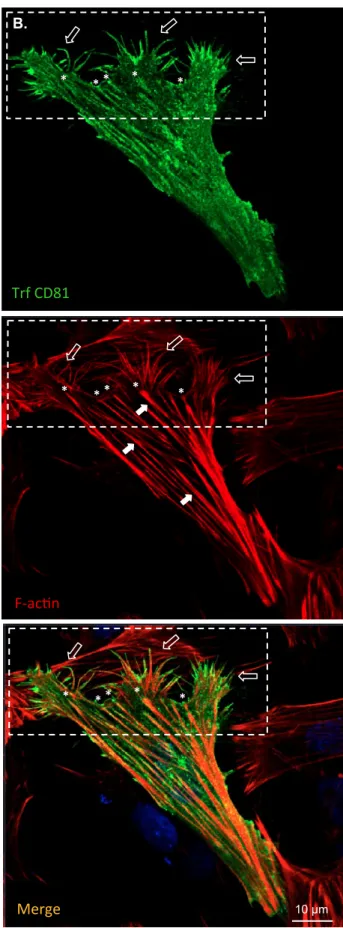
was performed in SH-SY5Y cells. Results show that CD81 was highly located at the cells’ 146
membranes, including in structures also stained for filamentous actin (F-actin), when at 147
endogenous levels (Figure 1A) but particularly when overexpressed (Figure 1B). Moreover, 148
CD81 was particularly enriched in structures related to cell migration, such as filopodia 149
(Figure 1B, open arrows in dashed rectangle), stress fibres (Figure 1B, full arrows, as in 150
(Auer et al. 2017)) and stress fibres’ terminals, most likely adhesion sites (Figure 1B, 151
asterisks). These are subcellular regions where a high degree of co-localization between F-152
actin and transfected CD81 could be observed (Figure 1B, orange staining in ‘Merge’). 153
154
CD81 overexpression promotes the acquisition of a migratory phenotype
155
A first observation of the CD81 overexpressing neuroblastoma population denoted the 156
presence of a high number of cells with a migratory phenotype. This includes a more 157
triangular shape, F-actin concentration at the leading front and at the cell’s rear, and a typical 158
array distribution of the stress fibres (Bari et al. 2011; Qian et al. 2005). The number of 159
CD81 overexpressing cells with this phenotype was scored, and compared to the number of 160
migratory-like non-transfected cells in the neighbourhood. Data shows that CD81 161
9 overexpression doubled the number of cells with this migratory phenotype (Figure 1C). 162
These results suggest a role for CD81 in promoting cellular mechanisms that underlie the 163
acquisition of a migratory phenotype. Further, they suggest that actin, a known interactor of 164
CD81 (Perez-Hernandez et al. 2013), is involved in the CD81 effects on cell morphological 165
transformation. 166
167
CD81 co-localizes with AKT, in transfected and non-transfected SH-SY5Y cells
168
We further analysed if another CD81 interactor, AKT, could also be involved in CD81-169
promotion of neuronal-like cells migratory phenotype. AKT (also known as protein kinase B) 170
is a serine/threonine protein kinase that regulates many processes, including metabolism, 171
proliferation, cell survival, growth and angiogenesis (Yu & Cui 2016). When activated by 172
PI3K, AKT phosphorylates various substrates involved in cytoskeleton remodelling, cell 173
growth and cell survival in neurons. Moreover, the PI3K/AKT signalling pathway is essential 174
for the modulation of the cytoskeleton during cell migration (Xue et al. 2013). As AKT is a 175
CD81 interactor protein, and also the best-known target of PI3K, we investigated if the 176
activation of the AKT/PI3K pathway is part of the mechanism by which CD81 promotes 177
actin remodelling by blocking PI3K to prevent activation and phosphorylation of AKT. 178
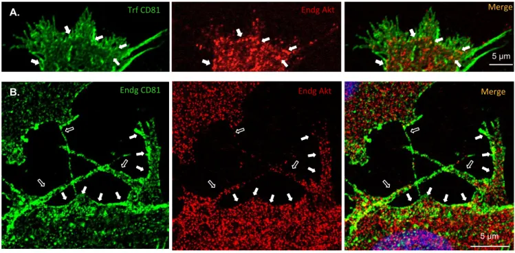
Co-localization analyses, both in CD81 transfected and non-transfected cells (Figure 2), 179
showed that AKT1 was present in the cytosol and at the plasma membrane of the SH-SY5Y 180
cells. Moreover, it could be observed that AKT1 mainly co-localized with CD81 at cellular 181
projections (Figure 2B, open arrows) and at the inner leaflet of the plasma membrane (Figure 182
2, full arrows). Scarcer co-localization between these proteins was observed in cytoplasmic 183
zones further away from the PM. Since AKT is cytoplasmic and translocates to the plasma 184
membrane when activated, this co-localization suggests that CD81 interacts particularly with 185
10 the active form of AKT, and that AKT signalling might be involved in CD81 functions in cell 186
motility. 187
188
PI3K/AKT signalling is involved in CD81-mediated actin remodelling and migratory
189
phenotype
190
PI3K is a canonical upstream activator of AKT, and a possible functional interaction between 191
CD81 and the PI3K-AKT pathway was pursued by treating CD81 transfected cells with 192
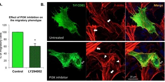
LY294002, a PI3K inhibitor. Inhibition of PI3K resulted in a ~40% decrease in the 193
percentage of transfected cells with a migratory phenotype, when compared to non-treated 194
control cells (Figure 3A). PI3K inhibition also inhibited the acquisition of a migratory 195
phenotype in non-transfected cells, although only by ~20% (data not shown). These results 196
strengthen the hypothesis that the PI3K-AKT pathway mediates the CD81 promotion of 197
migration in these neuronal-like cells. 198
Further, when treated with LY294002 (Figures 3B and 4), CD81 still strongly co-localized 199
with F-actin in CD81 overexpressing cells as it did in unexposed cells of Figure 1B. 200
Nevertheless, contrary to untreated cells where stress fibres and normal F-actin distribution is 201
observed (Figure 1B), the F-actin distribution was now altered. CD81 overexpressing cells 202
exposed to the PI3K inhibitor presented less stress fibres (Figures 3B and 4, arrowheads) and 203
a redistribution of the CD81 staining pattern (Figures 3B and 4 open arrows). A more 204
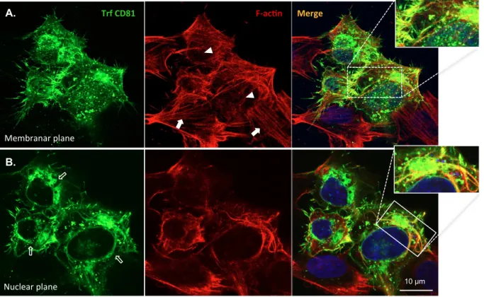
thorough analysis of the CD81 and F-actin distribution in SH-SY5Y cells exposed to the 205
PI3K (Figure 4) revealed that CD81 overexpressing cells had a high number of CD81- and F-206
actin positive filopodia (or less and longer filopodia), but very few stress fibres (arrowheads 207
in Figures 3B and 4). Moreover, CD81 was less enriched at the cell periphery, and was 208
redistributed from the cell membrane to the perinuclear region and to cytoplasmic 209
agglomerates (Figure 4, arrowheads and zoom-in of the nuclear plane). The percentage of 210
11 CD81 transfected cells with a perinuclear ring-like enrichment triplicated when PI3K was 211
inhibited (from ~10% to ~30%), and the number of smaller differentiated cells, with 212
filopodia and cortical F-actin but no stress fibres, duplicated (data not shown). Taken 213
together, these results indicate that the PI3K/AKT signalling pathway is part of the CD81 214
mechanism of F-actin remodelling into cell motility-related structures. 215
12
Discussion
217 218
CD81 is a tetraspanin protein that has been mainly studied in the immune system. Few 219
functions have been attributed to it, but it has been implicated in processes such as cell 220
adhesion and migration. Its expression levels, subcellular distribution and functions in other 221
cells, such as neuronal cells, are still barely known. In the present work, we aimed to evaluate 222
the role of CD81 in the migratory phenotype of these neuronal-like cells. 223
We first confirmed that CD81 is endogenously expressed in the nervous system-related SH-224
SY5Y cell line. This is not surprising since CD81 has been already implicated in the nervous 225
system physiology, being required for the normal development of the brain (Geisert et al. 226
2002). Geisert et al. (2002) studied the effects of a CD81 -/- mutation on the CNS of mice 227
and reported that these mice have extremely large brains. This was as a result of an increased 228
number of astrocytes and microglia, with no apparent effect on the number of neurons and 229
oligodendrocytes (Geisert et al. 2002). Accordingly, CD81 was reported to control astrocytes 230
and microglia cell number by suppressing cell proliferation, in a cell-cell contact-dependent 231
manner (Kelić et al. 2001). Potentially related to this, CD81 is found concentrated at regions 232
of cell-cell contact in cultured astrocytes, and may play a central role in the process of CNS 233
scar formation in spinal cord injury (Dijkstra et al. 2000). 234
In the work here presented we additionally observed that CD81 is mainly present at the 235
plasma membrane of SH-SY5Y neuroblastoma cells, as expected for a protein of the 236
tetraspanin family. CD81 was also found in some spot-like structures of the cytoplasm, with 237
the smaller of these potentially being exosomes, since CD81 was recently observed to be 238
enriched in exosomes-like vesicles (Andreu et al. 2014). 239
More importantly, CD81 was found to clearly promote the number of migrating neuronal-like 240
cells, similarly to its role in immune cells. Cell migration is a highly coordinated cellular 241
13 event, key for various physiological and pathological major processes, including embryonic 242
development, wound healing, immune response (Qian et al 2004). Migration is highly based 243
on actin filament polymerization and remodelling, and motile cells have characteristic 244
discrete actin structures at the cell periphery for attachment to the substratum: focal adhesion, 245
stress fibres, lamellipodia, filopodia, and membrane ruffles (Hall 1998). Overexpression of 246
CD81 alone altered the cells morphology, inducing a reorganization of the actin filaments 247
into cell motility-related structures. 248
A previous bioinformatics analysis of CD81-interacting proteins performed by our group 249
(unpublished data) showed that CD81 was linked to intracellular signalling components 250
involved in cytoskeletal regulation. Included in this group were the following key proteins: 251
AKT1, Rac and cytoskeleton-related proteins, including actin and tubulin. Actin itself is a 252
CD81 interacting protein, as described by Perez-Hernandez et al. (2013). Our results 253
demonstrated that CD81 perfectly co-localizes with F-actin in stress fibres and filopodia at 254
the leading edge of migrating SH-SY5Y cells. We have also observed that CD81 is highly 255
abundant in cytoplasmic cellular spots at the end of the stress fibres that transverse the cell, 256
most probably mature focal adhesions. These are key subcellular locations for a protein with 257
regulatory role on cell migration, and agree with such a role for CD81 on neuronal cells. The 258
CD81 role in neuronal cell migration seems not only to involve F-actin, but also other actin 259
regulators, such as AKT1. The AKT kinase plays a crucial role in neurogenesis by activating 260
the proliferation, migration and differentiation of neural stem and other cells (Koh & Lo 261
2015; Qian et al. 2005). AKT1 is an AKT isoform involved in a variety of signalling 262
pathways related to cell motility and cytoskeleton remodelling. When AKT1 is activated by 263
phosphorylation in the cytoplasm, it is targeted to the inner leaflet of the plasma membrane 264
and phosphorylates a number of subtracts, including actin (Xue et al. 2013). In the present 265
work, we performed an ICC in SH-SY5Y cells to access the subcellular co-localization of 266
14 CD81 and AKT1. AKT1 presented the expected subcellular localization along the entire 267
cytoplasm but, interestingly, CD81 and AKT1 mainly co-localized at the inner leaflet of the 268
plasma membrane and at some cellular projections. In CD81 overexpressing migrating cells, 269
AKT1 co-localized with CD81 at the leading edge. These suggest that CD81 co-localizes 270
with active, membrane-recruited AKT1, and raises the hypothesis that a tri-complex of 271
CD81/AKT/actin may exist and function in neuronal-like cell motility. Activated AKT at the 272
leading edge of the cell is already known to participate in the regulation of cell polarity and in 273
the reorganization of the cytoskeleton, mediating contraction of the cellular body that 274
facilitates directed cell migration (Xue et al. 2013). AKT is one of the best-known targets of 275
PI3K, and the PI3K/AKT pathway has role in neural migration by e.g. enhancing the 276
secretion of matrix metalloproteinase (MMP)-2 and MMP-9 (Koh et al. 2015). In chicken 277
embryo fibroblast (CEF) cells, the expression of active PI3K forms alone is enough to induce 278
the remodelling of actin filaments towards the formation of cell’s motility structures (Qian et 279
al 2004). The authors further reported that either the inhibition of PI3K activity with 280
LY294002, or the disruption of AKT activity in CEF cells inhibited both actin remodelling 281
and PI3K-induced cell migration (Qian et al. 2004). In our work, further support to the 282
existence of a CD81/AKT/actin tri-complex active in cell motility comes from our PI3K 283
pharmacological inhibitor data. The blocking of this signalling pathway, and thus of AKT 284
activation, partially impaired the CD81 positive effect on cell motile phenotype. Further, 285
PI3K inhibition leads to altered CD81 and actin subcellular distribution in the transfected SH-286
SY5Y neuroblastoma population (Figures 3 and 4). This population comprises two types of 287
cells: the larger S-type cells that are more neuroepithelial-related, and the smaller more 288
neuronal-related N-type cells (da Rocha et al. 2015). In all CD81 overexpressing cells there 289
was a decrease in F-actin stress fibres. Moreover, CD81 overexpressing larger cells 290
(potentially S-type) presented and intracellular accumulation of CD81/F-actin into 291
15 perinuclear ring-like structures, cytoplasmic agglomerates and/or protruding filaments. Other 292
authors have reported that CD81 overexpression promoted the formation of microvilli in B-293
cells, via reorganization of the cortical actin cytoskeleton (Bari et al. 2011). Further, CD81 294
overexpressing smaller (potential N-type) cells increased their neuronal-like differentiated 295
phenotype. 296
The fact that F-actin co-localizes with several of the CD81 intracellular agglomerates agrees 297
with the hypothesis that CD81-actin interaction is part of the mechanism by which CD81 298
regulates cell migration. Since impairing AKT activation resulted both in CD81 and actin re-299
location and in decreased CD81-induced motile phenotype (Figures 3 and 4), and since AKT 300
is known to bind CD81 and to phosphorylate actin to promote it’s remodelling, the 301
hypothesis that a complex of active AKT-CD81-actin exists is very reasonable, and would 302
partially explain the role of CD81 in cell migration. We hypothesize that if such a complex 303
exists it is necessary for, at least, actin polymerization and the polarized distribution of F-304
actin into motile structures such as stress fibres and adhesion sites. 305
In addition to AKT, the molecular mechanism by which CD81 promotes neuronal-like cells 306
motility may also involve other CD81 interactors and actin regulator such as Rac. This small 307
G protein of the Rho family is known to promote actin polymerization and to be involved in 308
cell migration. Rac may act downstream CD81, since a direct association between CD81 and 309
Rac in TEM was reported, with CD81-Rac complexes being most prominent at motile cells’ 310
leading edge (Tejera et al. 2013). These authors hypothesize that CD81 regulates Rac1 311
dynamics and localization at the cell membrane during membrane protrusion and during the 312
formation of adhesion complexes (Tejera et al. 2013). Noteworthy, besides activating AKT, 313
PI3K can also activate Rac, and both molecules can be involved in PI3K-induced cell motile 314
phenotype (Qian et al. 2004). Other authors have already hypothesized that CD81 may 315
coordinate cell migration via the regulation of key migration-related proteins with which 316
16 CD81 interacts in the ‘tetraspanin web’(Boucheix & Rubinstein 2001; Hemler 2005; Jiang et 317
al. 2015). Since Rac, AKT and actin are CD81-binding proteins, and CD81 is involved in 318
tetraspanin microdomains that may ‘catalyse’ cellular processes, we propose a working 319
scenario where, in TEM, CD81 serves as an anchor for proteins such as AKT, Rac and actin, 320
to promote their interaction and the actin cytoskeleton remodelling that will lead to the 321
acquisition of the polarized cell motile morphology. Although the SH-SY5Y cell line has 322
been extensively used in the study of neuronal cell cultures, it is tumour-derived and cannot 323
fully recapitulate the properties of the neuronal cells in vivo (Gordon et al. 2013). Thus our 324
data should be validated using primary neuronal cells or human induced pluripotent stem cell 325
(iPSC)-derived neurons. The confirmation of our hypothesis will improve our knowledge of 326
the molecular mechanisms behind neuronal cell migration, an important event underlying 327
various major neurological processes, such as neural development and neuroregeneration. 328
329
Conclusion
330
In neuronal-like cells, similarly to its role in immune cells, CD81 promotes cell 331
transformation events that underlie the acquisition of a migratory phenotype. The mechanism 332
behind CD81-enhancement of SH-SY5Y motility passes by the remodelling of the actin 333
cytoskeleton. Moreover, our findings indicate that the PI3K-AKT signalling mediates this 334
CD81 role in actin remodelling and subsequent polarization into a motile cell, and support the 335
existence of a CD81-AKT-actin complex as a key molecular effector of this CD81 role. 336
337
Acknowledgments
338
This work was supported Fundação para a Ciência e Tecnologia (Portuguese Ministry of 339
Science and Technology), Centro 2020 and Portugal2020, the COMPETE program, QREN, 340
and the European Union (FEDER program) via Institute for Biomedicine iBiMED 341
17 UID/BIM/04501/2013, fellowship SFRH/BD/90996/2012, project PTDC/CVT-342
CVT/32261/2017, and the support of the LiM facility of iBiMED, a member of the 343
Portuguese Platform of BioImaging (PPBI- POCI-01-0145-FEDER-022122). 344
18
References
346
Andreu, Z., & Yáñez-Mó, M. (2014). Tetraspanins in extracellular vesicle formation and 347
function. Frontiers in Immunology, 5, 442. 348
Auer, S., Rinnerthaler, M., Bischof, J., … Breitenbach, M. (2017). The Human NADPH 349
Oxidase, Nox4, Regulates Cytoskeletal Organization in Two Cancer Cell Lines, HepG2 350
and SH-SY5Y. Frontiers in Oncology, 7, 111. 351
Bari, R., Guo, Q., Xia, B., … Zhang, X. A. (2011). Tetraspanins regulate the protrusive 352
activities of cell membrane. Biochemical and Biophysical Research Communications, 353
415(4), 619–626.
354
Boucheix, C., & Rubinstein, E. (2001). Tetraspanins. Cellular and Molecular Life Sciences : 355
CMLS, 58(9), 1189–205. 356
da Rocha, J. F., da Cruz e Silva, O. A. B., & Vieira, S. I. (2015). Analysis of the amyloid 357
precursor protein role in neuritogenesis reveals a biphasic SH-SY5Y neuronal cell 358
differentiation model. Journal of Neurochemistry, 134(2), 288–301. 359
Dijkstra, S., Geisert EE, J. R., Gispen, W. H., Bär, P. R., & Joosten, E. A. (2000). Up-360
regulation of CD81 (target of the antiproliferative antibody; TAPA) by reactive 361
microglia and astrocytes after spinal cord injury in the rat. The Journal of Comparative 362
Neurology, 428(2), 266–77. 363
Geisert, E. E., Williams, R. W., Geisert, G. R., … Levy, S. (2002). Increased brain size and 364
glial cell number in CD81-null mice. The Journal of Comparative Neurology, 453(1), 365
22–32. 366
Gordon, J., Amini, S., & White, M. K. (2013). General overview of neuronal cell culture. 367
Methods in Molecular Biology (Clifton, N.J.), 1078, 1–8. 368
Hall, A. (1998). Rho GTPases and the actin cytoskeleton. Science (New York, N.Y.), 369
279(5350), 509–14.
370
Hemler, M. E. (2005). Tetraspanin functions and associated microdomains. Nature Reviews. 371
Molecular Cell Biology, 6(10), 801–11. 372
Jiang, X., Zhang, J., & Huang, Y. (2015). Tetraspanins in cell migration. Cell Adhesion and 373
Migration, 9(5), 406–415. 374
19 Kelić, S., Levy, S., Suarez, C., & Weinstein, D. E. (2001). CD81 regulates neuron-induced 375
astrocyte cell-cycle exit. Molecular and Cellular Neurosciences, 17(3), 551–560. 376
Koh, S. H., & Lo, E. H. (2015). The role of the PI3K pathway in the regeneration of the 377
damaged brain by neural stem cells after cerebral infarction. Journal of Clinical 378
Neurology (Korea), pp. 297–304. 379
Kovalevich, J., & Langford, D. (2013). Considerations for the use of SH-SY5Y 380
neuroblastoma cells in neurobiology. Methods in Molecular Biology (Clifton, N.J.), 381
1078, 9–21.
382
Levy, S., Todd, S. C., & Maecker, H. T. (1998). CD81 (TAPA-1): a molecule involved in 383
signal transduction and cell adhesion in the immune system. Annual Review of 384
Immunology, 16, 89–109. 385
Marín, O., Valiente, M., Ge, X., & Tsai, L.-H. (2010). Guiding neuronal cell migrations. Cold 386
Spring Harbor Perspectives in Biology, 2(2), a001834. 387
Perez-Hernandez, D., Gutiérrez-Vázquez, C., Jorge, I., … Yáñez-Mó, M. (2013). The 388
intracellular interactome of tetraspanin-enriched microdomains reveals their function as 389
sorting machineries toward exosomes. The Journal of Biological Chemistry, 288(17), 390
11649–61. 391
Qian, Y., Corum, L., Meng, Q., … Jiang, B.-H. (2004). PI3K induced actin filament 392
remodeling through Akt and p70S6K1: implication of essential role in cell migration. 393
American Journal of Physiology-Cell Physiology, 286(1), C153–C163. 394
Qian, Y., Zhong, X., Flynn, D. C., … Jiang, B.-H. (2005). ILK mediates actin filament 395
rearrangements and cell migration and invasion through PI3K/Akt/Rac1 signaling. 396
Oncogene, 24(19), 3154–3165. 397
Shoham, T., Rajapaksa, R., Kuo, C.-C., Haimovich, J., & Levy, S. (2006). Building of the 398
tetraspanin web: distinct structural domains of CD81 function in different cellular 399
compartments. Molecular and Cellular Biology, 26(4), 1373–85. 400
Tejera, E., Rocha-Perugini, V., López-Martín, S., … Yáñez-Mo, M. (2013). CD81 regulates 401
cell migration through its association with Rac GTPase. Molecular Biology of the Cell, 402
24(3), 261–73.
403
Xue, G., & Hemmings, B. A. (2013). PKB/Akt-dependent regulation of cell motility. Journal 404
20 of the National Cancer Institute, 105(6), 393–404.
405
Yu, J. S. L., & Cui, W. (2016). Proliferation, survival and metabolism: the role of 406
PI3K/AKT/mTOR signalling in pluripotency and cell fate determination. Development, 407
143(17), 3050–3060.
408 409 410
21
Figures Captions
411
412
Figure 1. CD81 transfected neuronal-like cells have increased migratory phenotype. A.
413
and B. Confocal micrographs of SH-SY5Y neuroblastoma cells non-transfected (A.) or 414
transfected (B.) with CD81 for 24h. Cells were fixed and subjected to ICC with an anti-CD81 415
antibody (labeled with AlexaFluor488, in green), red AlexaFluor568 Phalloidin to stain F-416
actin, and DAPI to stain the cells’ nuclei (in blue). Dashed rectangle in B. images – cell 417
leading edge. Full arrows – stress fibres. Open arrows – filopodia and similar cell protrusions. 418
Asterisks – potential focal adhesion sites. Endg, endogenous; Trf, transfected. C. Graphic 419
representation of the percentages of cells with a migratory phenotype, visually scored in SH-420
SY5Y cells overexpressing or not CD81. *, p < 0.05, statistical significance determined by 421
either the independent sample t-test or the Mann-Whitney U test (p = 0.012 in both); n=5. 422
423
Figure 2. Co-localization between CD81 and AKT1. Confocal microscopy analysis of the
424
subcellular co-localization of CD81 (in green), either exogenous (A., transfected cells) or 425
endogenous (B., parental cells), with AKT1 (in red). ICC was performed using an anti-CD81 426
antibody (green, secondary antibody AlexaFluor488) and anti-AKT1 antibody (red, 427
secondary AlexaFluor594). Open arrows – filopodia and similar cell protrusions. Full arrows 428
– location at the cell membrane. 429
430
Figure 3. PI3K inhibition decreases the percentage of migrating cells. A. SH-SY5Y cells
431
transfected with CD81 cDNA were incubated with 10 μM LY294002, a PI3K inhibitor for 432
18h. The effect of PI3K inhibition on the number of CD81-transfected cells with migratory 433
phenotype was quantified, taking the number of CD81-transfected migratory cells in control 434
22 (untreated) cells as 100%. *, p < 0.05, statistical significance according to the one sample t-435
test; p = 0.0504 when using the non-parametric Wilcoxon test; n=3. B. Confocal microscopy 436
images of transfected CD81 and F-actin staining, in control cells (untreated; upper panel) and 437
in cells incubated with the LY294002 PI3K inhibitor (lower panel). Full arrows indicate F-438
actin stress fibres. Arrowheads indicate a decrease in stress fibres in LY294002 treated cells. 439
Open arrow indicates cell with a CD81 ‘ring-like’ distribution. 440
441
Figure 4. PI3K inhibition alters CD81 and F-actin subcellular distribution.
CD81-442
transfected SH-SY5Y cells were incubated with 10 μM of the PI3K inhibitor LY294002, for 443
18h. Confocal microphotographs at the plasma membrane (A.) and nuclear (B.) focal planes 444
show the abnormal concentration of CD81 internally, at cytoplasmic agglomerates and 445
internal filaments, besides filopodia. CD81 and F-actin co-localize extensively in these 446
structures (zoom-ins). Full arrows indicate F-actin stress fibres in non-transfected cells. 447
Arrowheads indicate a decrease in stress fibres in transfected cells. Open arrows indicate the 448
CD81 ‘ring-like’ distribution. 449
Figure 1. CD81 transfected neuronal-‐like cells have increased migratory phenotype. A. and B. Confocal micrographs of SH-‐SY5Y neuroblastoma cells non-‐ transfected (A.) or transfected (B.) with CD81 for 24h. Cells were fixed and subjected to ICC with an anG-‐CD81 anGbody (labeled with AlexaFluor488, in green), red AlexaFluor568 Phalloidin to stain F-‐acGn, and DAPI to stain the cells’ nuclei (in blue). Dashed rectangle in B. images – cell leading edge. Full arrows – stress fibres. Open arrows – filopodia and similar cell protrusions. Asterisks – potenGal focal adhesion sites. Endg, endogenous; Trf, transfected. C. Graphic representaGon of the percentages of cells with a migratory phenotype, visually scored in SH-‐SY5Y cells overexpressing or not CD81. *, p < 0.05, staGsGcal significance determined by either the independent sample t-‐test or the Mann-‐ Whitney U test (p = 0.012 in both); n=5.
* * * * * Trf CD81 F-‐acGn * * * * * 10 µm Merge * * * * * B. 0 10 20 30 40 50 60 70 80 90 Non-transfected
cells CD81 transfected cells
% o f ce lls w / mi gra to ry ph en ot yp e * C. A. F-‐ac@n Endg CD81 DAPI 5 µm
Figure 2. Co-‐localiza@on between CD81 and AKT1. Confocal microscopy analysis of the subcellular co-‐localizaGon of CD81 (in green), either exogenous (A., transfected cells) or endogenous (B., parental cells), with AKT1 (in red). ICC was performed using an anG-‐CD81 anGbody (green, secondary anGbody AlexaFluor488) and anG-‐AKT1 anGbody (red, secondary AlexaFluor594). Open arrows – filopodia and similar cell protrusions. Full arrows – locaGon at the cell membrane. Endg Akt 5 µm Trf CD81 Merge Endg Akt Endg CD81 5 µm Merge A. B.
Figure 3. PI3K inhibi@on decreases the percentage of migra@ng cells. A. SH-‐SY5Y cells transfected with CD81 were incubated with 10 μM LY294002, a PI3K inhibitor, for 18h. The effect of PI3K inhibiGon on the number of CD81-‐transfected cells with migratory phenotype was quanGfied, taking the number of CD81-‐ transfected migratory cells in control (untreated) cells as 100%. *, p < 0.05, staGsGcal significance according to the one sample t-‐test; p = 0.0504 when using the non-‐parametric Wilcoxon test; n=3. B. Confocal microscopy images of transfected CD81 and F-‐acGn staining, in control cells (untreated; upper panel) and in cells incubated with the LY294002 PI3K inhibitor (lower panel). Full arrows indicate F-‐acGn stress fibres. Arrowheads indicate a decrease in stress fibres in LY294002 treated cells. Open arrow indicates cell with a CD81 ‘ring-‐like’ distribuGon. A. B. Trf CD81 F-‐ac@n Merge Untreated PI3K inhibitor 0 20 40 60 80 100 120 Control LY294002 % o f mi gra to ry ce lls
Effect of PI3K inhibition on the migratory phenotype
*
Figure 4. PI3K inhibi@on alters CD81 and F-‐ac@n subcellular distribu@on. CD81-‐transfected SH-‐SY5Y cells were incubated with 10 μM of the PI3K inhibitor LY294002, for 18h. Confocal microphotographs at the plasma membrane (A.) and nuclear (B.) focal planes show the abnormal concentraGon of CD81 internally, at cytoplasmic agglomerates and internal filaments, besides filopodia. CD81 and F-‐ acGn co-‐localize extensively in these structures (zoom-‐ins). Full arrows indicate F-‐acGn stress fibres in non-‐transfected cells. Arrowheads indicate a decrease in stress fibres in transfected cells. Open arrows indicate the CD81 ‘ring-‐like’ distribuGon.
Membranar plane Nuclear plane F-‐ac@n Trf CD81 Merge 10 µm A. B.