Maria Clara Ferreira de
Almeida Cardia Gomes
Galactoporfirinas catiónicas na inactivação de
microrganismos
Maria Clara Ferreira de
Almeida Cardia Gomes
Galactoporfirinas catiónicas na inactivação de
microrganismos
Cationic Galactoporphyrins on Microorganisms
Photoinactivation
Dissertação apresentada à Universidade de Aveiro para cumprimento dos requisitos necessários à obtenção do grau de Mestre em Biologia Aplicada com especialização em Microbiologia Clínica e Ambiental, realizada sob a orientação científica do Doutor João Paulo Costa Tomé, Investigador Auxiliar do Departamento de Química da Universidade de Aveiro e sob a co-orientação científica da Doutora Maria Ângela Sousa Dias Alves Cunha, Professora Auxiliar do Departamento de Biologia da Universidade de Aveiro.
o júri
Presidente Doutor António José Arsénia Nogueira
Prof. Associado c/ Agregação do Departamento de Biologia da Universidade de Aveiro
Vogais Doutora Rosa Cristina Simões Fernandes
Investigadora Auxiliar da Faculdade de Medicina da Universidade de Coimbra Doutor João Paulo Costa Tomé (orientador)
Investigador Auxiliar do Departamento de Química da Universidade de Aveiro Doutora Maria Ângela Sousa Dias Alves Cunha (co-orientadora)
agradecimentos Ao Doutor João Paulo Tomé, orientador da dissertação, por aceitar colaborar neste trabalho, pelo incentivo e confiança.
À Professora Doutora Ângela Cunha, co-orientadora da dissertação, pelo seu rigor científico, pelas críticas construtivas, dedicação e amizade.
Ao Professor José Cavaleiro, por me ter permitido descobrir um novo mundo.
À Professora Doutora Adelaide Almeida, pela disponibilidade e sugestões pertinentes.
À Professora Doutora Graça Neves e ao Professor Doutor Augusto Tomé, pela disponibilidade, pelas sugestões e por toda a ajuda.
Aos colegas do laboratório de Química Orgânica, principalmente àqueles que me receberam e partilharam comigo os seus conhecimentos.
Aos colegas de laboratório de Microbiologia Ambiental e Aplicada, por toda a ajuda e simpatia.
À Ana Luísa, por toda a disponibilidade, simpatia, por todos os conhecimentos que me transmitiu.
À Joana Brás, a minha companheira nesta nova descoberta de novos mundos, por toda a amizade.
Ao Bruno, por todo o apoio ao longo destes anos e por ter-me sempre incentivado
Aos meus pais, pelo carinho, motivação e amor incondicional por tudo. À Universidade de Aveiro pelo financiamento do QOPNA e CESAM. À FCT (Lisboa) e FEDER pelo financiamento do QOPNA.
Ao Projecto PTDC/QUI/65228/2006 pelo financiamento parcial do trabalho experimental.
palavras-chave Terapia fotodinâmica antimicrobiana, Porfirinas, Glicoporfirinas, Oxigénio singuleto, Oxidação lipídica, Oxidação proteica
resumo A terapia fotodinâmica antimicrobiana (aPDT) está-se a tornar uma
alternativa promissora para inactivar microrganismos patogénicos. Esta terapia consiste na combinação de três elementos não tóxicos, fotosensibilizadores (PS), luz e oxigénio que, quando combinados conduzem à formação de espécies reactivas de oxigénio altamente citotóxicas, nomeadamente o oxigénio singuleto. Estas espécies podem oxidar muitos tipos de moléculas biológicas, como o caso de proteínas, ácidos nucleicos e lípidos. A combinação de grupos com cargas positivas e hidratos de carbono com derivados porfirínicos resulta num aumento de reconhecimento celular e solubilidade em água, melhorando a penetração na membrana celular e acumulação em compartimentos sub-celulares.
O objectivo deste trabalho foi sintetizar novos derivados porfirínicos meso-glicosil substituídos e avaliar a eficácia destes compostos como PS na fotoinactivação de duas bactérias gram positivas ambientais, Brevibacterium sp. e Micrococcus sp. e uma bactéria gram negativas, Escherichia coli bioluminescente. Brevibacterium sp. e Micrococcus sp. foram escolhidas para este estudo por serem, respectivamente, representantes de tipos muito sensíveis e resistentes a experiências de irradiação UV-B. Também foi avaliado o efeito do 1O2 a nível de oxidação de lipídios e proteínas, sobre as
duas bactérias gram positivas.
Os derivados porfirínicos meso-tetrapiridil foram cationizados com iodeto de metilo e unidades glicosídicas. Todos os compostos sintetizados foram caracterizados por ressonância magnética nuclear dos protões e fluor e por espectrometria de massa. Dois desses compostos sintetizados, 5,10,15,20-tetrakis(N-methilpiridinium-4-il)porfirina tetra-iodeto (PS 1) e a 5-[N- (isopropilidene-6-deoxi-galactopiranos-6-il)piridinium-4-il]-10,15,20-tris(N-metilpiridinium-4-il)porfirina tetra-iodeto (PS 2), foram usados como PS nos estudos de aPDT. Nos estudos de aPDT foram irradiadas, após uma pré-incubação no escuro, suspensões bacterianas puras com 0.5, 1 e 5 µmol dm-3 de PS no caso das bactérias gram positivas, e 5 µmol dm-3 no caso da E. coli. A cinética de irradiação foi avaliada através da quantificação de unidades formadoras de colónias (UFC) colhidas durante os 15 minutos de irradiação, sob 150 mW cm-2. Foram incluídos controlos claros e escuros em todas as experiências. Foram também realizados estudos fotofísicos (Fotoestabilidade e Geração de 1O2). A oxidação lipídica foi avaliada através do ensaio com ácido
tiobarbitúrico (TBA) e os resultados expressos em termos de malondialdeído (MDA) (nmol dm-3). A oxidação de proteínas foi avaliada através do 2,4-dinitrofenilidrazina (DNPH) e os resultados expressos em termos de teor de proteína carbonilada (nmol cm-3).
Os ensaios de aPDT revelaram que o PS 2 foi mais eficaz (3,0 log de recução) do que o PS 1 (2,0 log) com 5 !mol dm-3 contra E. coli. No caso das bactérias gram positivas, ambos os PS demonstraram o mesmo efeito de fotoinactivação, apresentando uma inativação completa após 2 minutos de irradiação. No entanto, nas menores concentrações o PS 1 mostrou ser mais eficaz do que o PS 2 em ambas as bactérias gram positivas. Os estudos fotofísicos demonstraram que ambos os PS são fotoestáveis e bons produtores de 1O2. Os ensaios de peroxidação lipídica apresentaram
resultados diferentes para ambas as bactérias ambientais. No Micrococcus não foi observada oxidação lipídica com PS 1, enquanto com o PS 2 foi observado cerca de 31% de oxidação lipídica (0,083 nmol dm-3). No
Brevibacterium o PS 1 causou 28% (0,063 nmol dm-3) e o PS 2 50% (0,093 nmol dm-3) de peroxidação lipídica. De acordo com os resultados de oxidação de proteínas, o Micrococcus apresentou cerca de 2,1 nmol mL-1 e 6,2 nmol mL
-1 de carbonilação de proteínas com o PS 1 e 2, respectivamente. No caso de
Brevibacterium foram observados 5,0 nmol mL-1 e 4,8 nmol mL-1 com o PS 1 e 2, respectivamente.
Ambas as porfirinas mostraram bons resultados tanto na fotoinactivação bactérias gram negativa como nas gram positivas. A suscetibilidade do
Brevibacterium sp. e Micrococcus sp. na aPDT foi diferente dos resultados
observados na radiação UV-B, por estas mesmas bactérias. Os ensaios de oxidação lipídica permitiu concluir que, no caso Brevibacterium ambos os PS actuam na membrana plasmática, enquanto que no Micrococcus estes danos só acontecem com o PS 2. A oxidação de proteínas levou à conclusão que os danos a nível das proteínas podem ter ocorrido devido à oxidação lipídica, ou interação directa entre o 1O2 e as proteínas.
keywords Antimicrobial Photodynamic Therapy, Porphyrin, Glycoporphyrin, Singlet oxygen, Lipid oxidation, Protein oxidation
abstract Antimicrobial photodynamic therapy (aPDT) is becoming a promising alternative to inactivate microbial pathogens.This therapy combines three non-toxic components, a photosensitizer (PS), light and oxygen, that when combined leads to the formation of highly cytotoxic reactive oxygen species, mainly singlet oxygen (1O2). This specie can oxidize many types of biological
molecules, such as proteins, nucleic acids and lipids. The combination of positively charged groups and carbohydrate moieties with porphyrin derivatives results in an increased cell recognition and water solubility, which improves cell membrane penetration and accumulation in sub-cellular compartments.
The aim of this work was to synthesize new meso-substituted glycosyl porphyrins derivatives and evaluated the efficacy of these compounds as PS in the photoinactivation of two environmental gram positive bacteria,
Brevibacterium sp. and Micrococcus sp., and one gram negative bacteria,
bioluminescent Escherichia coli. Brevibacterium sp. and Micrococcus sp were chosen for these studies because they were, respectively, representative of very sensitive and very resistant types to UV-B irradiation experiments. It was also evaluated the effect of 1O2 at the lipid and protein oxidation level,
generated during the aPDT assay, on the two gram positive bacteria.
The derivatives of meso-tetrapyridyl porphyrin were cationized by methyl iodide or by carbohydrate moieties. All synthesized compounds were characterized by proton and fluor nuclear magnetic resonance and by mass spectrometry. Two of the compounds synthesized 5,10,15,20-tetrakis(N-methylpyridinium-4-yl)porphyrin tetra-iodide (PS 1) and 5-[N-(Isopropylidene-6- deoxy-galactopyranos-6-yl)pyridinium-4-yl]-10,15,20-tris(N-methylpyridinium-4-yl)porphyrin tetra-iodide (PS 2) were used as PS in the aPDT assays. For the aPDT assays pure bacterial suspensions were irradiated after pre-incubation in the dark, at concentrations of 0.5, 1 and 5 µmol dm-3 of PS in the case of gram positive bacteria, and 5 µmol dm-3 in the case of gram negative bacteria. The kinetics of irradiation was evaluated by the quantification of colony forming units in aliquots collected during 15 minutes of irradiation, under 150 mW cm-2. Light and dark controls were included in all experiments. Photophysical testes (photostability and 1O2 genereation studies) were also performed. Lipid
oxidation was assessed by Tiobarbituric acid (TBA) assay and results were expressed in terms of Malondialdehyde (MDA) (nmol dm-3). Protein oxidation was evaluated by 2,4-dinitrophenylhydrazine (DNPH) assay and results were expressed in terms of Protein carbonyl concentration (nmolcm-3).
aPDT assays revealed that PS 2 was more effective (3.0 log of reduction) than the PS 1 (2.0 log) with 5 µmol dm-3 against E. coli. In the case of gram positive bacteria, both PS showed the same photoinactivation effect, presenting complete inactivation after 2 minutes of irradiation. However, with lower concentration PS 1 showed to be more effective than PS 2 in both gram positive bacteria. Photophysical studies showed that both PS are photostable and good 1O2 producers.
!
Lipid peroxidation assays displayed different results for both environmental bacteria. In Micrococcus no lipid oxidation was observed with PS 1 while with PS 2 was observed around 31% of lipid oxidation (0.083 nmol dm-3). In
Brevibacterium PS 1 caused 28% (0.063 nmol dm-3) and PS 2 50% (0.093 nmol dm-3) of lipid peroxidation. According to the protein oxidation results,
Micrococcus showed around 2.1 nmol mL-1 and 6.2 nmol mL-1 of protein carbonyls with PS 1 and 2, respectively. In the case of Brevibacterium 5.0 nmol mL-1 and 4.8 nmol mL-1 were observed with PS 1 and 2, respectively.
Both porphyrins showed good photoinactivation results on gram negative and gram positive bacteria. Susceptibility of Brevibacterium sp. and
Micrococcus sp. to aPDT were different to those showed in UV-B irradiation by
these same bacteria. Lipid oxidation assays allowed to conclude that, in
Brevibacterium, both PS act in plasma membrane while in Micrococcus this
only happens with the PS with a carbohydrate moiety. Protein oxidation led to the conclusion that protein damage may have occurred due to lipid oxidation or direct interaction of 1O2 with proteins.
i
TABLE OF CONTENTS
LIST OF FIGURES v
LIST OF TABLES vii
LIST OF ACRONYMS AND ABBREVIATIONS ix
Chapter I - Introduction
1
1.1 Photodynamic effect – Brief history 3
1.2 Photodynamic therapy 4
1.3 Antimicrobial photodynamic therapy 5
1.4 Porphyrins 6
1.4.1 Overview 7
1.4.2 Nomenclature of porphyrin derivatives 8
1.4.3 Chemical and physical properties 9
1.4.4 Synthesis of meso–tetra-substituted porphyrins 11
1.4.5 General applications 14
1.5 Porphyrins in aPDT – applications 15
1.6 Factors affecting aPDT efficiency 16
1.6.1 Light source 16
1.6.2 Porphyrins as photosensitizers 17
1.6.3 Mechanism of photosensitization: the role of the oxygen 21
1.7 Thesis outline 22
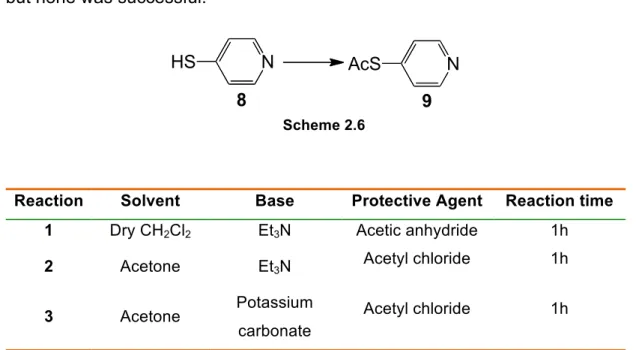
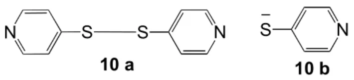
Chapter II – Synthesis of galactoporphyrin derivative
25
2.1 Glycoporphyrins – Overview 27
2.2 Experimental procedures 29
2.3 Initial working plan 34
2.4 Synthesis of template compounds used for biological
studies 36
2.4.1 Synthesis of 5,10,15,20-tetrakis(pentafluorophenyl)porphyrin
!
2.4.2 Synthesis of
5,10,15,20-tetrakis[2,3,5,6-tetrafluor-4-(4-pyridylsulfanyl)phenyl]porphyrin 37
2.4.3 Synthesis of 6-iodo-1,2:3,4-Di-O-isopropylidene-!-D
-galactopyranose 38
2.5 Synthesis of the galactoporphyrins 39
2.5.1 Coupling of 5,10,15,20-tetrakis[2,3,5,6-tetrafluor-4-(4-phyridylsulfanyl)phenyl]porphyrin to galactose iodide derivative 39 2.5.2 Coupling of galactose to 5,10,15,20-tetrakis(4-pyridyl) porphyrin 42
2.6 Methylation of compounds to use in biological assays 44
2.6.1 Methylation of glycoporphyrin 12 44
2.6.2 Methylation of porphyrin 5,10,15,20-tetrakis(4-pyridyl)porphyrin 45
Chapter III – In vitro effects of galactoporphyrins on
Environmental bacterial strain
47
3.1 Introduction 49
3.2 Materials and Methods 52
3.2.1 Photosensitizers 52
3.2.2 Bacterial strains, growth conditions and preparation of stock-
suspensions 52
3.2.3 aPDT experimental setup 54
3.2.4 Irradiation conditions 54
3.2.5 Bioluminescence screening assays 54
3.2.6 Quantification of colony forming units 55
3.2.7 Controls 56
3.2.8 Photostability studies of PS 1 and 2 56
3.2.9 Singlet oxygen generation studies. 56
3.2.10 Indicators of bacterial oxidative stress 57
3.2.10.1 Lipid oxidation 57
3.2.10.2 Protein oxidation 58
3.3 Results and Discussion 60
3.3.1 Photoinactivation experiments 60
iii
3.3.1.2 Photodynamic inactivation of Micrococcus sp. and
Brevibacterium sp. 61
3.3.2 Photostability of PS 1 and 2 64
3.3.3 Singlet oxygen production of PS 1 and 2 65
3.3.4 Lipid oxidation in Micrococcus sp. and Brevibacterium sp. 66 3.3.5 Protein oxidation of Micrococcus sp. and Brevibacterium sp. 69
Chapter IV – Main Conclusions
73
v
LIST OF FIGURES
Figure 1.1 First documented patient treated with eosin and sunlight for skin
lesions. Left picture before, and right picture after the treatment. 3
Figure 1.2 Some tetrapyrrolic units. 7
Figure 1.3 Natural tetrapyrrolic macrocycles: a) Heme, b) Chlorophyll. 8 Figure 1.4 Left: Nomenclature proposed by Hans Fisher. Right: Nomenclature
proposed by IUPAC. 9
Figure 1.5 Structure of porphyrin Core. 9
Figure 1.6 UV-vis spectra of porphyrin derivatives. 10 Figure 1.7 Molecular structures of porphyrins used by Caminos et al. 19 Figure 1.8 Molecular structures of porphyrins used by Cormick et al. 20
Figure 1.9 Modified Jablonski diagram. 21
Figure 2.1 Some glycoporphyrin derivatives already synthesized. 28 Figure 2.2 Compounds obtained during the reaction 1, 2 and 3. 40
Figure 2.3 1H NMR of compound 12. 42
Figure 2.4 1H NMR of compound 13. 44
Figure 3.1 Photosensitizer tested in aPDT assays.
5,10,15,20-tetrakis(N-methylpyridinium-4-yl)porphyrin tetra-iodide (1) and 5-[N-(Isopropylidene-6-deoxy-
galactopyranos-6-yl)pyridinium-4-yl]-10,15,20-tris(N-methylpyridinium-4-yl)porphyrin tetra-iodide (2). 52
Figure 3.2 Relationship between bioluminescence and viable counts of a growing
culture of recombinant E. coli. Bioluminescence is expressed in relative light units
(RLU) and viable counts in CFU mL!1. 55
Figure 3.3 Relationship between absorbance and MDA concentration. MDA
concentration is expressed in nmol dm-3. 58
!
Figure 3.5 Variation of the concentration of viable bioluminescent E. coli during
PDI experiments with 5 !mol dm-3 of PS 1 and 2 using white light at a fluence rate of 150 mW.cm-2. Values correspond to the average of two independent experiments. Error bars represent the standard deviation. (—!—light control, — ! —dark control PS 1, —!—dark control PS 2, — " — PS 1, — x —PS 2). 60
Figure 3.6 Variation of the concentration of CFU in Micrococcus sp. (upper charts)
and Brevibacterium sp. (lower charts) cell suspensions during PDI experiments with 0.5, 1 and 5 !M of PS 1 and PS 2, using white light at a fluence rate of 150 mW.cm-2. Values correspond to the average of two independent experiments. Error bars represent standard deviation. (—!—light control, — " —dark control,
—!—0.5 !mol dm-3, —x—1 !mol dm-3, — " —5 !mol dm-3). 62
Figure 3.7 Absorbance spectra of PS 1 and 2 after irradiation with white light (150
mW.cm-2) for different periods of time. 64
Figure 3.8 Relative decrease in absorption as a measure of photooxidation of
DPiBF (50 µmol L-1) in DMF/H
2O (9:1) upon irradiation with white light filtered
through a cut-off filter for wavelengths <540 nm (9 mW cm-2) with or without
photosensitizer 1 and 2 (0.5 µmol L-1). Values correspond to the average of two
independent experiments. (—!—DPiBF, —!—PS 1, —!—PS 2). 66
Figure 3.9 Lipid peroxidation kinetics in Micrococcus sp. (up charts) and
Brevibacterium sp. (down chart) by PS 1 and 2. Cell suspensions were treated with PS 1 and 2 (5 µmol dm-3) and white light (150 mW cm-2) for a maximum of 15 minutes. Lipid peroxidation was quantified by the TBA assay. Values correspond to the average of 2 independent experiments. Error bars represent the standard deviation. [Left to Right: Light Control (Blue), Dark Control (Red) and 5 µmol dm
-3
µM PS (Green)]. 67
Figure 3.10 Protein oxidation kinetics in Micrococcus sp. (up charts) and
Brevibacterium sp. (down charts) by PS 1 and 2. Cell suspensions were treated with 5 µM of PS 1 and PS 2 and white light (150 mW cm-2) for a maximum of 15 minutes. Protein oxidation was quantified using DNPH. Values correspond to the average of two independent experiments [left to right: Light Control (Blue), Dark
vii
LIST OF TABLES
Table 1.1 Some non-cancer diseases where PDT is applied. 5 Table 2.1 Reaction conditions used to attempt compound 9. 40 Table 3.1. Photostability of the photosensitizers after irradiation with white light
(150 mW cm-2) for different periods of time. The results are presented in
percentage calculated by the ratio of residual absorbance at different periods of time and the absorbance before irradiation. The absorbance was measured at the
corresponding Soret band wavelength. 65
Table 3.2 Concentration of MDA at 0 and 15 minutes and the concentration of
MDA produced during the aPDT assays (difference between the final and initial
ix LIST OF ACRONYMS AND ABBREVIATIONS
19F NMR Fluor Nuclear Magnetic Ressonance
1H NMR Proton Nuclear Magnetic Ressonance
1O
2 Singlet Oxygen
3O
2 Ground stated molecular oxygen
Amp Ampicillin
aPDT Antimicrobial Photodynamic Therapy
BCl Chloride born
BF3 Boron trifluoride
CFU Colony-forming units
Cm Chloramphenicol DMF Dimethylformamide DMSO Dimethylsulfoxide DNP 2,4-dinitrophenylhydrazone DNPH 2,4-dinitrophenylhydrazine DPiBF Diphenylisobenzofuran e- Electron eq. Equivalent
ESI-MS Electrospray ionization mass spectrometry
g Centrifugal force
H2O Water
H2O2 Hydrogen Peroxide
HPD Hematoporphyrin derivative
Hz Hertz
IUPAC International Union of Pure and Applied Chemistry
J Coupling constant
LB Luria Broth
Log Logarithm
m meso position
m/z mass-to-charge ratio
!
MDA Malondialdehyde
mg Milligram
mL Milliliter
mmol Millimoles
MRSA Methicillin-resistant Staphilococcus aureus
mW m-2 Miliwates per square meter
Na2HPO4 Disodium hydrogen phosphate
Na2S2O3 Sodium thiosulfate
NaCl Sodium chloride
NaHCO3 Sodium hydrogen carbonate
nm Nanometer
NMR Nuclear Magnetic Ressonance
o ortho position
O2 Molecular Oxygen
O2- Superoxide
ºC Degree Celsius
OD Optical density
OH# Hydroxyl radicals
PBS Phosphate buffered saline
PCA Plate Count Agar
PDI Photodynamic inactivation
PDT Photodynamic therapy
ppm parts per million
PS Photosensitizer PS 1 5,10,15,20-Tetrakis(N-methylpyridinium-4-yl)porphyrin tetra-iodide PS 2 5-[N-(Isopropylidene-6-deoxy-galactopyranos-6-yl)- -pyridinium-4-yl]-10,15,20-tris(N-methylpyridinium-4- -yl)porphyrin tetra-iodide
RLU Relative light units
ROS Reactive Oxygen Species
xi
S0 Photosensitizer Ground stated
S1* Photosensitizer excited ground state
T1* Photosensitizer excited triplet state
TBA Thiobarbituric acid
TFA Trifluoroacetic acid
THF Tetrahydrofuran
TLC Thin layer Chromatrografy
TMS Tetramethylsilane
TPP meso-tetraphephylporphyrin
TSB Triptic Soy Broth
UV Ultraviolet
UV-Vis Ultraviolet Visivel
VRE Vancomycin-Resistant enterococci
VRSA Vancomycin-Resistant Staphylococcus aureus
W Watt
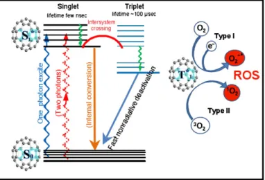
3 1.1 Photodynamic Effect – Brief history
Light is essential for life however, we took it for granted and forget all its benefits. Sunlight was employed to treat skin diseases, rickets and even psychosis thousands years ago, in ancient civilizations. However, only recently the benefits of light were applied in medicine. In the early 90’s, a medical student, Oscar Raab, observed the dead of Paramecium caudatum after light exposure in the presence of a dye, acridine orange [1]. Raab discovered that the conjugation of these two factors (light and dye) was greater together than separately. During his researches he concluded that acridine possess some product that induced in vitro toxicity, suggesting that this effect was caused by the energy transfer from light to the chemical molecule. With this discovery von Tappeiner, one of the pioneers of photobiology introduced in 1907 the term “photodynamic effect”. Photodynamic effect depends on a light source, an oxidizing agent (molecular oxygen [O2]) and
an intermediary agent (photosensitizer [PS]) to the formation of highly cytotoxic species (singlet oxygen [1O
2], hydrogen peroxide [H2O2], superoxide [O2-],
hydroxyl radicals [-OH!]) causing damage or destruction of live tissues or cells [2,3,4]. In fact, von Tappeiner also attempted the first known clinical application, in 1903, with the treatment of malignant skin lesions (Figure 1.1) [5].
Figure 1.1 First documented patient treated with eosin and sunlight for skin lesions. Left picture before, and right picture after the treatment [5].
Among these are Foscan or mTHPC, the most potent photosensitizer, and 5-ALA, which probably causes the least side effects.21–23The light-induced fluorescence of ALA in particular has been studied for its potential diagnostic value.24–26
PDT and fluorescence diagnosis (FD) using ALA offer several clinically significant advantages, and they have been studied extensively in preclinical and clinical investigations since 1987.27,28The first indications that ALA might
be a promising drug for PDT came from five independent sources:
In 1987, Zvi Malik and H. Lugaci29reported the use of 5-ALA to induce
endogenous porphyrin synthesis in Friend erythroleukaemic cells. Photoirra-diation of the cells with ‘black light’-induced deformations and cell disintegra-tion in more than 95% of the cells when examined by scanning electron microscopy (SEM) (Figure 2). The dependence of the process on the dose of light showed a relationship between the photodynamic effect and porphyrin accumulation. Both necrotic and apoptotic features were expressed, including disintegration of the plasma membrane (shown in Figure 2d and e), mi-tochondrial damage (Figure 3b), chromatin condensation and blebbing of the nuclear envelope (Figure 2e and f).
Mohammed El-Far had discussed in meetings30the possibility of using ALA,
but had not published his results.
Also in 1987, Johan Moan reported an evaluation of 5-ALA as a photosen-sitizer in mice. However, he failed to find photosensitizing concentrations of porphyrins in either tumours or normal tissues.31
Having investigated ALA-stimulated porphyrin synthesis in plant tissues as early as 1975,32Alcira del Batlle in Buenos Aires reported on tumor selective
build-up of porphyrins in ALA-incubated tissue-explants in 1988.33
Figure 1. ‘Photograms’ of the first-documented patient treated with eosin and sunlight for ‘multiple carcinomas’ of the skin (70-year-old ‘daytaller’s widow’). Left picture was
taken on Sept. 10, 1903 and right picture on November 14, 1903.
19 BASIC PRINCIPLES
Chapter I - Introduction
1.2 Photodynamic Therapy
The use of photodynamic therapy (PDT) as a therapeutic modality gained relevance in the last decades and it was approved for the treatment of several tumours in many countries (Europe, Japan, Canada and USA) [2]. In fact, the greater advances in this therapy were achieved in 1976 with a study involving patients with bladder cancer [6]. This therapy involved a systemic, topical or direct administration into the organ of a chemical compound (photosensitizer) and the irradiation of target cells with appropriate wavelength (normally visible light and laser light directed via optical fibre). The combination of these two non-toxic elements – light and photosensitizer – in the presence of an oxygenated environment results on the selective destruction of tissues or cells through a localized cytotoxic effect [7,8]. These damages are due to the formation of cytotoxic reactive oxygen species (ROS) generated by the transference of light energy to molecular oxygen, by the photosensitizer [9].
In the last two decades, significant improvements in the field of PDT made possible the use of this therapy in clinical treatments. The recognition of the need for a larger variety of photosensitizers with improved optical and target localization properties and a more complete understanding of biological mechanisms, increased the potential applications of PDT. As a consequence, PDT is now considered as a platform technology with broad applications in different medical specialties [10]. Most of clinical trials have been made with Photofrin", the first PS
approved for the treatment of some cancers. Photofrin" is a slightly purified form of
a hematoporphyrin derivative (HPD), a mixture of porphyrin polymers formed by acid treatment of hematoporphyrin [10]. While the initial focus of PDT was the treatment of cancer, a rapid increase in studies investigating the use of this therapy for the treatment of non-cancer disease was registered [10]. In fact, the use of PDT to treat age-macular degeneration is one of the most successful applications until now. Table 1.1 summarizes some of these applications, in various pre-clinical and clinical stages.
5 Table 1.1 Some non-cancer diseases where PDT is applied [10].
Cardiology Atherosclerosis
Dermatology Psoriasis
Actinic keratocis
Gynecology Endometriosis
Microbiology Infection control
Peridontal disease
Ophthalmology Age related macular degeneration
Blood banking Sterilization of blood products
1.3 Antimicrobial Photodynamic Therapy
Despite the enormous advances in medicine over the past 100 years, microbial diseases are still a major problem for human health. New approaches that are effective, affordable and widely applicable and that are not susceptible to resistance are urgently needed.
Antimicrobial photodynamic therapy (aPDT) appears for the first time in the 90’s with the inactivation of Paramecium caudatum. This therapy follows the same concept as PDT, using a photosensitizer subsequently activated by low doses of visible light, of an appropriate wavelength, to generate free radicals or singlet oxygen to inactivate microorganisms [4,2,11,12,13,14]. However, the potential of aPDT was not exploited until the middle of last century due to the discovery of antibiotics. The appearance of antibiotics raised the belief that pathogenic microorganisms would have been reduced to a level that no longer had serious impacts to human health. The widespread use and the inappropriate or excessive prescription of antibiotics made possible for some pathogenic microorganisms to develop resistance against those who were initially effective [2]. This, combined with the appearance of microbial cells with a large variety of mechanisms against external insults is putting an end to the “antibiotic era” [15,16]. In the 60´s, gram positive bacteria and methicillin-resistant Staphylococcus aureus (MRSA) acquired resistance against all $-lactam antibiotics. Years later, two other cases of bacteria
Chapter I - Introduction
that proved to be resistant to a new glycopeptide antibiotic, vancomycin, vancomycin-resistant enterococci (VRE) and vancomycin-resistant S. aureus (VRSA) emerged [17]. These three strains are presently major subjects of concern in human health.
An enormous effort was made to find alternatives for rapid and effective inactivation of pathogenic microorganisms without development of new resistances [8]. aPDT regained interest in the search of new solutions. This technique is now back on focus as a promising alternative against various pathogenic microorganisms. Since the mode of action of the PS on microbial cells involves oxidative modification of vital cellular constituents, markedly different from that typical of most antibiotic drugs, many scientists hypothesize that development of resistance can be overcome [2,7,13,18].
Currently, the most important application of aPDT is in the clinical area, used for sterilization of blood and blood products, as a measure of prevention of viral contamination [4,19,20]. Recent studies also demonstrated effectiveness as alternative for skin surface disinfection and treatment of superficial skin wounds, oral cavity infections (such as periodontitis and endodontitis) and acne vulgaris [8,21,22]. Several approaches have been also made for its application in environmental area were, several researchers groups reported effectiveness on the destruction of faecal bacteria, bacterial endospores, helminthes eggs and viruses in environmental waters [23,24,25,26,27,28,29]. This approach has been considered as a possible technology to treat drinking water disinfection and wastewater treatment plants [23,25,30]. aPDT approach represents lower costs, when compared with chemical compounds normally used, and is conceived to be environmentally-friendly and to exhibit a high level of safety of various ecosystems, as well for humans, animal and plants [31].
1.4 Porphyrins
Porphyrins are a class of aromatic heterocyclic compounds with unique physico-chemical properties. Actually, these compounds are being used in artificial photosynthesis, oxidation catalysis, sensors, PDT and aPDT [32].
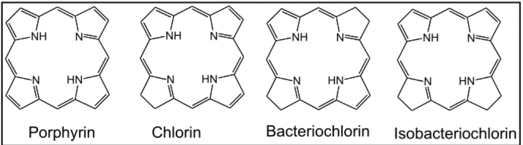
7 1.4.1 Overview
Many biologic functions depend on tetrapyrrolic macrocycles compounds for their accomplishment. These pigments, on its reduced, rusty or complex form, may be designated by porphyrins, chlorins, bacteriochlorins and isobacteriochlorins (Figure 1.2).
Figure 1.2 Some tetrapyrrolic units.
Respiration, photosynthesis and electron transport chains are the most important energetic processes carried in which tetrapyrrolic macrocycles namely porphyrins, are involved. The heme group (Figure 1.3), a complex iron of protoporphyrin-IX, discovered by Fisher in 1929, is found in proteins like hemoglobin, mioglobin and cytochroms. In hemoglobin and mioblogin, this tetrapyrrolic structure makes oxygen transport possible in the blood stream. In proteins like cytochrome, the heme group participates in the transfer of electrons and cellular production of energy [33].
N NH N HN N NH N HN N NH N HN N NH N HN
Chapter I - Introduction
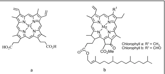
Figure 1.3 Natural tetrapyrrolic macrocycles: a) Heme, b) Chlorophyll.
Hans Fischer characterized the structure of hemoglobin and chlorophyll (Figure 1.3) demonstrating the existence of the same porphyrinic scaffold that, in the case of chlorophyll, is reduced and coordinated with magnesium [34].
1.4.2 Nomenclature of porphyrin derivatives
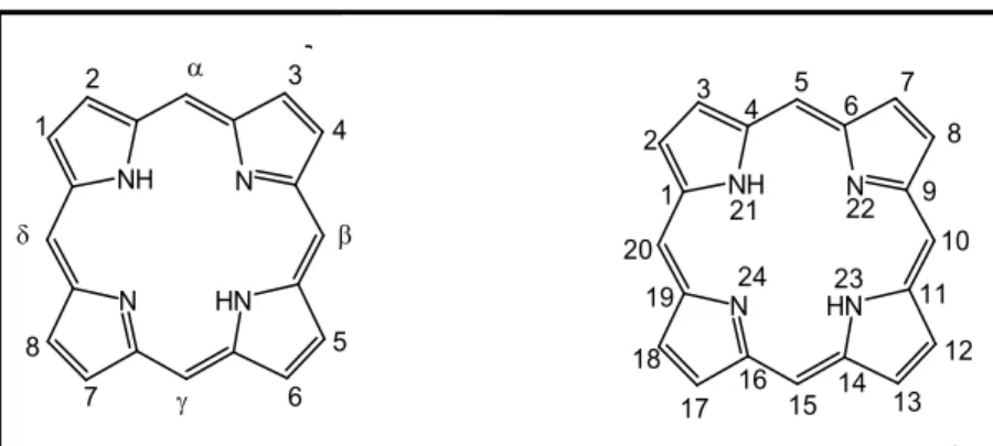
There are currently two systems of nomenclature that can be used for porphyrin macrocycles, one proposed in the 30’s by Hans Fischer and another, more recently proposed by the International Union of Pure and Applied Chemistry (IUPAC) [35]. Under the system proposed by Fischer, four pyrrole rings are designated by the letters A, B, C and D and their external positions are designated by $-pyrrolic positions. Methine bridges (inter-pyrrolic positions) are designated by Greek letters %, $, & and ' and are commonly known as meso positions (Figure 1.4). In the IUPAC nomenclature all the carbon and nitrogen atoms, that are part of the porphyrin macrocycle, are numbered from 1 to 24. The hydrogen atoms linked to nitrogen atoms are in N-21 and N-23, by convention. However, meso and $-pyrrolic designations are still widely in use [35,36].
3
1.1 Macrociclos tetrapirrólicos – Considerações gerais
Man cannot give a true reason for the grass under his feet why it should be green rather than red or any other color.
Sir Walter Raleigh
History of the World: Preface (1614)
Os macrociclos porfirínicos, nas suas várias formas, reduzidas, oxidadas e complexadas estão presentes em funções vitais como a respiração, o transporte de electrões, a fotossíntese, diversas acções enzimáticas e ainda na desintoxicação de drogas.1,2 A existência de vida, tal como a concebemos hoje, requer a presença de porfirinas.3
Na respiração estão envolvidas as hemoproteínas, designação dada às proteínas que apresentam, ligado à respectiva cadeia polipeptídica, um ou mais grupos prostéticos conhecidos por heme 1.1 (fig. 1.1).
Na fotossíntese a luz é absorvida por centenas de moléculas de clorofila 1.2 (fig. 1.1) que transferem a sua energia, resultante da fotoexcitação, para um “centro de reacção” onde ocorre a transformação de energia luminosa em energia química, necessária para converter o dióxido de carbono e água em hidratos de carbono essenciais à vida.
N N N N HO2C CO2H Fe N N N N R1 Mg CO2MeO C O O clorofila a: R1 = CH3 clorofila b: R1 = CHO
cadeia lateral de fitilo 1.1
1.2
Figura 1.1 Estruturas do grupo heme 1.1 e das clorofilas a e b 1.2.
Os citocromos, responsáveis pelo transporte de electrões, são também eles hemoproteínas, fazendo parte do grupo das biomoléculas mais antigas.
Chlorophyll a: R1 = CH
3
Chlorophyll b: R1 = CHO
Chapter I - Introduction
9 Figure 1.4 Left: Nomenclature proposed by Hans Fisher. Right: Nomenclature proposed by
IUPAC.
1.4.3 Chemical and Physical properties
Porphyrins are heteroaromatic compounds characterized by a tetrapyrrole structure that consists in four pentagonal pyrroles linked by four methine bridges (Figure 1.5). This structure presents twenty-two # electrons conjugated. However, only eighteen electrons confer the aromatic character to this molecule. The four # electrons that do not contribute to the aromaticity of the macrocycle presenting a dual character. The elevated conjugation of # electrons system gives to porphyrins and derivatives an intense coloration.
Figure 1.5 Structure of porphyrin Core.
The aromatic character of the macrocycle can be explained by the planar topology and the participation, for example, in electrophilic substitution reactions typical of these aromatic compounds [37,38]. Typical electrophilic substitution reactions of this class of compounds are for example: reactions of nitration, halogenation, sulfonation, formylation, acylation and deuteration [35].
+*********** * *,"-./$0-&1&/*&/*2./-$%*********************************************************** "%.10*3%$4-$-"10*/*%*&/0/".%5.-6/"#%*&/*#78"-810*&/*1"95-0/*:!"#"*;<=*&/*>?@*%'* ;1-%*ABC*$/./5%'*D'/*%*0-0#/61*3$%3%0#%*3%$*E-08F/$*")%*/$1*%*61-0*8%"./"-/"#/G* @%6*%*0-0#/61*3$%3%0#%*3/51*!,H2@C*4%$16*-"#$%&'I-&10*$/J$10*61-0*851$10*D'/* 8%"#/6351.16* #%&%0* %0* 9#%6%0* &%* 1"/5G* 200-6C* %0* .-"#/* /* D'1#$%* 9#%6%0* 3/$#/"8/"#/0*1%*618$%8-85%*4%$16*"'6/$1&%0*&/*>*1*KLG*M0*F-&$%J7"-%0*5-J1&%0* 1%0*9#%6%0*&/*1I%#%*/"8%"#$16N0/*"%*=NK>*/*=NK?C*3%$*8%"./"()%G*=%*/"#1"#%C* 10*&/0-J"1(O/0*$!%&*/*PN3-$$Q5-810*8%"#-"'16*1*0/$*51$J/6/"#/*'#-5-I1&%*:E-J'$1* >GKBG>RC*>S* * * * ! ! ! ! ! ! "#$%&'!()*)*=%6/"851#'$1*&/*3%$4-$-"10*3%$*TG*E-08F/$*/*3/51*!,H2@G* * ()()+)!!,-$%.'/!0&10'32/!45/#6'/!2!7%5.#6'/!32!01&4#'/!! * 20*3%$4-$-"10*0)%*8%63%0#%0*D'/*13$/0/"#16*81$98#/$*1$%69#-8%C*%"&/*13/"10* >U* /5/8#$O/0* VC* &%0* KK* D'/* 3%00'/6C* 0)%* $/03%"09./-0* 3/51* 1$%61#-8-&1&/G* 20* 1"95-0/0* 3%$* ;1-%* A* &/* 3%$4-$-"10* /* &/* 6/#15%3%$4-$-"10* 0'J/$/6* D'/* %* /0D'/5/#%* 3%$4-$W"-8%*7*351"1$C*%*D'/*/.-&/"8-1*%*81$98#/$*1$%69#-8%G>U*
2* /5/.1&1* 1$%61#-8-&1&/* 13$/0/"#1&1* 3%$* /0#/* #-3%* &/* 8%63%0#%0* 7* $/03%"09./5* 3/5%* 131$/8-6/"#%* /6* /03/8#$%0* &/* ;<=* &/* >T* &/* 0-"1-0* 8%$$/03%"&/"#/0* X* $/00%"Y"8-1* &%0* 3$%#O/0* -"#/$"%0* Z=T* 1* 8163%0* /5/.1&%0* :.15%$/0* &/.* [* /"#$/* NK* /* NR* 336BC* /"D'1"#%* D'/* %0* 3$%#O/0* 6/#W"-8%0* 0'$J/6* 1* = =T = T= > K ? L R + S U H%0-()% !N3-$$Q5-81 " ! # $ = =T = T= > K ? L R + S U \ >] >> >K >? >L >R >+ >S >U >\ K] K> KK K? KL
=%6/"851#'$1 &/ TGE-08F/$ =%6/"851#'$1 &1 !,H2@
!!!!!!!!!!!!!!!!!!!!!!!!!!!!!!!!!!!!!!!!!!!!!!!!!!!!!!!!!!!!!!!!!!!!!!!!!!!!!!!!!!!!!!!!!!!!!!!!!!!!!!!!!!!!!!!!!!!!!!!!!!!!!!!!!!!!!!!!!!!!!!!!!!!!!! ! "#$%&'($)*)&!)&!+%&$',!! -! !"#$%&'()*+,-.*'#/*+,('# # !"!"#0%+1(+(&,'#2#/*&*+,3(),)*'# # +!.*/*%'*!.,'0$'$#*!1!,'$2$#3'$*!)*!*#4$2*!5'16$*!,#)&!*!.*/*%'*!!"#!$%#&'&'*!7(*)*! .*'*!)&($2#*'!*!6,'!.8'.7'*9!:(4*!)&($2#*;<,!0*=!4,),!,!($),!7>*!%&=!?7&!*(!.,'0$'$#*(! (<,!.$2>,(!0,'4&>&!6,'*),(!&!?7&!*.'&(*>!*!6,'!.8'.7'*9@! A7$>$6*>&B!*(!.,'0$'$#*(!*.'&(*>!7>!>*6',6$6/,!C*(&B!6,>.,(4,!.,'!?7*4',! *#1$(!)&!4$.,!.$'',/!7#$),(!'&!($!.,'!7#$)*)&(!)&!6*'C,#,!(.D!E.,#4&(!>&4F#$6*(G!EH$29!@G9D! ! ! " #" " "# ! H$27'*!@!I!:(4'747'*!)&!7>!#86/&,!.,'0$'F#$6,! !
+(! .,'0$'$#*(! 4J>! ,6,''J#6$*! #*47'*/! &! *! (7*! &K$(4J#6$*! 1! 07#)*>*/! L! %$)*9! M! $>.'&(($,#*#4&!6,>,!.&?7&#*(!>,)$0$6*;N&(!#,!>*6',6$6/,!4&4'*.$''O/$6,!C3($6,!,'$2$#*>! 07#;N&(!C$,?7F>$6*(!4<,!)$%&'(*(9!+!(7*!.'$#6$.*/!07#;<,!#*!#*47'&=*!1!*!6,,')&#*;<,!6,>! >&4*$(!?7&!*647*>!6,>,!6',(!&>!&%,(!C$,?7F>$6,(!$>.,'4*#4&(9@!P*!'&(.$'*;<,!&(4<,! &#%,/%$)*(! Q&>,.',4&F#*(! E.',4&F#*(! 6,>! 7>! ,7!>*$(!2'7.,(! Q&>&! /$2*),(! L! (7*! 6*)&$*! .,/$.&.4F)$6*G! 6,>,! (&R*>! *! Q&>,2/,C$#*! &! *! >$,2/,C$#*9! S! 2'7.,! Q&>&! E6,>./&K,! )&! 0&'',!)*!.',4,.,'0$'$#*!TUG!EH$29D*G!/$2*!'&%&'($%&/>&!,!,K$21#$,!>,/&67/*'B!?7&!)&(4*! 0,'>*!.,)&!(&'!4'*#(.,'4*),!.,'!4,),!,!6,'.,!E*4'*%1(!)*!Q&>,2/,C$#*G!,7!*'>*=&#*),!#,(! 4&6$),(!>7(67/*'&(!E*4'*%1(!)*!>$,2/,C$#*G9!P*!0,4,((F#4&(&!,(!>*6',6$6/,(!&#%,/%$),(!(<,! ),!4$.,!6/,'$#*!EH$29-*GB!,7!(&R*!>*6',6$6/,(!6,>!7>*!7#$)*)&!.$''O/$6*!'&)7=$)*!6,>,!1!,! 6*(,!)*(!6/,',0$/*(!*!&!C!EH$29!DCG9!+(!6/,',0$/*(!(<,!*(!'&(.,#(3%&$(!.&/*!*C(,';<,!)*!/7=! !!!!!!!!!!!!!!!!!!!!!!!!!!!!!! !!!!!!!!!!!!!!!!!! @!V9W9!X$/2',>B($)'*"+"%#,'"-'+.-)/!!445B!SK0,')!"#$%&'($4Y!Z'&((9# D!59Z9!X,((B!0%#)'1!!+'*$)2B!!465B![\B!]B!^^\9! ! Dual character
Chapter I - Introduction
In addition, nucleophilic substitution, reduction and oxidation reactions can also occur [35].
Relatively to the transformations promoted by the nitrogen atoms of the macrocycle, acid-base and complexation reactions with metal ions may occur. The acidic and basic character of the macrocycle is strongly influenced by the presence or absence of substituents on the periphery of the macrocycle. Protonation or deprotonation of the macrocycle can occur, depending if the macrocycle is in acidic or basic media (Scheme 1.1) [39,40].
Scheme 1.1
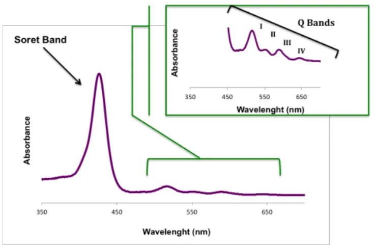
The photophysical and photochemical aspects, related with the structure as well as the aromatic character, are the most important properties of porphyrins. UV-Vis spectra are the most important characteristics of these compounds allowing their instant and precise identification. This type of spectrum presents two distinct regions. One absorption band, with major intensity around 390 – 425 nm, designated as Soret band, and two or four lower intensity bands – Q bands – in the region between 500 – 650 nm (Figure 1.6) – according to the existing macrocycle [41,42].
Figure 1.6 UV-vis spectra of porphyrin derivatives. Strong Bases
11
However, the intensity, number and position of these bands can be influenced by many factors such as number, relative position (meso or $-pyrrolic positions) or type of substituting groups that are in the peripheral positions of the macrocycle.
An NMR spectrum also allows the identification of porphyrins revealing the aromatic nature of the macrocycle. Free porphyrins show one signal generated by the resonance of the central protons N-H between -2 and -3 ppm compared with the signal generated by tetramethylsilane (TMS) protons. This phenomenon is due to a strong shield promoted by the electronic current around the macrocycle. This same current unprotects the meso and $-pyrrolic protons dislocating it resonances to lower fields, at % 7 and 9 ppm [41].
1.4.4 Synthesis of meso–tetra-substituted porphyrins
meso-Tetra-substituted porphyrins can be obtained in two different ways: by modifying a natural porphyrin or by synthesizing de novo from basic units (pyrrole and aldehyde). Depending on the intended porphyrin (symmetric or asymmetric), a mixture of aldehydes can be used. Aldehyde condensation with pyrrole proved to be the main procedure to synthesize meso-substituted porphyrins (Scheme 1.2).
Scheme 1.2
Capítulo I
6
Dada a existência de variadíssimas rotas de síntese de macrociclos tetrapirrólicos,e consequentemente uma vastíssima bibliografia sobre a síntese deles,22-25 vamos de uma forma breve, apresentar a evolução das rotas de síntese que aplicámos na preparação dos macrociclos usados neste trabalho.
1.2.1 Síntese de porfirinas meso-substituídas
Algumas das porfirinas meso-substituídas exigem processos de síntese bastante elaborados,26,27 no entanto outras são de síntese fácil, podendo mesmo prepararem-se num único passo. A partir da condensação de pirrol com um aldeído obtêm-se porfirinas simétricas (esquema 1.1). No entanto, se na reacção de condensação for usada uma mistura de aldeídos obtêm-se porfirinas assimétricas. Esta grande versatilidade reaccional permite a construção de várias porfirinas sem haver necessidade de elaborar rotas de síntese complexas. HN N N NH R R R R N H RCHO + H + Esquema 1.1
1.2.1.1 Síntese de porfirinas meso-tetra-substituídas simétricas
A síntese da meso-tetrametilporfirina foi efectuada pela primeira vez por Rothemund em 1935. Rothemund preparou mais de trinta porfirinas, entre as quais a meso-tetrafenilporfirina (TPP) 1.4 (esquema 1.2). O método era bastante simples, baseando-se no aquecimento de pirrol com os correspondentes aldeídos. A mistura dos dois reagentes em piridina era aquecida a 220 ºC durante 48 horas, num tubo selado sob atmosfera de azoto.2,21,28,29 Os rendimentos eram, na generalidade, inferiores a 5% e
Chapter I - Introduction
12
In the last decades, many procedures were used to synthesize these compounds allowing the optimization of procedure and improvement of reaction yields. Rothemund developed the first method in 1935 when synthesizing, for the first time, the meso-tetraphenylporphyrin (TPP) (Scheme 1.3) [43,44]. However, this method presented low yields (around 5%) and also produces the chlorin form.
Scheme 1.3
Some others methods were presented by different researchers, along the years, showing improved yields and solutions for minimizing chlorin formation in the reaction. Alder and Longo altered the condensation conditions in order to solve the problems that arose with the Rothmund protocol. In this case, the reaction occurred in acidic medium and under aerobic conditions. The TPP was then achieved by reflux of pyrrole and benzaldehyde in propionic acid for 30 minutes, rendering the final product in crystalline form directly from the reaction medium (Scheme 1.3b). However, the porphyrin macrocycle was equally contaminated with the corresponding chlorin, although in smaller quantities [44]. Between 1979 and 1986, Lindsey and his colleagues developed a new synthesis methodology based on the fact that the intermediary, the meso-tetraphenylporphyrinogen is thermodynamically favourable when the pyrrole and benzaldehyde are condensed in appropriate conditions (Scheme 1.3c). This type of synthesis is performed in two stages: initially the pyrrole and aldehyde are condensed in a dry dichloromethane solution, in the presence of
a: Rothmund conditions: Pyridine, 120°C, 48h
b: Alder and Longo conditions: CH3CH2CO2H, 141°C, 30 min c: Lindsey conditions: CH2Cl2, r.t., BF3, N2, 2.DDQ
d: Rocha Gonçalves conditions: C6H5NO2, CH3CO2H, 120°C, 1h
! +(! /.)0&)&%,*! %12,! /)./.)34.! 51(! +(/(%+(! +.! %62().! +(! ,7+(8+.*! 19&7&:,+.;! +,! )(,<9&'&+,+(!+(*9(*!(!+,*!51,%9&+,+(*!(*9(51&.2=9)&<,*!19&7&:,+,*>! ?!-)@ABC D ? ! " !"#EFA D ?!B#C AGBHIC#;!ABJAC#B !" !" "! ! #$ #$ #$ #$ ! ! K*51(2,!?! % !"!"#"$%&'()*+,-.$/-$&012(1(3*.$ !
L4.! &%62(),*! ,*! ,/7&<,3M(*! +,*! /.)0&)&%,*! (! *(1*! +()&',+.*;! %.2(,+,2(%9(! (2! 9(),/&,!0.9.+&%N2&<,!OPQRS;!(2!9(),/&,!0.9.+&%N2&<,!,%9&2&<).T&,%,!OP-ARS!(!<,9U7&*(>!"E! ! $2,! '(:! 51(! .! 9),T,7V.! +(! &%'(*9&W,34.! ,/)(*(%9,+.! %(*9,! +&**()9,34.! 9(2! <.2.! .TX(<9&'.! 679&2.! ,! ,',7&,34.! +,! (0&<U<&,! 0.9.&%,<9&',+.),! +.*! +()&',+.*! *&%9(9&:,+.*! (2! 2&<).)W,%&*2.*;! 9.)%,@*(! &2/)(*<&%+8'(7! +(*<)('()! .*! /)&%<8/&.*! TU*&<.*! +.! /).<(**.! 0.9.+&%N2&<.>! I.! <,/&917.! YYY;! +,)@*(@U! /,)9&<17,)! &2/.)9N%<&,! ,.! <,*.! (*/(<80&<.! +,! 0.9.&%,<9&',34.!+(!2&<).)W,%&*2.*;!/(7.!51(!%12,!/)&2(&),!,T.)+,W(2!&)4.!*()!+(*<)&9.*! ,7W1%*!+.*!,*/(<9.*!01%+,2(%9,&*!+,!PQR>!
! -! PQR! =! 12,! 0.)2,! (*/(<&,7! +(! 0.9.9(),/&,>! C! 9()2.!0.9.9(),/&,! &%<71&! 9.+.*! .*! 9),9,2(%9.*! 51(! 1*,2! ,! 71:! /,),! &%+1:&)! )(,<3M(*! %12! /,<&(%9(! +(! 2.+.! ,! 9),:()@7V(! T(%(08<&.*>!-!PQR!=!12,!9=<%&<,!(2!+(*(%'.7'&2(%9.!(!.!*(1!W),%+(!.TX(<9&'.!=!+(*9)1&)! <=717,*!&%+(*(XU'(&*!O!"#"!<=717,*!<,%<()8W(%,*S;!*(2!+(*9)1&)!9(<&+.*!*,1+U'(&*>!Z,*(&,@*(!
%.! 1*.! +(! 12,! +).W,! <V,2,+,! 0.9.**(%*&T&7&:,+.)! OPLS! ,+2&%&*9),+.! ,.! /,<&(%9(! %.)2,72(%9(! /.)! &%X(<34.>! C! PL! /.)! *&! *[! %4.! 9(2! 51,751()! (0(&9.! 9[\&<.! %.*! 9(<&+.*>! A.%91+.;!*(!0.)!,/7&<,+,!71:!%.!9(<&+.!<.%9(%+.!.!0U)2,<.;!.!0U)2,<.!=!,<9&',+,!(!.!9(<&+.! =!),/&+,2(%9(!+(*9)18+.!%.!7.<,7!.%+(!,!71:!=!,/7&<,+,>!L(!0.)!(*<.7V&+,!,!),+&,34.!<()9,;!,! 9=<%&<,! /.+(! *()! T,*9,%9(! *(7(<9&',! )(7,9&',2(%9(! ,.! 9(<&+.! +(*9)18+.>! L()U! 9,2T=2! <.%'(%&(%9(! 51(! .! PL! *(! <.%<(%9)(! *(7(<9&',2(%9(! %.! 9(<&+.! 912.),7! (].1! 9(<&+.! V&/()@ /).7&0(),9&'.>! -! 2,&.)! +(*',%9,W(2! +(*9(! 2=9.+.! ,+'=2! +,! 19&7&:,34.! +(! !!!!!!!!!!!!!!!!!!!!!!!!!!!!!! !!!!!!!!!!!!!!!!!!
"E!^>!_>!^,+&*V;!^>!_>!L2&9V;!$%!&'()*%+),-&./-01((2&3&4*5,6/7,(-89&'/87:&')!8!-7&/-0&;<7<)!;!`>!a1&7,)+!
13
an acid catalyst (BF3, BCl or TFA), in catalytic amount, at room temperature,
under nitrogen atmosphere. In a second phase, the formed porphyrinogens are oxidized by the addition of an oxidant agent (DDQ or p-chloranil), resulting in the corresponding reduced free porphyrin derivatives [45,46].
Rocha Gonsalves and his colleagues proved to be possible to prepare, in one step, meso-tetra-arylsubstituted porphyrins in good yields and without their chlorin derivatives. Currently, this method is widely used to synthesize meso– substituted porphyrins and consists in a condensation of pyrrole with benzaldehyde, in presence of acetic acid with 30% of nitrobenzene at reflux (120 ºC) for 1 hour (Scheme 1.3d) [47]. The acetic acid acts as solvent and catalyst, and the nitrobenzene as oxidizing agent. Due to the simplicity of this approach, it can be applied to produce a wide range of tetra-substituted derivatives comprising four identical aryl units [34]. This method also allows the synthesis of asymmetric meso-substituted porphyrins, by condensation of pyrrole with a mixture of aldehydes. The proportion of the different compounds formed depends on the proportion of aldehydes present in the reaction mixture and their reactivity. The following scheme (Scheme 1.4) helps to visualize the combination of possible porphyrins, if we consider a mixture of two aldehydes (B-CHO and A-CHO) in 1 to 3 eq., respectively, with equal reactivity.
Chapter I - Introduction
Scheme 1.4
1.4.5 General applications
Currently, porphyrins derivatives have applications in different areas, such as catalysis, enzyme catalyzes, models of photosynthetic systems, artificial oxygen carriers, chemical sensors, optoelectronic systems, semiconductors, superconductors, liquid crystals, biocides agents in PDT and aPDT [48,49,50]. !"#$%&'()%* ! 4 5 6 37859 6 :7859 4 45 4 54 3 3 3 3 4 45 4 54 : 3 3 3 4 45 4 54 3 : 3 : 4 45 4 54 3 3 : : 4 45 4 54 3 : : : 4 45 4 54 : : : :
3;<=+>?;@A 3=: <;,>+B@A #$2"173,:,<C>D=@A
E.173,:,<+;>D?@A 3:=<;>?B @A :;<D>=B @A * ! "#$%&'(!)*+! !
)*)*,*!!-./01(23&#!4&!.5670608(#!&'!'&40108(!
*
F0/.&%* G1* 1'21* H$%H$.0&2&01* I'JK.E21* 0* LJ1.E21>* %1* E%KH%1#%1* H%$L.$J".E%1* H%&0K* 10$* 2HM.E2&%1* 0K* G$021* &.1#."#21>* E%K%* NG* L%.* $0L0$.&%* "%* H%"#%* +O+O* 4%* 0"#2"#%>* P* "2* * K0&.E."2* I'0* %* '1%* &01#01* E%KH%1#%1*Q2"R2* K2.%$* &01#2I'0>* %"&0* 1)%* 2HM.E2&%1* E%K* 1'E011%* 0K* SFT>* "2* &0#0E()%* &0* #0E.&%1* "0%HMG1.E%1* 0* "2* .".U.()%* &0* 2Q0"#01* /.$2.1* H$010"#01* "%* 12"Q'0O* T0K* 1.&%* #2KUPK* 2/2M.2&2* 2* 1'2* 2HM.E2U.M.&2&0* E%K%* 2Q0"#01* ."#0$E2M2"#01* &%* F43>* "%* #$2#2K0"#%* &0* &%0"(21* &2* H0M0* <2E"0>* 0EV0K2>* H1%$J210>* 0#EOA>* 2$#0$.%1EM0$%10>* 2$#$.#01* 0* "2* &0Q0"0$2()%* &2* KGE'M2*&2*$0#."2O;>?>C>B>++>+,>,?*
15 1.5 Porphyrins in aPDT – applications
Although various tumors can be successfully treated with PSs and light, the application on the inactivation of microorganisms has not yet found specific medical applications and still remains as an open field. Due to their interesting physical and chemical properties, porphyrins proved to be good PS for antimicrobial photodynamic therapy. During the last years, the application of these compounds against several infectious agents has been gaining importance, as demonstrated in several in vivo and in vitro studies. As mentioned earlier, the major use of aPDT is in the clinical area. Unfortunately, due to the dependence on light, this therapy can only be applied on localized infections [8]. ALA-based PS showed very good results against herpes simplex virus, Trichophyton rubrum and cutaneous leishmaniosis, is tests carried in vivo [5,51]. The in vitro tests show that porphyrins can also used as efficient PS for bacterial, viral and fungal photoinactivation [7,12,26,52,53,54]. For a porphyrin derivative to be used as PS in the inactivation of microorganisms of clinical importance, it must fullfill some requirements [8]:
1. be effective in destroying the disease-causing pathogens; 2. have selectivity towards the target microorganisms; 3. minimize the regrowth of pathogens;
4. be effective against clinical multi-resistant microbial stains 5. be effective in the presence of biological products.
In addition to clinical applications, porphyrins started to be used in environmental technology, specifically, in water disinfection, sewage treatment plants and in fish-farming plants [27,55,56,57]. The potential for wastewater disinfection has been addressed in several studies, with different porphyrins and microorganisms (bacteria and bacteriophages) and promising results were obtained [55,27,25,58]. In fact, Alves et al. demonstrated an efficient inactivation of gram negative and gram positive faecal bacteria with tri- and tetracationic porphyrin derivatives, with porphyrins dissolved in the water sample or immobilized on solid matrixes [59]. In order to a sustainable use of this therapy for environmental applications, some aspects must be considered [27,58,59]:
Chapter I - Introduction
1. The possibility to remove de PS after the photodynamic action in order to recover/reuse and prevent the release of the PS to the environment; 2. The stability of PS under the irradiation source and conditions;
3. The toxicity of the PS in aquatic organisms, having in attention the impact of this therapy on the natural non-pathogenic microbial communities.
1.6 Factors affecting aPDT efficiency
As mentioned before, aPDT requires three non-toxic elements – light, photosensitizer and oxygen – to cause oxidative damage to cells. However, some types of cells have ability to repair slight oxidative damage but, if a large number of oxidative events takes place within a relatively short period of time, the capacity of a cell to recover can be exceeded. For an effective aPDT, the oxidative damage must be produced faster than damage repair, making the damage to extensive for cell survival. The rate at which damage is produced depends upon a number of interacting factors.
1.6.1 Light source
By definition, aPDT requires a light source to provide energy capable of inducing the in situ formation, of singlet oxygen, which causes oxidative damage to cells. Generally, every visible light source within the appropriate wavelength range can be used in aPDT. However, three important factors should also be considered: the wavelength(s) of the photoactivating light (light source), the fluence rate (irradiation power) and the total dose of light at the target site (light dose) influence the aPDT effectiveness [5]. Over the years, a variety of light sources (artificial) have been tested for microorganism photoinactivation.
The light source used in an aPDT approach depends on the wavelength that is necessary to activate the PS. This is ultimately determined by the
17
structure and electron absorption spectrum of the PS used [60]. Porphyrin derivatives, have a Soret band between 390 – 425 nm and four Q-bands between 500 – 650 nm [61]. Therefore, the irradiation light spectrum should cover all or a part of the porphyrin absorption spectrum. Several studies show that, for the same PS and microorganism, the photodynamic effect varies with the light source used [62,63]. As an alternative to artificial light sources, sunlight has also been used on aPDT, mainly in environmental approaches [23,26,55,62]. Several studies revealed that light source influences the rate and the extent of microbial photoinactivation [26,62,64,65].
Light dose, used during irradiation, is also important in the outcome of microbial photoinactivation in a fluence rate-time dependent manner [62]. According to the law of reciprocity, a certain biological effect is directly proportional to the total energy dose, regardless the deliverance regime. However, very few studies have addressed the role of the law of reciprocity in aPDT [61,66,67]. The studies about the influence of light parameters currently available in the literature show that a higher fluence rate over a shorter time period may produce different results, in terms of microbial inactivation, from those of a low fluence rate over a longer exposure time, corresponding to the same light dose [4]. Costa et al. showed that when the same light dose (using the same light sources) was obtained under different fluence rates (150; 300; 600 and 1200 W m-2), the T4-like bacteriophage inactivation was dependent on the fluence rate used [62].
1.6.2 Porphyrins as Photosensitizers
A photosensitizing agent with potentiality in aPDT should be skilled with specific features in terms of photophysical and chemical properties. When a new porphyrin is synthesized it is expected that it fulfils some photophysical requirements such as a high quantum yield for the generation of both the long-lived triplet state and the cytotoxic singlet oxygen species, a good absorption capacity at the wavelength of the spectral region where the light source is emitted, a good efficiency to generate ROS and good photostability [11,34].
Chapter I - Introduction
A large number of porphyrins have been tested as PSs and a great deal of work has been carried out to evaluate the correlation between antimicrobial efficiency and the structure of these compounds [1]. Several lines of evidence indicate that the nature of the meso-substituted groups, the presence of charge and the charge number, and the amphiphilicity of porphyrins have potential impact on the efficiency of photosensitization [59,68]. Over the years, many porphyrins with different meso-substituted groups were synthesized and tested in aPDT. Tricationic porphyrins differing in a meso-aryl group, showed different photoinactivation results in gram positive and gram negative bacteria, as well as in bacteriophages and viruses of eukaryotes [27,54,59]. Several studies have proved that cationic meso-substituted porphyrin derivatives are more efficient in the microbial photoinactivation than the neutral or anionic derivatives [27]. Both gram negative and gram positive bacteria showed to be more susceptible to cationic porphyrins than to neutral and anionic molecules, to which gram negative bacteria are usually resistant [4,13,14,8,69]. The existence of an outer membrane, which acts as a permeability barrier in gram negative bacteria, difficult the entry of the PS [13]. The insertion of positively charged substituents in the peripheral position of the tetrapyrrolic macrocycle may largely affect the kinetics and extent of binding to the microbial cells [2]. In fact, some researchers hypothesized that this phenomenon may be due to the electrostatic attraction between the cationic porphyrin derivatives and the negatively charged membrane of the microorganisms [70]. Not only the existence of positive charges is important, but also the number and the distribution of these charges must be taken into consideration. Meso-substituted cationic porphyrins with different charge number/distribution (mono-, di-, tri- or tetracationic) have already been compared on viral, bacterial, and fungal photoinactivation, with very different results [13,27,52,59,56]. Caminos et al., showed that the tricationic porphyrin with a trifluoromethyl group (A3B3+) were more efficient in
the photoinactivation against Escherichia coli than the tetracationic porphyrin (TMAP4+) (Figure 1.7). This can be justified by the presence of a highly lipophilic trifluorophenyl group (A3B3+) that increases the amphiphilic character of the
19 Figure 1.7 Molecular structures of porphyrins used by Caminos et al [12].
However, in other studies, dicationic porphyrin derivatives showed to be more efficient in the photoinactivation of several bacteria when compared with tri- and tetracationic ones [71,72]. Costa et al. obtained results in the photoinactivation of a T4-like phage similar to those reported by Caminos et al. The photoinactivation was more efficient in the presence of a tricationic porphyrin than with tetracationic or dicationic porphyrins [27]. In the case of antifungal aPDT, Cormick et al. showed that tricationic porphyrin TFAP3+ presented better results than the tetracationic TMAP4+ in the photoinactivation of Candida albicans [73] (Figure 1.8).
N NH N HN R1 R2 R1 R1 A3B3+ R1: -OCH2CH2CH2N+ (CH3)3 I- R2: -CF3 TMAP4+ R1=R2: -N+(CH 3)3 p-Tos -TPPS4- R1=R2: -SO 3-Na+
Chapter I - Introduction
Figure 1.8 Molecular structures of porphyrins used by Cormick et al [73].
Knowing that the lipid bilayer of microbial cell membrane allows the passive transport of hydrophobic molecules, several investigation groups synthesized hydrophobic PS hoping that the PS diffusion would be facilitated, thus improving aPDT results. However, to aPDT assays, PS must be in solution, needing for that a hydrophilic character. Therefore, the ideal PS must have both hydrophilic and hydrophobic properties, making it amphiphilic. Some of the factors that are referred in the literature as increasing the amphiphilic character of the porphyrins are the asymmetric charge distribution at the peripheral position of the porphyrin, cationic charges combined into different patterns with highly lipophilic groups (e.g., trifluoromethyl groups), the introduction of aromatic hydrocarbon side groups and the modulation of the number of positive charges on the PS [72,74,75,76,77,78]. This increases the amphiphilic character of the PS and seems to enhance its affinity for bacteria, which ultimately improves its accumulation in the cells and is accompanied by an increase in the photoinactivation efficiency [72,74,79,77].
N NH N HN R1 R2 R1 R1 TFAP3+ R1: -N+(CH 3)3 I- R2: -CF3 TMAP4+ R1=R2: -N+(CH 3)3 p-Tos -TPPS4- R1=R2: -SO 3-Na+
![Figure 1.1 First documented patient treated with eosin and sunlight for skin lesions. Left picture before, and right picture after the treatment [ 5 ]](https://thumb-eu.123doks.com/thumbv2/123dok_br/15905702.1092014/29.892.291.688.735.970/figure-documented-patient-treated-sunlight-lesions-picture-treatment.webp)
![Figure 1.8 Molecular structures of porphyrins used by Cormick et al [73].](https://thumb-eu.123doks.com/thumbv2/123dok_br/15905702.1092014/46.892.313.591.134.458/figure-molecular-structures-porphyrins-used-cormick-et-al.webp)