Setembro 2017
Marta Sofia Barbosa Fernandes
Licenciada em Bioquímica
Tauroursodeoxycholic Acid Drives Mitochondrial
Bioenergetics Toward Neural Stem Cell Proliferation
Dissertação para obtenção do Grau de Mestre em
Genética Molecular e Biomedicina
Orientador: Susana Solá, Professora Doutora, Faculdade
de Farmácia, Universidade de Lisboa
Co-orientador: Cecília M. P. Rodrigues, Professora Doutora,
Faculdade de Farmácia, Universidade de Lisboa
Júri:
Presidente: Doutora Paula Maria Theriaga Mendes Bernardes Gonçalves Arguente: Doutora Joana Paiva Gomes Miranda
Vogal: Doutora Susana Zeferino Solá da Cruz
i
Tauroursodeoxycholic Acid Drives Mitochondrial Bioenergetics Toward Neural Stem Cell Proliferation
Copyright © Marta Sofia Barbosa Fernandes, Faculdade de Ciências e Tecnologia, Universidade Nova de Lisboa.
A Faculdade de Ciências e Tecnologia e a Universidade Nova de Lisboa têm o direito, perpétuo e sem limites geográficos, de arquivar e publicar esta dissertação através de exemplares impressos reproduzidos em papel ou de forma digital, ou por qualquer outro meio conhecido ou que venha a ser inventado, e de a divulgar através de repositórios científicos e de admitir a sua cópia e distribuição com objetivos educacionais ou de investigação, não comerciais, desde que seja dado crédito ao autor e editor.
iii
Marta B. Fernandes, Maria F. Ribeiro, Tânia Genebra, Cecília M.P. Rodrigues, Susana Solá. Driving Mitochondrial Proteins Toward Neural Differentiation. Jornadas Intercalares das Dissertações Anuais dos Mestrados, Faculdade de Ciências e Tecnologia, Universidade Nova de Lisboa, 2nd February 2017, Caparica, Portugal. [Oral Communication]
Marta B. Fernandes, Maria F. Ribeiro, Tânia Genebra, Cecília M.P. Rodrigues, [Susana Solá]. Driving Mitochondrial Bioenergetics Toward Neural Proliferation and Differentiation. MitoPorto – Advances in mitochondrial research International Symposium, 26th May 2017 Porto, Portugal. [Abstract and Oral Communication]
Marta B. Fernandes, Maria F. Ribeiro, Tânia Genebra, Cecília M.P. Rodrigues, Susana Solá. Driving Mitochondrial Bioenergetics Toward Neural Stem Cell Proliferation. 9th iMed.ULisboa Postgraduate Students Meeting and 2nd i3DU Meeting, 13-14th July 2017, Lisbon, Portugal. [Abstract and Poster]
Marta B. Fernandes, Maria F. Ribeiro, Tânia Genebra, Joana Martins, Ana Varela, Cecília M.P. Rodrigues, Susana Solá. Tauroursodeoxycholic Acid Drives Mitochondrial Bioenergetics Toward Neural Stem Cell Proliferation. 10th International Meeting of the Portuguese Society for Stem Cells and Cell Therapies, 12-14th October 2017, Covilhã, Portugal. [Abstract and Poster]
This work was developed at the Research Institute for Medicines (iMed.ULisboa), Faculty of Pharmacy, Universidade de Lisboa and received funding support from Fundação para a Ciência e a Tecnologia (FCT) (UID/DTP/04138/2013), and from European Structural and Investment Funds through the COMPETE Programme and from National Funds through FCT (SAICTPAC/0019/2015).
v
Agradecimentos
As minhas primeiras palavras de agradecimento são dirigidas à Professora Doutora Cecília Rodrigues pela oportunidade que me concedeu em realizar a dissertação de mestrado no seu laboratório de investigação, por todo o apoio e pelos conselhos cruciais que conduziram ao progresso deste trabalho.
Um agradecimento especial à Professora Doutora Susana Solá que me acompanhou durante a realização deste trabalho. Por todos os conhecimentos transmitidos e por toda a atenção quando tudo parecia mais complicado. Igualmente pela simpatia, confiança e entusiasmo contagiante mas acima de tudo pelo apoio constante.
Quero agradecer à Professora Doutora Margarida Silva e ao Doutor Marco Moedas por toda disponibilidade na obtenção de resultados de grande relevância contribuindo para o enriquecimento deste trabalho.
À Professora Doutora Elsa Rodrigues e à Professora Doutora Maria João Nunes pelo interesse, conselhos e disponibilidade em ajudar.
Às minhas irmãs das NSCs pelo companheirismo, pelos conhecimentos transmitidos e por toda a ajuda fulcral. À Tânia Genebra por toda disponibilidade, pela boa energia e pela simpatia constante. À Maria Ribeiro, pela troca de ideias e cumplicidade, também pela paciência, mas acima de tudo, pelo apoio e alegria essenciais em muitas ocasiões. Um obrigada do tamanho de todos os bons momentos que partilhámos.
A todos os meus colegas do laboratório, ao Pedro Rodrigues, à Marta Afonso, à Diane Pereira, à Sofia Gomes, ao André Simão, ao Pedro Dionísio, ao Hugo Brito, à Sara Oliveira, à Vanda Marques, também aos Doutorados Joana Amaral, Rui Castro, André Santos e Alexandra Rosa pelo grande espírito de entreajuda que senti, por toda a simpatia e por todos os momentos de boa disposição que são uma constante neste grupo. Foi uma excelente experiência, por tudo o que aprendi convosco tanto a nível profissional como pessoal, muito obrigada!
Aos meus colegas quase mestres. Ana Poim, desejo-te muita sorte e agradeço a ajuda em várias ocasiões.
Carolina Tavares, que boa surpresa e que seja o começo de uma boa amizade, obrigada por tudo!
vi
obrigada por me teres aturado! Já tinhas um lugarzinho reservado há muito tempo mas nunca é demais dizer que prezo muito a nossa amizade.
Dário Valezim, também gostei de ter novamente por perto amigo, muito obrigada por todos os momentos de boa disposição!
À Teresa Duarte, a companhia e alegria lá de casa, todo o apoio por entre desabafos e gargalhadas.
Às minhas amigas Margarida Pita, Rita Garcia Marques e Andreia Henriques pela força, preocupação e compreensão pela ausência.
Um enorme obrigada ao Miguel Palhas por acreditar sempre em mim e me transmitir sempre o melhor. Toda a paciência e amor. Obrigada por seres uma fonte de inspiração e por teres a capacidade de tornar tudo sempre tão mais simples.
À minha família, estou-vos eternamente grata por todo o amor e apoio incondicional. Aos meus queridos avós toda a preocupação e por me encorajarem sempre. Finalmente, aos meus pais e ao meu irmão Guilherme que são o meu suporte e que nunca me deixam vacilar, o maior dos obrigadas!
vii
Resumo
A neurogénese ocorre ao longo da vida em áreas específicas do cérebro de mamíferos adultos. Infelizmente, não existe uma regeneração eficaz durante o envelhecimento ou após lesão. Deste modo, potenciais estratégias responsáveis por um aumento da neurogénese endógena, poderão constituir um importante contributo. Recentemente, diversos estudos têm demonstrado que a bioenergética mitocondrial e a lipogénese de ácidos gordos (FA) determinam o potencial de proliferação e diferenciação das células estaminais neurais (NSCs).
Curiosamente, o ácido tauroursodesoxicólico (TUDCA), um ácido biliar neuroprotector endógeno, é um regulador do metabolismo energético e um inibidor dos eventos apoptóticos que ocorrem no início da diferenciação das NSCs, estimulando a proliferação e a conversão neuronal destas células.
Neste trabalho pretendemos esclarecer o impacto do TUDCA no proteoma mitocondrial de NSCs de ratinho em autorrenovação e/ou em diferenciação, recorrendo à proteómica de expressão diferencial por cromatografia líquida acoplada a espectrometria de massa (LC-MS). A validação da análise do perfil proteómico mitocondrial foi realizada através de Western blot em duas linhas de NSCs e revelou que o TUDCA diminui significativamente os níveis mitocondriais da proteína acil-coA desidrogenase de cadeia longa (LCAD), uma enzima crucial na β-oxidação de FAs de cadeia longa, em NSCs em diferenciação. Por outro lado, os níveis nucleares da proteína de ligação a elementos reguladores de esteróis (SREBP-1), um fator de transcrição crucial para biossíntese de lípidos, encontraram-se aumentados, tal como, os níveis de FAs, como o ácido palmítico e esteárico.
Curiosamente, os níveis mitocondriais da subunidade E1-α da enzima piruvato desidrogenase (PDHE1-α), a qual pertence ao metabolismo da glucose, também se encontraram significativamente aumentados pelo TUDCA. Além disso, o TUDCA promoveu a translocação da PDHE1-α para o núcleo das NSCs. Assim, o papel proliferativo deste ácido biliar poderá dever-se, em parte, ao aumento das reservas de acetil-CoA mitocondrial e/ou nuclear para garantir a progressão do ciclo celular das NSCs. Os perfis de expressão das proteínas LCAD, SREBP-1 e PDHE1-α nos primeiros estadios de diferenciação neural foram, também, avaliados, revelando escolhas metabólicas das NSCs durante o processo de diferenciação.
Os nossos resultados esclarecem o mecanismo de ação do TUDCA no destino das NSCs, demonstrando que este ácido biliar não só induz condições mitocondriais mais vantajosas, mas é também um importante regulador da plasticidade metabólica.
Palavras-chave: Ácido tauroursodesoxicólico; Células estaminais neurais; Diferenciação;
ix
Abstract
Neurogenesis occurs throughout life in discrete areas of the adult mammalian brain. Unfortunately, there is a lack of effective regeneration during aging or after injury. Therefore, life-long potentiation of endogenous neurogenesis represents a major issue. Curiously, proliferation and differentiation potential of neural stem cells (NSCs) were recently shown to be highly dependent on mitochondrial bioenergetics and fatty acid (FA) lipogenesis.
Furthermore, tauroursodeoxycholic acid (TUDCA), an endogenous neuroprotective bile acid, considered a regulator of energy metabolism and an inhibitor of early differentiation-associated apoptosis events in NSCs, stimulates proliferation and neuronal conversion of these cells.
We aimed to clarify the impact of TUDCA on the mitochondrial proteome in self-renewing or differentiating mouse NSCs, using liquid chromatography coupled with mass spectrometry (LC-MS) based detection of differential proteomics. Validation of mitochondrial proteomic analysis by Western blot in two different NSC lines revealed that TUDCA significantly decreases the mitochondrial levels of long-chain acyl-CoA dehydrogenase (LCAD) protein upon differentiation, an enzyme crucial for β-oxidation of long-chain FAs. Further, nuclear levels of sterol regulatory element-binding protein (SREBP-1), a major transcription factor of lipid biosynthesis, were also found significantly increased, as the levels of palmitic and stearic FAs raise up. Interestingly, mitochondrial levels of pyruvate dehydrogenase E1-α (PDHE1-α), an enzymatic subunit belonging to glucose metabolism, were also markedly enhanced by TUDCA. Of note, TUDCA promoted mitochondria-nucleus translocation of PDHE1-α. Therefore, the proliferative role of this bile acid may rely, in part, in increasing the pool of mitochondrial and/or nuclear acetyl-CoA to assure NSC cycle progression. Finally, LCAD, SREBP-1, and PDHE1-α expression profiles were also assessed during early stages of neural differentiation bringing novel insights to NSC metabolic choices throughout differentiation.
Altogether, our results unravel the metabolic impact of TUDCA in controlling NSC fate, demonstrating that this bile acid not only induces mitochondrial advantageous conditions but also metabolic plasticity.
Keywords: Differentiation; Lipid metabolism; Mitochondria; Neural stem cells; Proliferation;
xi
Table of Contents
Agradecimentos ... v
Resumo ... vii
Abstract ... ix
Figure Index ... xiii
Table Index ... xv
List of Abbreviations ... xvii
Introduction ... 1
Neural Stem Cells ... 1
Adult neurogenesis ... 2
Therapeutic relevance of NSCs ... 5
Mitochondrial control of NSC activity ... 6
General aspects of mitochondrial structure, function, and biogenesis ... 7
Mitochondria nucleus cross-talk ... 8
Mitochondrial dynamics ... 10
Metabolic regulation of NSC fate... 11
The emerging role of lipid metabolism in NSC behavior ... 13
The potential of tauroursodeoxycholic acid and its unexpected effects ... 16
Bile acids and cell function ... 16
TUDCA affords neuroprotection ... 17
Regenerative potential of TUDCA ... 18
Motivation and Aims ... 21
Materials and Methods ... 23
Ethics statement ... 23
Mouse NSC lines ... 23
Cell line handling, maintenance, and differentiation ... 23
Cellular treatments ... 24
Mitochondrial, cytosolic, and nuclear protein extraction ... 25
Histone purification ... 26
Total protein extraction ... 26
Immunoblotting ... 26
Oil-Red-O staining ... 27
Immunocytochemistry ... 28
GC-MS determination of free-FAs ... 29
Densitometry and statistical analysis ... 29
Results and Discussion ... 31
Validation of the TUDCA impact on the mitochondrial proteome in NSCs ... 31
ATP synthase β subunit ... 31
xii
Heterogeneous nuclear ribonucleoprotein A2/B1 ... 34
Long-chain acyl-CoA dehydrogenase ... 36
TUDCA promotes de novo lipogenesis in early differentiation of NSCs ... 38
TUDCA increases SREBP-1 expression ... 38
TUDCA increases the expression of palmitic and stearic acid... 40
TUDCA increases PDHE1-α expression in NSCs ... 41
TUDCA potentiates a novel mechanism for mitochondria-nucleus PDC cross-talk in early differentiating NSCs ... 43
TUDCA induces nuclear translocation of PDHE1-α ... 43
TUDCA significantly increases histone 3 content and acetylation levels ... 46
TUDCA increases VDAC expression in differentiating NSCs ... 47
Metabolic protein expression profile in early stages of neural differentiation ... 49
Conclusions and Future Perspectives... 53
References ... 57
Annexes ... A
xiii
Figure Index
Figure 1.1 - Potential behavior of adult NSCs over cell life cycle………...………1 Figure 1.2 - Adult NSC niches………..…………3 Figure 1.3 - Schematic diagram illustrating specific stages of adult hippocampal neurogenesis……….4 Figure 1.4 – Dynamic translocation of mitochondrial PDC to the nucleus………10 Figure 1.5 - Schematic diagram of mitochondrial dynamic changes occurring as NSCs differentiate..11 Figure 1.6 - Major cellular metabolic pathways……….12 Figure 1.7 - Schematic representation of the major changes in metabolic pathways from qNSPCs to their neuronal progeny………16 Figure 1.8 - The role of the bile acid TUDCA in early neural differentiation………19 Figure 1.9 – Representative results of TUDCA impact on the mitochondrial proteome in self-renewing or differentiating NSCs………...21 Figure 2.1 – Graphical scheme of NS-TGFP and CGR8 cells treatment course………25 Figure 3.1 – TUDCA does not modulate the expression of β-F1-ATPase in self-renewing NSCs……32 Figure 3.2 – TUDCA does not modulate the expression of Gβ2 in differentiating NSCs……….33 Figure 3.3 – TUDCA increases hnRNP A2/B1 expression in mitochondria of differentiating NSCs...34 Figure 3.4 – TUDCA modulates hnRNP A2/B1 expression in different cellular compartments of NSCs………...35 Figure 3.5 – TUDCA decreases LCAD expression in differentiating NSCs……….….36 Figure 3.6 - TUDCA does not cause intracellular lipid accumulation in differentiating NSCs……….38 Figure 3.7 – TUDCA increases SREBP-1 nuclear levels in differentiating NSCs……….39 Figure 3.8 – TUDCA increases the cellular amount of palmitic and stearic acid in differentiating NSCs………...40 Figure 3.9 – TUDCA increases mitochondrial levels of PDHE1-α either during self-renewal or differentiation of NSCs in two different mouse NSC lines………41 Figure 3.10 – TUDCA promotes nuclear traffic of PDHE1-α and increased mitochondrial expression of Hsp70 in NSCs………...………44 Figure 3.11 - TUDCA increases mitochondrial and nuclear levels of PDHE1-α in differentiating NSCs………...45 Figure 3.12 – TUDCA increases total histone 3 acetylation levels in differentiating NSCs…….…….46 Figure 3.13 – TUDCA increases VDAC levels in two different mouse NSCs lines………..48 Figure 3.14 – NSC differentiation is associated with increased LCAD and decreased SREBP-1 levels………...50 Figure 3.15 – PDHE1-α protein expression is not affected during neural differentiation………..51
xiv
Figure 4.1 – Graphical abstract……….……….……….56 Figure A.1 –Total protein profile of histone purified extracts of NSCs………..A Figure A.2 - Immunoblots showing isolated pure mitochondria of NSCs………..A Figure A.3 - Immunoblots showing isolated pure nuclei of NSCs………..A
xv
Table Index
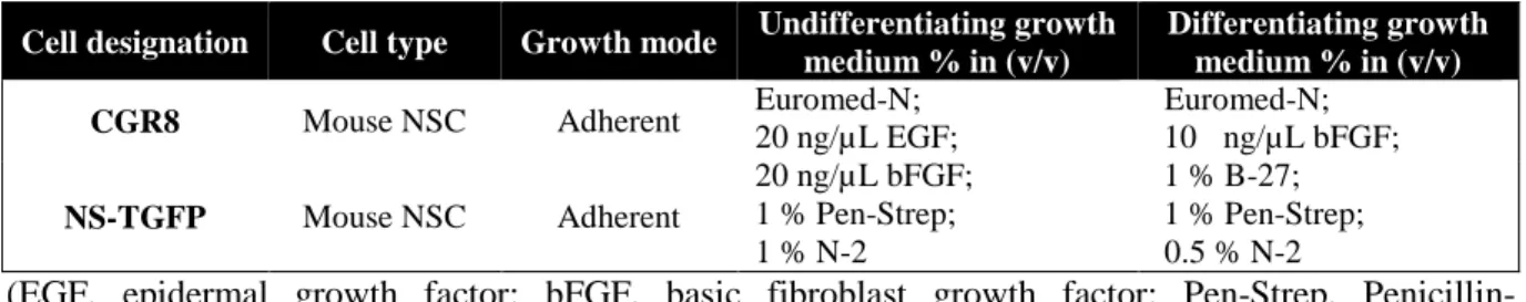
Table 2.1 – Details and characteristics of mouse NSC lines used in this study………….………24
Table 2.2 – Details of the primary antibodies used for Western blot……….27
Table 2.3 – Details of the secondary antibodies used for Western blot………...……….27
Table 2.4 – Details of the primary antibody used for immunocytochemistry…………....………29
xvii
List of Abbreviations
ACC Acetyl-H3 ADP ATP BA BBB bFGF BSA CNS CoA DG DNA Drp1 EGF ER ERR α ESC ETC FA FAO FASN FGF GAPDH GC-MS Gβ2 H3 hnRNP A2/B1 Hsp70 iPSC LCAD LC-MS Mfn 2 mPTP mRNA Acetyl-CoA carboxylase Acetylated histone 3 Adenosine diphosphate Adenosine triphosphate Bile acidBlood brain barrier
Basic fibroblast growth factor Bovine serum albumin Central nervous system Coenzyme A
Dentate gyrus
Deoxyribonucleic acid Dynamin-related protein 1 Epidermal growth factor Endoplasmic reticulum Estrogen-related receptor α Embryonic stem cell Electron transport chain Fatty acid
Fatty acid oxidation Fatty acid synthase Fibroblast growth factor
Glyceraldehyde-3-phosphate dehydrogenase Gas chromatography-mass spectrometry G-protein β2 subunit
Histone 3
Heterogeneous nuclear ribonucleoprotein A2/B1 Heat shock protein 70
Induced pluripotent stem cells Long-chain acyl-CoA dehydrogenase Liquid chromatography-mass spectrometry Mitofusin 2
Mitochondrial permeability transition pore Messenger ribonucleic acid (RNA)
xviii NADH NADPH ND NPC NSC NSPC NS-TGFP OMM ORO OXPHOS PDC PDH PDHE1-α PDK PGC-1α PPAR PUFA qNSC RNA ROS RT SC SGZ SREBP1 SVZ TCA TUDCA UDCA VDAC β-F1-ATPase
Nicotinamide adenine dinucleotide (reduced form)
Nicotinamide adenine dinucleotide phosphate (reduced form) Neurodegenerative disease
Neural progenitor cell Neural stem cell
Neural stem/progenitor cell
tau-green fluorescent protein (GFP) mouse neural stem cell line Outer mitochondrial membrane
Oil-Red-O
Oxidative phosphorylation Pyruvate dehydrogenase complex Pyruvate dehydrogenase
Pyruvate dehydrogenase E1-α subunit Pyruvate dehydrogenase kinase
Proliferator-activated receptor gamma coactivator 1 α Peroxisome proliferator-activated receptor
Polyunsaturated fatty acid Quiescent neural stem cell Ribonucleic acid
Reactive oxygen species Room temperature Stem cell
Subgranular zone
Sterol regulatory element-binding protein 1 Subventricular zone
Tricarboxylic acid
Tauroursodeoxycholic acid Ursodeoxycholic acid
Voltage-dependent anion channel ATP synthase β subunit
1
Introduction
Neural Stem Cells
Since the second half of the XX century, neural stem cells (NSCs) in the adult mammalian brain have been playing a major role in enriching our understanding about the plastic nature of the mammalian brain (Ma et al., 2009), opening gates to investigate future therapeutic approaches in the field of neuroscience. The discovery of these powerful cells fostered whole new perspectives to treat many neurodegenerative diseases (NDs) due to their identity and pertinent properties. (Filippis & Binda, 2012)
NSCs are the most primitive cells residing in the central nervous system (CNS). They have a huge relevance in sustaining the development and homeostasis of nervous tissue by giving the possibility to create a dynamic equilibrium between self-renewal and differentiation without depleting stem cell (SC) pool and to replace the most mature neural cells. This potential neuroregeneration is progressively and temporally restricted and it has been difficult to understand the temporal control of NSCs output. Under physiological conditions, in vivo, adult NSCs basically rest in a state of quiescence, which allow them to bear up metabolic stress and to preserve genome integrity during their lifetime. However, once activated to entering the cell cycle, NSCs must choose between its self-renewal and a mode of dividing. According to this idea, a NSC can divide symmetrically, yielding two progenitors or two NSCs, or asymmetrically, generating one NSC and one progenitor cell. Then, neural progenitor cells (NPCs) can acquire a more committed state to give rise to a specific cell type, including neurons, or glial cells, such as astrocytes or oligodendrocytes. Nevertheless, in this last scenario, they lose their capacity to self-renewal or proliferate (Figure 1.1). (Filippis & Binda, 2012; Bond et al., 2015)
Introduction
2
throughout life but once activated they choose between symmetric or asymmetric division. Both modes of dividing can give rise to progenitor cells, which differentiate into a specific cell type. Intermediate progenitor cells (IPCs) are a type of neurogenic transient amplifying cells that finally become neurons. NG-2 oligodendrocyte precursor cells become oligodendrocytes. Neural progenitor cells can generate astrocytes as well. Adapted from Bond et al., 2015.
Therefore, adult NSCs are self-renewing and multipotent progenitors, also differentiating into a wide-range of cell types. Meanwhile, the identification of trophic and mitogenic actions of growth factors, including epidermal growth factor (EGF) (Morrison et al., 1987) and fibroblast growth factor (FGF) (Nurcombe et al., 1993) family proteins enabled to culture and maintain NSC expansion in both floating and adherent conditions. (Ma et al., 2009) These two in vitro culture systems have several controversies, in terms of conservation of the molecular and biological properties of genuine NSCs. NSCs can form neurospheres in culture (Reynolds & Weiss, 1992), neurospheres mimic physiological conditions because of their three-dimensional structure, although they are not considered a good model for efficient neuronal generation due to its highly-associated heterogeneity. In contrast, expanding NSC cultures in monolayer conditions were shown to assure almost pure NSC populations, with a negligible differentiated component and a higher neurogenic potential in comparison to neurosphere models. (Conti & Cattaneo, 2010)
NSCs can be derived in vitro from embryonic stem cells (ESCs), derived from the inner cell mass of blastocysts; as from induced pluripotent stem cells (iPSCs), acquired by reprogramming from somatic cell; and also, isolated from fetal tissue and adult brain samples of germinative areas. Current NSC systems are not perfect. Therefore, determining the best sources for the in vitro derivation of NSCs and optimizing protocols for stable and clonal proliferation are still central goals of SC research. (Conti & Cattaneo, 2010) Cell propagation sets close to homogeneity in appropriate models for studying the neurogenesis process.
Adult neurogenesis
Neurogenesis was only believed to occur during embryonic stages in the CNS of mammalians. (Ramon y Cajal, 1913) The very first evidence for the existence of adult NSCs in mammalian brain was provided by pioneering studies with tritiated thymidine (thymidine-H3), a thymidine analogue that has been used as a label of proliferating cells. (Altman & Das, 1965) Other progresses were made but, for decades, the concept of adult neurogenesis has been ignored until it was rediscovered in the early 1990s. The introduction of 5-bromo-2-deoxyuridine (BrdU), other thymidine analogue which incorporates into the nuclear DNA during the S-phase of the cell cycle, demonstrated that in aged mammals, including humans, many newly generated cells were in fact neurons, and the field of adult neurogenesis took off. (Eriksson et al., 1998; Ma et al., 2009)
In most regions of the mammalian brain, the production of neurons is largely confined to the prenatal period as adult neurogenesis shows an age-dependent decline. Although it recapitulates the complete process of neuronal development, adult neurogenesis takes place in unique local niches that
3
support distinct NSCs behaviors, including their maintenance, self-renewal, fate specification and development. (Kuhn et al., 1996; Ming & Song, 2011; Gage & Temple, 2013) There are four processes critical to the overall levels of neurogenesis, cell survival, proliferation, neuronal differentiation, and migration. Moreover, quiescent neural SCs (qNSCs) can be localized in several regions across the adult brain. There is evidence to suggest the presence of neurogenesis in other regions of the adult brain. But neurogenesis is known to happen mainly in restricted zones with a distinct microenvironment. These germinative zones are evolutionarily conserved in mammals and have been identified in the subventricular zone (SVZ) lining the lateral ventricles and the subgranular zone (SGZ) within the dentate gyrus of the hippocampus (Figure 1.2 (A)). (Bond et al., 2015; Ernst & Frisén, 2015) Several cellular components integrate these neurogenic niches underlining, mature neurons, NSC progeny, astrocytes, endothelial cells, microglia and the blood vascular system itself (Figure 1.2 (B)). (Ma et al., 2009; Gage & Temple, 2013)
Figure 1.2 - Adult NSC niches. (A) A sagittal plane of the adult rodent brain, highlighting the two
major niches where adult neurogenesis occurs, the lateral ventricle (LV) in the subventricular zone (SVZ) to the rostral migratory stream (RMS) to the olfactory bulb (OB) and the dentate gyrus (DG) in the subgranular zone (SGZ) of the hippocampal formation (Hipp). There are also signalized the brain areas of the corpus callosum (CC) and the striatum (St). (B) A schematic diagram depicting cellular and molecular components of the SVZ and SGZ niches. CSF, cerebrospinal fluid; ECM, extracellular matrix; EZ, ependymal zone; GCL, granule cell layer; ML, molecular layer. Adapted from Bond et al., 2015.
Adult SVZ neurogenesis comprises different developmental stages, starting with the activation of radial glia-like cells (Type B cells) in the subventricular zone which gives rise to rapidly dividing
A
Introduction
4
transient amplifying cells (Type C cells) and leads to the generation of neuronal progenitor cells (Type A cells or neuroblasts). Then, these neuroblasts tangentially migrate along the rostral migratory stream (RMS) to the olfactory bulb. During this migration, these cells continue to divide, but the cell cycle is lengthened and the synaptic integration and maturation of granule cells and periglomerular neurons only occur when these cells reside in the core of olfactory bulb (OB). (Li et al., 2008; Ming & Song, 2011) It is important to note that the direction of neuroblast migration is the OB in most mammals, but in adult humans striatal neuroblast migration is most pronounced. Further, neuroblasts are not restricted to the lateral ventricles, but are also present beyond the neurogenic niche. (Ernst & Frisén, 2015)
In the SGZ of the hippocampus, NSCs migrate into the granule cell layer, a shorter distance when compared with the extensive migration undertaken by OB neurons. These cells give rise to mature granule neurons in the dentate gyrus. Analogous to the type B cells in the SVZ, they generate transient amplifying progenitor cells (Type IIa, IIb, and III cells) that then differentiate into immature neurons. At last, immature neurons become mature granule neurons and the sequential process of synaptic integration takes place. From the beginning of NSC differentiation to the synaptic integration of newly generated neurons takes approximately 2-4 weeks. Throughout the neurogenesis process, both in SVZ and SGZ regions, different cell stages and cell lineages can be detected by stage-specific markers, as illustrated in Figure 1.3. (Li et al., 2008; Ming & Song, 2011)
Figure 1.3 - Schematic diagram illustrating specific stages of adult hippocampal neurogenesis.
The neuronal differentiation cascade, from radial glia-like cells (RGL) to mature neurons, is represented by the major stage-specific markers: GFAP, glial fibrillary acidic protein; Sox2, sex determining region Y (SRY)-box 2; DCX, doublecortin; PSA-NCAM, polysialylated form of the
5
neural cell adhesion molecule; TuJ1, β-tubulin III; NeuN, neuronal nuclear antigen. Adapted from Lucassen et al., 2010; Kang et al., 2016.
Therapeutic relevance of NSCs
The discovery of NSCs and neurogenesis in the adult mammalian brain has changed our view about the plasticity and function of the human brain bringing up new expectations for rescuing brain function after injury or NDs. (Casarosa et al., 2014)
Following brain injury adjacent NSCs have the capacity to proliferate and to migrate to the local lesion. Indeed, NSCs in the SVZ and DG are stimulated after traumatic brain injury or seizures, revealing that adult neurogenesis may be relevant in self-recovery mechanisms of the brain. However, far from obtaining regenerative effects, the amount of produced neuroblasts after brain injury as well as their survival and differentiation into mature neurons is highly limited. (Casarosa et al., 2014)
Aging is the primary risk factor for prevalent NDs. (Mattson & Magnus, 2006) However, irrespective of their specific etiology, NDs eventually lead to loss or functional alteration of neurons and glia in the brain or spinal cord either in acute or chronic cases. In acute cases, like ischemic stroke or spinal cord injury, different types of neurons and glial cells die within a restricted area over a short time period. Whereas in chronic cases, usually there is a progressive loss of a specific population of cells, such as motor neurons in amyotrophic lateral sclerosis (ALS) and dopamine neurons in Parkinson disease (PD), or an extensive degeneration of many types of neurons in Alzheimer disease (AD). NDs currently lack effective treatments and SC research could lead to the development of new powerful therapies. (Lindvall & Kokaia, 2010)
Over the past few years, and based on the pathology of each disease, there has been a continuous progress in developing approaches to generate the types of human-derived neurons and glial cells for cell replacement therapy. Indeed, there are three principal mechanisms by which SC-based therapies could be helpful: cell replacement, where cell transplants are given to directly replace cells that are damaged or lost; trophic support, where transplanted cells are used to promote endogenous repair of specific diseased brain areas; and, modulation of inflammation, frequently involved in the disease process and where transplanted cells are used to release anti-inflammatory agents. (Lindvall & Kokaia, 2010)
Different sources of SCs have been proposed to be used for the recovery of CNS injuries. (Lodi et al., 2011) Recently, the discovery iPSCs, by Yamanaka in 2006, opened new possibilities for autologous transplantation. (Takahashi & Yamanaka, 2006) Human iPSC-derived neural precursor cells can be transplanted into a rat model of PD and ameliorate the motor-sensory behavior by increasing the number of midbrain dopamine neurons. (Wernig et al., 2008) Although, the major drawback of these genetically manipulated cells is the high risk of cancer formation, mainly due to possible uncontrolled integration of viral vectors and non-specific recombination events. In fact, due to their high proliferative potential, iPSCs were shown to induce tumor formation and impair neural
Introduction
6
outgrowth and their use in the clinical therapy must be very cautious. (Lodi et al., 2011) ESCs have also indefinite self-renewal capabilities as well as the ability to differentiate into all cell types derived from the three embryonic germ layers. However, there are major challenges for the safety of using them in any kind of transplantation therapy, also involving the risk for unregulated cell growth. Therefore, the most suitable candidates for SC therapies in the context of neurological disorders are adult SCs, particularly NSCs. NSCs can potentially be an autologous SC source for transplantation with minimal risk of tumor formation. (Yu et al., 2008)
The therapeutic use of NSCs is being actively investigated, once they show considerable promise for regenerative repair of CNS and there are present in a considerable number of ongoing clinical trials. (Trounson & McDonald, 2015) Plus, for a realistic exploitation of NSCs in cell therapies, they must have a reproducible safe behavior as well as long-term survival and ability to differentiate. The underlying problem in NSC use is the short time window following neural induction during which the cells can be directed toward specific neuronal lineages. Ultimately, increasing the number of cells capable of differentiating might prove useful for in vitro or in vivo expansion of NSCs. (Yu et al., 2008)
Collectively, a more comprehensive understanding of the regulatory mechanisms involved in self-renewal, proliferation and differentiation of NSCs will certainly help to ensure well-established methods to manipulate these cells in vitro and in vivo and improve the successful use of NSCs in clinics.
Mitochondrial control of NSC activity
The relevance of mitochondrial function and energy metabolism in determinating NSC fate is being currently widely discussed. In fact, the whole integration of different mitochondrial events is yet to be revealed as the metabolic force driving NSC identity. In this regard, it might be worth increasing the knowledge on the influence of cell metabolism on NSC behavior during CNS diseases. (Wanet et al., 2015; Xavier et al., 2015; Ottoboni et al., 2017)
Mitochondria are classically known to be the powerhouse of eukaryotic cells. Despite their major function as cell energy producer, mitochondria regulate intracellular calcium and redox homeostasis, playing important roles in biosynthetic processes, including lipid, cholesterol, nucleotide, heme, and steroid synthesis, as in amino acid metabolism and ion homeostasis. This dynamic organelle has even gained more attention over the years by being implicated in signaling pathways of cell death and survival. (Hock & Kralli, 2009; Murphy, 2009)
The CNS has an extraordinary high metabolic rate. It consumes about 20% of oxygen inspired at rest, whereas accounting for only 2% of the body weight. CNS depends upon glucose as its main source of energy and most of neuronal adenosine triphosphate (ATP) is generated through oxidative phosphorylation (OXPHOS) in mitochondria. Neurons, in turn, are highly differentiated cells, consuming much of this energy to sustain cell membrane ionic gradients and neurotransmission.
7
Notably, they display a complex morphology and are highly compartmentalized in a complex network, where mitochondrial trafficking matches the energy demand throughout the long neuronal segments. (Kann & Kovács, 2007)
Cellular specialization relies on specific mitochondrial specialization and maturation. Indeed, several neural development processes rely on mitochondrial regulation: self-renewal and differentiation of NSCs, as neurogenesis it-self within axonal and dendritic growth, synaptic formation, and reorganization. Moreover, mitochondrial dynamics and bioenergetics are closely related to NSC fate and behavior also, intriguingly, providing signal to the nucleus to modulate differentiation and development. (Kann & Kovács, 2007; Wanet et al., 2015; Xavier et al., 2015) In this regard, mitochondrial dysfunction can be an underlying problem, namely in the eventual depletion of the SC pool and impaired neurogenesis, as observed during aging and degenerative or metabolic diseases. (Wallace, 2005; Khacho et al., 2017) In addition, mitochondria are responsible for long-term survival, differentiation and synaptic integration of newborn neural cells. (Xavier et al., 2015) Therefore, it is likely that mitochondria and its regulatory network will have major implications toward a more efficient use of neural replacement therapies.
General aspects of mitochondrial structure, function, and biogenesis
Mitochondria have two membranes, an intermembrane space, where there are important enzymes, and an internal matrix. The outer mitochondrial membrane (OMM) is permeable to ions and small molecules. The inner mitochondrial membrane (IMM), in turn, is almost impermeable and forms a tight barrier between the mitochondrial matrix and the cytoplasm, being composed by different ion channels and transporters, such as the Ca2+ uniporter, K+ATP channels, and Na+/Ca2+ exchanger, and mitochondrial enzyme systems, such as the molecular machinery for energy production, the electron transport chain (ETC). The ETC consists of five protein complexes. Three of the complexes (I, III, and IV) pump protons (H+) across the inner membrane to establish a H+ gradient with a potential difference of 150–180 mV (negative with respect to cytosol). This potential difference sets the driving force for protons (along with ΔpH) that actuate F1F0-ATP synthase (complex V) to generate ATP and for cytosolic Ca2+ ions to accumulate in the matrix via the mitochondrial Ca2+ uniporter. Nicotinamide adenine dinucleotides (NADH) and flavin adenine dinucleotides (FADH2) serve in energy transfer to the ETC, via the complexes I (NADH-ubiquinone oxidoreductase), II (succinate dehydrogenase), III (ubiquinol cytochrome c oxidoreductase) and IV (cytochrome c oxidase), electrons are transferred from NADH and FADH2 to O2. During electron transport, the free radical superoxide (O2-.) is also generated. (Kann & Kovács, 2007; Mattson et al., 2008)
Importantly, voltage-dependent anion channel (VDAC) is a key protein complex involved in ATP rationing, Ca2+ homeostasis, protection against oxidative stress and regulation of apoptosis. VDAC is located at OMM and helps to form the mitochondrial permeability transition pore (mPTP) where mitochondrial metabolites and ions enter and exit. During apoptosis, the opening of mPTP
Introduction
8
results in release of cytochrome c into the cytoplasm. (Shoshan-Barmatz et al., 2010)
Recently, it has become clear that Bcl-2 family members are implicated in mitochondria-mediated death during neurogenesis and neuronal differentiation processes. Direct interactions between mitochondria and the endoplasmic reticulum (ER) have also a role to play. For example, during apoptosis, cytochrome c released from mitochondria can trigger the release of Ca2+ from the ER. (Mattson et al., 2008)
Undouthbly, mitochondrial function is connected to a wide range of cell signaling pathways through changes in redox and phosphate pairs (NAD/NADH; ATP/adenosine diphosphate (ADP) vs. adenosine monophosphate (AMP)), reactive oxygen species (ROS) production and critical metabolite concentrations, such as acetyl-CoA. (Cagin & Enriquez, 2015)
Interestingly, mitochondrial biogenesis is regulated to respond to energetic and metabolic demands of the cell and is influenced by environmental stress, such as exercise and caloric restriction, cell division, renewal, and differentiation. In fact, throughout these processes, mitochondria variations in number and size and mass occur. Thus, mitochondrial dynamic is a complex process that requires the synthesis, import, and incorporation of new proteins and lipids to the existing mitochondrial reticulum. Intriguingly, mitochondria have their own genome and can autoreplicate, which is related to their bacterial origin. The mitochondrial proteome comprises ∼1100 to 1500 proteins and the vast majority of them are encoded by nuclear genes. However, the mitochondrial genome encodes 13 essential proteins that are components of OXPHOS. Indeed, mitochondrial biogenesis is regulated by a transcriptional network in between the coordination of the two genomes. (Hock & Kralli, 2009; Jornayvaz & Shulman, 2010)
Mitochondria nucleus cross-talk
Mitochondrial function and activity are under strong nuclear control and several signaling pathways coordinate the communication between mitochondria and the nucleus. The so called ‘anterograde regulation’ is based mainly on the expression of several nuclear-encoded mitochondrial proteins, transcription factors, and co-regulators. (Quirós et al., 2016) The expression of nuclear-encoded mitochondrial proteins is principally mediated by nuclear transcription factors, such as nuclear respiratory factor 1 (NRF-1), which controls the expression of the vast majority of nuclear-encoded subunits that are involved in OXPHOS. In fact, Dhar et al. demonstrated that NRF-1 regulates all ten nuclear-encoded subunits of cytochrome c oxidase in neurons. (Dhar et al., 2008) Moreover, nuclear receptor factors, which include peroxisome proliferator-activated receptors (PPARs), are nuclear receptors that sense lipids and control lipid homeostasis. PPARα and PPARδ are primary regulators of lipid oxidation, whereas PPARγ promotes lipid synthesis and storage. The estrogen-related receptors (ERRs) - ERRα, ERRβ, and ERRγ, in turn, are associated with the expression of nuclear-encoded mitochondrial proteins that are involved in the tricarboxylic acid (TCA) cycle, also in OXPHOS and fatty acid oxidation (FAO). Importantly, proliferator-activated receptor
9
gamma coactivators 1 α and β (PGC-1α/β) are master regulators of mitochondrial biogenesis known to activate the NRF, ERR and PPAR factors, which may lead to induced mitochondrial biogenesis by increasing the expression of mitochondrial proteins encoded by nuclear DNA, such as the mitochondrial transcription factor A (TFAM). Upregulation of TFAM drives an increase in replication and expression of mtDNA. (Picca & Lezza, 2015; Quirós et al., 2016)
Conversely, mitochondria can create a ‘retrograde response’ signaling the nucleus to alter the expression of nuclear genes in order to modify cell function or/and metabolism. Retrograde signals are generally classified as energetic stress responses. This retrograde response has been classically linked to mammalian target of rapamycin (mTOR) and AMP-activated protein kinase (AMPK). A decrease in mitochondrial ATP synthesis stimulates AMPK which, in turn, promotes the activation of PGC-1α. Additionally, AMPK triggers the mitochondrial quality control system, interfering with mitochondrial dynamics to induce mitophagy, leading to either an elimination of mitochondrial content or damaged mitochondria. (Ashrafi & Schwarz, 2013; Quirós et al., 2016) Similarly, stress reduced mTOR activity facilitates mitochondrial retrograde signaling, while its activation inhibits retrograde response. (Quirós et al., 2016) Furthermore, this organelle has also been implicated in the regulation of cell cycle progression in NSCs through this kind of mechanism. (Owusu-Ansah et al., 2008; Xavier et al., 2014) Apart from complex signaling pathways responsible for mediating the communication between nucleus and mitochondria a more direct way of interorganellar coordination has emerged, the redistribution of nuclear or mitochondrial proteins between these two compartments. (Lionaki et al., 2016) Of the dual-localized proteins, a good number of predominantly nuclear proteins are now known to be localized to and function at the mitochondria. Prominent among these are those whose activities are related to cell death and survival, as well as senescence and aging. For example, the tumor suppressor p53, ERR α and β, Sirtuin 1 (Sirt1) and PGC-1α, among others with or without mitochondrial defined activities. (Tang, 2015; Lionaki et al., 2016)
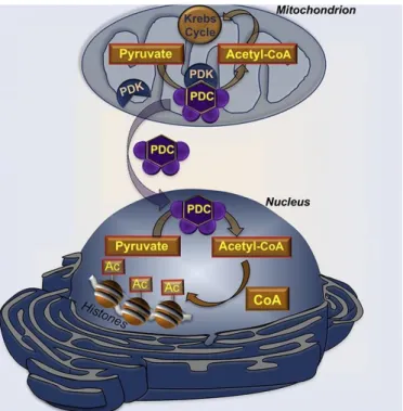
On the other hand, several recent findings have indicated that a small number of classically mitochondrial-localized proteins could be also found in the nucleus, performing non-transcriptional activities. (Tang, 2015; Lionaki et al., 2016) Surprisingly, one of the largest multiprotein complexes known, the pyruvate dehydrogenase complex (PDC), is one of them. (Sutendra et al., 2014) In mitochondria matrix, the PDC catalyzes multiple enzymatic steps to generate acetyl-CoA from glycolysis-derived pyruvate that, then, fuels the TCA cycle. The mammalian PDC is about 8-10 MDa in size and has 3 enzyme activities (E1, E2, and E3), each comprising of multiple polypeptide subunits, which makes PDC even larger than the largest complex in the mitochondrial ETC. (Tang, 2015) Interestingly, nuclear PDC is functional in generating acetyl-CoA from pyruvate (Figure 1.4). (Sutendra et al., 2014) Another interesting example of bi-organellar protein distribution is fumarase, a TCA cycle enzyme, now known to be also important for DNA repair in the nucleus. There are other examples of proteins whose nuclear function and mechanism driving nuclear localization remains elusive. (Tang, 2015; Lionaki et al., 2016)
Introduction
10
Figure 1.4 – Dynamic translocation of mitochondrial PDC to the nucleus. The PDC plays a central
role in cellular metabolism by catalyzing the irreversible conversion of pyruvate into acetyl-CoA in mitochondria. The activity of PDC is tightly controlled via reversible inactivating phosphorylation due to the activity of specific pyruvate dehydrogenase kinases (PDKs). Mitochondrial PDC translocates to the nucleus of mammalian cells, where is functional, providing a novel pathway for nuclear acetyl-CoA synthesis required for histone acetylation and epigenetic regulation. Adapted from Sutendra et al., 2014.
Ultimately, to a great extent, the integration of anterograde (from nucleus to mitochondria) and retrograde (from mitochondria to nucleus) signals and this dual localization of proteins is a level of interorganellar communication and coordination that allows for direct and finely tuned responses of both organelles, leading to an enhanced and precise defense of cell homeostasis. (Lionaki et al., 2016; Quirós et al., 2016)
Mitochondrial dynamics
The ultrastructure, morphology and intracellular distribution of mitochondria undergo significant changes during cellular life. Somatic cells exhibit mature elongated mitochondria, with numerous cristae and an electron-dense matrix. (Varum et al., 2011) However, the first observations of mitochondria in mouse and human ESCs (mESCs and hESCs) using transmission electron microscopy surprisingly showed characteristics that are not related to the well-known ultrastructure of mitochondria. Indeed, in these cases, mitochondria morphology appeared globular displaying a perinuclear localization and containing poorly developed cristae, along with an electron-lucid matrix. (Baharvand & Matthaei, 2003; Prowse et al., 2012; Wanet et al., 2015)
11
these two opposing processes dictates the connectivity and the overall length of mitochondria. Mitochondrial elongation requires the coordinated process of outer and inner membrane fusion. Mitochondrial fusion is mainly mediated by three dynamin-related large GTPases. Mitofusin 1 (Mfn1) and 2 (Mfn2) mediate the fusion of the OMM, and the optic atrophy 1 (Opa1) protein and Mfn1 mediate the fusion of the IMM. The fission process, on the other hand, involves the division of mitochondria and is regulated by the cytosolic dynamin-related protein 1 (Drp1). (Khacho & Slack, 2015)
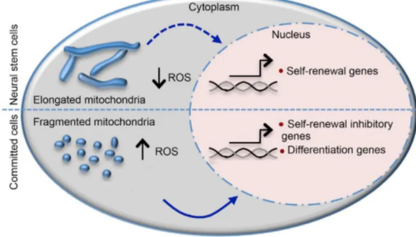
Notably, changes in mitochondrial dynamics regulate NSC fate decisions by interfering in physiological levels of ROS, which in turn, suppressed self-renewal and promoted differentiation through transcriptional programming. Recently, the view about the classical mitochondrial dynamics regulation has been modified. Mitochondrial fragmentation has been demonstrated to be required for the transient passage of NSCs to committed progenitors. (Figure 1.5) (Khacho et al., 2016) Although, it is generally perceived as a sign of mitochondrial dysfunction toward a chronical imbalance that is observed during aging and many NDs. (Khacho & Slack, 2015)
Figure 1.5 - Schematic diagram of mitochondrial dynamic changes occurring as NSCs differentiate. In NSCs, elongated mitochondria maintain low ROS levels and promote self-renewal,
while a more fragmented state of mitochondria is normally associated with increase of ROS levels, inhibition of self-renewal and induction of cell differentiation. Adapted from Khacho et al., 2016.
Fission and fusion events do not only dictate the structural morphology of mitochondria but also regulate cell metabolism, playing a major role in NSC differentiation and survival.
Metabolic regulation of NSC fate
Increasing evidence suggests that metabolic plasticity is essential in the transition between stemness maintenance and lineage specification. Indeed, adult NSCs may rely on different metabolic pathways (Figure 1.6) to keep up with cell-specific bioenergetic demands. Energy generation is always fundamental to cell homeostasis, although SCs must fine-tune the balance between catabolism to
Introduction
12
generate ATP, and anabolism to create biomass. (Folmes et al., 2013; Knobloch & Jessberger, 2017)
Figure 1.6 - Major cellular metabolic pathways. Glucose is taken up and metabolized through
glycolysis. The final product, pyruvate, can be either fermented into lactate, which is secreted by the cell or can be shuttled into the mitochondria and converted into acetyl-CoA by PDC. This acetyl-CoA is subsequently used in the TCA cycle to generate NADH and reduced FADH, which are necessary for the OXPHOS process. Overall, this results in ATP generation. As a side product, ROS can also be produced. NADH and FADH are also generated in large amounts by FAO. Resulting acetyl-CoA levels enter into the TCA cycle for further energy production, and as a carbon source, or even be exported from mitochondria via citrate for other use, like lipid biosynthesis. Fatty acid synthase (FASN) is involved in this process, yielding palmitate, which can be used to generate more complex fatty acids. The reduced nicotinamide adenine dinucleotide phosphate (NADPH) required for lipid synthesis can be generated during the pentose phosphate pathway (PPP), a metabolic pathway parallel to glycolysis. Adapted from Knobloch & Jessberger, 2017.
NSCs reside in hypoxic niches where low oxygen tensions contribute to the maintenance of an undifferentiated state, influencing cell fate and proliferation. (Ito & Suda, 2014) Numerous studies show that mouse and human ESCs and iPSCs rely principally on glycolysis under aerobic conditions. (Folmes et al., 2011; Varum et al., 2011; Panopoulos et al., 2012) Glycolysis generates reducing equivalents through the pentose phosphate cycle and, by attenuating mitochondrial activity, directly diminishes the generation of ROS, thus, favoring NSC self-renewal and long-term maintenance.
13 (Wanet et al., 2015)
Metabolic changes between SCs and progeny suggest that mitochondrial mass and activity, as well as, OXPHOS increase with lineage progression whereas elevated anaerobic glycolytic activity is rather considered a metabolic hallmark of cell stemness. (Wanet et al., 2015) Although, it has been demonstrated, in NSCs, that inhibition of glycolytic pathways, even when oxidizable substrate was provided, greatly impaired their survival. (Candelario et al., 2013) An interesting study in Drosophila during metamorphosis suggests a direct regulation of NSC differentiation via OXPHOS. (Homem et al., 2014) Also, differentiation of hNSCs into motor neurons stimulated mitochondrial biogenesis and decreased glycolytic flux. (O’Brien et al., 2015) Being now well-stabilized that the metabolic transition profile of NSCs to neurons relies on a switch from glycolysis to mitochondrial OXPHOS to meet the robust energy demands associated with differentiation. (Hu et al., 2016)
The identity of stage-specific metabolic programs and their impact on adult neurogenesis are largely unknown, we are now starting to understand mitochondria and its stage-specific molecular program adaptation of metabolic circuits under this scenario. The adult hippocampal neurogenic lineage is critically dependent on the mitochondrial ETC and OXPHOS machinery at the stage of the fast proliferating neural stem/progenitor cell (NSPC). (Beckervordersandforth et al, 2016)
As mentioned above, ROS are naturally produced by OXPHOS in mitochondria under physiological conditions. (Murphy, 2009) In a mouse model of adult hippocampal neurogenesis, a peak in mitochondria number and ROS levels occurred immediately after inducing differentiation, in a highly proliferative, intermediate progenitor state, but not in undifferentiated NSCs or postmitotic neurons. (Walton et al., 2012) Notably, ROS-mediated process triggers a dual program to suppress self-renewal and promote differentiation via retrograde signaling in NSCs. ROS force SCs out of hypoxia-dependent quiescence into a more proliferative state, promoting cellular commitment and differentiation. (Ito & Suda, 2014; Khacho et al., 2016)
Under the road of cellular metabolic pathways, lipid metabolism has also been largely neglected for the role it may play in neurogenesis process. Lipids emerge in NSC life as building blocks of membranes, an alternative energy source and as signaling entities. (Knobloch, 2016) The current knowledge of lipid metabolism in NSC regulation and neurogenesis will be discussed ahead.
The emerging role of lipid metabolism in NSC behavior
Lipid metabolism plays a crucial role in tissue physiology and cell signaling, being important for the CNS, as lipids make up roughly 50% of brain dry weight. In fact, brain is the organ with the second highest lipid content next to adipose tissue. (Mitchell & Hatch, 2011) On the other hand, altered lipid metabolism is believed to contribute to CNS injury and is linked to many NDs. Lipids are classified into eight categories (fatty acyls, glycerolipids, glycerophospholipids, sphingolipids, sterol lipids, prenol lipids, saccharolipids, and polyketides) comprising a large number of chemically distinct molecules that arise from combinations of fatty acids (FAs) with different backbone structures. These
Introduction
14
FAs can be used as raw materials to build cell membranes, be metabolized into bioactive agents or be degraded. (Adibhatla & Hatcher, 2008) FAs are present in the bloodstream of adult mammals, commonly bound to serum albumin. For a long period of time, it was thought that FAs do not penetrate the blood brain barrier (BBB); however, there is a slow speed passage of FAs across the BBB. Although FA import mechanism into the BBB is still uncertain (Hamilton & Brunaldi 2007; Mitchell & Hatch, 2011), during brain development, FAs are necessarily taken up because they are critically involved in neurodevelopment, neurotransmission and repair processes. (Schönfeld & Reiser, 2013)
FA import into cells has also been a source of constant debate. FAs can enter a cell through protein-mediated mechanisms involving “FAs transporters” or, much like other hydrophobic molecules, can cross lipid bilayers by passive diffusion using a ‘flip-flop’ mechanism independent of proteins. (Mitchell & Hatch, 2011) After entry into the cell, FAs are activated to acyl-CoA esters by acyl-CoA synthetases and can be targeted to esterification to be stored as triglycerides or be an alternative source of energy when submitted to a mitochondrial degradation process called β-oxidation. Mitochondrial β-oxidation, in turn, can be conceptually divided into two major phases: the process of getting acyl groups into the mitochondria for oxidation and the intramitochondrial chain shortening. Indeed, medium- and short-chain FAs can cross the mitochondrial membrane directly and be oxidized as their CoA (coenzyme A) esters. In contrast, long-chain FAs are imported into the mitochondria in a carnitine palmitoyl transferase (CPT)-1 dependent manner, through a “carnitine shuttle”. Once inside mitochondria, FAs are then oxidized to acetyl-CoA units. Chain length is reduced by two carbons in every cycle of β-oxidation and the acetyl-CoA produced from FAO enters the TCA cycle. At the same time, there is production of reducing equivalents (FADH2 and NADH), which are utilized in the ETC contributing toward the production of ATP. Subsequent rounds of β-oxidation generate successively shorter fatty acids. The first step is catalysed by acyl-CoA dehydrogenase enzymes, like long-chain acyl-CoA dehydrogenase (LCAD) that have preference for acyl-CoA substrates of long-chain length. (Bartlett & Eaton, 2004; Kompare & Rizzo, 2008)
The relation between FAO and SC identity was initially suggested by the observation that, in mice, disruption of promyelocytic leukemia gene (PML) (which regulates FAO), or direct inhibition of FAO, decreases the hematopoietic SC pool. (Ito et al., 2012) Consistently, congenital defects in mitochondrial FAO in NSCs promote symmetric differentiation division at the expense of NSC self-renewal in development of neocortical mouse brain. (Xie et al., 2016) It seems that this pathway is specifically up-regulated in qNSCs. Notice that, upon its activation, NSCs increase translational capacity, followed by cell-cycle entry with G1 to S transition. Furthermore, new data from single-cell transcriptomes of adult hippocampal qNSCs and their immediate progeny revealed that oxidative metabolism through FAO in qNSC may represent an alternative energy fuel to glucose. During the transition from quiescent to active NSCs, glycolysis and FAO tend to gradually decrease while there is an increased dependence on glucose to supply OXPHOS for energy generation. (Shin et al., 2015;
15 Fidaleo et al., 2017)
FAs can also be produced by the cell itself. In fact, FAs have been shown to be produced endogenously in adult NSCs; a novel mechanism governing adult neurogenesis has been identified, where lipogenesis determines the proliferative activity of NSPCs. Lipogenesis is dependent upon the rate-limiting enzyme for FA synthesis, fatty acid synthase (FASN), as well as on the availability of acetyl-CoA and NADPH and, consequently, on acetyl-CoA carboxylase (ACC) for the synthesis of malonyl-CoA (Figure 1.6). (Folmes et al., 2013) Importantly, sterol regulatory element binding protein-1c (SREBP-1c) exerts a positive transcriptional regulation on FASN and ACC expression, the sterol regulatory elements binding protein (SREBP), a family of transcriptional activators plays a crucial role in both FA and cholesterol homeostasis in the brain. (Kim et al., 2007) Curiously, it has been noticed FASN is preferably distributed in major areas of neurogenesis (SVZ and DG) within the adult murine brain. Expression is high in proliferating NSPCs and reduced in differentiated progeny. Accordingly, inhibition or deletion of FASN reduces proliferation of NSCs. (Knobloch et al., 2012) FASN expression in support of anabolic lipogenesis enables the production of lipid membranes required to sustain high SC proliferation. (Folmes et al., 2013) This mechanism is very similar to that of cancer cells, which produce the majority of their lipids de novo. (Menendez & Lupu, 2007)
Indeed, lipids are synthesized by de novo lipogenesis with the exceptions of two essential FAs, α-linolenic acid (ALA, an omega-3 FA) and linoleic acid (LA, an omega-6 FA), which are polyunsaturated fatty acids (PUFAs) and must be obtained via nutrition. Long-term culture of neurons in vitro, in turn, requires serum-free neurobasal medium with B-27 supplement, which includes lipids such as ALA and LA. In addition, many studies have demonstrated a beneficial role of FA precursors for neurogenesis both during development and in the adult. These are examples of bioactive lipid signaling playing a key role in neurogenesis. Bioactive lipid signaling, for instance, has recently emerged, as a lipid second messenger to regulate the energy and redox balance of differentiating NSCs. (Bieberich, 2012; Knobloch, 2016) Furthermore, recently, the receptor 1 for lysophosphatidic acid (LPA), a phospholipid, has been suggested as a novel NSC marker. LPA plays signaling functions, is important for brain development and neurogenesis, and shows an interesting expression pattern in the adult DG. (Walker et al., 2016) This example illustrates an emerging and complex topic bringing also the question if lipids can be novel NSC markers. (Knobloch, 2016)
Not less important, mechanistic studies have recently demonstrated that lipid accumulation perturbs the neurogenic niche microenvironment and restrains neurogenesis in diseased brains. For example, a FA-mediated mechanism suppressed NSC activity, showing potential relevance in cases of AD risk and cognitive decline in obesity and type 2 diabetes. (Hamilton et al., 2015; Fidaleo et al., 2017)
Cell metabolism has undoubtedly a central role in determining NSC physiology and behavior on multiple levels either by changing energy state, fuel source, biomass production, or epigenetics. (Knobloch & Jessberger, 2017) Figure 1.7 attempts to summarize the current knowledge on the
Introduction
16
metabolic switches ruling NSC transformation into immature neurons.
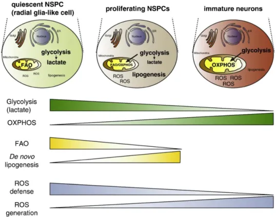
Figure 1.7 - Schematic representation of the major changes in metabolic pathways from qNSPCs to their neuronal progeny. Fateful metabolic shifts, occurring in quiescent and proliferating NSPCs
as well as in immature neurons, control NSC identity through glycolysis versus OXPHOS, FAO versus lipogenesis, and ROS defense versus generation. Adapted from Knobloch & Jessberger, 2017.
Specific modulation of metabolic pathways might be awaiting future discovery, with the potential to improve adult neurogenesis and revolutionize regenerative medicine.
The potential of tauroursodeoxycholic acid and its unexpected effects
Bile acids and cell function
Bile acids (BAs), the major constituents of bile, are mainly produced in the liver. Bile acid synthesis is the primary pathway for cholesterol catabolism. Thus, BAs are end products of cholesterol, water-soluble as result of their amphipathic structure, helping to balance cholesterol levels in the body. They are usually conjugated with glycine or taurine groups and, then, secreted into the small intestine, where they assume an important role in the solubilization of lipids. They act as detergents forming mixed micelles with dietary lipids, thereby promoting their uptake. (Russell & Setchells, 1992) Minor changes in the chemical structure of bile acids determine their mechanism of action either by increasing or decreasing their hydrophobicity. Hydrophobic bile acids can not only destabilize biological membranes but they can also activate cell death pathways, while hydrophilic bile acids may be cytoprotective triggering survival pathways and inhibiting induced cellular toxicity.
17 (Amaral et al., 2009)
Importantly, BAs are increasingly being comprehended not just as lipid solubilizers and simple regulators of BA homeostasis but as complex metabolic integrators and signaling factors. BAs are ligands for G‑protein-coupled receptors (GPCRs), such as TGR5, as well as for nuclear hormone receptors, such as farnesoid X receptor-α (FXR-α), also known as the ‘nuclear bile-acid receptor’, because BAs are the best-characterized ligands. Through activation of signaling pathways, BAs have been shown to regulate triglyceride, cholesterol, energy and glucose homeostasis, thus becoming attractive therapies for metabolic disorders, such as obesity and type 2 diabetes, as well as other associated chronic diseases, including non-alcoholic steatohepatitis. (Thomas et al., 2008)
Ursodeoxycholic acid (UDCA) is an endogenous hydrophilic bile acid US Food and Drug Administration (FDA)–approved for the treatment of certain cholestatic liver diseases. Curiously, UDCA is one of the major components of bear bile and has been used in Chinese medicine for centuries to treat numerous health problems. (Vang et al., 2014) In humans, it is also produced endogenously but at very low concentrations. (Bentayeb et al., 2008) It is widely known to be a cytoprotective agent that strongly detain programmed cell death through modulation of classical mitochondrial pathways, also preventing unfolded protein response dysfunction and ameliorating ER stress. (Amaral et al., 2009; Vang et al., 2014) Accordingly, UDCA inhibits several typical apoptotic events by stabilizing the mitochondrial membrane. (Rodrigues et al., 1998a; Rodrigues et al., 1998b; Rodrigues et al., 1999) Moreover, DNA microarray analysis revealed that UDCA could significantly regulate the expression of 96 different genes, involved not only in apoptosis but also in cell cycle regulation, proliferation and cell metabolism, in primary rat hepatocytes. (Castro et al., 2005) Tauroursodeoxycholic acid (TUDCA) is the taurine-conjugated form of UDCA. Importantly, after conjugation with taurine, UDCA administrated in high doses can be delivered to other tissues, including the brain. Thus, TUDCA is orally bioavailable and able to penetrate the CNS through systemic circulation and, by crossing the BBB. (Keene et al., 2002) This is possible due to the small size and relative hydrophobicity of TUDCA, and probably because of the existence of transporters for the taurine-conjugated molecule in the brain. (Pow et al., 2002) Gene expression microarray analysis demonstrated that TUDCA specifically modulates transcripts for proteins with kinase activity, diverse transcription factors and several enzymes involved in FA metabolism in primary rat hepatocytes. (Castro et al., 2005) Importantly, TUDCA has potential therapeutic effects on a wide variety of non-liver diseases (Vang et al., 2014), which will be described ahead.
TUDCA affords neuroprotection
Neurobiological disorders are caused by many factors directly related to mitochondrial dysfunction, such as oxidative stress, misfolded proteins, impairment of ECT complexes and Ca2+ imbalance. (Bredesen et al., 2006) Over the past few years, the protective role of TUDCA has been demonstrated in a wide range of models of neurological disorders, including Alzheimer’s (Dionísio et