https://doi.org/10.1080/20477724.2017.1299831
Association of enteric parasitic infections with intestinal inflammation and
permeability in asymptomatic infants of São Tomé Island
Marisol Garzóna , Luis Pereira-da-Silvab,c , Jorge Seixasa , Ana Luísa Papoilab,d , Marta Alvesb , Filipa Ferreiraa and Ana Reisa
atropical Clinic teaching and Research Unit, Instituto de higiene e Medicina tropical, Universidade noVa de lisboa, lisboa, Portugal; bResearch Unit, Centro hospitalar de lisboa Central, lisboa, Portugal; cWoman, Children and adolescent’s Medicine teaching and Research area, noVa Medical school, Universidade noVa de lisboa, lisbon, Portugal; dnoVa Medical school, Universidade noVa de lisboa, lisbon, Portugal
ABSTRACT
The cumulative effect of repeated asymptomatic enteric infections on intestinal barrier is not fully understood in infants. We aimed to evaluate the association between previous enteric parasitic infections and intestinal inflammation and permeability at 24-months of age, in asymptomatic infants of São Tomé Island. A subset of infants from a birth cohort, with intestinal parasite evaluations in at least four points of assessment, was eligible. Intestinal inflammatory response and permeability were assessed using fecal S100A12 and alpha-1-antitrypsin (A1AT), respectively. The cutoff <–1SD for weight-for-length and length-for-age was used to define wasting and stunting. Multivariable linear regression analysis explored if cumulative enteric parasitic infections explained variability of fecal biomarkers, after adjusting for potential confounders. Eighty infants were included. Giardia duodenalis and soil-transmitted helminths (STH) were the most frequent parasites. The median (interquartile range) levels were 2.87 μg/g (2.41–3.92) for S100A12 and 165.1 μg/g (66.0–275.6) for A1AT. Weak evidence of association was found between S100A12 levels and G. duodenalis (p = 0.080) and STH infections (p = 0.089), and between A1AT levels and parasitic infection of any etiology (p = 0.089), at 24-months of age. Significant associations between A1AT levels and wasting (p = 0.006) and stunting (p = 0.044) were found. Previous parasitic infections were not associated with fecal biomarkers at 24 months of age. To summarize, previous asymptomatic parasitic infections showed no association with intestinal barrier dysfunction. Notwithstanding, a tendency toward increased levels of the inflammatory biomarker was observed for current G. duodenalis and STH infections, and increased levels of the permeability biomarker were significantly associated with stunting and wasting.
Introduction
Enteric infections, including those caused by parasites, are defined as pathogen-associated disrupted intestinal absorptive and/or barrier function, with or without overt diarrhea [1] and may have devastating consequences on the infant growth [2,3], and neurodevelopment [4,5]. Two recent comprehensive multinational studies Global Enteric Multicenter Study [6] (GEMS) and Malnutrition and Enteric Disease (Mal-ED) Study [7], carried out in developing countries, highlight the role of protozoan as etiologic agents of enteric infections in the first two years of life, particularly Cryptosporidium spp. associated to moderate-to-severe diarrhea and Giardia duodenalis in asymptomatic infected infants. The soil transmitted helminths (STH) Ascaris lumbricoides, Trichuris trichiura, and hookworms, highly prevalent in infants from poor settings, also may be a cause of enteric infections [8].
Increased intestinal permeability and local inflamma-tory response are mechanisms by which enteric infec-tions can cause epithelial damage [9,10]. The assessment of these phenomena is important for the understanding of gut-infection interaction. Parasitic enteric pathogens can disrupt the intestinal barrier directly, by binding to cell surface molecules, causing cell damage and apop-tosis, or by disrupting tight junctions and cell cytoskele-ton [9,10] as described in Cryptosporidium spp. [11,12], G.
duodenalis [13,14] and STH infections [15,16]. The sever-ity of the intestinal inflammatory response is variable and dependent on the immune status of the host, parasite invasive potential, and ecological niche [17]. At intesti-nal level, parasite may induce a robust innate mucosal immune response that includes activation of neutrophils and other cells which may participate in the intestinal lesion [18], as described in Cryptosporidium spp. [19], G.
duodenalis [20,21], and STH infections [22].
© 2017 Informa UK limited, trading as taylor & Francis group
KEY WORDS
asymptomatic infection; enteric parasite; fecal alpha-1-antitrypsin; fecal s100a12; infants; stunting; wasting
Tomé, Island that belongs to the Republic of São Tomé and Príncipe, a low-middle income country of sub-Sa-haran Africa. The birth cohort included 500 appro-priate-for-gestational age infants (>10th and <90th percentiles) recruited within the first 28 postnatal days, from March to June 2013, and followed-up to 24 months of age. Neonates with low birth weight (<2500 g), born preterm (<37 weeks of gestation), without gestational age information, or with major congenital malformations were excluded. Infants were recruited at the mother-in-fant health care center in the main district Água Grande, and in the local hospitals in Lembá and Caué districts.
In this cohort study, follow-up was scheduled for anthropometric measurements, neurodevelopment assessment, and intestinal parasites examination, approximately at 3, 6, 9, 12, 16, 18, and 24 months of age. To assess the cumulative exposure to enteric para-sitic infections, a minimum of four points of assessment (at 6, 12, 18 and 24 months) were required, which coin-cided with semestral appointments for feeding advice and scheduled immunizations. The subset of infants complying with this criterion was selected for a single measurement of fecal biomarkers of intestinal barrier function at 24 months of age.
Written informed consent in the national official language (Portuguese) was obtained from parents or caregivers. A local nurse in each health care setting repre-sented the parents or caregivers and signed the consent in case of subjects having language/literacy difficulties. The study was approved by the Ministry of Health of São Tomé and Principe and by the Institute of Tropical Medicine and Hygiene ethics committee.
Clinical data and anthropometry
Socio-demographic data regarding mothers’ educa-tional level and household data were recorded, includ-ing improved drinkinclud-ing water source and sanitation availability [43]. In each visit, feeding practices (breast-feeding, formula (breast-feeding, and complementary feeding), and clinical events were recorded if they occurred or were still occurring in the week prior to visit, including acute diarrhea (lasting < 14 days), persistent diarrhea (lasting > 14 days), respiratory symptoms, and malaria (confirmed by Rapid Diagnostic Test and/or blood smear microscopic identification).
Anthropometry was performed in duplicate by the same trained observer (MG) in each point assessment. Infants were weighed using an electronic baby scale to the nearest decigram and crown-heel length measured using an infantometer, to the nearest millimeter. The
z-scores for weight-for-age (WAZ), weight-for-length
(WLZ) and length-for-age (LAZ) were calculated using the WHO Anthro software v.3.2.2. Wasting and stunting were defined by WLZ and LAZ, respectively; in this study, the cut-off <–1SD was chosen by convenience to allow The aforementioned mechanisms were investigated
using in vitro and animal models, which may not accu-rately reflect the intricate in vivo dynamics of the mucosal immune system in humans [10]. Several clinical stud-ies have been carried out in children from developing countries to explore intestinal injury associated with enteric parasitic infections. The intestinal inflammatory response has been studied using systemic biomarkers (e.g., alpha1-antichymotrypsin, alpha-1-acid glycopro-tein, and cytokines) [23–25], or fecal biomarkers (e.g., cytokines, lactoferrin, calprotectin, neopterin, myelop-eroxidase) [26–30]. Fecal S100A12 is a calcium-bind-ing pro-inflammatory protein restrictelly secreted by activated neutrophils, used as a noninvasive specific biomarker of intestinal inflammation [31]. In children, it is reported to have higher sensitivity and specificity in inflammatory bowel disease than other markers [32]. To the best of our knowledge fecal S100A12 was never used as biomarker of inflammation in enteric parasitic infections. For intestinal permeability, biomarkers such as urinary lactulose/mannitol absorption test, fecal A1AT, serum endotoxin core antibody, and zonuline test are the most frequently used biomarkers [30,33–35]. Fecal A1AT is classically used as a simple diagnostic method for protein-losing enteropathies [36]. In children, fecal A1AT was shown to be a convenient, cheap and sensitive method to indirectly assess the intestinal permeability in several gastrointestinal diseases, particularly in enteric infections [30,37,38], and environment enteropathy dys-function [30,39,40].
Infancy is a period of rapid gastrointestinal develop-ment and the mucosal barrier function may not be fully established until after the second year of life [41]. The first 2 years of age is a critical period in which impaired intesti-nal absorptive function may result from repeated enteric infections, even in those without overt liquid diarrhea [42]. The less explored cumulative effect of exposure to repeated asymptomatic parasitic infections on the intes-tinal barrier motivated our study.
We aimed to evaluate the association between pre-vious exposure to enteric parasitic infections and intes-tinal inflammation and permeability at 24-months of age, in asymptomatic infants from São Tomé Island. Fecal S100A12 and A1AT were used as biomarkers for intestinal inflammation and permeability, respectively. We hypoth-esize that previous exposure to asymptomatic enteric parasitic infections is associated with local inflammation and increased intestinal permeability at 24 months of age.
Methods
This study is nested within a birth cohort study aimed to determine the association between enteric parasitic infections and nutritional status, intestinal barrier func-tion, and neurodevelopment in infants living in São
the inclusion of infants with mild-to-moderate under-nutrition [44].
Parasite examination techniques
For each point assessment, parents collected a single stool sample at home on the day before or on the same day of the evaluation visit, using a sterile container pro-vided by the research team. Collected samples were stored at 4 °C in the local laboratory and processed on the same day of reception.
Microscopic ova and parasite examination was per-formed in iodine-stained wet mounts of feces dissolved in saline and after formol-ether concentration proce-dure [45]. A cold acid-fast Kinyoun stain (Biomerieux®) was used for Cryptosporidium spp. and coccidian spe-cies (Cystoisospora and Cyclospora) detection. The same trained observer (MG) performed these microscopic examinations. A Rapid test for G. duodenalis detection (STICK Giardia/simple Giardia Operon, Immune and Molecular diagnostics) was used for liquid stool sam-ples. Examinations for bacterial and viral enteropatho-gens were not performed due to logistical and economic constraints.
Additionally, three aliquots of each stool sample were transported to the Institute of Tropical Medicine and Hygiene Laboratory in Lisbon. Two aliquots (one pre-served in Protofix TM® Alphatec, and another obtained from the formol–ether sedimentation) were stored at 4 °C for a second microscopic exam by an independent expe-rienced observer (AR) at the Institute of Tropical Medicine and Hygiene Laboratory. The third aliquot was stored for up to 6 months at −20 °C without preservative, for molecular characterization of G. duodenalis and detec-tion of fecal markers by enzyme linked immunosorbent assay (ELISA). The aforementioned time of storage and temperature are reported to have low impact on protein (S100A12 and A1AT) concentrations [46].
Microscopically positive stool samples for G.
duode-nalis at 24 months were processed for molecular
charac-terization. DNA was extracted from the stools stored at −20 °C, using the QIAamp DNA Stool Mini Kit (Qiagen). Amplification of the fragments from ssurRNA (175 bp) and β-giardin (511 bp) genes was performed accord-ing to previously described protocols [47]. PCR prod-ucts were purified using illustra GFX PCR DNA and Gel Band Purification Kit (GE HealthCare Life Sciences) and sequenced from both strands. The obtained sequences were aligned with published sequences of G. duodenalis isolates available in the GenBank database, using Clustal Omega and BioEdit 7.0.9 software for subassemblage determination.
It should be noted that after delivery of stool sam-ples at each point evaluation, infants older than 1 year received mebendazole every four months, in compliance with the WHO preventive chemotherapy strategy for STH
[48] implemented by the Health Ministry of São Tomé and Principe. Additionally, infants were treated for G.
duodenalis with metronidazole suspension (provided
by the research team) in case of microscopic detection of trophozoite (independently of symptoms) or in case of detection of cysts or a positive rapid test only in sympto-matic infants, according to the current recommendations [49].
Fecal biomarkers
The analysis of fecal biomarkers was performed in stool samples stored at –20 °C. Temperatures during transpor-tation never exceed 18 to 25 °C as recommended. Stool samples were processed for S100A12 (Inflamark F-INFL-ELISA Cisbio Bioassays) and for A1AT (RIDASCREEN α1 -An-titrypsin R-Biopharm) determination by using the ELISA technique following the manufacturers’ instructions. Samples out of the range of standard curve were run at higher or lower concentrations as appropriate. For S100A12, the absorbance was read at wavelength of 450 nm; final concentrations, expressed in μg/g, were derived from a calibration curve using a 3rd-degree pol-ynomial extrapolation. For A1AT, the absorbance was read at 450 nm with a reference wavelength of 620 nm; final concentrations expressed in μg/g were obtained using a four-parameter logistic-log model. As the afore-mentioned tests measure protein (A1AT and S100A12) concentrations, these are more accurately determined using dry weight or standardized dilution of specimens [30]. Therefore, watery or diarrheal stool samples were excluded from the analysis.
Statistical analysis
Socio-demographic characteristics, feeding practices, clinical events, anthropometric measures, and labora-tory findings were described with frequencies (percent-ages) and with mean (SD: standard deviation) or with median and interquartile range (P25 – P75), as appropriate. Linear regression analysis was used to explore if cumu-lative enteric parasitic infections (including previous and current infections, etiology, and single or multiple infections) explained the variability of fecal markers, measured at 24 months. Potential confounders such as sex, feeding practices, and nutritional status were con-sidered in this analysis. In the univariate regression anal-ysis, all the variables with a p-value < 0.25 were selected for the multivariable models. Normality assumption of the residuals was verified using Kolmogorov–Smirnov goodness-of-fit test with Lilliefors correction. A loga-rithmic transformation of S100A12 and A1AT values was performed as this assumption has been violated. A level of significance of α = 0.05 was used, although p-values
greater than 0.05 and lower than 0.1 (weak evidence of the difference/association) were still considered [50].
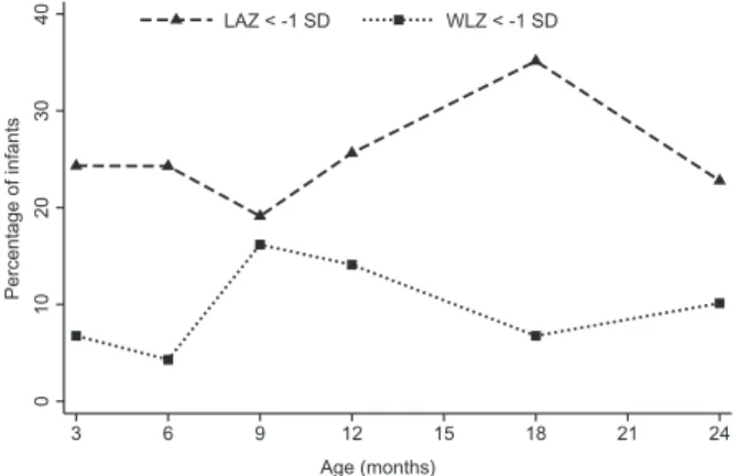
variation preceded the LAZ variation (Figure 2). At the end of the study at 24 months of age, 23% of infants were stunted and 10% wasted; from these, only five infants were moderately-to-severely (<–2SD) stunted and none was moderately-to-severely (<–2SD) wasted.
Parasitic infection
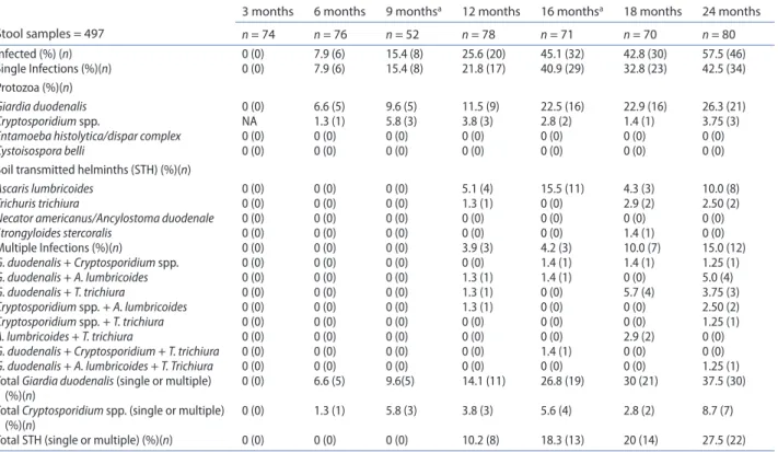
During the study period 497 stool samples were exam-ined for intestinal parasites. The frequencies of intes-tinal pathogenic parasites at each point assessment are shown in Table 1. Parasites were not detected at 3 months of age; subsequently, the frequency of par-asitic infection increased progressively with age, from 7.9% at 6 months to 57.5% at 24 months of age. Single parasitic infections predominated. Co-infections were detected from 12 months of age and their frequency increased up to the end of the study. The three most fre-quent pathogenic parasites, either as single or multiple infection, were by decreasing order G. duodenalis, STH, and Cryptosporidium spp. (Table 1). Entamoeba
histolyt-ica/ dispar complex was not detected. Other pathogenic
parasites detected are shown in Table 1.
In the entire study period, 1 to 2 episodes of parasitic infection occurred in 39 (48.8%) infants, 3 to 4 episodes in 26 (32.5%) infants, and more than 4 episodes in 5 (6.3%) infants, independently of the etiology, either as single or multiple agent infection. In 11 (13%) infants no parasite was found.
From the 30 stool samples with a positive micros-copy for G. duodenalis at 24 months of age, only 28 had enough material to be processed for molecular charac-terization. Twelve samples were successfully amplified for β-giardin, 14 for ssu-rRNA fragment gene, and two failed to amplify. Eighty percent (20/25) of samples belonged to Assemblage B and 20% (5/25) to Assemblage A. Additionally, β-giardin sequences were analyzed for sub-assemblage discrimination according to the described genetic polymorphisms. Nine isolates belonged to sub-assemblage B3, and 3 to subsub-assemblage A3.
Data were analyzed using the software SPSS 22.0 (SPSS for Windows, Rel. 22.0.1. 2013. SPSS Inc., Chicago, IL, EUA) and Stata (Stata Statistical Software: Release 13. College Station, TX: StataCorp LP).
Results
From the birth cohort, 283 infants completed 24 months of follow–up; from these, 82 were eligible for fecal bio-markers determinations, but two were subsequently excluded due to acute severe conditions. Thus, a final sample of 80 infants (Figure 1) was included, in which 57.5% were females. Sex distribution and anthropomet-ric data did not significantly differ between included and excluded infants (data not shown). In the final sample, 81.3% of mothers had more than five years of school edu-cation, 98.8% of the households had improved drinking water source, and 72.5% improved sanitation. In relation to feeding practices, 98.8% of infants were breastfed at some time point, 77.5% were exclusively breastfed at 6 months, and 10% maintained breastfeeding at 24 months; the mean (SD) duration of breastfeeding was 16.7 (2.9) months, and the mean (SD) age of intro-duction of complementary feeding was 5.7 (1.0) months. The frequency of clinical events varied along the study period, the respiratory symptoms (most frequent) varied between 10.5 and 37.2% of cases, and acute diarrhea between 1.3 and 9.3%; one case of persistent diarrhea and one of malaria were registered. In most infants the anthropometric measures were within the normal range at neonatal period, with a mean (SD) weight of 3.46 (0.46) kg and length of 50.2 (1.95) cm. After the neonatal period and up to 24 months of age, the rate of wasted infants varied between 4.3% and 16.2%, and of stunted infants between 19.1 and 35.1% (Figure 2). Noteworthy, the WLZ
Figure 1. Flow-chart of infants enrolled in the study.
0 10 20 30 40 Percentage of infants 3 6 9 12 15 18 21 24 Age (months) LAZ < -1 SD WLZ < -1 SD
Figure 2. Wasting (WlZ) and stunting (laZ) patterns up to 24 months of age. WlZ weight-for-length z-score; laZ length-for-age z-score.
Discussion
This prospective study, conducted in a low-middle income African country, showed that 87% of infants were infected by at least one pathogenic parasite in the first two years of life. Enteric parasitic infection was diagnosed as early as at 6 months of age, with frequency increasing with age and affecting around half of the infants at 24 months. G. duodenalis was the most fre-quently isolated parasite followed by STH, only detected after 12 months of age. Low frequencies were found for
Cryptosporidium spp. In Africa, few longitudinal studies
have assessed the prevalence of enteric parasitic infec-tions in infants and recognized a high burden of para-sitism during the first years of life [8,23], as in our study. During the study, most of the infants were asympto-matic, suggesting colonization and the development of a possible mutualistic or commensal relationship with the host. Moreover, the pathogenic role of parasites isolated in asymptomatic infants is difficult to interpret, because it may represent convalescent excretion of enteropatho-gens after acute diarrhea rather than true asymptomatic infection [51].
Regarding intestinal inflammation, a weak evidence of association was found between fecal S100A12 levels and current G. duodenalis and STH infections at 24 months of age. This association may be clinically meaningful, since this tendency toward increased fecal S100A12 levels was noticeable with the most frequent parasites. Classically,
G. duodenalis infection is characterized by little or no
inflammatory intestinal response [52]. In young children
Fecal biomarkers
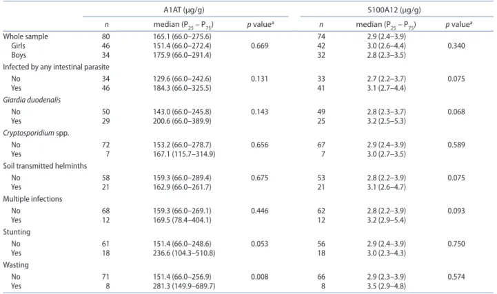
Fecal biomarkers were tested in 80 infants for A1AT, and in 74 for S100A12 due to unsolvable technical con-straints. The fecal S100A12 median (interquartile range) level was 2.87 (2.41–3.92) μg/g and of A1AT was 165.1 (66.0–275.6) μg/g (Table 2). A descriptive statistics of both biomarkers by the categories of several variables and distribution of their values is shown in Table 2 and Figure 3, respectively.
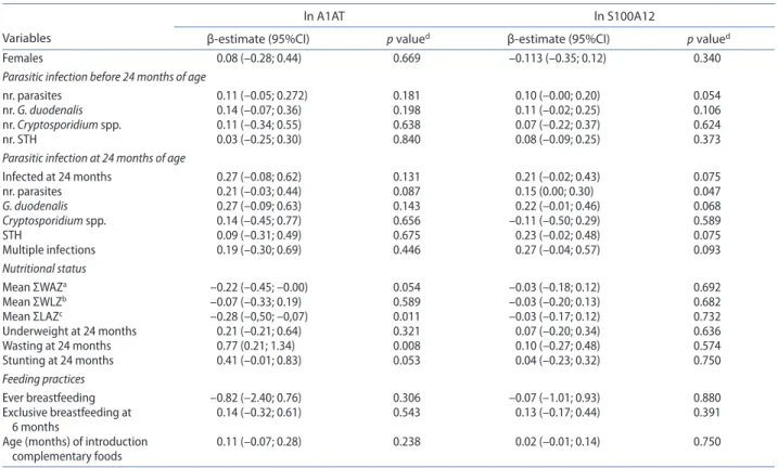
Variables with a p-value < 0.250 obtained in the uni-variable analysis (Table 3) were selected for the multi-variable analysis. Results from the multimulti-variable models (Table 4) showed a weak evidence of an association between fecal S100A12 levels and current G.
duode-nalis infection (p = 0.080) and between fecal S100A12
levels and current STH infection (p = 0.089). This sug-gests an increasing tendency of 23.6% and 24.1% in fecal S100A12 levels in infants infected with G.
duode-nalis and STH, respectively. Similarly, a weak evidence
of an association was found between fecal A1AT levels and current parasitic infection of any etiology (p = 0.089), suggesting an increasing tendency of 33.6% higher A1AT levels in infected infants. Remarkably, significant associa-tions were found between fecal A1AT levels and wasting (p = 0.006) and stunting (p = 0.044) at 24 months of age; specifically, fecal A1AT levels were twice higher in wasted infants and 50% higher in stunted infants. No significant associations between either fecal S100A12 or fecal A1AT and parasitic infection before 24 months of age (includ-ing number and etiology), were found.
Table 1. Frequency of intestinal pathogenic parasites by age.
note: na not available.
aParasite examination was included in the analysis of the cumulative effect of enteric parasitic infections before 24 months of age. Stool samples = 497
3 months 6 months 9 monthsa 12 months 16 monthsa 18 months 24 months
n = 74 n = 76 n = 52 n = 78 n = 71 n = 70 n = 80 Infected (%) (n) 0 (0) 7.9 (6) 15.4 (8) 25.6 (20) 45.1 (32) 42.8 (30) 57.5 (46) single Infections (%)(n) 0 (0) 7.9 (6) 15.4 (8) 21.8 (17) 40.9 (29) 32.8 (23) 42.5 (34) Protozoa (%)(n) Giardia duodenalis 0 (0) 6.6 (5) 9.6 (5) 11.5 (9) 22.5 (16) 22.9 (16) 26.3 (21) Cryptosporidium spp. na 1.3 (1) 5.8 (3) 3.8 (3) 2.8 (2) 1.4 (1) 3.75 (3)
Entamoeba histolytica/dispar complex 0 (0) 0 (0) 0 (0) 0 (0) 0 (0) 0 (0) 0 (0)
Cystoisospora belli 0 (0) 0 (0) 0 (0) 0 (0) 0 (0) 0 (0) 0 (0)
soil transmitted helminths (sth) (%)(n)
Ascaris lumbricoides 0 (0) 0 (0) 0 (0) 5.1 (4) 15.5 (11) 4.3 (3) 10.0 (8)
Trichuris trichiura 0 (0) 0 (0) 0 (0) 1.3 (1) 0 (0) 2.9 (2) 2.50 (2)
Necator americanus/Ancylostoma duodenale 0 (0) 0 (0) 0 (0) 0 (0) 0 (0) 0 (0) 0 (0)
Strongyloides stercoralis 0 (0) 0 (0) 0 (0) 0 (0) 0 (0) 1.4 (1) 0 (0) Multiple Infections (%)(n) 0 (0) 0 (0) 0 (0) 3.9 (3) 4.2 (3) 10.0 (7) 15.0 (12) G. duodenalis + Cryptosporidium spp. 0 (0) 0 (0) 0 (0) 0 (0) 1.4 (1) 1.4 (1) 1.25 (1) G. duodenalis + A. lumbricoides 0 (0) 0 (0) 0 (0) 1.3 (1) 1.4 (1) 0 (0) 5.0 (4) G. duodenalis + T. trichiura 0 (0) 0 (0) 0 (0) 1.3 (1) 0 (0) 5.7 (4) 3.75 (3) Cryptosporidium spp. + A. lumbricoides 0 (0) 0 (0) 0 (0) 1.3 (1) 0 (0) 0 (0) 2.50 (2) Cryptosporidium spp. + T. trichiura 0 (0) 0 (0) 0 (0) 0 (0) 0 (0) 0 (0) 1.25 (1) A. lumbricoides + T. trichiura 0 (0) 0 (0) 0 (0) 0 (0) 0 (0) 2.9 (2) 0 (0)
G. duodenalis + Cryptosporidium + T. trichiura 0 (0) 0 (0) 0 (0) 0 (0) 1.4 (1) 0 (0) 0 (0) G. duodenalis + A. lumbricoides + T. Trichiura 0 (0) 0 (0) 0 (0) 0 (0) 0 (0) 0 (0) 1.25 (1) total Giardia duodenalis (single or multiple)
(%)(n) 0 (0) 6.6 (5) 9.6(5) 14.1 (11) 26.8 (19) 30 (21) 37.5 (30)
total Cryptosporidium spp. (single or multiple)
(%)(n) 0 (0) 1.3 (1) 5.8 (3) 3.8 (3) 5.6 (4) 2.8 (2) 8.7 (7)
explanation for our findings may be the predominant existences of assemblage B (80.0%) in our sample, as described in other African countries [55,56]. This gen-otype has been associated to extensive damage of the mucosal architecture and infiltration of inflammatory cells [21,57]. Very few studies have explored the effects of STH infections on mucosal immunity in infants [58]. In early STH infections, Zanzibari infants showed a reg-ulatory Th2 pattern of peripheral cytokine responses to
Ascaris and hookworm antigens [59], without association with acute phase proteins [60]. In our study, the tendency toward increased fecal S100A12 levels in current STH infections may suggest the presence of local inflam-matory response mediated by neutrophils. In murine models using Heligmosomoides polygyrus, an early and pronounced infiltration of neutrophils and macrophages from developing countries G. duodenalis is usually
asymptomatic, probably as a result of modulation of the innate immune system [53] or a decrease of inflammatory response in subsequent infections [20,29]. Previous stud-ies reported the association between G. duodenalis and biomarkers of intestinal inflammation in children [28,29], while others did not [30]. In the recent MAL-ED study, fecal myeloperoxidase, a marker of neutrophil inflam-mation, was surprisingly lower in children with Giardia [30]. In our study, despite the fact that most infants with
Giardia infection were asymptomatic, the tendency
toward increased fecal S100A12 levels may suggest local inflammatory response with participation of neutrophils. Histopathological studies using murine models showed intestinal infiltration of neutrophils that persisted several days after G. duodenalis infection [21,54]. One plausible
Table 2. Fecal values of alpha1-anti-trypsin (a1at) and s100a12 at 24 months of age, considering sex, parasite agent, and nutritional status categories.
ap values obtained by student’s t-test after logarithmic transformation of a1at and s100a12.
A1AT (μg/g) S100A12 (μg/g)
n median (P25 – P75) p valuea n median (P
25 – P75) p valuea
Whole sample 80 165.1 (66.0–275.6) 74 2.9 (2.4–3.9)
girls 46 151.4 (66.0–272.4) 0.669 42 3.0 (2.6–4.4) 0.340
boys 34 175.9 (66.0–291.4) 32 2.8 (2.3–3.5)
Infected by any intestinal parasite
no 34 129.6 (66.0–242.6) 0.131 33 2.7 (2.2–3.7) 0.075 Yes 46 184.3 (66.0–325.5) 41 3.1 (2.7–4.4) Giardia duodenalis no 50 143.0 (66.0–245.8) 0.143 49 2.8 (2.3–3.7) 0.068 Yes 29 200.6 (66.0–389.9) 25 3.2 (2.5–5.3) Cryptosporidium spp. no 72 153.2 (66.0–278.7) 0.656 67 2.9 (2.4–3.9) 0.589 Yes 7 167.1 (115.7–314.9) 7 3.0 (2.7–3.5)
soil transmitted helminths
no 58 159.3 (66.0–289.4) 0.675 53 2.8 (2.2–3.9) 0.075 Yes 21 162.9 (66.0–261.7) 21 3.1 (2.6–4.7) Multiple infections no 68 159.3 (66.0–269.1) 0.446 62 2.8 (2.2–3.9) 0.093 Yes 12 169.5 (78.4–404.1) 12 3.2 (2.9–5.4) stunting no 61 151.4 (66.0–248.6) 0.053 56 2.9 (2.4–3.9) 0.750 Yes 18 236.6 (104.3–510.8) 18 3.0 (2.3–4.3) Wasting no 71 151.4 (66.0–256.9) 0.008 66 2.9 (2.3–3.9) 0.574 Yes 8 281.3 (149.9–689.7) 8 3.5 (2.9–4.8) 0 20 40 60 Number of infants 0 5 10 15 20 ug/g stool S100A12 0 20 40 60 Number of infants 0 500 1000 1500 ug/g stool Alpha-1 anti-trypsin
children from a developed country [65,66], and meas-ured using different ELISA method [67]. To the best of our knowledge, this is the first time that S100A12 is used to assess intestinal inflammatory response in enteric para-sitic infection in children. This biomarker was chosen tak-ing into account its advantages in field studies involvtak-ing children. This protein is restrictively secreted by activated neutrophils and is strongly correlated with histologically intestinal inflammation and neutrophil infiltration [68]. Fecal S100A12 has a sensitivity of 96% and a specificity of 92% in distinguishing healthy children from those with inflammatory bowel disease, using the threshold 10 mg/kg [32,67]. It is evenly distributed throughout feces and is stable at a wide range of temperatures (4 to 20 °C) for several days [67]. These characteristics make fecal S100A12 a convenient biomarker, facilitating samples collection and transportation to a laboratory for meas-urement, avoiding resource-consuming storage needs [67]. Several commercial ELISA kits are available for fecal S100A12 analysis requiring a small stool sample (approx-imately 100 mg). Other fecal neutrophil-derived proteins used as biomarkers of intestinal inflammation may have limitations compared with S100A12, particularly in infants. Calprotectin, another calcium and zinc-binding protein, is present in neutrophils, but also in monocytes and macrophages [69]. Fecal calprotectin correlates well with endoscopic and histological inflammatory bowel in regions immediately adjacent to the parasite has been
described [61]. Furthermore, primary infections by STH, as occurring in younger children, may stimulate a strong inflammatory response in the mucosa [58]. Young chil-dren in developing countries frequently respond to
Cryptosporidium infection with intestinal inflammation
[27]. In our study Cryptosporidium spp. was not associ-ated with the fecal inflammatory biomarker, probably explained by its low frequency and less severe infections in asymptomatic infants.
The fecal biomarkers have the advantage of measur-ing proteins originatmeasur-ing in the intestinal mucosa, more accurately reflecting local inflammation [62]. Several fecal biomarkers of neutrophil activity (e.g., lactoferrin, myeloperoxidase, and calprotectin) or of activated cell mediated immunity (e.g., neopterin) have been used to assess intestinal inflammation in infants [63]. High lev-els of these biomarkers were reported in infants from developing countries associated either to enteric par-asite infections [24,27–30] or to environmental enteric dysfunction [30,39,40,64]. In accordance, we found high median fecal levels of S100A12 in all infants at 24 months of age, five times above those described for healthy chil-dren (0.5 mg/kg), but below the threshold 10 mg/kg used for inflammatory bowel disease [65,66]. This comparison should be interpreted with caution, since the reported values were obtained from a limited number of healthy
Table 3. Univariable analysis for fecal alpha1-anti-trypsin (a1at) and s100a12, considering sex, parasitic infection before and at 24 months of age, nutritional status, and feeding practices.
notes: ln logarithmic transformation, sth soil transmitted helminths.
aΣWaZ bΣWlZ cΣlaZ sum up of weight-for-age, weight-for-length, and length-for-age z-scores, respectively, at all point assessments dp values obtained by linear regression models after logarithmic transformation of a1at (ln a1at) and s100a12 (ln s100a12) values.
Variables
ln A1AT ln S100A12
β-estimate (95%CI) p valued β-estimate (95%CI) p valued
Females 0.08 (–0.28; 0.44) 0.669 –0.113 (–0.35; 0.12) 0.340
Parasitic infection before 24 months of age
nr. parasites 0.11 (–0.05; 0.272) 0.181 0.10 (–0.00; 0.20) 0.054
nr. G. duodenalis 0.14 (–0.07; 0.36) 0.198 0.11 (–0.02; 0.25) 0.106
nr. Cryptosporidium spp. 0.11 (–0.34; 0.55) 0.638 0.07 (–0.22; 0.37) 0.624
nr. sth 0.03 (–0.25; 0.30) 0.840 0.08 (–0.09; 0.25) 0.373
Parasitic infection at 24 months of age
Infected at 24 months 0.27 (–0.08; 0.62) 0.131 0.21 (–0.02; 0.43) 0.075 nr. parasites 0.21 (–0.03; 0.44) 0.087 0.15 (0.00; 0.30) 0.047 G. duodenalis 0.27 (–0.09; 0.63) 0.143 0.22 (–0.01; 0.46) 0.068 Cryptosporidium spp. 0.14 (–0.45; 0.77) 0.656 –0.11 (–0.50; 0.29) 0.589 sth 0.09 (–0.31; 0.49) 0.675 0.23 (–0.02; 0.48) 0.075 Multiple infections 0.19 (–0.30; 0.69) 0.446 0.27 (–0.04; 0.57) 0.093 Nutritional status Mean ΣWaZa –0.22 (–0.45; –0.00) 0.054 –0.03 (–0.18; 0.12) 0.692 Mean ΣWlZb –0.07 (–0.33; 0.19) 0.589 –0.03 (–0.20; 0.13) 0.682 Mean ΣlaZc –0.28 (–0,50; –0,07) 0.011 –0.03 (–0.17; 0.12) 0.732 Underweight at 24 months 0.21 (–0.21; 0.64) 0.321 0.07 (–0.20; 0.34) 0.636 Wasting at 24 months 0.77 (0.21; 1.34) 0.008 0.10 (–0.27; 0.48) 0.574 stunting at 24 months 0.41 (–0.01; 0.83) 0.053 0.04 (–0.23; 0.32) 0.750 Feeding practices ever breastfeeding –0.82 (–2.40; 0.76) 0.306 –0.07 (–1.01; 0.93) 0.880 exclusive breastfeeding at 6 months 0.14 (–0.32; 0.61) 0.543 0.13 (–0.17; 0.44) 0.391
age (months) of introduction
two inflammatory biomarkers (myeloperoxidase and neopterin) in a score to predict growth deficit in infants [40]; specifically, fecal A1AT levels at or above 75th were reported to predict a loss of 0.152 LAZ in the subse-quent six months [40]. Similar results were described in Brazilian infants [39]. In our study, the convenience threshold of 1SD for WLZ and LAZ was used to define wasting and stunting. A higher number of undernour-ished infants due to the inclusion of mild-to-moderate degrees may be responsible for the significant associa-tion we found. In fact, the hazardous effects of undernu-trition happen along a continuum spectrum, in which mild-to-moderate degrees may be associated with oth-erwise-unobserved changes in disease exposure [44,80]. Marginally nourished children may have inadequate or rate-limiting stores of key nutrients to repair the mucosal damage [42]. Despite the aforementioned association of fecal A1AT levels with stunting and wasting, the median A1AT level of 165.1 μg/g found in our sample was below the mean 299 μg/g described in infants from develop-ing countries [30]. Most of the studies have used urinary lactulose:mannitol ratio to assess intestinal permeability [81]. In our study, fecal A1AT was preferred instead of lactulose:mannitol test considering some advantages. The AAT is neither degraded by intestinal proteases nor reabsorbed and it is notably stable in stool samples [82]. Furthermore, large size molecules such as A1AT (50,000 Da), transported through the paracellular route, may better reflect structural damage of tight junctions than small size molecules such as lactulose (342 Da) and mannitol (182 Da) [83]. Moreover, the lactulose:mannitol test has inconveniences that may limit its use in infants, particularly in field settings, such as the requirement of fasting before testing, the need for several hours for urine collection, the lack of standardized procedures, and of reference values for children [81].
The suspected effect of repeated enteric infections on intestinal function [42] was recently confirmed by a better correlation of fecal biomarkers with cumulative pathogen burden than with a single ongoing infection [30]. In spite of this evidence, we found no associations between previous exposure to asymptomatic parasitic infections and fecal biomarkers of intestinal inflamma-tion and permeability at 24 months of age. Since these biomarkers were not measured longitudinally, a revers-ible process of intestinal epithelial injury [33] cannot be excluded; otherwise, asymptomatic infections may induce less severe intestinal barrier dysfunction than symptomatic infections [26,76].
The following constitute strong points of our study. The measurement of intestinal barrier biomarkers at 24 months, and not before, reflects more accurately the impact of parasite infection, not biased by a potential effect of an incomplete development of mucosal barrier function [30,41]. The longitudinal search for intestinal parasites included not only protozoa but also STH, providing a broader view of the association between disease activity [70] but a meta-analysis showed a
rel-atively low pooled specificity (0.76) in children [71]. It seems to be a less accurate biomarker for inflammatory bowel disease than fecal S100A12 in pediatric population [32]. In healthy infants, fecal calprotectin levels are higher than in older children [72], and in those breastfed [73], Lactoferrin is likewise not specific, as it is produced by neutrophils and epithelial cells [74], It may be inaccurate to determine intestinal inflammation in infants using this biomarker, since breast milk may contribute to increase its fecal levels [75]. Additionally, fecal lactoferrin may be less sensitive in malnourished children [76]. Myeloperoxidase is produced by neutrophils, but it is found at lower con-centrations in monocytes and macrophages [77]. In spite of a good correlation of fecal myeloperoxidase with labo-ratory and endoscopic parameters of inflammation [77], recent breastfeeding is also associated with increased fecal levels [30]. Furthermore, gender differences, with lower intracellular neutrophil myeloperoxidase levels in boys, have been reported [78]. Neopterin, an indicator of T-helper cell 1 activity, is used as a biomarker of intestinal inflammation [28,30,39,40]. Its fecal levels were found to be much higher (26 times) in infants from developing countries than from non-tropical countries [30,40].
Regarding intestinal permeability, a weak evidence of association was found between fecal A1AT levels and current parasitic infection of any etiologic at 24 months of age. This association may have limited clinical rele-vance, since the tendency toward increased fecal A1AT levels was associated with unspecific etiology. Several studies (most using lactulose:mannitol test) described the association between enteric parasitic infection and increased intestinal permeability [23,30,33,34], while others did not [25,28]. Significant associations were found between increased fecal A1AT levels and both acute (wasting) and chronic (stunting) undernutrition at 24 months of age, independently of intestinal par-asitic infection. The association between increased intestinal permeability and undernutrition has been pre-viously described in infants from developing countries [25,28,35,39,40]. It has been suggested that increased intestinal permeability explains at least 40% of growth faltering [79]. Recently, fecal A1AT was combined with
Table 4. Multivariable regression models for alpha1-anti-trypsin (a1at) and s100a12.
notes: β regression coefficient, CI confidence interval.
ap values obtained by linear regression models after logarithmic transfor-mation of a1at(ln a1at) and s100a12 (ln s100a12) values.
Variables β-estimate (95%CI) p value a
ln a1at
Parasitic infection at 24 months
of age 0.29 (–0.05; 0.62) 0.089
Wasting at 24 months of age 0.78 (0.23; 1.33) 0.006 stunting at 24 months of age 0.41 (0.01; 0.80) 0.044
ln s100a12
Giardia duodenalis at 24 months
of age 0.21 (–0.03; 0.45) 0.080
soil transmitted helminths at
Acknowledgments
This study is part of the PhD thesis in Tropical Medicine of one of the authors (Marisol Garzón), supervised and co-supervised by two authors (Luis Pereira-da-Silva and Jorge Seixas) from Universidade NOVA de Lisboa, Lisbon, Portugal. The authors are very grateful to Sonia Centeno Lima, PhD, the former PhD thesis supervisor, for the valuable contribution in the design and implementation of the study, and also to the logistic local support from members of the non-governmental organiza-tion Marquês de Valle Flôr Institute, namely Paulo Telles de Freitas, Ahmed Zaky, Edgar Neves, and António Lima.
Disclosure statement
No potential conflict of interest was reported by the authors. Funding
This study is supported by the Fundação para a Ciência e Tecnologia [grant number SFRH/BD/81431/2011].
ORCID
Marisol Garzón http://orcid.org/0000-0001-7793-6948 Luis Pereira-da-Silva http://orcid.org/0000-0001-7069-6451 Jorge Seixas http://orcid.org/0000-0002-3110-6583 Ana Luísa Papoila http://orcid.org/0000-0002-2918-8364 Marta Alves http://orcid.org/0000-0002-7421-8550 References
[1] Petri WA, Miller M, Binder HJ, et al. Enteric infections, diarrhea, and their impact on function and development. J Clin Invest. 2008;118:1277–1290.
[2] Farthing MJ, Mata L, Urrutia JJ, et al. Natural history of Giardia infection of infants and children in rural Guatemala and its impact on physical growth. Am J Clin Nutr. 1986;43:395–405.
[3] Checkley W, Gilman RH, Epstein LD, et al. Asymptomatic and symptomatic cryptosporidiosis: their acute effect on weight gain in Peruvian children. Am J Epidemiol. 1997;145:156–163.
[4] Guerrant DI, Moore SR, Lima AA, et al. Association of early childhood diarrhea and cryptosporidiosis with impaired physical fitness and cognitive function four-seven years later in a poor urban community in northeast Brazil. Am J Trop Med Hyg. 1999;61:707–713.
[5] Berkman DS, Lescano AG, Gilman RH, et al. Effects of stunting, diarrhoeal disease, and parasitic infection during infancy on cognition in late childhood: a follow-up study. Lancet. 2002;359:564–571.
[6] Kotloff KL, Nataro JP, Blackwelder WC, et al. Burden and aetiology of diarrhoeal disease in infants and young children in developing countries (the Global Enteric Multicenter Study, GEMS): a prospective, case-control study. Lancet. 2013;382:209–222.
[7] Platts-Mills JA, Babji S, Bodhidatta L, et al. Pathogen-specific burdens of community diarrhoea in developing countries: a multisite birth cohort study (MAL-ED). Lancet Glob Heal. 2015;3:e564–e575.
[8] LaBeaud AD, Nayakwadi Singer M, McKibben M, et al. Parasitism in children aged three years and under: relationship between infection and growth in rural coastal Kenya. PLoS Negl Trop Dis. 2015;9:e0003721.
these enteric parasites and intestinal barrier dysfunc-tion in younger children. Fecal S100A12 was herein firstly used as a biomarker of intestinal inflammatory response in enteric parasitic infection, and it appears to be a convenient and accurate tool for field studies in infants. Finally, analyzing mild-to-moderate degrees of undernutrition allowed early detection of its associ-ation with intestinal barrier dysfunction related with parasitic infection, before severe undernutrition was established [42,44].
Limitations of this study should be acknowledged. Firstly, this study may be underpowered once the sam-ple size was not calculated. By the time the study started no reference values were found in the literature to pro-vide an idea about the mean and variability of the two biomarkers under study specifically in enteric parasitic infection in children. In spite of the convenience size of the sample, most of the associations obtained have rela-tively narrow confidence intervals. Secondly, microscopic parasite examination using a single stool specimen have low sensitivity [84]; which may however be increased when it is performed by a trained laboratory technician in high prevalence settings [85], as in our study. Thirdly, since bacterial and virus were not analyzed, changes in fecal markers attributable to these enteropathogens can-not be discarded. In children, transient high intestinal permeability may occur in rotavirus infection [33] and to a lesser degree in bacterial infections [86]. Intestinal inflammatory response is common in invasive bacterial enteric infections [30,87], but not in viral infections [87]. Noteworthy, all these studies assessed enteric infections in children with diarrhea, and we only selected asymp-tomatic infants with non-diarrheic stools to minimize the probability of coexisting viral and bacterial enteric infections. Fourthly, anthelminthic and anti-Giardia treatments might have biased the results, although it is described that treatment does not have a significant impact on small intestine function [88]. Finally, the deter-mination of environmental enteric dysfunction [89] was beyond the scope of our study. Thus, it is not possible to exclude that parasitic enteric infections may have con-tributed to the development or exacerbation of a poten-tial environmental enteric dysfunction [90] or that both conditions coexisted.
To conclude, the hypothesized association between previous exposure to asymptomatic enteric parasitic infections and intestinal barrier dysfunction at 24 months was not confirmed. Notwithstanding, an observed ten-dency toward increased fecal levels of inflammatory biomarker associated with the most prevalent parasitic infections in asymptomatic infants may have clinical relevance. Although our study could be underpowered to assess the aforementioned associations, it was pow-ered enough to demonstrate a significant association of increased intestinal permeability with wasting and stunting, including of mild-to-moderate degrees, in 24-months aged infants.
[27] Kirkpatrick BD, Daniels MM, Jean SS, et al. Cryptosporidiosis stimulates an inflammatory intestinal response in malnourished Haitian children. J Infect Dis. 2002;186:94– 101.
[28] Campbell DI, McPhail G, Lunn PG, et al. Intestinal inflammation measured by fecal neopterin in Gambian children with enteropathy: association with growth failure, Giardia lamblia, and intestinal permeability. J Pediatr Gastroenterol Nutr. 2004;39:153–157.
[29] Kohli A, Bushen OY, Pinkerton RC, et al. Giardia duodenalis assemblage, clinical presentation and markers of intestinal inflammation in Brazilian children. Trans R Soc Trop Med Hyg. 2008;102:718–725.
[30] McCormick BJ, Lee GO, Seidman J, et al. Dynamics and trends in fecal biomarkers of gut function in children from 1-24 months in the MAL-ED study. Am J Trop Med Hyg. 2016;19:16–0496.
[31] Meijer B, Gearry RB, Day AS. The role of S100A12 as a systemic marker of inflammation. Int J Inflam. 2012;2012:907078.
[32] Sidler MA, Leach ST, Day AS. Fecal S100A12 and fecal calprotectin as noninvasive markers for inflammatory bowel disease in children. Inflamm Bowel Dis. 2008;14:359–366.
[33] Zhang Y, Lee B, Thompson M, et al. Lactulose-mannitol intestinal permeability test in children with diarrhea caused by rotavirus and cryptosporidium. Diarrhea Working Group, Peru. J Pediatr Gastroenterol Nutr. 2000;31:16–21.
[34] Goto R, Panter-Brick C, Northrop-Clewes CA, et al. Poor intestinal permeability in mildly stunted Nepali children: associations with weaning practices and Giardia lamblia infection. Br J Nutr. 2002;88:141–149.
[35] Mondal D, Minak J, Alam M, et al. Contribution of enteric infection, altered intestinal barrier function, and maternal malnutrition to infant malnutrition in Bangladesh. Clin Infect Dis. 2012;54:185–192.
[36] Thomas DW, Sinatra FR, Merritt RJ. Random fecal alpha-1-antitrypsin concentration in children with gastrointestinal disease. Gastroenterology. 1981;80:776–782.
[37] Amemoto K, Nagita A, Matsuse R, et al. Clinical evaluation of fecal lactoferrin and α-1-antitrypsin in pediatric gastrointestinal infections. Pathophysiology. 1996;3:87– 90.
[38] Weizman Z, Binsztok M, Fraser D, et al. Intestinal protein loss in acute and persistent diarrhea of early childhood. J Clin Gastroenterol. 2002;34:427–429.
[39] Guerrant RL, Leite AM, Pinkerton R, et al. Biomarkers of environmental enteropathy, inflammation, stunting, and impaired growth in children in Northeast Brazil. PLoS One. 2016;11:e0158772.
[40] Kosek M, Haque R, Lima A, et al. Fecal markers of intestinal inflammation and permeability associated with the subsequent acquisition of linear growth deficits in infants. Am J Trop Med Hyg. 2013;88:390–396.
[41] Brandtzaeg, P. The innate and adaptive immune system of the intestinal epithelium. In: Heidt, PJ, Bienenstock, J, Midtvedt, T, Rusch, V, van der Waaij, D, editors. Defence mechanisms of the innate system: influence of microbes. Herborn: Herborn litterae; 2006. p. 55–88.
[42] Guerrant RL, Oriá RB, Moore SR, et al. Malnutrition as an enteric infectious disease with long-term effects on child development. Nutr Rev. 2008;66:487–505.
[43] World Health Organization. Progress on sanitation and drinking water – 2015 update and MDG assess-ment. Geneva: UNICEF and World Health Organization; 2015.
[9] Berkes J, Viswanathan VK, Savkovic SD, et al. Intestinal epithelial responses to enteric pathogens: effects on the tight junction barrier, ion transport, and inflammation. Gut. 2003;52:439–451.
[10] Di Genova BM, Tonelli RR. Infection strategies of intestinal parasite pathogens and host cell responses. Frontt Microbiol. 2016;7:256. DOI:10.3389/fmicb.2016.00256 [11] Buret AG, Chin AC, Scott KG. Infection of human and
bovine epithelial cells with Cryptosporidium andersoni induces apoptosis and disrupts tight junctional ZO-1: effects of epidermal growth factor. Int J Parasitol. 2003;33:1363–1371.
[12] de Sablet T, Potiron L, Marquis M, et al. Cryptosporidium parvum increases intestinal permeability through interaction with epithelial cells and IL-1β and TNFα released by inflammatory monocytes. Cell Microbiol. 2016;18:1871–1880.
[13] Teoh DA, Kamieniecki D, Pang G, et al. Giardia lamblia rearranges F-actin and alpha-actinin in human colonic and duodenal monolayers and reduces transepithelial electrical resistance. J Parasitol. 2000;86:800–806. [14] Troeger H, Epple HJ, Schneider T, et al. Effect of chronic
Giardia lamblia infection on epithelial transport and barrier function in human duodenum. Gut. 2007;56:328– 335.
[15] McDermott JR, Bartram RE, Knight PA, et al. Mast cells disrupt epithelial barrier function during enteric nematode infection. Proc Natl Acad Sci. 2003;100:7761–7766. [16] Su CW, Cao Y, Kaplan J, et al. Duodenal helminth infection
alters barrier function of the colonic epithelium via adaptive immune activation. Infect Immun. 2011;79:2285– 2294.
[17] Farthing MJ. Immune response-mediated pathology in human intestinal parasitic infection. Parasite Immunol. 2003;25:247–257.
[18] Kasper LH, Buzoni-Gatel D. Ups and downs of mucosal cellular immunity against protozoan parasites. Infect Immun. 2001;69:1–8.
[19] Laurent F, Eckmann L, Savidge TC, et al. Cryptosporidium parvum infection of human intestinal epithelial cells induces the polarized secretion of C-X-C chemokines. Infect Immun. 1997;65:5067–5073.
[20] Hanevik K, Hausken T, Morken MH, et al. Persisting symptoms and duodenal inflammation related to Giardia duodenalis infection. J Infect. 2007;55:524–530.
[21] Chen TL, Chen S, Wu HW, et al. Persistent gut barrier damage and commensal bacterial influx following eradication of Giardia infection in mice. Gut Pathog. 2013;5:26. DOI:10.1186/1757-4749-5-26
[22] Falcone FH, Rossi AG, Sharkey R, et al. Ascaris suum-derived products induce human neutrophil activation via a g protein-coupled receptor that interacts with the interleukin-8 receptor pathway. Infect Immun. 2001;69:4007–4018.
[23] Lunn PG, Erinoso HO, Northrop-Clewes CA, et al. Giardia intestinalis is unlikely to be a major cause of the poor growth of rural Gambian infants. J Nutr. 1999;129:872–877. [24] Kirkpatrick BD, Noel F, Rouzier PD, et al. Childhood
cryptosporidiosis is associated with a persistent systemic inflammatory response. Clin Infect Dis. 2006;43:604–608. [25] Goto R, Mascie-Taylor CG, Lunn PG. Impact of intestinal permeability, inflammation status and parasitic infections on infant growth faltering in rural Bangladesh. Br J Nutr. 2009;101:1509–1516.
[26] Newman RD, Sears CL, Moore SR, et al. Longitudinal study of cryptosporidium infection in children in Northeastern Brazil. J Infect Dis. 1999;180:167–175.
[62] Assche GV. Fecal biomarkers for the diagnosis and management of inflammatory bowel disease. Gastroenterol Hepatol. 2011;7(6):396–398.
[63] Däbritz J, Musci J, Foell D. Diagnostic utility of faecal biomarkers in patients with irritable bowel syndrome. World J Gastroenterol. 2014;20:363–375.
[64] Naylor C, Lu M, Haque R, et al. Environmental enteropathy, oral vaccine failure and growth faltering in infants in Bangladesh. EBioMedicine. 2015;2:1759–1766.
[65] Day AS, Ehn M, Gearry RB, et al. Fecal S100A12 in healthy infants and children. Dis Markers. 2013;35:295–299. [66] Ehn M. Levels of fecal S100A12 in normal children and
children with Inflammatory Bowel disease. Degree Project in Medicine. Uppsala: Uppsala University; 2011.
[67] de Jong NS, Leach ST, Day AS. Fecal S100A12: a novel noninvasive marker in children with Crohn’s disease. Inflamm Bowel Dis. 2006;12:566–572.
[68] Foell D, Kucharzik T, Kraft M, et al. Neutrophil derived human S100A12 (EN-RAGE) is strongly expressed during chronic active inflammatory bowel disease. Gut. 2003;52(6):847–853.
[69] Lehmann FS, Burri E, Beglinger C. The role and utility of faecal markers in inflammatory bowel disease. Therap Adv Gastroenterol. 2015;8(1):23–36.
[70] Berni Canani R, Terrin G, Rapacciuolo L, et al. Faecal calprotectin as reliable non-invasive marker to assess the severity of mucosal inflammation in children with inflammatory bowel disease. Dig Liver Dis. 2008;40:547– 553.
[71] van Rheenen PF, Van de Vijver E, Fidler V. Faecal calprotectin for screening of patients with suspected inflammatory bowel disease: diagnostic meta-analysis. BMJ. 2010;341:c3369.
[72] Li F, Ma J, Geng S, et al. Fecal calprotectin concentrations in healthy children aged 1-18 months. PLoS One. 2015;10(3):e0119574.
[73] Li F, Ma J, Geng S, et al. Comparison of the different kinds of feeding on the level of fecal calprotectin. Early Hum Dev. 2014;90(9):471–475.
[74] González-Chávez SA, Arévalo-Gallegos S, Rascón-Cruz Q. Lactoferrin: structure, function and applications. Int J Antimicrob Agents. 2009;33(4):301e1–301e8. DOI: 10.1016/j.ijantimicag.2008.07.020
[75] Perrin MT, Fogleman AD, Newburg DS, Allen JC. A longitudinal study of human milk composition in the second year postpartum: implications for human milk banking. Matern Child Nutr. 2017;13(1). DOI:10.1111/ mcn.12239
[76] Opintan JA, Newman MJ, Ayeh-Kumi PF, et al. Pediatric diarrhea in Southern Ghana: etiology and association with intestinal inflammation and malnutrition. Am J Trop Med Hyg. 2010;83:936–943.
[77] Saiki T. Myeloperoxidase concentrations in the stool as a new parameter of inflammatory bowel disease. Kurume Med J. 1998;45:69–73.
[78] Nikulshin S, Tolstikova I, Bartule A, et al. Intracellular neutrophil myeloperoxidase level in pediatric patients: significant age and gender variability. Int J Lab Hematol. 2015;37:120–124.
[79] Lunn PG. The impact of infection and nutrition on gut function and growth in childhood. Proc Nutr Soc. 2000;59:147–154.
[80] Bhagowalia P, Chen SE, Masters WA. Effects and determinants of mild underweight among preschool children across countries and over time. Econ Hum Biol. 2011;9:66–77.
[44] Pelletier DL, Frongillo EA Jr, Schroeder DG, et al. The effects of malnutrition on child mortality in developing countries. Bull WHO. 1995;73:443–448.
[45] World Health Organization. Bench aids for the diagnosis of intestinal parasites. Geneva: World Health Organization; 1994.
[46] Morris LS, Marchesi JR. Assessing the impact of long term frozen storage of faecal samples on protein concentration and protease activity. J Microbiol Methods. 2016;123:31– 38.
[47] Cacciò SM, De Giacomo M, Pozio E. Sequence analysis of the beta-giardin gene and development of a polymerase chain reaction-restriction fragment length polymorphism assay to genotype Giardia duodenalis cysts from human faecal samples. Int J Parasitol. 2002;32:1023–1030. [48] World Health Organization. Preventive chemotherapy in
human helminthiasis: coordinated use of anthelminthic drugs in control interventions: a manual for health professionals and programme managers. Geneva: World Health Organization; 2006.
[49] American Academy of Pediatrics. Giardia intestinalis (formerly Giardia lamblia and Giardia duodenalis) infections. In: Kimberlin DW, Brady MT, Jackson MA, Long SS, editors. Red Book: 2012 Report of the Committee on Infectious Diseases. 29th ed. Elk Grove Village, IL: American Academy of Pediatrics; 2015. p. 353–355
[50] Bland M. An introduction to medical statistics. 3rd ed. Oxford: Oxford University Press; 2000.
[51] Levine MM, Robins-Browne RM. Factors that explain excretion of enteric pathogens by persons without diarrhea. Clin Infect Dis. 2012;55:S303–S311.
[52] Roxström-Lindquist K, Palm D, Reiner D, et al. Giardia immunity-an update. Trends Parasitol. 2006;22:26–31. [53] Muhsen K, Levine MM. A systematic review and
meta-analysis of the association between Giardia lamblia and endemic pediatric diarrhea in developing countries. Clin Infect Dis. 2012;55:S271–S293.
[54] Abd-Al-Zahra E, Sadoon W, Khalil M. Local and systemic immune responses in rats infected with Giardia lamblia. Med J Basrah Univ. 2012;30(1):60–73.
[55] Ignatius R, Gahutu JB, Klotz C, et al. High prevalence of Giardia duodenalis Assemblage B infection and association with underweight in Rwandan children. PLoS Negl Trop Dis. 2012;6:e1677.
[56] Mbae C, Mulinge E, Guleid F, et al. Molecular characterization of Giardia duodenalis in children in Kenya. BMC Infect Dis. 2016;16:210. DOI:10.1186/s12879-016-1436-z
[57] Barlet L, Roche J, Kolling G, et al. Persistent G. lamblia impairs growth in a murine malnutrition model. J Clin Invest. 2013;123(6):2672–2684.
[58] Cooper PJ. Mucosal immunology of geohelminth infections in humans. Mucosal Immunol. 2009;2:288–299. [59] Wright VJ, Ame SM, Haji HS, et al. Early exposure of
infants to GI nematodes induces Th2 dominant immune responses which are unaffected by periodic anthelminthic treatment. PLoS Negl Trop Dis. 2009;3:e433.
[60] Kung’u JK, Goodman D, Haji HJ, et al. Early helminth infections are inversely related to anemia, malnutrition, and malaria and are not associated with inflammation in 6- to 23-month-old Zanzibari children. Am J Trop Med Hyg. 2009;81:1062–1070.
[61] Morimoto M, Morimoto M, Whitmire J, et al. Peripheral CD4 T cells rapidly accumulate at the host:parasite interface during an inflammatory Th2 memory response. J Immunol. 2004;172:2424–2430.
[87] Sýkora J, Siala K, Huml M, et al. Evaluation of faecal calprotectin as a valuable non-invasive marker in distinguishing gut pathogens in young children with acute gastroenteritis. Acta Paediatr. 2010;99:1389– 1395.
[88] Goto R, Mascie-Taylor CG, Lunn PG. Impact of anti-Giardia and anthelminthic treatment on infant growth and intestinal permeability in rural Bangladesh: a randomised double-blind controlled study. Trans R Soc Trop Med Hyg. 2009;103:520–529.
[89] Watanabe K, Petri WA Jr. Environmental Enteropathy: elusive but significant subclinical abnormalities in developing countries. EBioMedicine. 2016;10:25–32.
[90] Bartelt LA, Lima AA, Kosek M, et al. “Barriers” to child development and human potential: the case for including the “neglected enteric protozoa” (NEP) and other enteropathy-associated pathogens in the NTDs. PLoS Negl Trop Dis. 2013;7:e2125.
[81] Denno DM, VanBuskirk K, Nelson ZC, et al. Use of the lactulose to mannitol ratio to evaluate childhood environmental enteric dysfunction: a systematic review. Clin Infect Dis. 2014;59:S213–S219.
[82] Erickson JA, Jensen RA, Grenache DG. Performance evaluation of an ELISA for the quantitative measurement of α 1 -antitrypsin in stool. J Appl Lab Med An. 2016;1:60–66. [83] Vojdani A. For the assessment of intestinal permeability,
size matters. Altern Ther Health Med. 2013;19:12–24. [84] Cartwright CP. Utility of multiple-stool-specimen ova and
parasite examinations in a high-prevalence setting. J Clin Microbiol. 1999;37:2408–2411.
[85] Branda JA, Lin TY, Rosenberg ES, et al. A rational approach to the stool ova and parasite examination. Clin Infect Dis. 2006;42:972–978.
[86] Kukuruzovic R, Robins-Browne RM, Anstey NM, et al. Enteric pathogens, intestinal permeability and nitric oxide production in acute gastroenteritis. Pediatr Infect Dis J. 2002;21:730–739.