UNIVERSIDADE DE LISBOA
Faculdade de Ciências
Departamento de Biologia Vegetal
The role of auxin in the initiation of seed coat
development in Arabidopsis thaliana
Rita Adriano Batista
Dissertação
Mestrado em Biologia Molecular e Genética
UNIVERSIDADE DE LISBOA
Faculdade de Ciências
Departamento de Biologia Vegetal
The role of auxin in the initiation of seed coat
development in Arabidopsis thaliana
Rita Adriano Batista
Dissertação orientada por:
Professora Doutora Claudia Köhler
Professor Doutor Jorge Marques da Silva
Mestrado em Biologia Molecular e Genética
Agradecimentos - Acknowledgements
First and foremost, I would like to thank Prof. Claudia Köhler for accepting me in her group and giving me the opportunity to work in such a wonderful lab. Your support and enthusiasm were constant, and I always felt very motivated by the nice discussions. Thank you for creating such a stimulating and pleasant working environment.
I would also like to thank Prof. Jorge Silva for accepting to be my supervisor and for the help with the manuscript, the paperwork and for organizing the defense.
I am truly grateful for all the people at VBSG, especially for the past and present members of the Köhler and Hennig groups. Whenever I needed anything in the lab you were always there to answer my questions, and your help was unconditional. It was great to work with all of you, and I learned a lot through our stimulating conversations. I was lucky to find very kind, supportive, fun and interesting people that besides being such awesome colleagues, are terrific friends! You were always there when I needed a cheer-up or a laugh, and I really enjoyed every dinner, late beer, movie, ice-skating session and so on... Soon after arriving in Sweden I knew I had chosen the right place to be and indeed, I had a great year, much thanks to you! I look forward to continue sharing my time with you, inside and outside the lab.
Duarte had a crucial role in this thesis and I owe him a huge thank you – I could not ask for a better boss! ;) Even before arriving in Sweden you gave me helpful advice, and throughout this whole year you have been always available for whatever I needed. You sacrificed a lot of your time to teach me many things, to answer my infinite questions and to guide me through this project. I always felt extremely supported and very confortable to discuss ideas with you. Also, your help in reviewing this manuscript was essential: thank you for the very insightful tips and corrections. It has been a pleasure to work with you because you are such a competent and dedicated person, but at the same time so much fun and easy-going! I am happy to have the opportunity to keep working with you in the future. Besides all the help at the lab, I am also really lucky to have you around as a friend, and as a fellow compatriot!
I would also like to thank Pilar Testillano, head of the Pollen Biotechnology of Crop Plants group at the CIB in Madrid, for opening me the doors of her lab during the time I was there, learning the immunolocalization technique. I felt very welcomed and I am very grateful for your availability and all the nice discussions and helpful tips. To Hector, a big thank you! We had three very intensive weeks, but we also had lots of fun. I enjoyed working alongside you, and absorbing all your knowledge. In the end, I left Spain very tired but also very happy with everything I learned and the results we obtained. I am very thankful for your full dedication, since you spent all your precious time helping me. I hope maybe someday I can return the favor. And remember, you are always welcome in Sweden! I would also like to thank the remaining group members for being so nice and helpful, whenever I needed.
Finding somewhere to live in Uppsala can be difficult, luckily I found the perfect place at the first attempt. I could not ask for a better person to share a house than Agneta, we had some great moments together, and you always helped me with everything I needed and beyond! I feel like you and Bengt are my “family away from my family”. It is
very good to come home, tired from work, and to find someone there, asking how my day was and truly caring for my well-being. Thank you for that, for all the nice conversations, and also for our fun culinary “exchanges”.
To my colleagues at the IGC, where I had my first full-time research experience, I must say that all of you had a great influence on me, so I would like to use this opportunity to formally thank you. You taught me many things during the year I worked there and contributed to my decision to pursue a career as a researcher. I am also grateful for your friendship and for the happy moments we shared together, and I hope we still have many more to come in the future. A special thank you to Estelle, the most hardworking and enthusiastic person I had the opportunity to work with. Looking in retrospect, I realized you had a huge impact in my life! You had the patience not only to teach me so many practical things about working in the lab, but also many other important skills like critical thinking and creativity. You supported me and believed in me since the beginning and I think we made a great team! I will always remember your excellent work ethic and use it as an inspiration in the future.
Aos meus colegas de mestrado tenho a agradecer pelo tempo que passámos juntos. Foi um ano difícil, por vezes desesperante, mas com um grande espírito de entreajuda e com muita boa disposição conseguimos ultrapassar todos os momentos mais desafiantes, sempre na potência máxima! ;)
À Joana, um obrigado muito especial! Primeiro, por teres aceite tratar de toda a burocracia enquanto estive fora, sei que foi deveras excitante e fico para sempre grata pela tua ajuda preciosa! Mas sobretudo, pela tua amizade e pelas longas conversas que tivemos, por vezes em pessoa, por vezes através de extensos e-mails. É optimo poder desabafar contigo e contar com o teu apoio e compreensão. Fico à espera de uma visita na Suécia!
Ao João, obrigado pela tua amizade ao longo destes anos e pelos bons e divertidos momentos que passámos juntos. Fico também à espera de uma visita tua na Suécia!
Um grande obrigado a toda a minha família pelo apoio e pelo carinho que me deram. Mesmo longe, estão sempre comigo. Aos meus avós, um especial obrigado pela grande ajuda financeira durante a minha estadia na Suécia, mas principalmente por se preocuparem comigo e por me receberem sempre de braços abertos. É sempre um conforto voltar a casa!
Por fim, dedico esta tese aos meus pais e à minha irmã. A excelente educação que me deram tornou-me na pessoa que sou hoje, e todos os meus feitos são também vossos. O vosso apoio sempre foi incondicional, sempre confiaram em mim e nas minhas escolhas, e sei que fizeram um esforço enorme para que pudesse estar na Suécia. Estou muito orgulhosa e feliz com a família que tenho! Muito obrigado por tudo o que fizeram e continuam a fazer por mim, por estarem sempre presentes e por serem tão bons amigos.
Though my name is on the cover, each and every one of you contributed to this thesis, so ultimately, I feel like this was a collaborative work among all of us.
Table of Contents
Abbreviations ... i
Abstract ... v
Resumo ... vii
1. Introduction ... 1
1.1. Ovule and female gametophyte development in Arabidopsis ... 1
1.2. Double fertilization ... 2
1.3. Seed development ... 3
1.3.1. Embryo ... 3
1.3.2. Endosperm ... 4
1.3.3. Seed coat ... 5
1.4. Interactions between endosperm and seed coat ... 5
1.5. Polycomb Group proteins in seed development ... 6
1.6. Auxin ... 8
1.6.1. Biosynthesis ... 8
1.6.2. Signaling ... 9
1.6.3. Transport ... 9
1.7. Auxin in ovule and early seed development ... 10
1.8. Aim of the thesis ... 10
2. Materials and Methods ... 11
2.1. Plant material and growth conditions ... 11
2.2. Cloning ... 11
2.3. Generation of transgenic plants ... 12
2.4. Gene expression analysis ... 12
2.5. Chemical treatments ... 13
2.6. Microscopy ... 13
3. Results ... 14
3.1. Auxin is present in the integuments of unfertilized ovules ... 14
3.2. YUC10 and YUC11 are active after fertilization and auxin is present both in the sexual endosperm and the seed coat ... 16
3.3. Auxin triggers autonomous central cell division and seed coat development in unfertilized ovules ... 18
3.4. Seed coat development is not affected by inhibitors of polar auxin transport or of auxin biosynthesis ... 19
3.5. The agl62 mutant phenotype is partially rescued by auxin ... 20
3.6. Increased auxin in the integuments does not significantly influence autonomous seed development in fis2 mutants ... 21
3.7. Reduced PcG function and increased auxin in the integuments lead to autonomous seed coat development ... 22
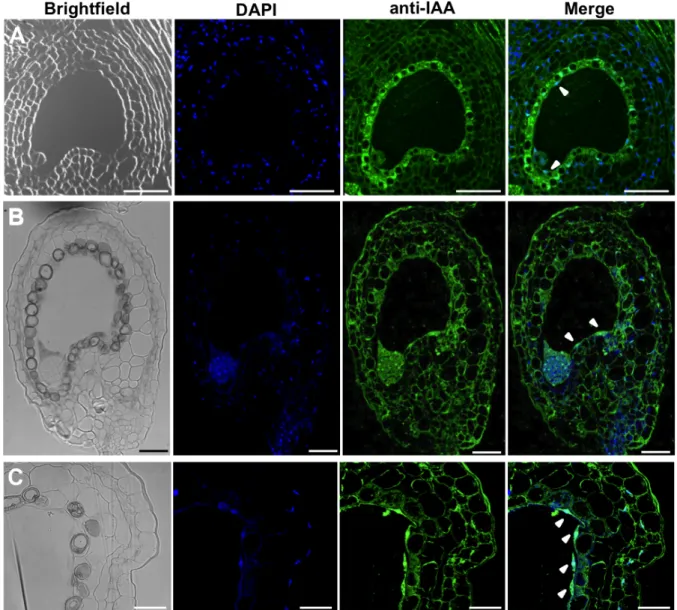
3.8. Impaired auxin signaling in the integuments leads to developmental aberrations in the female gametophyte ... 23
4. Discussion ... 26
4.1. Seed coat development might be triggered by increased auxin production after fertilization ... 26
4.2. Seed coat development in fertilized seeds is not exclusively dependent on auxin ... 27
4.3. PRC2 repression is bypassed by auxin ... 27
4.4. Auxin acts non-cell autonomously to regulate female gametophyte development ... 28
4.5. Concluding remarks ... 29
5. References ... 30
List of figures
Figure 1-1 Megasporogenesis ... 1
Figure 1-2 Megagametogenesis ... 2
Figure 1-3 Seed development ... 3
Figure 1-4 Seed coat structure ... 5
Figure 1-5 PRC2-like complexes in Arabidopsis ... 7
Figure 3-1 GH3.3pro:GUS expression pattern and IAA immunolocalization in unfertilized ovules ... 15
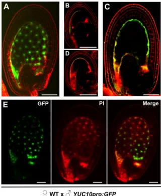
Figure 3-2 YUC10 and YUC11 promoter activity in ovules and seeds ... 16
Figure 3-3 GH3.3pro:GUS expression pattern in developing seeds ... 16
Figure 3-4 IAA Immunolocalization in developing seeds ... 17
Figure 3-5 Vanillin staining of 2,4-D and NPA treated ovules ... 18
Figure 3-6 Clearings and measurements of 2,4-D and NPA treated ovules ... 19
Figure 3-7 Seed abortion phenotype of agl62-2 plants. ... 20
Figure 3-8 Clearings and vanillin staining of yucca6-2D fis2-5 ovules ... 21
Figure 3-9 Clearings and vanillin staining of yucca6-2D vrn2-1 emf2-5 ovules ... 22
Figure 3-10 Ovules and seeds of dominant-negative Aux/IAA plant lines ... 25
Figure 6-1 IAA immunolocalization controls ... 42
Figure 6-2 Reciprocal crosses between YUC10pro:GFP transgenic lines and WT. ... 42
Figure 6-3 Transcript analysis of upregulated genes in microarray data of vrn2-1 emf2-5 ovules ... 43
Figure 6-4 Transcript analysis of downregulated genes in microarray data of vrn2-1 emf2-5 ovules ... 43
List of Tables
Table 6-1 Mutant lines used in this study ... 39
Table 6-2 Reporter lines used in this study ... 39
Table 6-3 Primers used in this study ... 39
i
Abbreviations
2,4-D 2,4-Dichlorophenoxyacetic acid
CaMV35S Promoter of the 35S RNA from Cauliflower Mosaic Virus
4-Cl-IAA 4-Chloroindole-3-acetic acid
ABCB ATP-BINDING CASSETE B
AFB AUXIN SIGNALING F-BOX
AGL62 AGAMOUS –LIKE 62
APEX (3-Aminopropyl)triethoxysilane
ARF AUXIN RESPONSE FACTOR
Aux/IAA Auxin/INDOLE-3-ACETIC ACID
AUX1 AUXIN RESISTANT1
AuxRE Auxin Response Element
bps Base pairs
CLF Col-0
CURLY LEAF
Arabidopsis thaliana ecotype Columbia
DAE Days after emasculation
DAP Days after pollination
DAPI 4',6-Diamidino-2-phenylindole
ddH2O Double deionized water
DIC Differential interference contrast
Dicamba 3,6-Dichloro-2-methoxybenzoic acid
DR5 Synthetic auxin response element
EDTA Ethylenediaminetetraacetic acid
EMF2 EMBRYONIC FLOWER2
EMS Ethyl methanesulfonate
FCS FIE FIS2
Fetal calf serum
FERTILIZATION INDEPENDENT ENDOSPERM FERTILIZATION INDEPENDENT SEED2
ii
FLC FLOWERING LOCUS C
GFP Green Fluorescent Protein
GH3 GRETCHEN HAGEN3
GUS β-glucuronidase
H3K27me3 Trimethylation of Lysine 27 on Histone 3
IAA Indole-3-acetic acid
IAM Indole-3-acetamide
IAOx Indole-3-acetaldoxime
IPA Indole-3-pyruvic acid
KLU CYTOCHROME P450 KLUH
LAX LIKE AUX
Ler Arabidopsis thaliana ecotype Landsberg erecta
MADS Acronym derived from four members of the family: MCM1, AGAMOUS, DEFICIENS and SRF
MEA MEDEA
MES 4-Morpholineethanesulfonic acid
MINI3 MINISEED3
MS-medium Murashige and Skoog medium
MSI1 MULTICOPY SUPRESSOR OF IRA1
NAA 1-Naphthaleneacetic acid
NLS Nuclear localization signal
NPA 1-N-Naphthylphthalamic acid
PA Protoanthocyanidins
PAA 2-Phenylacetic acid
PAT Polar auxin transport
PBS Phosphate buffer saline
PcG Polycomb group
iii
PCR Polymerase Chain Reaction
PEG Paternally expressed gene
PGP P-GLYCOPROTEIN
PHD Plant Homeo Domain
PHE1 PHERES1
PI Propidium iodide
PIN PIN-FORMED
PP2A PROTEIN PHOSPHATASE 2A
PRC Polycomb Repressive Complex
pro Promoter
RT-qPCR Reverse-transcription quantitative (real-time) PCR
SCF Skp, Cullin, F-box containing complex
SHB1 SWN
SHORT HYPOCOTYL UNDER BLUE1 SWINGER
TAA1 TRYPTOPHAN AMINOTRANSFERASE OF ARABIDOPSIS
TAR TRYPTOPHAN AMINOTRANSFERASE RELATED
TIR1 TRANSPORT INHIBITOR RESPONSE1
Trp Tryptophan
TTG2 TRANSPARENT TESTA GLABRA2
VRN2 VERNALIZATION2
WT Wild-type
v
Abstract
Seed formation in Arabidopsis thaliana requires a coordinated development between embryo, endosperm and seed coat. While the first two structures are originated by a simultaneous fertilization event of egg and central cell, the seed coat is solely of sporophytic origin. Still, initiation of its development is triggered by the presence of the sexual endosperm. In fact, the type I MADS-box transcription factor, AGL62 is necessary to form an endosperm-derived signal that upon fertilization, triggers seed coat development. The nature of such signal is however still unknown. Symplastic connections between endosperm and seed coat are not known to exist, therefore phytohormones, due to their ability to actively cross cell membranes, are good candidates to be involved in this signaling process. Molecular evidence, such as gene expression studies, suggests that auxin is involved in seed coat development. In this study the role of this phytohormone in seed development, and more particularly in seed coat initiation were investigated.
Reporter gene analysis revealed that auxin is present in all tissues of the ovule and seed throughout their development, and suggest that a fertilization-dependent increase in biosynthetic activity may be the initial drive for seed development. Additionally, it was found that auxin has the ability to induce autonomous seed formation, bypassing the developmental repression exerted by PcG proteins, in the absence of fertilization. Also, auxin partially rescued the phenotype of the agl62 mutant, which fails to develop a seed coat. Moreover, the results obtained here show that auxin is essential for correct cell specification and positioning in the embryo sac. Altogether, these results indicate that auxin has a predominant role in ovule and seed development and that this hormone seems to be involved in the pathway that leads to seed coat formation, most likely not as an exclusive participant.
Keywords
vii
Resumo
Durante o desenvolvimento das plantas observa-se a alternância entre uma fase esporofítica diplóide e uma fase gametofítica haplóide. Em angiospérmicas, ambos os gametófitos unissexuais são constituídos por um pequeno conjunto de células que se encontram envolvidos pela flor. O gametófito feminino forma-se em duas fases distintas: megaesporogénese e megagametogénese e após o desenvolvimento estar completo, o óvulo possui um conjunto de células gametofíticas envolvidas pelo tecido maternal esporofítico – o integumento. A composição final do gametófito feminino inclui uma oosfera, uma célula central formada a partir da fusão dos dois núcleos polares, duas sinérgides e duas antípodas. Na ausência de fertilização, proteínas PcG reprimem o desenvolvimento da semente. Estas proteínas estão organizadas em complexos que inibem a transcrição de vários loci através da sua metilação na lisina 27 da histona 3 (H3K27) e cada complexo actua em tecidos específicos durante o desenvolvimento. Assim, no óvulo, o complexo FIS (FERTILIZATION INDEPENDENT SEED) inibe a divisão autónoma da célula central, enquanto que os complexos VRN (VERNALIZATION) e EMF (EMBRYONIC FLOWER) inibem o desenvolvimento do invólucro da semente - o tegumento. A actividade destas proteínas permite portanto que a transição de óvulo para semente aconteça apenas mediante a fertilização.
A fertilização dá-se quando os dois gâmetas masculinos se fundem com a oosfera e a célula central: a oosfera haplóide origina um embrião diplóide, enquanto que a célula central homodiplóide origina o endosperma triplóide. O crescimento simultâneo destas estruturas é essencial já que, apesar de apenas o embrião contribuir para estabelecimento da nova geração, o endosperma é indispensável para nutrir o embrião, assegurando que este possa completar a sua maturação. Para além do embrião e do endosperma, o tegumento da semente, originado a partir do integumento materno, é também essencial, já que mantém a integridade da semente durante o desenvolvimento, protegendo fisicamente os seus conteúdos. Adicionalmente, é também importante para promover a dormência e permitir a dispersão das sementes. Apesar de a fertilização apenas iniciar directamente o desenvolvimento do embrião e do endosperma, o tegumento começa também o seu desenvolvimento após a fertilização, desenvolvimento esse que é coordenado com os restantes componentes da semente. Isto sugere que exista um mecanismo de comunicação entre os tecidos gametofíticos e o tegumento esporofítico. De facto, o uso de mutantes de Arabidopsis que apenas produzem um gâmeta masculino – fertilizando unicamente a oosfera ou a célula central – demonstrou que o endosperma é necessário e suficiente para a formação do sinal que inicia o desenvolvimento do tegumento da semente. Para além disto, sabe-se que este sinal está dependente da actividade do factor de transcrição AGL62, expresso especificamente no endosperma. No entanto, a natureza deste sinal ainda é desconhecida.
A comunicação entre endosperma e tegumento parece ser restrita, dado que não existem ligações simplásticas descritas entre estes dois compartimentos da semente. Desta forma, a natureza molecular
viii
do sinal que promove o desenvolvimento do tegumento está limitada a moléculas que consigam atravessar a membrana celular activamente, tais como hormonas. A análise de dados de expressão génica revela que genes de biossíntese de auxina estão sobre-regulados em mutantes de proteínas PcG, que produzem tegumento na ausência de fertilização. Adicionalmente, uma sub-regulação destes genes é observada no mutante agl62, cujas sementes não desenvolvem tegumento. Sabe-se também que a aplicação de auxina exógena induz partenocarpia em diversas espécies, incluindo Arabidopsis, e recorrendo a genes repórter que respondem à presença de auxina, viu-se que a concentração desta hormona é aumentada pouco após a fertilização. Todos estes dados sugerem a auxina como uma potencial candidata para o sinal que inicia o desenvolvimento do tegumento da semente. Neste estudo pretendeu-se não só melhorar o conhecimento actual sobre o papel da auxina no desenvolvimento dos óvulos e sementes de Arabidopsis, como também determinar se esta hormona está envolvida na via de sinalização que promove o desenvolvimento do tegumento.
Os resultados obtidos neste estudo, através de genes repórter e da imunolocalização de auxina, indicam que esta hormona está presente em óvulos, tanto nos integumentos como na célula central. Além disso, a manipulação das vias de sinalização da auxina, permitiu perceber que a presença desta hormona nos integumentos é essencial para a formação do gametófito feminino. A disrupção da sinalização, especificamente nos integumentos, originou defeitos na diferenciação e no posicionamento das células do gametófito feminino e inclusivamente, em alguns dos óvulos, a formação do gametófito não ocorreu, pelo que estas estruturas possuíam apenas os integumentos. Daqui se conclui que a auxina dos tecidos esporofíticos tem um efeito indirecto mas crucial na formação do gametófito feminino.
Após a fertilização, a expressão dos genes de síntese de auxina, YUC10 e YUC11, é induzida e restrita ao endosperma. De modo concordante, a presença de auxina foi detectada no endosperma, bem como no tegumento de sementes em crescimento.
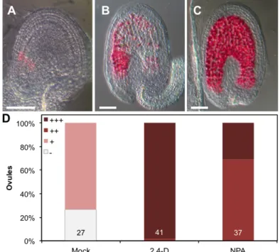
Observou-se também que a aplicação de um composto análogo à auxina, 2,4-D, em óvulos não fertilizados induz a formação de sementes autónomas. Nos óvulos tratados com este composto não só o tegumento se desenvolve, como também ocorre a divisão da célula central, formando assim endosperma assexual. Quando a aplicação deste composto é efectuada no mutante agl62, uma recuperação parcial do fenótipo é observada, indicando que a auxina é importante para o desenvolvimento do tegumento.
A aplicação de NPA, um inibidor do transporte polar de auxina que causa a acumulação desta hormona nos locais da sua síntese, produziu resultados semelhantes aos descritos para a aplicação de 2,4-D, o que reforça a ideia de que a presença de auxina, acima de uma determinada concentração, induz a formação de sementes autónomas. Para além disto, e em concordância com os dados obtidos com os genes repórter e a imunolocalização de auxina, o fenótipo resultante da aplicação de NPA indica também que esta hormona é produzida na célula central e nos integumentos dos óvulos não fertilizados, se bem que em menores quantidades do que após a fertilização. É também interessante
ix
notar que a auxina tem a capacidade de ultrapassar a repressão ao desenvolvimento da semente, exercida pelas proteínas PcG.
O mutante yucca6-2D, que apresenta níveis aumentados de auxina nos integumentos, e o mutante para as proteínas PcG VRN e EMF, vnr2-1 emf2-5, apresentam fenótipos semelhantes: ambos desenvolvem um tegumento autónomo, na ausência de fertilização. A análise dos fenótipos do triplo mutante yucca6-2D vnr2-1 emf2-5, e dos níveis de expressão de genes seleccionados nos mutantes yucca6-2D e vnr2-1 emf2-5 revelou que as proteínas PcG e a auxina têm a capacidade de regular processos semelhantes durante o desenvolvimento da semente. Contudo, nem todos os processos em que estão envolvidos serão comuns, já que diferenças na regulação da expressão de genes, bem como relações não-aditivas entre os fenótipos, são observadas nestes mutantes.
Assim, os resultados obtidos neste estudo indicam que a auxina tem um papel fundamental no desenvolvimento dos óvulos e das sementes de Arabidopsis thaliana. Por um lado, a presença desta hormona durante a megagametogénese é essencial para a correcta formação do gametófito feminino, por outro, o aumento da síntese de auxina após a fertilização parece ser o factor que comanda e estimula o desenvolvimento da semente, possivelmente através da modulação da actividade das proteínas PcG. O facto de a expressão dos genes de síntese de auxina ser altamente induzida após a fertilização, bem como o facto de esta hormona induzir a formação de sementes autónomas, suportam esta hipótese. Apesar de o sinal que induz o desenvolvimento dos tegumentos ainda não ter sido identificado, os resultados aqui apresentados levam a crer que a auxina tem um papel importante na activação das vias moleculares que promovem o crescimento desta estrutura, quer seja fazendo parte de um sinal complexo criado no endosperma e que se desloca para o tegumento ou, alternativamente, sendo uma consequência directa de um outro sinal distinto.
Palavras-chave
Auxina; semente; tegumento; endosperma; gametófito feminino; proteínas PcG; AGL62; Arabidopsis thaliana
1
1. Introduction
1.1. Ovule and female gametophyte development in Arabidopsis
During plant development an alternation between diploid sporophytic and haploid gametophytic generations occurs. In angiosperms both unisexual gametophytes comprise a small set of cells, which are surrounded by the sporophytic tissues. The male gametophyte originates from a microspore mother cell that undergoes a meiotic division to form the haploid microspores, which after two mitotic divisions form the functional male gametophyte (pollen). The female gametophyte, on the other hand, is formed from a megaspore mother cell that also undergoes a meiotic division followed by mitotic divisions, as detailed below.
The complete development of the female gametophyte can be divided in two distinct phases: megasporogenesis (Fig. 1-1) and megagametogenesis (Fig. 1-2). During megasporogenesis a finger-like protrusion, the ovule primordium, emerges from the placental tissue, where three major areas along the proximal-distal axis can be distinguished (Schneitz et al., 1995): in the proximal end is the funiculus which connects the ovule to the maternal plant and allows communication between the two, followed by the chalaza, from where the integuments derive, and in the distal end, the nucellus. Within the nucellus, a subepidermal cell differentiates into a functional megaspore mother cell that will undergo meiosis and originate four haploid cells. Among these, three degenerate through programmed cell death and only one functional megaspore remains (Christensen et al., 1997)
Megagametogenesis is initiated by a succession of mitotic divisions of the functional megaspore that are not followed by cytokinesis. During the first division, a central vacuole is formed isolating the two nuclei on each pole of the coenocyte. Another two rounds of mitosis occur, leading to the formation of a total of eight nuclei. During cellularization, six of these nuclei become surrounded by cell walls whereas two of them, each on one pole, migrate to the center of the female gametophyte and fuse, giving rise to the homodiplod central cell (Drews and Koltunow, 2011). Thus, the final composition of the female
Figure 1-1 Megasporogenesis. (A) Ovule primordium with subepidermal megaspore mother cell. L1 represents the epidermal
layer that will be a part of the innermost layer of the inner integument. (B) Megasporogenesis starts with a protrusion from the placenta. In the nucellus region the megaspore undergoes meiosis to originate a tetrad of which only the functional megaspore prevails. From the chalaza area of the ovule primordium epidermal cells divide and start formation of the inner and outer integuments. ch: chalaza region; dm: degenerated megaspores; f: funiculus region; fm: functional megaspore; ii: inner integument; L1: L1 epidermal layer; mmc: megaspore mother cell; mt: meiotic tetrad; nu: nucellus region; oi: outer integument. Adapted from Drews and Koltunow (2012).
2
gametophyte comprises seven cells: one egg cell, one central cell, two synergid cells and three antipodal cells (Bajon et al., 1999). Polarization is extremely important so that synergids, central cell and egg cell are maintained close to each other, facilitating the double fertilization event.
The sporophytic integuments are derived from the chalazal zone of the ovule primordium and their development starts with the division of the epidermal cells, creating a ring-like structure that gradually envelopes the nucellus, forming the inner integument. Later, outer integument growth starts in a similar fashion, surrounding the inner integument. While the inner integument grows symmetrically, growth of the outer integument is more pronounced on the apical side, causing the ovule to be slightly bent. Two distinct cells layers form both the inner and outer integuments, and the innermost layer of the inner integument undergoes an additional periclinal division. Thus, mature ovules contain five distinguishable cell layers (Robinson-Beers et al., 1992). Nevertheless, the female gametophyte is never fully enclosed by the integuments, since a small opening on the apical side of the ovule, the micropyle, allows the penetration of the pollen tube.
1.2. Double fertilization
The Arabidopsis male gametophyte consists of a pollen grain that contains two sperm cells and one vegetative cell. The pollen grain develops in the anthers, and during anthesis it reaches full maturity, allowing its release onto the papillae of the female receptive organ, the stigma. Here, rehydration of the pollen grain triggers the growth of the pollen tube, which is promoted by the vegetative cell. Numerous factors contribute to the guidance of the pollen tube, which grows inside the placental tissue and eventually enters the female gametophyte through the ovule micropyle (reviewed by Drews and Koltunow, 2011). The female gametophyte has a crucial role in ensuring correct guidance of the pollen tube, and synergid cells were shown to be essential to this process (Hulskamp et al., 1995; Higashiyama et al., 2001).
After entering the female gametophyte, the pollen tube contacts with the synergid cells and its growth is arrested. One of the synergid cells undergoes cell death and shortly after, the pollen tube releases the two sperm cells into the cytoplasm of the degenerating synergid. Then, the sperm cells approach and
Figure 1-2 Megagametogenesis. (A) The functional megaspore divides, producing a two-nucleated coenocyte, with two nuclei at
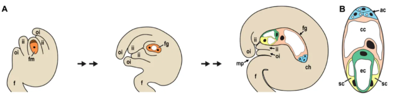
each pole, separated by a large vacuole. During cellularization differentiation of embryo sac cells occurs. Inner and outer integuments grow, surrounding the female gametophyte, though incomplete growth leaves a small opening, the micropyle. (B) Female gametophyte composition. Perpendicular cross-section of that in (A). ac: antipodal cells; cc: central cell; ch: chalazal region of the ovule; ec: egg cell; f: funiculus; fg: female gametophyte/ embryo sac; fm: functional megaspore; ii: inner integument; mp: micropyle; oi: outer integument; sc: synergid cells. Adapted from Drews and Koltunow (2012).
3
subsequently fuse with egg and central cells. Both sperm cells have the ability to fertilize either egg or central cell, suggesting that they are equivalent and that this is a random event (Ingouff et al., 2009; Hamamura et al., 2011).
1.3. Seed development
Production of seeds is an evolutionary landmark, which allows plants to endure harsh environmental conditions by suspending their life cycle, and resuming it when more favorable conditions are met (Bentsink and Koornneef, 2008). Seed development is tightly regulated and several specific structures are essential to ensure that a quality seed is produced, so that establishment of the new generation is not compromised (Santos-Mendoza et al., 2008).
The double fertilization event initiates the development of two independent structures – embryo and endosperm – that grow simultaneously (Fig. 1-3). Though only the embryo contributes to the next generation, endosperm development is responsible for the nourishment of the embryo, which is essential so that it reaches a mature stage. Besides these two structures, the sporophytic seed coat is also a key element in seed development since it protects the fertilization products from exterior disruptions (Nowack et al., 2010).
1.3.1. Embryo
Embryogenesis starts with a first asymmetric division of the egg cell, originating a smaller apical cell and a larger vacuolated basal cell. The latter undergoes several divisions, forming the embryo suspensor, which supports the embryo and connects it to the maternal tissues. Meanwhile, division of the apical cell leads to its differentiation into the eight-cell proembryo. At this stage, three areas can be defined along the apical-basal axis: the apical tier of the proembryo, which gives rise to the cotyledons and the shoot apical meristem; the lower tier that forms the hypocotyl, root and root apical meristem and finally, the extraembryonic suspensor cells (Möller and Weijers, 2009).
Maturation is achieved through multiple divisions and ultimately, the fully developed embryo fills the seed with its grown hypocotyl and cotyledons. In dicot plants like Arabidopsis, this fast growth is supported by the consumption of the nutritious endosperm, which by the end of seed development is
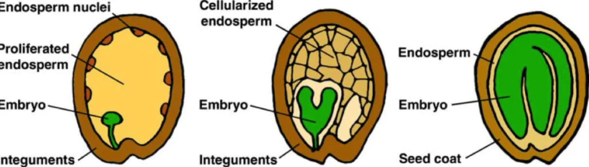
Figure 1-3 Seed development. The double fertilization event initiates embryo and endosperm development, which
are accompanied by seed coat growth. In the syncytial phase the endosperm proliferates rapidly which is followed by cellularization. As the embryo grows it consumes the surrounding endosperm and eventually, the seed cavity is completely filled with the mature embryo. Adapted from Sun et al. (2010).
4
almost absent. At this point, the seed enters the desiccation phase, where the embryo is prepared for dormancy (Bentsink and Koornneef, 2008).
1.3.2. Endosperm
In the majority of flowering plants, endosperm development starts with the syncytial phase, where several rounds of mitotic divisions are not followed by cell wall formation. During the first divisions, endosperm nuclei migrate from the micropylar zone to the chalazal region and subsequent divisions originate specific domains within the syncytium. In these domains, different cytoplasmic and cytoskeletal properties are observed, and division is autonomously regulated. Three areas can be defined: the micropylar endosperm (MCE) that surrounds the embryo, the central or peripheral endosperm (PEN) and the chalazal endosperm (CZE) (Costa et al., 2004; Olsen, 2004).
The following cellularization phase starts first in the MCE, where cell walls are deposited between neighboring cytoplasmic domains that surround each nucleus. The cellularization then spreads to the PEN but not to the CZE. Instead, this domain persists as a syncytium throughout seed development. Mitotic divisions in this domain are not followed by nuclear divisions, and large nuclei form as a result of endoreduplication. The CZE possesses specialized extensions that allow penetration in the sporophytic tissue close to the funiculus, thus it is hypothesized that this specific endosperm domain is responsible for loading the nutrients coming from the maternal vascular bundle (Nguyen et al., 2000).
The cellularized endosperm still goes through additional rounds of division and eventually fills the seed cavity. In dicot plants the endosperm is consumed by the embryo and only its outermost layer, the aleurone, persists (Olsen, 2004).
The endosperm is generated by fertilization of the homodiploid central cell. Thus, the triploid endosperm contains two copies of the maternal genome and one copy of the paternal genome. Maintenance of the correct genome dosage is very important, and changes in the parental genome dosage causes abnormal endosperm development (Scott et al., 1998).
Parent-of-origin-dependent gene expression is an epigenetic phenomenon causing maternal and paternal alleles to be differentially expressed. This phenomenon, also termed genomic imprinting, occurs in the endosperm and probably underlies the sensitivity of the endosperm in response to changes of the parental genome dosage (Schatlowski and Köhler, 2012). One interpretation for this is that maternal and paternal alleles are subject to different evolutionary forces: maternal interests rely on equal distribution of resources among the progeny, whereas paternal interests favor allocation of as many resources as possible into one individual offspring (Haig and Westoby, 1991). Recent studies identified a broad array of imprinted genes in the endosperm, both maternally and paternally expressed, many of which are transcriptional regulators, suggesting that imprinting has a crucial role in regulation of endosperm and seed development (Hsieh et al., 2011; Wolff et al., 2011).
5
1.3.3. Seed coat
The seed coat has a crucial function in maintaining seed integrity throughout its development by physically protecting the embryo. Furthermore, it is important in promoting dormancy and allowing seed dispersal (Haughn and Chaudhury, 2005).
While integument growth relies mostly on cell division, seed coat growth is mainly achieved by cell elongation, which has a predominant role over cell division in determining final seed size (Garcia et al., 2005; Roszak, 2012). Five cell layers are present in the unfertilized ovule: three belonging to the inner integument, and two to the outer integument. The same layers are present in the early seed (Fig. 1-4), but as the seed matures, each layer acquires a specific fate.
Shortly after fertilization, the innermost layer of the inner integument starts accumulating protoanthocyanidins (PA). Later in development PAs are released, oxidized, and spread to the remaining seed coat layers conferring a brown color to the seed (Lepiniec et al., 2006). The other two layers of the inner integument do not differentiate further, go through programmed cell death, and are eventually crushed together by the pressure exerted by the growing embryo (Haughn and Chaudhury, 2005; Nakaune et al., 2005).
In the two layers of the outer integument, an initial deposition of amyloplasts is observed, but as development progresses, each layer diverges in fate. While cells of the subepidermal layer produce thickened cell walls, the epidermal layer secretes high quantities of mucilage to the apoplast. Mucilage accumulation leads to contraction of the cytoplasm that remains confined to the center of the cell. With time, the cytoplasm columns are replaced by cell walls, thus forming the columella, and when all the cell layers collapse, these structures, together with the mucilage, hold the seed shape (Haughn and Chaudhury, 2005). When imbibition occurs, the mucilage is released promoting a moist environment during seed germination (Haughn and Western, 2012).
1.4. Interactions between endosperm and seed coat
Seed growth requires regulatory mechanisms to ensure simultaneous development of embryo, endosperm and seed coat. The seed coat is a sporophytic tissue but initiation of its development is dependent on the fertilization event. This suggests that a signal originated in the embryo or endosperm might be the trigger of seed coat development. The use of an Arabidopsis mutant that only produces a single sperm cell showed that only the fertilized endosperm is able to induce seed coat growth (Roszak and Köhler, 2011), revealing that the necessary signal for seed coat growth is generated by the sexual
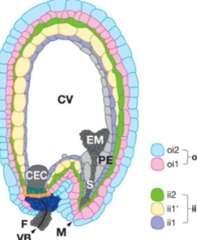
Figure 1-4 Seed coat structure. Shortly
after fertilization the seed coat comprises five cell layers. Three layers of the inner integument (ii): ii1, ii1’ and ii2; And two layers from the outer integument (oi): oi1,
oi2. CEC: chalazal endosperm cyst; CV:
central vacuole; EM: embryo; F: funiculus;
M: micropyle; PE: peripheral endosperm; S:
embryo suspensor; VB: vascular bundle. Adapted from Lepiniec et al. (2006).
6
endosperm. In agreement with that, ablation of the endosperm inhibits seed coat development (Weijers et al., 2003).
In fact, several studies have shown that endosperm development directly impacts seed coat growth. Mutants for genes belonging to the HAIKU family (iku1 and iku2) produce seeds with reduced endosperm growth and early cellularization, which results in a decreased seed size (Garcia et al., 2003). Similar phenotypes were described for mini3 and shb1 and the proposed pathway suggests that SHORT HYPOCOTYL UNDER BLUE1 (SHB1) is a direct upstream regulator of IKU2 and MINISEED3 (MINI3), promoting their expression (Zhou et al., 2009). Specific expression of these genes in the endosperm, and absence of maternal sporophytic effects are strong indications that the endosperm regulates seed size.
Another indication that the endosperm regulates seed coat growth comes from the characterization of the agl62 mutant (Kang et al., 2008; Roszak and Köhler, 2011). AGAMOUS-LIKE 62 (AGL62) codes for a type I MADS-box transcription factor, expressed specifically in the endosperm. Seeds of the agl62 mutant show early endosperm cellularization and fail to develop a seed coat, despite the presence of dividing endosperm. This suggests that AGL62 is crucial for the formation of the signal that initiates seed coat development.
Several studies have also shown that not only does the endosperm control seed coat growth, but that the reverse also happens. Influence of the maternal sporophytic tissues on endosperm development is illustrated for example, by the ttg2 mutant. TRANSPARENT TESTA GLABRA2 (TTG2) is strongly expressed in all seed coat layers and is known to be a part of the PA and mucilage synthesis pathways (Johnson et al., 2002). Similarly to haiku mutants, ttg2 causes defects in endosperm growth and cellularization and decreased seed coat cell elongation (Garcia et al., 2005). This mutation has a strict maternal sporophytic effect and the defects observed in endosperm growth are a direct consequence of the defective seed coat cell elongation, suggesting that maternal tissues also have a significant impact in seed size control, through interaction with endosperm and regulation of its development.
1.5. Polycomb Group proteins in seed development
Polycomb group (PcG) proteins are chromatin-associated factors that repress transcription of several specific target loci. These proteins were initially identified in Drosophila, where they control homeotic genes by ensuring that their expression is restricted to specific tissues and time-points in development, thus being important to maintain cell identity (Jürgens, 1985). PcG proteins are organized in multimeric complexes, of which Polycomb Repressor Complex 1 and 2 (PRC1 and PRC2) are the best characterized ones that are present in multicellular animals and plants (reviewed by Müller and Verrijzer, 2009).
PRC2 proteins are highly conserved in plants and due to genome duplication events the Arabidopsis PRC2 complexes have many homologous subunits, with specific and partially redundant functions in distinct phases of development. Transcriptional repression by these complexes is achieved through
7
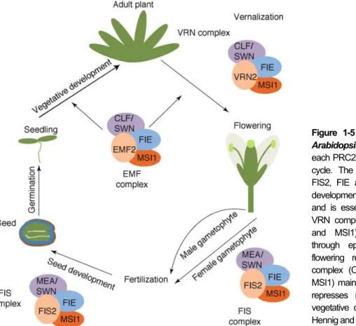
association with Plant Homeo Domain (PHD)-finger proteins and subsequent trimethylation of lysine 27 of histone 3 (H3K27me3) at target loci (reviewed by Hennig and Derkacheva, 2009). There are three different PRC2 complexes in Arabidopsis: EMBRYONIC FLOWER (EMF), VERNALIZATION (VRN) and FERTILIZATION INDEPENDENT SEED (FIS) and their role in plant development is illustrated in
Figure 1-5.
The first subunits of the FIS-PRC2 were identified in a screen for non-sexual seed formation (apomixis) (Ohad et al., 1996; Chaudhury et al., 1997). Mutations in any of the four subunits - FIS2 (FERTILIZATION INDEPENDENT SEED2), MEA (MEDEA), FIE (FERTILIZATION INDEPENDENT ENDOSPERM) and MSI1 (MULTICOPY SUPRESSOR OF IRA19) - lead to the formation of autonomous seeds containing proliferating endosperm and, in some cases, initiation of seed coat development. Additionally, post-fertilization phenotypes, such as non-cellularized endosperm and seed arrest, are also observed (Chaudhury et al., 1997; Kiyosue et al., 1999). This, coupled with the fact that FIS subunits are expressed in the central cell, as well as in the developing endosperm (Luo et al., 2000), indicates that this complex has a crucial role in female gametophyte and seed development. In fact, it is believed that the FIS complex suppresses central cell division in absence of a fertilization event (Ohad et al., 1996; Chaudhury et al., 1997).
Despite the specific action of the FIS-PRC2 in gametophytic tissues, mutations in some of its subunits (FIE and MSI1) have a sporophytic effect and phenocopy to some extent the seed coat initiation phenotype observed in emf2 and vrn2 mutants. Both FIE and MSI1 subunits are common to the PCR2
Figure 1-5 PRC2-like complexes in Arabidopsis. Composition and roles of each PRC2-like complex in the plant life cycle. The FIS complex (MEA/SWN, FIS2, FIE and MSI1) represses seed development in absence of fertilization and is essential for seed growth. The VRN complex (CLF/SWN, VRN2, FIE and MSI1) controls flowering time through epigenetic silencing of the flowering repressor FLC. The EMF complex (CLF/SWN, EMF2, FIE and MSI1) maintains cell differentiation, and represses reproduction by promoting vegetative development. Adapted from Hennig and Derkacheva (2009).
8
sporophytic complexes EMF and VRN, suggesting a role for these complexes in regulation of seed growth, specifically, by suppressing seed coat development in unfertilized seeds (Roszak and Köhler, 2011).
1.6. Auxin
Auxin is an essential phytohormone that controls multiple processes during plant growth and development. It is known to be a key regulator of critical aspects such as cell division, elongation and differentiation, among others. Thus, auxin responses both on the cellular and on the tissue level have to be tightly regulated, which is achieved by modulating its biosynthesis, downstream signaling and transport.
1.6.1. Biosynthesis
The main active auxin form is free IAA (Indole-3-acetic acid), but other naturally occurring forms, such as PAA and 4-Cl-IAA, as well as synthetic molecules, such as 2,4-D, NAA and dicamba, are also able to generate auxin responses. Besides active forms, the auxin pool is also composed of many inactive auxins that are thought to help maintain homeostasis: these include precursors and storage forms (often auxin conjugated with amino acids, peptides or carbohydrates) (reviewed in Woodward and Bartel, 2005; Korasick et al., 2013).
Synthesis of free IAA occurs via Tryptophan (Trp)-dependent and Trp-independent pathways. Although still little is known about Trp-independent pathways, experimental evidence strongly supports their existence (reviewed by Normanly et al., 2004). Analysis of trp mutants, both in Arabidopsis and maize, show that these plants have similar free IAA levels when comparing with WT. Also, feeding assays with labeled Trp support the idea that IAA synthesis is not exclusively dependent on Trp presence (Normanly et al., 1993). Still, many components of these pathways remain unknown and further studies are required.
Nonetheless, Trp-dependent pathways are better characterized and three main routes, named according to their main IAA intermediate, are known: indole-3-acetaldoxime (IAOx), indole-3-acetamide (IAM) and indole-3-pyruvic acid (IPA). In Arabidopsis the main contributor to free IAA is the IPA pathway, where production of IAA is a two-step process mediated by two different groups of enzymes (Mashiguchi et al., 2011; Stepanova et al., 2011; Zhao, 2012). The first step is the conversion of Trp to IPA by Trp aminotransferases: TAA1, TAR1 and TAR2. In the second step, flavin monooxygenases from the YUCCA (YUC) family convert the produced IPA into IAA. Activity of Trp aminotransferases, as well as of YUCs, has been shown to be essential in many developmental processes, such as embryogenesis, lateral root formation, hypocotyl elongation, among others (Cheng et al., 2007; Stepanova et al., 2008). Furthermore, it is proposed that regulation of auxin biosynthesis, essential for vascular and flower tissue formation, can be achieved by spatial and temporal control of YUC gene expression (reviewed by Korasick et al., 2013).
9
1.6.2. Signaling
Several auxin signaling pathways that involve perception of extracellular and cytoplasmic auxin are proposed to exist, but the best understood pathway is the Aux/IAA, localized in the nucleus. This pathway is dependent on the activity of transcriptional repressors belonging to the Aux/IAA family. These repressors have four conserved domains through which they interact with several other regulatory proteins of this pathway (Pierre-Jerome et al., 2013). Domain III and IV, for instance, allow dimerization of Aux/IAAs with Auxin Response Elements (ARF). ARFs are transcription factors that bind to the auxin responsive cis-elements (AuxRE) present in the promoters of different auxin responsive genes, and whose transcription is repressed by the formation of ARF-Aux/IAA dimers (Leyser, 2010).
When intracellular levels of auxin increase, this hormone binds to the SCFTIR1/AFB complex. This
promotes and stabilizes the interaction of Aux/IAAs (through its domain II) with the TIR1 and AFB subunits of the SCFTIR1/AFB complex (Tan et al., 2007). Through ubiquitination, this complex targets
Aux/IAAs for degradation by the 26S proteasome, leaving ARFs free to form ARF-ARF dimers, which in turn, allow transcription of several auxin responsive genes (Leyser, 2010). The common characteristic among those genes is the presence of AuxREs in their regulatory regions, the specific conserved sequence that allows auxin-dependent transcriptional regulation. Based on these sequences, genome-wide studies identified many putative auxin-responsive genes, reinforcing the role of auxin as a powerful regulator of plant development (reviewed in Hagen and Guilfoyle, 2002; Woodward and Bartel, 2005).
1.6.3. Transport
On the whole-plant level, auxin is largely synthesized in shoot apical regions, but since it regulates a wide variety of processes in many other tissues, transport of this hormone is required. In fact, auxin is effectively transported between cells across long distances and directionality of its flow is regulated by polar localization of the transporters, allowing fine-tuning of auxin contents between cells, and establishment of auxin gradients.
Intercellular auxin transport is best described by the chemiosmotic model, which integrates several molecular components known to participate in auxin transport and proposes a physiological basis for auxin movement. Free IAA can enter the cell by passive diffusion, while auxin anions (IAA-), along with
H+ are imported to the cell by influx carriers of the AUX1/LAX family (Yang et al., 2006). The intracellular alkaline environment promotes IAA dissociation and efflux is achieved through PIN (Petrásek et al., 2006) and ABCB/PGP active transporters (Noh et al., 2001). The asymmetric positioning of both importers and exporters is responsible for the directional flows of auxin. In fact, polar localization of the AUX1 importer, as well as ABCB/PGP exporters is often observed, but PIN positioning on the cellular membrane is the main factor influencing the direction of polar auxin transport. Apical or basal positioning is determined by phosphorylation of these transporters by the PINOID protein kinase, whose transcription is, together with PINs, upregulated by auxin (Benjamins et al., 2001). Furthermore, the eight
10
members of the PIN family have specific roles and their expression patterns are highly dependent on the developmental stage and tissue in question (Friml, 2010).
1.7. Auxin in ovule and early seed development
Although the role of auxin in ovule and seed development is not fully understood, several studies have supported the idea that this phytohormone is, as in many other developmental processes, essential.
During the first phases of female gametophyte formation, TAA1 is expressed in the chalazal area of the ovule primordium (Nole-Wilson et al., 2010), where the integuments later arise. Simultaneously, PIN expression is detected in the same region, as well as in the nucellus (Pagnussat et al., 2009; Ceccato et al., 2013), along with YUC expression (Pagnussat et al., 2009; Bencivenga et al., 2011). In addition, auxin reporter activity (DR5pro:GUS) is also strong in the nucellus, suggesting that auxin is important for ovule development, since its onset (Pagnussat et al., 2009; Bencivenga et al., 2011). Furthermore, Pagnussat and colleagues (2009) have reported that female gametophyte cellularization is directly dependent on the establishment of an auxin gradient inside the female gametophyte. This gradient defines the fates of the female gametophyte cells: the highest auxin concentration originates synergids, followed by egg cell and finally, the lowest concentrations originate central cell and antipodals. Hence, this model proposes two essential roles for auxin during ovule development: first it is necessary for induction of embryo sac development, and in a later phase controls gametophyte cell differentiation and specification.
During early seed development, the role of auxin in embryogenesis has been thoroughly investigated and numerous studies show that control of auxin biosynthesis, transport and signaling is absolutely required for correct cell specification and patterning. Furthermore, disruption of auxin-related genes often leads to severe embryo defects (Möller and Weijers, 2009). However, how this hormone specifically influences development of the remaining seed components - endosperm and seed coat - is poorly understood. Analysis of auxin reporter activity suggests that after fertilization, there is an increase of auxin contents in the seed (Dorcey et al., 2009). Also, it is known that in several species, including Arabidopsis, application of exogenous auxin leads to parthenocarpic fruit formation (Vivian-Smith and Koltunow, 1999; Dorcey et al., 2009; Pandolfini, 2009). This suggests that auxin might be the fertilization-associated trigger that stimulates seed and fruit development, though further evidence is required to substantiate this hypothesis.
1.8. Aim of the thesis
The synchronized growth of the different seed components requires signaling mechanisms between them and, while the simultaneous fertilization event of both egg and central cell triggers development of embryo and endosperm, the trigger of the sporophytic seed coat development is still unknown. Recent results published by Roszak and Köhler (2011) showed that an AGL62-dependent signal originated in the sexual endosperm is required for seed coat initiation. However, AGL62 expression is restricted to the
11
endosperm, excluding this transcription factor from the list of possible mobile signals that, upon fertilization, move to the seed coat and stimulate its development. Moreover, communication routes between endosperm and seed coat are scarce, and it appears that symplastic communication between the two tissues is not possible (Stadler et al., 2005; Ingram, 2010). This restricts the molecular nature of the signal to molecules that can actively cross membranes, such as hormones.
Analysis of gene expression data showed that auxin-related genes are not only upregulated in fertilized seeds when comparing to unfertilized ovules, but are also downregulated in the agl62 mutant that fails to develop a seed coat (Pawel Roszak, personal communication). In addition, previous studies also support the idea that auxin plays a role in seed and fruit development, since this hormone is able to induce parthenocarpy, and appears to be increased in early seeds. Thus, the major aim of this thesis was to test whether auxin could be the signal triggering seed coat development after fertilization. To achieve this, several experiments were planned to be performed: development of auxin reporter tools that are active in reproductive tissues, determination of the effects of exogenous application of auxin-related compounds in ovules and seeds, and generation of stable transgenic lines that allow modulation of auxin signaling in a tissue-specific manner. Additionally, analysis of the effect of auxin in the agl62 phenotype, as well as use of auxin overproducing and PcG mutants to identify the molecular relations between auxin and epigenetic regulation of seed development were also planned.
2. Materials and Methods
2.1. Plant material and growth conditions
Seeds were sterilized in 5% commercial bleach and 0.01% Tween-20 for 10 min and washed three times in sterile ddH2O. Sterile seeds were plated on ½ MS-medium (0.43% MS-salts, 0.8% Bacto Agar,
0.19% MES hydrate and 1% Sucrose; when necessary, the medium was supplemented with the appropriate antibiotics) and stratified at 4ºC in the dark for 48h. Plates were then transferred to a growth chamber (16h light / 8h dark; 110 µmol s-1 m-2; 21ºC; 70% humidity) where seedlings developed. After 10
days the seedlings were transferred to soil and grown in a growth chamber (16h light / 8h dark; 110 µmol s-1 m-2; 21ºC; 70% humidity).
All the mutant lines, as well as reporter lines used in this project are described in Table 6-1 and Table
6-2, respectively. Double and triple mutants were generated by crossing the corresponding single
mutants. Genomic DNA for PCR analysis was extracted as described in Edwards et al. (1991) and genotyping of segregating mutants was performed using the primers described in Table 6-3.
2.2. Cloning
All constructs were generated using the Gateway Cloning Technology (Invitrogen) following the manufacturer’s instructions. To obtain dominant-negative versions of the selected Aux/IAA proteins,
site-12
directed mutagenesis, on a highly conserved motif of domain II, was performed. The inserted point mutations were made in order to change the conserved proline of Domain II to a leucine: IAA5P58->L;
IAA10P53->L; IAA28P98->L. In the case of IAA32 no site-directed mutagenesis was performed because
domain II is not conserved. The coding regions of IAA5, IAA10 and IAA28 were amplified from cDNA of WT Col-0 plants using the flanking primers in combination with the mutation primers (Table 6-3): the right flanking primer (forward) was used with the reverse mutation primer, and the left flanking primer (reverse) was used with the forward mutation primer in two separate PCR reactions. Both PCR products for each IAA gene were then combined and a final PCR with the flanking primers was performed, thus originating a single fragment containing the desired mutation and the Gateway adaptors. The mutation primers had opposite directions but were designed to overlap each other (in approximately 10 bps) and the point mutation was present in the overlapping region of both primers. In the case of IAA32, only the flanking primers were used and WT Col-0 gDNA was used as a template.
The amplified fragments were then purified from gel, recombined into the donor vector (pDONR221) to create entry clones, and sequenced. Three different destination vectors derived from pB7WG2 (Karimi et al., 2002) were used. These vectors had the CaMV35S promoter replaced by the FIS2, PHE1 or KLU promoters (the first two vectors were provided by Pawel Roszak and the last one by Duarte Figueiredo). The cassettes including each IAA gene were recombined into the three different destination vectors to generate the following constructs: FIS2pro:IAA5P58->L, PHE1pro:IAA5P58->L, KLUpro:IAA5P58->L;
FIS2pro:IAA10P53->L, PHE1pro:IAA10P53->L, KLUpro:IAA10P53->L; FIS2pro:IAA28P98->L, PHE1pro:IA28P98->L,
KLUpro:IAA28P98->L; FIS2pro:IAA32, PHE1pro:IAA32, KLUpro:IAA32.
2.3. Generation of transgenic plants
Col-0 plants were transformed using the floral dip method described by Clough and Bent (1998) and transformants were selected with the appropriate antibiotics.
2.4. Gene expression analysis
Shortly before anthesis, flowers from WT, yucca6-2D and vrn2-1 emf2-5/+ plants were emasculated. The ovules of 25 emasculated siliques were harvested at 4 DAE in 20µL of RNAlater solution (Invitrogen) and ground for 2 min using a TissueLyser II (Qiagen). Total RNA was extracted using the Qiagen RNeasy kit, followed by DNase I treatment (Qiagen). RNA concentration and quality were assessed using a NanoDrop 1000 Spectrophotometer (Thermo Scientific). The same amount of RNA for each sample (1µg) was used to synthesize cDNA using the RevertAid First Strand cDNA Synthesis Kit (Thermo Scientific). Maxima SYBR Green qPCR Master Mix (Thermo Scientific) was used to perform the qPCR in an iQ5 qPCR system (Bio-Rad). The primers used for the RT-qPCR are described in Table
6-3. PP2A was used as the reference gene. Relative quantification of gene expression was performed
13
2.5. Chemical treatments
All chemicals were prepared in a solution containing 5% EtOH and 0,05%Tween-20. Final concentrations of the several compounds were as follows: 2,4-D – 200µM; NPA – 350µM; L-Kynurenine – 200µM and 400µM. In all experiments a mock control was run in parallel.
Flowers were emasculated shortly before anthesis and, at 2 DAE, the chemicals were applied by covering the whole silique with 2µL of the respective solution. The treatment was either done only once, or repeated every 24h until the silique was collected (this is designated as the continuous treatment). A similar procedure was used for pollinated siliques: flowers were emasculated and, at 2 DAE, the siliques were hand pollinated. The solutions were then applied 8h after pollination, as this is the determined time necessary for fertilization to occur (Duarte Figueiredo, personal communication). Treatment of pollinated siliques was also done either once or continuously. The treated siliques were then collected at the specified time-points, dissected, and the ovules or seeds were processed for DIC microscopy, vanillin staining or ovule measurements.
2.6. Microscopy
For DIC microscopy, siliques were opened lengthwise to expose the ovules/seeds, fixed overnight at 4ºC on a EtOH:Acetic Acid (9:1) solution and incubated 10 min with 90% EtOH and 10 min with 70% EtOH. Ethanol was then replaced by clearing solution (66.7g Chloralhydrate; 25g H2O; 8,3g Glycerol)
and samples were incubated overnight. The carpels were then removed and ovules/seeds were mounted in clearing solution.
For GUS staining, siliques were opened lengthwise, incubated in 90% acetone at -20ºC for 1h, washed twice for 15 min in 50mM sodium phosphate buffer and vacuum infiltrated for 20 min in GUS staining buffer (50mM sodium phosphate buffer; 10mM EDTA; 0,1% Triton X-10; 1mM potassium ferrocyanide III; 1mM potassium ferricyanide IV; 1mg/mL X-Gluc). Samples were kept in GUS staining buffer and incubated at 37ºC for the necessary time so that staining became visible, followed by an overnight incubation in clearing solution. Ovules/seeds were removed from the carpels and mounted on chloralhydrate clearing solution.
For vanillin staining, emasculated siliques were opened lengthwise, the carpels were removed and the ovules were mounted in vanillin solution (1% (w/v) vanillin (4-hydroxy-3-methoxybenzaldehyde) in 6N HCl). The slides were observed after 30 min.
All the samples described above were observed on a Zeiss Axioplan microscope or on a Leica DMI 4000 microscope, both equipped with DIC optics and digital cameras.
Confocal imaging was performed on ovules/seeds that were removed from the silique and mounted in PI solution (7% glucose; 0,1mg/ml PI) (Hélène Robert-Boisivon, personal communication). Pictures for ovule measurements were taken in the widest transversal plane, where the ovule appeared bigger. Ovule area was then measured with ImageJ software. A Zeiss LSM 700 or a Leica SP5 was used to image the samples. Excitation and emission wavelengths are detailed in Table 6-4.
14
2.7. IAA Immunolocalization
Siliques were emasculated or pollinated as described above and collected at the specified time points. Ovules or seeds were vacuum infiltrated in 4% paraformaldehyde and incubated overnight at 4ºC, followed by dehydration in a series of acetone dilutions (30%; 50%; 70%; 90%; 100%; each step was repeated once, for 1h, except the last one which was repeated three times). Samples were then embedded in Technovit 8100 (Electron Microscopy Sciences) following the manufacturer’s instructions. Sections with 2µm thickness were obtained using a LKB Ultratome III and placed on APEX covered microscope slides. The APEX-covered slides were prepared by submerging them in the following solutions: once in 2% APEX in acetone for 3 min, twice in 100% acetone for 2 min and twice in ddH2O
for 2 min. The slides were then dried at room temperature and stored.
Prepared sections of ovules/seeds were incubated in 10% FCS for 10 min, followed by incubation in primary antibody for 1 h (Mouse Monoclonal Anti-IAA (Sigma-Aldrich); 1/100 dilution in FCS) and a 10 min incubation in 5% FCS. Secondary antibody (Alexa Fluor 488 Rabbit Anti-Mouse IgG (Invitrogen); 1/10 dilution in PBS) was then applied for 45 min and washed with PBS (eight times for 3 min each). Sections were stained with DAPI (1 µg/mL in PBS), washed with PBS (six times for 3 min each), mounted on Mowiol 4-88 mounting medium (1gr Mowiol 4-88; 4mL PBS; 2mL glicerol), and observed on a Leica SP5 confocal microscope.
Controls for the immunolocalization protocol were run in parallel with the standard immunolocalization experiments (Fig. 6-1). Two different controls were performed: a negative control, where incubation with the primary antibody was substituted by incubation with 10% FCS solution and a pre-block of the primary antibody with IAA to assess its specificity to this antigen. For the pre-block experiment the primary antibody was incubated overnight at 4ºC with IAA (5 mg/mL) in a ratio of 1:2. This mixture was then diluted 100x in FCS and used in the immunolocalization experiment instead of the primary antibody. The fact that no signal was detected in both controls (Fig. 6-1) shows that both antibodies are highly specific to their antigens and that the signal detected in immunolocalization experiments corresponds to IAA presence.
3. Results
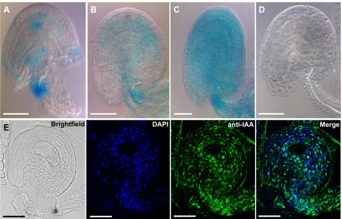
3.1. Auxin is present in the integuments of unfertilized ovules
To investigate the presence of auxin in the integuments of unfertilized ovules, plants expressing GH3.3pro:GUS were used, and the activity of this promoter was examined (Fig. 3-1). This construct was chosen because genes from the GRETCHEN HAGEN3 (GH3) family are known to respond to auxin due to the presence of auxin-responsive cis-elements (AuxRE) in their promoters (reviewed by Hagen and Guilfoyle, 2002). Promoter expression pattern in emasculated siliques was analyzed and a strong