Universidade de Lisboa
Faculdade de Farmácia
Multiple Sclerosis vs. Guillain-Barré syndrome: differences in
two autoimmune disorders with a common target in two different
regions
Inês Vaz Pésinho
Mestrado Integrado em Ciências Farmacêuticas
2019
Universidade de Lisboa
Faculdade de Farmácia
Multiple Sclerosis vs. Guillain-Barré syndrome: differences in
two autoimmune disorders with a common target in two different
regions
Inês Vaz Pésinho
Monogr
afia de Mestrado Integrado em Ciências Farmacêuticas
apresentada à Universidade de Lisboa através da Faculdade de Farmácia
Orientador: Doutora Adelaide Fernandes Borralho, Professora Auxiliar
Abstract
Multiple Sclerosis (MS) and Guillain-Barré Syndrome (GBS) are both demyelinating and autoimmune disorders affecting, respectively, the central nervous system (CNS) and the peripheral nervous system (PNS), which means they belong to a group of neurodegenerative diseases that involve inflammatory lesions associated with demyelination, inducing axonal damage and consequent neurodegeneration, leading to progressive loss of function.
MS is a chronic inflammatory disorder of the CNS and is assumed to be the most frequent cause of neurological disability in young adults. This disorder consists in inflammation, demyelination and variable levels of axonal loss. The etiology is still unknown but it is presumed to involve interaction between genetic and environmental factors that triggers an autoimmune attack, resulting in damaged myelin and axons. Clinically, most of the patients experience a relapsing-remitting phase, characterized by relapses followed by recovery. The majority of them, late on enter in a progressive phase called secondary progressive MS. The remaining patients pursue a progressive course that is called primary progressive MS. There is also clinically isolated syndrome corresponding to a first episode of neurologic symptoms in the CNS, and people who experience it may or may not develop MS. In terms of therapeutic options, disease-modifying treatments are approved specially to treat relapsing remitting form of the disease.
GBS is also an inflammatory demyelinating disease of the PNS and it is the most frequent cause of acute flaccid paralysis. This autoimmune disorder is, in most cases, preceded by viral or bacterial infections, such as Campylobacter jejuni or Influenza virus, that are capable of triggering an abnormal immune responses directed against components of the peripheral nerves by molecular mimicry. Clinically, the most frequent forms of GBS is acute inflammatory demyelinating polyradiculoneuropathy and acute motor axonal neuropathy, but there is also acute motor-sensory axonal neuropathy and Miller-Fischer syndrome. Patients with GBS commonly have respiratory insufficiency and autonomic dysfunction as associated complications. The treatment of this syndrome is composed by a multidisciplinary approach that includes general medical care and immunotherapies.
The priorities for MS and GBS investigation include establishment of biomarkers and an improved knowledge of the immunopathogenesis, to go towards personalized medicine.
Keywords: Multiple Sclerosis, Guillain-Barré syndrome, Inflammation, Demyelination, Neurodegeneration.
Resumo
A Esclerose Múltipla (EM) e o Síndrome Guillain-Barré (SGB) são ambos doenças autoimunes e desmielinizantes que afetam, respetivamente, o Sistema Nervoso Central (SNC) e o Sistema Nervoso Periférico (SNP), pertencendo ainda a um grupo de doenças neurodegenerativas que envolvem lesões inflamatórias associadas a desmielinização, induzindo dano no axónio e consequente neurodegeneração, o que leva a uma perda de função progressiva.
A EM é uma doença inflamatória crónica do SNC sendo a causa mais frequente de distúrbios neurológicos em jovens adultos. É uma doença que consiste na inflamação, desmielinização e uma variável perda axonal. A sua etiologia ainda não é completamente conhecida, mas presume-se que envolva a interação entre fatores genéticos e ambientais, estimulando um ataque autoimune e consequentes danos na mielina e nos axónios. Clinicamente, a maior parte dos doentes tem uma fase recidivante-remitente, caracterizada pela presença de surtos seguida de recuperação. Destes doentes, a maioria progride para uma doença secundária progressiva e os restantes doentes desenvolvem uma Esclerose Múltipla primária progressiva. Alguns doentes têm ainda um síndrome clinicamente isolado que corresponde a um primeiro episódio de sintomas neurológicos no SNC, sendo que estes podem ou não evoluir para Esclerose Múltipla. Em termos de tratamento, estão aprovados os medicamentos modificadores de doença, especialmente no caso de doença recidivante-remitente.
A SGB é uma doença inflamatória, mas do SNP, sendo a causa mais frequente de paralisia flácida aguda. Esta doença autoimune é antecedida por uma infeção viral ou bacteriana, como vírus Influenza ou Campilobacter jejuni, que são capazes de desencadear uma resposta imune anormal direcionada contra os componentes dos nervos periféricos, por mimetismo molecular. As formas mais frequentes são polineuropatia desmielinizante inflamatória aguda e neuropatia motora axonal aguda, existindo ainda a neuropatia motora sensorial axonal aguda e a síndrome de Miller Fisher. Os doentes com SGB têm insuficiência respiratória e disfunção autónoma como complicações associadas. O tratamento é composto por uma abordagem multidisciplinar que inclui cuidados médicos gerais e imunoterapia.
As prioridades na investigação da EM e da SGB incluem o desenvolvimento de biomarcadores e um melhor conhecimento da imunopatogénese, para que haja medicina personalizada.
Palavras-chave: Esclerose Múltipla, Síndrome Guillain-Barré, Inflamação, Desmielinização, Neurodegeneração.
Acknowledgement
This review represents the end of a very important chapter of my life, my master thesis and the end of my academic life. I believe that this was possible due to all the effort I put in this review but also to all of my friends and family.
First, I would like to thank especially to my mother, who was always there for me and for having been an example of resilience and persistence. To my friends, Leonor, Ana, Ema, Salgado, Rita, Rodrigo, Rodrigo Xavier, Gustavo and Tiago, thank you for all the concern and motivation and for understanding that I couldn’t be always present. To Carol, Cátia, Catarina and Marta, I would like to express my infinite gratitude, and thank them for being there for me in the past few years, you know that College wouldn´t be the same without you.
Finally, I would like to thank my supervisor, Professor Adelaide Fernandes, for all the important advices and interest.
Abbreviations
AIDP Acute Inflammatory Demyelinating Polyradiculoneuropathy AMAN Acute Motor Axonal Neuropathy
AMSAN Acute Motor-Sensory Axonal Neuropathy ARR Annualised Relapse Rate
BBB Blood-Brain Barrier CSF Cerebrospinal Fluid
CIS Clinically Isolated Syndrome CNS Central Nervous System GBS Guillain-Barré Syndrome IFN-β Interferon beta
IgG Immunoglobulin G
IL2RA Interleukin-2 receptor alpha gene IL7RA Interleukin-7 receptor alpha gene IVIg Intravenous Immunoglobulin MFS Miller Fisher Syndrome
MHC II Major Histocompatibility Complex, Class II MS Multiple Sclerosis
PNS Peripheral Nervous System
PPMS Primary Progressive Multiple Sclerosis RRMS Relapsing remitting Multiple Sclerosis SPMS Secondary Progressive Multiple Sclerosis Th T helper
VLA-4 Very Late Antigen-4
7
Table of contents
1. Introduction ... 8
1.1. Aim ... 9
2. Material and Methods ... 9
3. Multiple Sclerosis ... 9
3.1. Etiology of Multiple Sclerosis ... 10
3.2. Multiple Sclerosis Pathogenesis and immunopathogenesis ... 11
3.3. Multiple Sclerosis Clinical course ... 15
3.4. Therapeutic approach ... 17
4. Guillain-Barré Syndrome ... 21
4.1. GBS Etiology ... 22
4.2. GBS Pathogenesis and immunopathogenesis ... 22
4.3. GBS Clinical course ... 25
4.4. Therapeutic approach ... 28
5. Comparison between Multiple Sclerosis and Guillain-Barré syndrome ... 30
6. Conclusions and Future perspectives ... 32
7. References ... 34
Table of contents
Figure 1: Immunopathogenesis of MS. ... 128
1. Introduction
Multiple Sclerosis (MS) and Guillain-Barré Syndrome (GBS) are demyelinating diseases that affect, respectively, the central nervous system (CNS) and the peripheral nervous system (PNS), which means they belong to a group of neurodegenerative diseases that consists in inflammatory lesions associated with demyelination, inducing axonal damage, neurodegeneration and progressive loss of function (1). The term demyelination defines a pathologic process that implies destruction of myelin-supporting cells (oligodendrocytes in CNS and Schwan cells in PNS) and/or myelin sheath with some preservation of axons, and, therefore intrinsically normal myelin is destroyed (2). Central demyelination affects the CNS and peripheral demyelination involves the PNS, being that this division occurs depending on the site of the primary demyelination.
MS is a chronic inflammatory and autoimmune disorder of the CNS and is assumed to be the most frequent cause of neurological disability in young adults, being that affects mostly young women. This disorder consists in inflammation, demyelination and variable levels of axonal loss. Although the etiology is unknown, it is presumed to involve interaction between genetic, environmental and other factors that triggers an aberrant autoimmune attack resulting in damaged myelin and axons (3,4). Clinically, most of the patients (80-85%) experience a relapsing-remitting phase (RRMS), characterized by relapses and a functional recovery after that, but about 50% of RRMS patients enters in a progressive phase called secondary progressive MS (SPMS) after a variable period. The remaining 15-20% MS patients pursue a progressive course that is mentioned to as primary progressive MS (PPMS). There is also clinically isolated syndrome (CIS) corresponding to a first episode of neurologic symptoms in the CNS, and people who experience it may or may not develop MS (4).
GBS is an inflammatory demyelinating disease of the PNS which is caused by abnormal immune responses targeted against components of the peripheral nerves (5), therefore it is also recognized as acute autoimmune peripheral neuropathy and is the most frequent cause of acute flaccid paralysis after the eradication of Poliomyelitis (6). It affects more men than women and it can occur at any age (2,7). This autoimmune disorder is, in most cases, preceded by viral or bacterial infections, for instance Cytomegalovirus, Epstein-Barr Virus (EBV) and Influenza virus, or Campylobacter jejuni and Mycoplasma pneumoniae (2). Clinically, the most frequent form of GBS is acute inflammatory demyelinating polyradiculoneuropathy (AIDP), but there is also acute motor axonal neuropathy (AMAN), acute motor-sensory axonal neuropathy (AMSAN) and Miller-Fischer syndrome (MFS) (8).
9 In this review I will summarise the current information in terms of the immunopathogenesis, clinical course and characteristics as well as therapeutic approaches of both diseases. In the end I will present a comparison between MS and GBS.
1.1. Aim
The aim of this review is to compare MS and GBS considering the fact that they are both autoimmune and demyelinating diseases, but attacking different sites of the nervous system, by summarising the important differences between both diseases.
2. Material and Methods
The review was based on an exhaustive electronic and online research, using as keywords the following concepts: Multiple Sclerosis, Guillain-Barré Syndrome, Multiple Sclerosis and Guillain-Barré Syndrome, Demyelinating diseases, central and peripheral demyelinating diseases. The databases used were PubMed and Google Scholar and all the bibliographies in the reviews were read.
3. Multiple Sclerosis
Multiple Sclerosis (MS) is an inflammatory demyelinating disease of the CNS (9) affecting mostly young adults between 20 and 40 years old, with a female prevalence (10). The cause of MS is still unknown, but a multifactorial and heterogenous etiology is recognized, with a development of the disease in individuals with genetic susceptibility, who are exposed to various triggering environmental factors, for example Epstein-Barr virus and vitamin D (4,10). Pathologically, MS is described by inflammation, demyelination and axonal damage, with progressive neurodegeneration, caused by an autoimmune response to self-antigens, leading to loss of motor and sensory function (1,4,10–14), however immunopathogenesis is not completely known (15). Inflammation causes damage in the oligodendrocytes and demyelination, leading to neuronal damage (12). Additionally, the demyelinating lesion comprehends the immunologic response controlled by CD4 and CD8 T cells (9) and the involvement of B cells, that are also important in the pathogenesis of the disease (9). These lesions or plaques are commonly found in the optic nerves, brainstem, spinal cord and
10 cerebellum (16). In fact, disease progression might lead to permanent and irreversible disability and neuronal damage (12).
MS generally includes three phases: a pre-clinical phase, where an association of environmental and genetic factors trigger the disease; a clinical phase, in which patients have initially a relapsing remitting MS (RRMS), with relapses and episodes of neurologic dysfunction; and a progressive phase, with worsening of the episodes (4,17). The progressive MS can develop from RRMS, and in this case, it is called secondary progressive MS (SPMS), or it can be progressive from the beginning, named primary progressive MS (PPMS) (17). Patients with MS can also experience Clinically Isolated Syndrome (CIS), that corresponds to the first clinical event in the CNS (3,4). Clinical symptoms are different depending on the location of neurologic lesions and are related to the invasion of inflammatory cells through blood-brain barrier (BBB) (11).
The understanding of the immunopathogenesis of MS is important to contribute to the development of therapeutic options. In the past years, disease-modifying treatments were approved to treat MS, especially the relapsing remitting forms, with a possible decrease in the progression as well as an alteration in the course of the disease (3). However, there are few therapeutic options to treat progressive forms of the disease (17) and no options that promotes remyelination (9).
3.1. Etiology of Multiple Sclerosis
The etiology of MS is still unknown, although it is presumed to involve interaction between genetic and environmental factors that triggers an immune dysregulation. Even though MS is not an inherited disease, there are evidences showing a strong genetic factor (3): the risk of having MS among first-degree relatives of MS patient is 10-50 times higher than the general population with an absolute risk of 2-5% (18); genetic studies have revealed some gene loci as risk factors, being the major histocompatibility complex (MHC) HLA DR15/DQ6 allele the strongest one, but also the alleles of 2 receptor alpha gene (IL2RA) and interleukin-7 receptor alpha gene (ILinterleukin-7RA) have been recognised as inheritable risk factors (18–20).
Nevertheless, genetic susceptibility does not totally explain the etiology in MS, suggesting that environmental factors, such as Epstein-Barr virus infection and deficiency in vitamin D, have an important role in the risk of having MS (21,22). The incidence and prevalence of this disease, in regions of temperate climate, increase with latitude, both north and south of the
11 equator (21). In addition, there is an inverse correlation between MS risk and prevalence with sunlight exposure and vitamin D, which appears to have a protective effect and consequently help on reducing the risk of having MS (22).
3.2. Multiple Sclerosis Pathogenesis and immunopathogenesis
In MS, the innate and adaptative responses of the immune system are dysregulated, and it is thought that myelin reactive CD4+ T cells (T helper cells) and CD8+ T cells have a central role in the pathogenesis of this disease, with a contribution by B cells (3,15,17,23). In addition, it has been identified differences in the involvement of T lymphocytes specific to CNS antigens, activated macrophages and microglia, antibodies directed against CNS antigens and complement (15,24) (Figure 1). The immunological patterns in this disease, particularly, in early phases, are heterogeneous and patient dependent, leading to different pathogenic mechanisms and tissue targets (15,24). The diversity in the pathogenesis of demyelination is due to either models resembling an autoimmune antibody and T lymphocyte-mediated disorder directed against myelin, as well as oligodendropathy with consequent demyelination (24), whereas different susceptibility of target tissue has been found between subsets of patients, suggesting an interindividual disease heterogeneity (15,25).
The CD4+ T cells are found in a variable number in perivascular and meningeal inflammatory infiltrates but in a lower quantity comparing to CD8+ T cells (26). There are studies indicating that Th-1 cells, which release proinflammatory cytokines, including IL-2 and TNF-α, are the crucial participants in mediating inflammation in MS, as well as Th-17 cells, releasing IL-17 (3,27). It is also thought that T helper cells induce the formation of MS lesions due to recruitment of macrophages that consequently present antigens (26). CD8+T cells are also thought to be involved in the pathogenesis of MS by inducing axonal pathology by direct injury of neurons and oligodendrocytes (3). The involvement of B cells in this pathogenesis is probably through autoantibody secretion and antigen presentation to T cells, and it has been recognized recently due to observed pathological heterogeneity of lesions and the success of B cell-based immunotherapies (3). B cells and plasma cells are mostly present in the perivascular and meningeal inflammatory infiltrates (26), and, in patients with evident inflammatory pathology, large B cell aggregates may occur (26,28). Although the exact function of these cells is still uncertain, it is known that B cells specific for myelin, when into the CNS, can lead to demyelination via IgG deposition and complement-mediated mechanisms, nonetheless, this mechanism remains controversial. Instead, antibody-independent mechanisms that could affect
12 T cells responses, including MHC II dependent antigen presenting cell functions (29) as well as IL-6 production, can have a crucial role in the pathogenic function of B cell (15).
The peripheral activation and consequent migration of autoreactive Th1 and Th17 cells into the CNS is considered to be a crucial phase in the pathogenesis of MS (23,30). It is thought that this loss of peripheral tolerance can cause dysregulation in CD4+ T cell responses, leading to the pathogenicity of these cells (15,31) and can also conduce to the activation and development of B cells, leading to BBB disruption (32), followed by multifocal infiltration of immune cells, oligodendrocyte loss, reactive gliosis and consequent axonal degeneration and demyelination, causing MS (15,17,33). The demyelination occurs in white matter as well as in the cortical and grey matter (17). In the cortex, different types of lesions have been described based on the location of the plaques, including subpial, intracortical and leukocortical (34). Furthermore, cortical lesions present in early MS are normally inflammatory and related with cognitive impairment (3,35–37).
Figure 1: Immunopathogenesis of MS. CNS antigens appear to be the expected targets of the autoimmune response. In genetically susceptible individuals, who are exposed to certain environmental factors, there is a dysregulation of the immune system with activation of autoreactive T cells, that migrate into the CNS, where they may become reactivated by antigen-presenting cells presenting CNS autoantigens on major histocompatibility complex molecules to the invading T cells. The released autoantibodies, complement proteins and proinflammatory cytokines lead to loss of oligodendrocyte, causing consequent demyelination and axonal loss. Abbreviations: Treg, regulatory T cell; NK cell, natural killer cell; IL, interleukin. From (27)
13 In the pathogenesis of MS plaques, it is thought that autoreactive myelin-specific lymphocytes are activated outside the CNS, cross the BBB by contact of integrins on the surface of lymphocytes with cell adhesion molecules on endothelial cells (38), leading to the formation of new inflammatory demyelinating lesions (26,39). This process requires interaction between very late antigen-4 (VLA-4) integrin, in T lymphocytes, and the vascular cell adhesion molecule-1 (VCAM-1), present on brain vascular endothelium (3,26,38,40). After entering in the CNS, the autoreactive peripherally activated T cells can be reactivated when crossing with the autoantigenic peptides in the brain parenchyma in the context of MHC II molecules expressed by local antigens presenting cell, including dendritic cells, macrophages and B cells. This triggers an inflammatory cascade, leading to the release of cytokines and chemokines, recruitment of additional inflammatory cells, like T cells, monocytes and B cells, as well as persistent activation of microglia and macrophages, causing myelin damage (14,41). Additionally, activation of CNS glial cells, such as microglia, results in continued inflammation even when there is no further infiltration of exogenous inflammatory cells (3).
The plaques or lesions in MS are focal areas of demyelination in association with variable inflammation and axonal loss, that mainly affect the white matter of the brain, spinal cord, optic nerves, brainstem and also cerebral cortex including subpial regions (3,42). The inflammatory infiltrates that predominates in the parenchyma of these plaques are CD8+ T cells, showing a clonal development persisting over time, although in some cases these lymphocytes were found to be in contact with oligodendrocytes or axons (26). These plaques can be characterized as classic active, smoldering, chronic inactive, demyelinating in the grey matter and remyelinated (26). Active plaques are present in relapsing remitting disease and are described by myelin degradation with axonal preservation, the presence of numerous macrophages, and a significant number of T cells and variable number of meningeal and peripheral B cells, and in the lesions it is possible to find oligodendrocytes activated possibly reflecting early remyelination (3,26,43). In acute lesions, the entire plaque is occupied with perivascular inflammatory cells, including macrophages, T cells, some B cells and plasma cells (26,42). On the contrary, in chronic active lesion the myelin-containing macrophages are densely found at the edge of the lesion with an inactive core (26). Smoldering lesion, or slowly expanding lesions, are principally seen in patients with progressive disease (44), having the periphery with activated microglia and the centre of the lesion inactive (26). Additionally, this lesions includes some macrophages digesting myelin components at the edge and dispersed T cells, indicating an outgoing expansion into the adjacent normal appearing white matter, as well as an acute axonal
14 injury at the edge (26). Furthermore, microglia dystrophy has been identified due to the accumulation of iron, whereas remyelination is absent (26). Nevertheless, the driving force causing a constant expansion is still unknown (26). The chronic inactive lesions are found in long standing MS. These lesions normally have thin myelin sheaths at the edge indicating remyelination, and few macrophages and T cells are found mostly in the perivascular space. There is as well evident axonal damage, leading to axonal loss (26). Demyelinating lesions in the grey matter occur in the cortex and are expected to be found in chronic disease phases (26,45) and are related to cognitive impairments (26). These lesions can be sited subpially, intracortically or overlap with the white matter, termed compound plaques (26). Finally, remyelination of plaques occurs spontaneously but is regularly incomplete. Remyelination is mediated by oligodendrocyte precursor cells that migrate into the lesion during the process (42). Remyelination fibres are described by thin myelin sheaths and may possibly appear in the periphery of lesions or may also completely remyelinate a lesion, consequently termed as shadow plaque, that appear in patients with relapsing or progressive disease, and that can go through new demyelination (3,26). The area of remyelination differs between patients and appears to be more frequent in the early stage of the disease and in lesions with inflammation and macrophage activity (26). In addition, the repair capacity is determined by the lesion location, with periventricular lesions revealing less extensive remyelination (26).
Inflammation is associated with demyelination and neurodegeneration in all forms of the disease, being more noticeable in acute and relapsing forms and showing also an association with T and B cell infiltrates (15,46). In cortical lesions, the molecular incidents that are responsible for demyelination and neurodegeneration are Th1 and Th17-mediated inflammation, microglia activation, oxidative stress and cell death (15,47). Consequently, besides the autoreactive T lymphocytes it can be assumed that other immune cells have an important part in the trigger of MS (15).
Even though demyelination is the major characteristic of this pathology, early axonal injury and loss occur and may lead to disability progression (3). The exact mechanisms of myelin and axonal injury are not entirely understood, but are likely to embrace direct injury to myelin, oligodendrocytes and axons by both activated CD4+ and CD8+ T lymphocytes, microglia and macrophages, antibodies and complement, as well as the indirect effects of proinflammatory cytokines (3,48).
Neurodegeneration is characterized by damaged axons, neurons and synapses, and it can occur in both early and late phases of MS (49). Axonal loss has been indicated to be the most
15 important factor for permanent motor disability in this disease (50), with acute axonal damage being more evident in the active demyelinating lesions (26). The loss of myelin also lead to axonal damage due to the loss of trophic factors, as well as oxidative injury that directly damage the axons (26). In chronic lesions, the axons are significantly decreased, contributing to failure in remyelination (26).
The most important event contributing to relapsing MS is the entry of immune cells in the CNS (51). Identical to disease beginning, relapses are described by combined Th1 and Th17 immune responses, with B cells being the antigen-presenting cells (17,51). However, relapses are not processes in persistent development and growth due to the activation of protective counterregulatory mechanisms in response to inflammatory demyelination, leading to disease remission (15). To continue, remission is described by remyelination, possibly in the early disease phase (52). This process is characterized by myelin internalization, induction of peroxisome proliferator-activated receptor, conversion of proinflammatory macrophages to anti-inflammatory macrophages responsible by clearing the myelin debris and stimulating oligodendrocyte differentiation during remyelination of the CNS (15,53,54).
The progressive phase of the disease is defined by the gradual development of pre-existence lesions, causing diffuse injury in cortical regions and in normal-appearing white matter (14). Cortical lesions are most pronounced in subpial cortical layers and are related to meningeal follicles that consist of T and B cells germinal centres as well as follicular dendritic cells. Throughout progression, demyelination and neurodegeneration are related to immune-dependent and inimmune-dependent mechanisms (15,17). In immune-immune-dependent way, an innate response is recognized and involves activated microglia and macrophages, B cells as well as activated T cells in meningeal follicles (55) and in the periphery (15,17,56). Additionally, disturbed axonal ion homeostasis, mitochondrial injury and iron accumulation, can also contribute to neurodegeneration by immune-independent mechanisms (15,17,55). The pathological aspects associated with progressive MS includes brain atrophy, which is linked to axonal loss, cortical demyelination, microglia activation and failure of remyelination (17).
3.3. Multiple Sclerosis Clinical course
MS signs and clinical symptoms vary depending on the area of the CNS affected, and may have the involvement of sensory, motor, visual, autonomic and brainstem pathways (26,57). The characteristics of MS includes optic neuritis, an inflammation in the optic nerve, Uhthoff’s
16 phenomenon, described as a momentary oscillation or worsening of symptoms in the presence of an increase in body temperature, and Lhermitte’s phenomenon, which is an atypical electric-shock like impression through the spinal or limbs on neck flexion (13,57–59). Established on symptoms beginning and their evolution, there are different phenotypes of this disease: CIS, RRMS, SPMS and PPMS (3,10,57). The progression of this disease is described as a permanent deterioration of neurologic symptoms with new lesions. The majority of patients with this disease begins with a relapsing remitting disease, converting it later to a SPMS (26). In PPMS, the disease is characterized by a continuous deterioration from the onset with no recovery in neurological function (1). The pathogenic process leading to progression is thought to result from a continuing inflammatory reaction (26). Patients with MS may present limb weakness, ataxia, sensory disturbances, diplopia and neurogenic bowel and bladder, and, with the progression of disease, patients may also develop cognitive impairment, depression, sensory loss and pain (16).
CIS is the term used to describe the first clinical event of the disease that by definition is isolated in time or not proceeded by any neurologic episode, and it may affect the optic nerves, the brainstem or the spinal cord causing an optic neuritis, a brainstem syndrome or an incomplete myelitis, respectively (3,10,60,61). In CIS patients, the most critical predictor of having a second relapse is the presence of classic demyelination lesions on baseline brain or spinal cord. In addition, cerebrospinal fluid (CSF) abnormalities, for example the presence of positive oligoclonal bands, may have additional prognostic value in patients with CIS (3,10,62). Following the occurrence of a CIS, the risk of developing MS is increased by the presence of a large number of lesions in the white matter (13,61,63), as well as younger age at the begging of this syndrome (10,63).
The typical form of MS is RRMS. This form of the disease is described by the development of relapses, characterized by discrete episodes of neurologic dysfunction due to focal areas of demyelination (58,64), followed by symptom recovery (10,42,57), with partial or complete remission between relapses (1). The disease has an asymptomatic period of undetermined duration that go before the initial presentation with an isolated syndrome. The majority of patients will continue to have relapses, either with complete remissions or with gradual accumulation of disability, that can lead to the development of other signs and symptoms due to irreversible CNS damage, particularly if the brainstem or spinal cord are involved (10,13,18,42). Through a severe exacerbation, inflammatory damage to myelin affects adjacent axons, leading to poor recovery and permanent disability (42).
17 SPMS may evolve from relapsing remitting disease (42), due to overlapping progressive axonal injury that exceed the capacity of the brain to compensate for further axonal loss (17,18,33,65). Also, at disease beginning, male sex, older age, polysymptomatic onset, sphincter and motor symptoms are predictors of the conversion to SPMS, whereas regular attacks of optic neuritis are predictor of lower probability of conversion to SPMS (18). There is gradually less inflammatory activity, with a decreasing number of new or no relapses and new or increasing lesions with progressive brain atrophy (18). Besides that, patients also present neurologic symptoms that progress over time, mostly gait loss, with or without superimposed relapses (3,10,66,67)
PPMS is characterized by progressive evolution and accumulation of disability from the beginning (3,26) with minor or no relapses, and normally develops with a progressive myelopathy (3,68). Furthermore, these patients have a slow progressive neurologic incapacity from the start of the disease and the majority of them (80%) present with a gait disorder due to a spastic paraparesis, which may be associated with sensory symptoms and sphincter dysfunction (10,68). Unlike relapsing remitting form, in primary progressive men and women are equally affected and the disease tend to develop at an older age (42).
3.4. Therapeutic approach
The treatments for MS can be divided into three categories, including acute relapse management, disease-modifying treatments and symptomatic treatments (57). In the acute exacerbations, the primary difficulty is identifying an episode as a true relapse or an exacerbation due to an existing demyelination lesion (57,58). In both cases, the priority is to reject or treat any associated infection that can cause this disturbances (57). If the relapse is of moderate or worse severity, the appropriate treatment should be high dose of corticosteroids, particularly, methylprednisolone, that tend to reduce the duration of the relapse but do not significantly influence the long-term effect of the exacerbation (42,57). Plasma exchange is sometimes used, when the relapse is severe or rapidly progressive, as an adjunctive therapy or alone (57). Additionally, physiotherapy should begin early to improve recovery (57,58).
In the last few years an increasing number of disease-modifying treatments have appeared for treating relapsing MS (10,57,69), and all of them have proved to be effective in decreasing clinical disease activity, and consequently modify the course of MS (10). However, for each treatment there is its own risk-benefit profile (10) and the decision of changing therapies depends on criteria based on clinical and radiologic features (57). Nevertheless, the suggestion
18 is to start treatment as early as possible to influence the frequency of relapses, stabilize disease activity and minimize long-term disability (42,57).
In CIS, disease-modifying treatments, particularly interferon beta and glatiramer acetate, have proven to be capable of delaying clinically definite MS by approximately 1 year (57). If injectable therapy begins before clinically definite MS, there may be progress in cognitive results (57).
For the progressive form of MS, Ocrelizumab is the only disease-modifying treatment approved (70), except for those patients with secondary progressive disease with two or more relapses in 2 years and slow progression, that may benefit with beta-interferon-1b or glatiramer acetate (57). In patients with SPMS with rapidly progressing disease, intravenous mitoxantrone can be considered, but it is necessary a tight monitorization of the cardiac side effects and acute myelogenous leukaemia (57). Intravenous methylprednisolone pulsed therapy can also be considered, but long-term information are absent (57,58,71). In fact, effective treatments for progressive MS will be based on the early treatment of inflammation to prevent development of compartmentalized inflammation in the CNS, as well as therapeutic agents capable of targeting the innate immune process in the brain and non-immune approaches that target neurodegeneration and axonal dysfunction (17).
Patients with MS experiences significant and incapacitating symptoms, that includes fatigue, cognitive impairment, pain, spasticity and bladder dysfunction (57). However, not all symptoms have effective therapies, so the essential methods includes eliminate co-existent causes, handle with triggering factors and use available treatments rationally and insistently until relief is obtained (57,58,64). For example, paresthesia may react to antidepressants and anticonvulsants, fatigue may necessitate amantadine and CNS stimulants and bladder function may improve with anticholinergic and β-blockers (42). Additionally, spasticity, spasms and muscle cramp react to stretching and to antispasticity medicines, including baclofen, tizanidine, benzodiazepines and, if necessary, botulinum toxin can be injected into specific muscles. If generalized spasticity is refractory to other treatments, intrathecal baclofen should be considered (42). However, there are no therapeutic options to treat muscle weakness, so physiotherapy is of most importance (42).
Immune modulatory therapy is responsible for reducing the activation and proliferation of immune cells and their passage into the CNS by increasing intrinsic suppressor activity or restricting the destruction caused by inflammatory processes (42). Consequently, the aim of
19 these therapies is to decrease the frequency of relapses, reduce the number of lesions and slow the disability progression (3). This disease-modifying treatments includes injectable medicines (interferon-beta and glatiramer acetate), monoclonal antibodies (natalizumab and alemtuzumab), oral medication (fingolimod, dimethyl fumarate and teriflunomide) and a chemotherapeutic agent (mitoxantrone and cladribine) (42). They can be divided into those of moderate efficacy, with a decrease in annualised relapse rate (ARR) of 30-50%, which includes interferons, glatiramer acetate, fingolimod, teriflunomide, and dimethyl fumarate, and those with higher efficacy, with a decrease in ARR of more than 50%, including alemtuzumab and natalizumab (57,69). Oral medications are presently considered second line because their safety profile is less favourable, due to an increased risk of serious side effects in patients with mild disease. This concern results from the possibility for prolonged immunosuppression to increase the risk of infections and other diseases as a result of different immune surveillance (3).
Interferons are endogenous proteins implicated in immune responses against infectious agents. In MS, beta-interferons (IFN-β) are related to the maintenance of BBB, reducing the entry of T cells into the CNS, as well as modifying T and B cells function and changing the expression of cytokines (3). This treatment with antiproliferative and immunomodulatory effects (10,72) was the first treatment to establish a decrease in the number of relapses, as well as a reduction in the development of new lesions in the brain (10). Additionally, it may delay progression of the disease and increase survival in patients with relapsing remitting disease (11,73,74), since therapy with IFN-β in secondary progressive disease neglects to modify disease progression (11,75). There are 4 formulations of IFN-β approved: subcutaneous IFN-β 1a and IFN-β 1b, intramuscular IFN-β 1a and subcutaneous pegylated IFN-β 1a (10).
Glatiramer acetate is a synthetic polymer composed by 4 amino acids, having a complex mechanism of action related to mimicking myelin basic protein, a autoantigen targeted by the T cells (3). Glatiramer acetate prevent the formation of myelin reactive T cells and activating specific regulatory T cell expression and Th2 anti-inflammatory cytokine production (3). This treatment have showed efficacy in treating relapsing remitting disease, but have limited effects on the progression of disability (3,10,76).
Natalizumab is a humanized monoclonal antibody directed against the alfa-4 subunit of the VLA-4 adhesion molecule, that is expressed on the surface of lymphocytes. This molecule prevent the interaction with VCAM-1 on endothelial cells, inhibiting the passage of lymphocytes into the CNS through BBB (3,10,77). Natalizumab has inhibitory effects on the inflammatory aspects of the disease, demonstrating a decrease of more than 65% in relapses in
20 2 years of treatment and about 90% suppression of new inflammatory lesions (11,78). The most serious side effect of this therapy is progressive multifocal leukoencephalopathy (PML), that results from an activation of the John Cunningham virus that will not be attacked by the immune system due to Natalizumab action (11,57). In addition, PML has also been observed in patients using fingolimod and dimethyl fumarate as oral therapies (3,57).
Alemtuzumab is a humanized monoclonal antibody directed against the CD52 antigen present in the B and T cells, macrophages and monocytes. The biding of this antibody with CD52 antigen will cause a deep and prolonged decrease of the cells mentioned above (10,79).
Ocrelizumab is a humanized anti-CD20 monoclonal antibody (17) that reduces B cells via antibody-dependent cell-mediated toxicity, leading to the decrease of side effects related to infusion (80,81). This therapy is the first to demonstrate efficacy in patients with PPMS, by reducing disability progression (80).
Fingolimod was the first oral treatment approved for treating relapsing remitting disease. In vivo, it converts to an analogue of sphingosine, downregulating receptor 1 of sphingosine 1-phosphate on lymphocytes and in the endothelium, thus blocking T lymphocytes in lymph nodes and preventing autoreactive lymphocytes to enter into the CNS (10,11,82). Consequently, this treatment supresses disease activity, with a reduction of 60% in relapses (11).
Teriflunomide is an active metabolite of leflunomide (3), that express its mechanism of action by inhibiting dihydroorotate-dehydrogenase, a mitochondrial enzyme implicated in the synthesis of pyrimidines in rapidly dividing cells (3,83). In MS, the anti-inflammatory effect of Teriflunomide is mediated by diminishing the activity of proliferating T and B cells (3,10,84).
Dimethyl fumarate is the latest oral therapy approved in MS treatment (3). It is a derivative from fumaric acid, with a mechanism of action not well-established, but it seems to have an immunomodulatory effect through cytokine modulation, as well as a neuroprotective effect by decreasing the oxidative cellular stress, and consequently reducing axonal damage (10,80,85,86).
Mitoxantrone is a synthetic antineoplastic agent with immunomodulatory effects, including suppression of T and B cells as well as macrophage proliferation. This therapy demonstrated reduction of relapse frequency and disability in patients with worsening of both relapsing remitting and secondary progressive disease (3).
Cladribine is a purine nucleoside, that suffers phosphorylation in cells, leading to nuclear accumulation and cell death, decreasing immune cells by lymphocyte apoptosis (17,70). This
21 therapy demonstrated reduction in ARR in RRMS, and consequently it is approved for this form of disease (17,70). However it is still unknown if cladribine may have positive long-term effects on progression of the disease (70).
4. Guillain-Barré Syndrome
Guillain-Barré syndrome (GBS) is as an inflammatory polyneuropathy and an antibody-mediated disease (87) characterized by an acute beginning, rapid progression, symmetric weakness, unstable gait and hyporeflexia or areflexia (7,88), and is the most common cause of acute flaccid paralysis (89).
The cause of this syndrome is still unknown, but in the majority of cases it appear 1 or 2 weeks after a respiratory or gastrointestinal infection or other immune stimulus, that triggers an autoimmune response directed to peripheral nerves and their spinal roots (7,90,91). The interaction between microbial and host factors that determines if and how the immune response is altered towards autoreactivity is not well understood, as well as the genetic and environmental factors that affect the patient’s susceptibility (7,90). However, it is known that molecular mimicry between microbial and nerve antigens is an important trigger to the development of this syndrome, especially in infections by Campylobacter jejuni (90). Additionally, the immune response is responsible for producing antibodies that crossreact with gangliosides of peripheral nerves, leading to nerve damage and impediment of nerve conduction (Figure 2) (89).
The clinical course, as well as severity and outcomes of GBS can be very different (89). The most common forms of this syndrome include acute inflammatory demyelinating polyneuropathy (AIDP), acute motor axonal neuropathy (AMAN) (89,92–94), acute motor-sensory axonal neuropathy (AMSAN) and Miller Fisher syndrome (MFS) (87,89,95,96). Even though the clinical presentation of the axonal and demyelinating forms are similar, improvement from axonal form is delayed and incomplete (97). Patients with GBS have bilateral, symmetric and ascending weakness of the limbs, numbness and paresthesias (93). They may also have related comorbidities, including respiratory insufficiency, autonomic dysfunction, venous thromboembolisms and neuropathic pain (16).
All patients with Guillain-Barre syndrome need careful monitoring and can benefit from general medical care and the early start of specific treatment (7,98), especially in patients with rapidly progressive weakness (90,99). The principal objective of treating patients with GBS is to decrease the duration of disease and minimize its severity (100).
22 4.1. GBS Etiology
Since the eradication of poliomyelitis, GBS has become the most common cause of acute or subacute flaccid weakness in the world (7,87,90), being more frequent in men rather than women, and the incidence increases with age (89,101). The exact cause of this syndrome is uncertain, but the majority of cases appear 10 to 14 days after a respiratory or gastrointestinal infection or another immune stimulus (87,89,90) and around 70% of patients can recognize a preceding disease even if it is habitually benign and may be underestimated or ignored by the patient (7,87,89). Various infections have been associated with GBS including Cytomegalovirus, Mycoplasma pneumoniae, Epstein-Barr virus, Influenza A, Haemophilus influenzae, Enterovirus and Campylobacter jejuni (88,102). In about 40% of patients it is possible to find antibodies against Campylobacter jejuni antigen, making it the most common pathogen associated with GBS (7,87). In addition, Zika virus can also trigger GBS and it is considered to be one of the most common causes, being that weakness starts a few days after infection (87,103).
Genetic and environmental factors that influence patients predisposition to develop the disease are still unknown (90,104). Furthermore, the geographical variety, regarding the incidence of the disease and its types in the world, is possibly related to differences in exposure to certain types of infections, in association with different genetic susceptibilities between individuals or groups of people living in different areas of the world (89). These differences might be linked to the development of a particular subtype of this syndrome as well as to the course and severity of the disease (89).
4.2. GBS Pathogenesis and immunopathogenesis
GBS is a post-infectious, immune-mediated nerve injury, presenting various phenotypes, comprehending, principally, AIDP, in which the immune-related injury disturbs the myelin sheath and related Schwann cells, and AMAN, in which the membranes of nerve axons (axolemma) are the main target, therefore the immunopathogenesis varies in each of these phenotypes (7,90,105). Even though both T-cells and B-cells play a role in the immune response, GBS is mostly an antibody-mediated disorder (7,87,89,105–107). In the demyelinating form of GBS, antigens, that are still not identified, are assumed to be responsible for complement activation myelin destruction and clean up by macrophages. In the axonal type and in MFS, specific gangliosides, GM1, GD1a and GQ1b, that are at nodal structures and roots, are targeted by immunoglobulins and share epitopes with the infectives agents (105).
23 Along with the characterization of AMAN came the understanding that the axons might be the main focus of injury by the autoimmune process. In patients with AMAN, there is a formation of biomarkers antibodies that crossreact with neuronal membrane gangliosides: GM1a, GM1b and GD1a (7,87,94,108). Not every antiganglioside antibodies are neurotoxic but the ones that bind to GM1 or GD1a gangliosides trigger myelin-destroying complement (105). These antibodies appear as a consequence of molecular mimicry between the lipooligosaccharide of the infective organism, C. jejuni, and the surface molecules on the motor axons. This theory is based on the resemblance that the epitopes on the surface of the infectious agent have with the epitopes on the surface of the peripheral nerves, resulting in a crossreactive autoimmune response (2,87,101,105). IgG1 and IgG3 immunoglobulins bind with GM1 and GD1a gangliosides and to stimulate axon injury by complement activation, membrane attack complex formation, disappearance of voltage-gated sodium channels, detachment of paranodal myelin and nerve conduction failure. Thereby, macrophages can invade from the nodes into the periaxonal space, scavenging the injured axons (7,87,89,105,109). This immunological cascade disturbs the anatomical and physiological integrity of exposed nerve membranes in nerve terminals and nodes of Ranvier, producing a nerve conduction blockade that is either reversible, or in severe cases, resulting in extensive axonal degeneration with reduced recovery (90).
The immunological mechanisms implicated in AIDP are less understood. First, a wide range of immune stimulants, including viruses and bacterial agents, can cause this disease, and secondly, specific antibody biomarkers are yet to be described (7,87,90,110). In fact, several anti-nerves autoantibodies targeted at both proteins and glycolipids could be responsible for AIDP immunopathology. Instead, nerve specific T cells, directed against unidentified antigens might play a more important role in AIDP than it is recognized now (90). Nerve glycolipids that are expressed in glial membrane and in myelin are the most important candidates as crucial antigens in AIDP (90,111). Besides being present in axonal membranes, certain gangliosides, including GM1 and GQ1b, are expressed in glial membranes at the node of Ranvier, where they might intervene in the paranodal demyelination that causes the pathophysiological characteristics of AIDP (90,112). These unknown antibodies can bind to the surface of Schwann cells, activating complement and leading to membrane attack complex formation and consequent vesicular degeneration, resulting in invasion of myelin by macrophages (89,92). In severe cases, the axons can be also damaged as a secondary consequence of the toxic enzymes and radicals that are released by immune mediated inflammatory response directed against the myelin (92).
24 The pathology in the AMSAN is similar to the axonal type, with the same model of macrophage invasion of the perinodal space. Although, in this type, the dorsal and ventral roots are both affected (92). Additionally, there is the same lack of lymphocytic inflammation expected in an antibody-mediated pathogenesis (92).
MFS is associated with antibodies against GD1b, GQ1b, GD3 and GT1a, which are related to ataxia and ophthalmoplegia (89,90,93,95,113). In this type, the macrophages clean up the axons fragments and come in from the nodes (105). Furthermore, there is an abnormality of sensory conduction that is characterized by the decrease in the amplitude of action-potential of the sensory nerve that return to normal with clinical improvement. This fact is consistent with sensory peripheral nerve demyelination or conduction failure along the axon, rather than axonal loss followed by regeneration (92).
To summarize, it is important to know that antibodies anti-GM1 and anti-GQ1b bind to peripheral nerves and neuromuscular junctions (114,115) and antibodies anti-GD1 bind to the nodes of Ranvier, paranodal myelin and neuromuscular junction as well (115,116).
A new area for studies, that appeared from Japanese GBS studies has exposed the concept that glycolipid domains, composed of multiple glycolipid and lipid components, can combine to create neoantigens that do not appear in any single molecule (117). Furthermore, these alleged anti-complex antibodies only bind heteromeric or multimeric lipid complexes and are hard to detect. They are found in certain cases of AMAN, however they might be commonly present, but yet unknown, in AIDP (90,118).
The nodal area is replete with possible antigens, including glycolipids and proteins, and is extremely susceptible to pathological disturbances stimulated by antibody deposits, complement activation and macrophage recruitment. Consequently, nodal conduction block, with origin in glial or axon, can occur rapidly, but functionality can be repaired in short periods of time through local repair of damaged membranes. On the contrary, complete axonal damage (119), particularly if located close to the nerve roots and at a long distance from the innervation target, will be a perpetual irreversible injury because regeneration cannot successfully occur over long distances. Even if these considerations have clinical importance, expectation of how they might affect outcome in individual cases is difficult (90).
25 Figure 2: Immunopathogenesis of GBS. Molecular mimicry and antiganglioside antibodies. Infections Campylobacter jejuni, may trigger humoral immune and autoimmune responses, resulting in nerve damage and consequently the symptoms of GBS. Lipooligosaccharide on the C. jejuni outer membrane may stimulate the production of antibodies that crossreact with gangliosides, such as GM1 and GD1a on peripheral nerves. The antigens targeted in AMAN are located at or near the node of Ranvier. The antibodies anti-GM1 and anti-GD1a bind to the nodal axolemma, leading to complement activation and MAC formation, followed by disappearance of voltage-gated sodium channels, causing detachment of the paranodal myelin and consequent nerve conduction failure. Macrophages then invade from the nodes to the periaxonal space, scavenging the injured axons. The antigens targeted in AIDP are, apparently, found on the myelin sheath. The antibodies can activate complement, leading to the formation of the MAC on the outer surface of Schwann cells, initiation of vesicular degeneration, and consequent invasion of myelin by macrophages. Abbreviations: AIDP, acute inflammatory demyelinating polyneuropathy; AMAN, acute motor axonal neuropathy; APC, antigen-presenting cell; GBS, Guillain– Barré syndrome; MAC, membrane attack complex. From (89)
4.3. GBS Clinical course
GBS is described as a rapidly progressive and ascending flaccid paralysis, with diminished or absent reflexes (120). However, this syndrome is extremely distinct regarding the presence, distribution and extent of sensory symptoms, weakness, ataxia, pain, cranial nerves involvement, autonomic dysfunction and also the course of the disease (89). The nature of preceding infection and the selectivity of the antiganglioside antibodies is another factor essential to establish the subtype as well as the clinical course of GBS (89,90). In fact, poorer outcomes are associated with an older age, C. jejuni infection in the four weeks prior the disease
26 and elevated degree of incapacity associated with maximal weakness (7,90). Additionally, preceding infection by C. jejuni is the most common cause associated with AMAN (89,90,105). In classic GBS the principal symptom, in the majority of patients, is rapidly progressive bilateral weakness (88,98,121,122). This type of weakness is characterized as ascending, usually from the distal lower extremities, but can also start more proximally in the legs or arms. A small number of patients might present with paraparesis, that can persist through the course of the disease and other patients might present with cranial nerve involvement culminating in facial, oculomotor or bulbar weakness, as in MFS (7), which might afterwards extend to the extremities. Additionally, patients might, at first, have sensory signs, ataxia and characteristics of autonomic dysfunction. Another common sign, in approximately 30% of patients, that precedes weakness is muscle or radicular pain, usually but not always in the spinal region (7,90,123). The majority of patients have or develop decreased tendon reflexes in the affected members. These reflexes can firstly be normal, particularly in pure motor and axonal types of Guillain-Barré, or in a few cases the reflexes can be hyper-reflexive (90,124). Patients can have progression of weakness in 4 weeks, but most patients get to the maximum severity (nadir) within 2 weeks (90,125). Furthermore, some patients present with paraparesis that may continue through the course of the disease (7,126). In rare cases of subacute GBS, progression can last up to 6 weeks after beginning, and through this phase around 20-30% of patients develop complications, including respiratory failure necessitating ventilation in intensive care unit (90,125). Additional complications are aspiration pneumoniae and sepsis (7,98). Patients might also have signs or symptoms of autonomic dysfunction, that are more common in severe weakness and in patients with respiratory failure (98) and they include cardiac arrythmia (tachycardia, bradycardia or asystole), arterial hypertension or hypotension, abnormal hemodynamic responses to drugs, excessive sweating and gastrointestinal dysfunction (including gastroparesis, paralytic ileus and diarrhoea) (7,89,90,127). Symptoms as urinary retention and constipation are rare at the beginning of the disease but generally appear at the nadir of the disease (7,98). Some studies showed that patients affected by autonomic failure and dysfunction have the recovery of these signs and symptoms similarly to the improvement of motor function (127).
The symptoms at the onset of typical GBS are pain, paresthesia, numbness and rapidly progressive bilateral limb weakness (90,99,128). In addition, patients usually exhibit “rubbery legs” or legs that tend to buckle, in the presence or not of numbness or tingling. The weakness begins in the distal lower extremities and continues upwards, in hours or days, until arms and
27 facial muscles become affected, resulting in bulbar weakness and respiratory problems (122). On the opposite, muscle weakness might start in the arms first, which means that is a descending type, or simultaneously in the arms and legs (7).
The duration and severity of this disease is extremely distinct in patients and can range from mild or moderate weakness, from which patients recover spontaneously, to patients becoming quadriplegic and ventilator-dependent without signs of recovery. Eventually, all patients begin to recover, even though recovery could follow an extended course and result in severe and persistent disability (90).
The AIDP type is a sensory motor form of this syndrome that sometimes is associated with cranial nerve involvement, autonomic dysfunction and pain (89,92). On the contrary, the AMAN form is a pure motor form, in which the axonal polyneuropathy has not the involvement of sensory symptoms and signs (87,89,94). Therefore, pure motor form can appear either in patients with AMAN or with AIDP (90). Cranial nerve involvement, in its turn, are less recurrent than in AIDP but patients may have autonomic dysfunction and pain (89,94). Abnormally, disease progresses more rapidly, and recovery is often prolonged, due to axonal degeneration (89). Nevertheless, patients with AMAN recover quickly, even from severe weakness (89). In fact, rapid recovery of children with AMAN is explained by the quick repair of nodal and paranodal conduction block (87).
The AMSAN can be thought as a severe form of AMAN, however, not only the motor fibres are affected but also sensory fibres, leading to sensory deficits (89,92–94). In addition, in this type of neuropathy, it may be present a slower or incomplete improvement (87,129).
Clinical presentation of MFS is remarkably different from classic Guillain Barré syndrome, in part due to the fact that patients present a triad of ophthalmoparesis or ophthalmoplegia, ataxia and areflexia (90,96,130), and in the majority of patients, diplopia is the presenting symptom (89,96). Patients with this syndrome normally have a good clinical outcome, even if is commonly associated with other cranial nerve involvement and can develop weakness of the limbs and respiratory failure as well, termed Miller Fisher-Guillain-Barré overlap syndrome (89,90,131). Additionally, some patients have lower brainstem involvement, including facial and pharyngeal fragility (87). The prognosis of this syndrome is positive, with patients improving in 1 to 2 months and having a completely recuperation after 6 months without specific treatment (87).
28 4.4. Therapeutic approach
The treatment of GBS necessitates a multidisciplinary approach involving general medical care and immunological treatment (7,87,90), since this syndrome has an uncertain course with fast deterioration (105). It is necessary to carefully monitor respiratory, cardiac and hemodynamic functions to prevent or manage complications, such as respiratory failure, cardiac arrhythmias and hypotension and hypertension (7,87,90). Additionally, care should also be taken to guarantee prophylaxis for deep vein thrombosis and management for potential bladder or bowel dysfunction (90,123,132). Early initiation of physiotherapy and rehabilitation as well as psychosocial support is of major importance (7,87,123,132). Pain management using either opioids or non-steroidal anti-inflammatory medicines for short-term treatment (7) or other pharmacologic agents such as gabapentin, pregabalin and low doses of tricyclic agents, should be considered to treat neuropathic pain (87).
There is no specific treatment for this syndrome, but some therapies have been used to target the components of the immune response (7). Immunomodulatory treatments, principally in the form of intravenous immunoglobulin (IVIg) therapy and plasma exchange, have demonstrated to be successful in accelerating recovery and improving outcome, being both equally efficacious (7,87,90,99,133). Immunotherapy is usually initiated when patients are not able to walk 10 m without help (89,134,135). One of these approaches should be initiated as early as possible, after the diagnosis of GBS is defined and before irreversible nerve damage happens (7,87,90).
Plasma exchange eliminates antibodies, complement components and other humoral mediators of inflammation (89,105,133,134) and may enhance T cell suppressor function, leading to rapid clinical improvement, reduced necessity for mechanical ventilation and quicker recovery (105). Plasma exchange is beneficial when started in the first 4 weeks after the beginning of weakness in patients incapable of walking unassisted, but the major effect is noticed when this therapy is initiated in the first 2 weeks (89,90,133,134). Additionally, the common regimen consists of 5 sessions of plasma exchange administered over 2 weeks (133,134). However, in mildly affected patients, who are able to walk, may improve more rapidly after 2 sessions than without therapy (89,90).
The mechanism of action of IVIg is not completely known but is related to his pleiotropic immune modulatory effects, that may inhibit of Fc-mediated activation of macrophages, impede binding of antibodies to neural targets and complement activation, preventing more nerve damage (89,99,105,136). This treatment is effective in patients who are incapable of walking
29 unaided, when initiated in the first 2 weeks after the beginning of weakness (89,90,99,134). In patients with persistent worsening, there are no evidence demonstrating that a second course of IVIg is effective (7,87,90). Whether IVIg dose (2 g/kg) is more effective in rapid treatment administered in 2 days (1 g/kg per day) or administered in 5 days (0,4 g/kg per day) is not determined (89,90). However, it is known that the total dose of IVIg (2 g/kg), is as effective as a complete regimen of 5 plasma exchange sessions, administered over 2 weeks (89,137).
Association of plasma exchange followed by IVIg has not demonstrated to be more effective than both in monotherapy (87,90). Over the years, IVIg treatment has become the preferred one because it has widespread availability, fewer associated complications and good tolerability, peripheral vein access and convenience of infusing at night and weekends, although IVIg therapy is more expensive than plasma exchange (7,87,90).
Oral and intravenous steroids, in monotherapy or associated with IVIg or plasma exchange, have not demonstrated efficacy in patients with GBS (7,87,138). However, intravenous methylprednisolone associated with IVIg may accelerate improvement, in a short-term, but does not significantly affect the long-term outcome of the disease or neuropathic pain (105,136,139).
There are approximately 10 % of patients with GBS (87), who will have worsening of weakness after an initial recovery or stabilisation, named treatment-related fluctuations (87,90,99,140,141). It normally occur in the first 2 months after treatment with plasma exchange or IVIg is initiated, and these patients may possibly have a prolonged autoimmune response, causing a persistent damage on the peripheral nerves (87,89,90). There are evidences that children receiving treatment with IVIg administered over 2 days have treatment-related fluctuations more frequently than those who receive treatment in 5 days, suggesting that a shorter treatment regimen is associated with an increased risk of developing a treatment-related fluctuations (89,90,128,136). These patients may require additional treatment with one more course of plasma exchange or IVIg, that have demonstrated to be beneficial with improvement of patients (87,89,90).
The majority of studies were realized in patients with AIDP and is indeterminate if patients with AMAN should receive the same treatment, however it is recommended to treat them similarly (89,90,136). In patients with MFS no clinical trials have been done, however there are evidences demonstrating that patients treated with IVIg have an earlier recovery than those treated with plasma exchange or even those who did not received immunotherapy
30 (89,136,142,143). Nevertheless, almost all patients with MFS have a completely recovery, regardless of whether they were treated with immunotherapy (143,144), so immunotherapy in these patients is suppressed (89,136). In Miller Fisher-Guillain-Barré overlap syndrome, patients may be extremely affected and, in these cases, treatment with plasma exchange and IVIg continues an option (89,136).
Outcomes of GBS are improved with both IVIg or plasma exchange, however many patients persist disabled, developing severe weakness, with a long disease course, sometimes with incomplete recovery and experiencing persistent fatigue or chronic pain (90,115).
A new approach for the treatment of GBS is being studied in clinical trials and is related to specific complement inhibition, since activated complement can lead to disruption of ion channels and nodal structures (101,115). Eculizumab is a humanized monoclonal antibody that binds with high affinity to the complement factor C5, blocking its cleavage to C5a and proinflammatory cytolytic C5b-9 complex (145,146), thus preventing the formation of membrane attack complex and nerve injury, presently in an animal model (6,90,136,145). This antibody was effective in a murine axonal model of GBS and demonstrated to be partially efficacious in a patient with severe disease (101,145). Consequently, complement-directed treatments are promising future treatments of GBS (101).
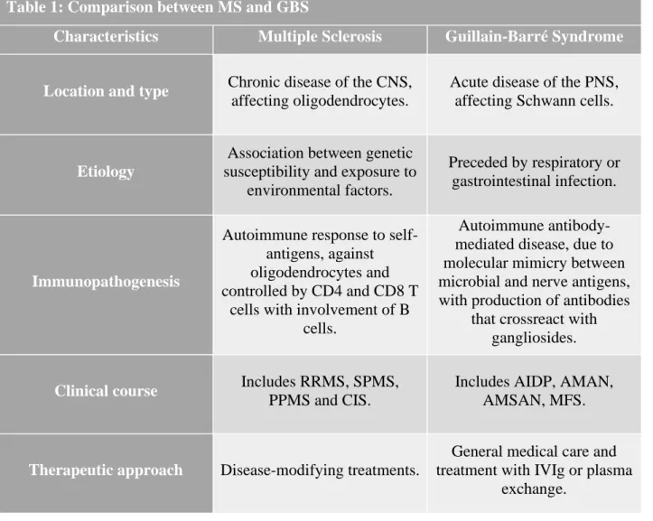
5. Comparison between Multiple Sclerosis and Guillain-Barré syndrome
MS and GBS are both complex demyelinating diseases, however MS is a chronic inflammatory disease that affects myelin of the CNS, whereas GBS is an inflammatory disease affecting myelin in the PNS (1,24). They are both characterized by inflammatory lesions and consequent demyelination, leading to axonal damage, neurodegeneration and progressive loss of function (1). The major difference in the demyelination process is the type of supporting cells affected, once that in the CNS there is the destruction of oligodendrocytes and in the PNS Schwann cells are the ones affected by the destruction of myelin (2). A detailed comparison may be observed in table 1.According to the etiology of the diseases, they are still unclear, but it is known that MS is present in susceptible genetic patients that develops the disease after exposure to various environmental factors, for instance EBV and vitamin D (4,10). On the other hand, GBS occurs, in most cases, after a respiratory or gastrointestinal infection, particularly by Campylobacter