FACULDADE DE CIÊNCIAS
DEPARTAMENTO DE QUÍMICA E BIOQUÍMICA
P
RODUCTION OF A
G
ENOMIC
CFTR
C
ONSTRUCT
I
NSERTED INTO A
H
UMAN
A
RTIFICIAL
C
HROMOSOME
(HAC)
AND
C
HARACTERIZATION OF ITS
E
XPRESSION
Carla Susana Rodrigues Braz
DOUTORAMENTO EM BIOQUÍMICA (Genética Molecular
)
FACULDADE DE CIÊNCIAS
DEPARTAMENTO DE QUÍMICA E BIOQUÍMICA
P
RODUCTION OF A
G
ENOMIC
CFTR
C
ONSTRUCT
I
NSERTED INTO A
H
UMAN
A
RTIFICIAL
C
HROMOSOME
(HAC)
AND
C
HARACTERIZATION OF ITS
E
XPRESSION
Carla Susana Rodrigues Braz
DOUTORAMENTO EM BIOQUÍMICA (Genética Molecular
)
Tese orientada pela Prof. Doutora Margarida D. Amaral
Carla Susana Rodrigues Braz foi bolseira de
Doutoramento da Fundação para a Ciência e Tecnologia do
Ministério da Ciência, Tecnologia e Ensino Superior
SFRH/BD/17912/2004
Graduados da Universidade de Lisboa, Deliberação nº 1506/2006, publicada no Diário da república, 2.ª série — N.º 209 — 30 de Outubro de 2006, foram utilizados nesta dissertação resultados do seguinte artigo:
Rocchi L*, Braz C*, Cattani S, Ramalho A, Christan S, Edlinger M, Ascenzioni F, Laner A, Kraner S, Amaral MD, Schindelhauer D (2010). Escherichia coli-cloned CFTR loci relevant for human artificial chromosome therapy. Hum Gene Ther. Jul 8. [Epub ahead of print]
* Joint first authorship
The manuscript is reproduced here with alterations. Data generated by other authors were included here (with permission) for clearer understanding, and this is acknowledged in the respective figure.
No cumprimento do disposto na referida deliberação, esclareço serem da minha responsabilidade a execução das experiências que estiveram na base dos resultados apresentados (excepto quando referido em contrário), bem como a sua interpretação, discussão e redacção.
T
ABLE OFC
ONTENTSPREFACE...X
ACKNOWLEDGEMENTS/AGRADECIMENTOS...XII
SUMMARY...XV
RESUMO...XVI
ABBREVIATIONS...XXIII
CHAPTER I.GENERAL INTRODUCTION... 3
I.OVERVIEW OF CYSTIC FIBROSIS (CF) AND THE CFTR GENE AND PROTEIN... 3
I.1.CYSTIC FIBROSIS... 3
I.1.1HISTORICAL BACKGROUND... 3
I.1.2DESCRIPTION OF THE DISEASE... 4
I.2THE CFTR GENE... 5
I.2.1THE CFTR GENE PROMOTER AND TISSUE-SPECIFIC CFTR EXPRESSION... 5
I.3 THE CFTR PROTEIN... 8
I.3.1CFTR: A MULTIDOMAIN MEMBRANE PROTEIN... 9
I.3.1.1THE ABC SUPERFAMILY... 9
I.3.2MOLECULAR FUNCTION... 10
I.4 CFTR FUNCTION AS A CHLORIDE/BICARBONATE CHANNEL... 10
I.5 OTHER CFTR FUNCTIONS –ONE CHANNEL TO RULE THEM ALL... 11
I.5.1CFTR AS A REGULATOR OF OTHER CHANNELS AND TRANSPORTERS... 11
I.5.2CFTR AS A PUTATIVE PSEUDOMONAS AERUGINOSA RECEPTOR... 13
I.6 CFTR MUTATIONS... 14
I.6.1F508DEL-CFTR ... 15
I.6.2OTHER CFTR MUTATIONS... 16
I.7 PHARMACOLOGICAL APPROACHES... 16
II.GENE THERAPY AND HUMAN ARTIFICIAL CHROMOSOMES (HACS)... 19
II.1GENE THERAPY FOR CF... 19
II.2 GENE DELIVERY SYSTEMS (GDS) ... 21
II.2.1VIRAL VECTORS... 23
II.2.2SYNTHETIC VECTOR SYSTEMS... 24
II.2.3.1YACS... 27
II.2.3.2BACS AND PACS... 28
II.2.4HUMAN ARTIFICIAL CHROMOSOMES (HACS) ... 29
II.2.4.1THE CENTROMERE... 32
OBJECTIVES... 34
CHAPTER II.RESULTS... 37
1.SUMMARY... 37
2.INTRODUCTION... 37
3.RESULTS... 39
3.1 CONSTRUCTION OF THE FUSION PACCF225 FROM CHARACTERIZED RESOURCE CLONES... 39
3.2 ANALYSIS OF THE CLONING STABILITY OF CF225 ... 43
3.3 STRUCTURAL ANALYSIS OF CLONED CFTR LOCUS... 44
3.3.1LR-PCR ... 44
3.3.2END SEQUENCING OF LOCUS CF225 ... 45
3.4 CO-TRANSFECTION EXPERIMENTS... 46
CHAPTER III.OVERALL DISCUSSION... 53
CHAPTER IV.MATERIALS AND METHODS... 59
1.PAC CLONES COVERING THE CFTR LOCUS... 59
2.E. COLI GROWTH AND AGAROSE PLUG PREPARATION... 59
3.PREPARATION OF INTACT DNA... 60
4.CONSTRUCTION OF CF225 ... 63
4.1 IN GEL RESTRICTION DIGESTION AND DEPHOSPHORYLATION... 63
4.2 PFGE SEPARATION AND DNA ISOLATION... 63
4.3 LIGATION AND ELECTROPORATION... 63
4.4 E. COLI SCREENING... 64
5.VERY LONG PCR... 64
6.GENERATION OF STABLE HT1080 LINES... 65
6.1 CELL CULTURE... 65
6.2LIPOFECTION... 65
6.3 CLONE EXPANSION AND ISOLATION... 65
7.EXPRESSION ANALYSIS AND SEQUENCING... 66
7.1 RT-PCR... 66
CHAPTER V.FUTURE DIRECTIONS... 71
P
REFACECystic fibrosis (CF), the most lethal genetic disease among caucasians is caused by mutations in the CFTR gene, which encodes the cystic fibrosis transmembrane conductance regulator (CFTR) protein. This makes CF a monogenetic disease and simultaneously a good candidate for gene therapy.
The focus of my research was not the CFTR protein but the gene that underlies it and the assembly of a human artificial chromosome (HAC) that could be used as a gene therapy tool for CF. Like the protein it encodes, the CFTR gene is not an easy one. The gene promoter has no TATA box and resembles, in its characteristics, a house keeping gene. However, it is regulated both temporally and spatially, an indication of complexity. Definitely, that gene and its regulatory sequences still have many secrets for scientists to uncover.
Let us now speak of gene therapy for CF, the main cause behind this work. Gene therapy is a science field still in its infancy, where scientists are groping their way in search of answers, it is a path full of difficulties. At the same time, anyone who believes seriously in gene therapy knows that quitting is out of the question, as the ones who are working in this field right now are aware that they are laying the foundations for a future cure of CF. As is mentioned in the General Introduction to this thesis, the CF disorder can be caused by a great variability of different mutations (the CF data base records over 1700 mutations and the number keeps rising), so it is going to be difficult to find a drug that is able to provide cure (and not just an alleviation of symptoms) for all cases of disease. Gene therapy, which implies the delivery of a normal copy of the CFTR gene to affected cells, on the other hand, is a treatment independent of any specific mutation, which makes it an all encompassing tool against the multimutation, life-shortening CF disease. However, the target of CF gene therapy, the airway epithelium, is well protected against foreign invaders (which may include vectors carrying therapeutic genes) by the mucociliary clearance system and the glycocalyx. Although these barriers are somehow weakened in CF patients, due to the characteristics of the disease, the delivery of a transgene by a gene therapy vector is further hindered, in the case of the CF lung, by the presence of a thick, dehydrated mucus inherent to the disease.
Yes, there is a huge amount of hard work ahead of gene therapy researchers but as I said above we simply cannot quit because more than just a research field, gene therapy is a question of humanity. This work is a humble step towards the
advancement of gene therapy and human artificial chromosomes and its parts shall be described below.
Chapter I is divided into two main sections. In section I an overview of CF and the CFTR gene and protein is given. Starting with an historical background regarding the identification of the CF disease, we then make a description of the disorder and its underlying gene and protein. We describe briefly CFTR protein function and how it regulates other channels and transporters, like a lord of the channels. Next for this multifunctional protein its proposed role as a putative Pseudomonas aeruginosa receptor is mentioned. A description of CFTR mutations, namely the most prevalent one, F508del, follows. Finally, an account of the pharmacological approaches towards a treatment of CF is given. Section II provides a small description of gene therapy for CF and gene delivery systems, including both viral and non viral vectors. Chapter I finishes by alluding to the centromere as an essential component in any human artificial chromosome.
Chapter II includes the results. A thorough description of the assembly of the fusion PAC CF225, carrying a reconstructed CFTR locus, followed by the analysis of its stability is made. Next comes the precise identification of the genomic insert/vector junctions as well as the two deletions in introns 9 and 10, which led to the subsequent determination of the length of the reconstructed locus. The results of co-transfecting a lung sarcoma cell line with CF225 and the TTE1 construct carrying a selectable marker and centromere to form a de novo human artificial chromosome (HAC) are described at the end of the chapter.
In Chapter III an overall discussion of this work is given. Chapter IV gives a description of the materials and methods used in this study. In Chapter V preliminary assays to construct a pre-fabricated HAC are mentioned and strategies for the continuation of the work are suggested. A pre-fabricated HAC carrying a copy of the CFTR gene and its regulatory sequences is essential to attain a CF cure through gene therapy.
A
CKNOWLEDGEMENTS/A
GRADECIMENTOSChegar a esta etapa implicou percorrer um caminho que nem sempre foi fácil e eu gostaria de agradecer aqui a todos aqueles que, directa ou indirectamente, possibilitaram a realização deste trabalho, nomeadamente:
À minha orientadora, a Prof. Doutora Margarida Amaral, agradeço muito especialmente ter apostado em mim e todo o apoio prestado, bem como os conselhos dados no sentido da melhor prossecução do estudo que é aqui apresentado e a possibilidade de ter desenvolvido parte do trabalho na Alemanha. O apoio e incentivo da Prof. Margarida foram cruciais para a conclusão deste trabalho e por isso lhe estarei sempre grata.
Ao Instituto Nacional de Saúde Dr. Ricardo Jorge (INSA), o bom acolhimento e as condições disponibilizadas, com especial destaque para as pessoas do seu Presidente, Prof. Doutor José Pereira Miguel e do responsável pela Unidade de Investigação e Desenvolvimento do Departamento de Genética, Dr. João Lavinha, por me terem aceite neste Instituto, onde realizei parte do meu trabalho experimental.
To Dr. Dirk Schindelhauer (then at Livestock Biotechnology, Technische Universität München (TUM), Freising, Germany) for his collaboration which allowed me to work in this exciting field of gene therapy and human artificial chromosomes. I would also like to thank Dr. Schindelhauer for all his support and help, as well as his teachings and for reviewing part of this dissertation.
To all the people at the Livestock Biotechnology, especially Dr. Simone Kraner, who supervised part of my work, and research assistants Sulith Christan and Marlene Edlinger for all their support and friendship. I must also thank Prof. Dr. Angelika Schnieke for her welcome, for allowing me to perform benchwork at her Chair and for her indispensable support. I absolutely loved my stay in Germany and I owe it to these persons that I shall never forget. Friendship has no frontiers.
À Fundação para a Ciência e Tecnologia (FCT), por me ter concedido uma Bolsa de Doutoramento (SFRH/BD/17912/2004), sem a qual este projecto não teria sido possível.
Ao Centro de Investigação BioFIG (Center for Biodiversity, Functional & Integrative Genomics), pelo financiamento de parte deste trabalho.
A todos os meus colegas do grupo de “Membrane Protein Disorders” do BioFIG por toda a sua simpatia e apoio, inclusive nos piores momentos. Tenho que referir aqui os nomes da Anabela Ramalho Venâncio, Ana Carina da Paula, Marisa Sousa e Inna Uliyakina por terem sido as pessoas que estiveram comigo no INSA e com quem convivi mais de perto após o meu regresso da Alemanha. Todas elas foram impecáveis, a Anabela carinhosa e com um sorriso caloroso, a Ana Carina de sorriso radiante, a Marisa é uma força da natureza e a Inna é uma pessoa extremamente simpática e atenciosa. Todas criaram um excelente ambiente de trabalho no laboratório e transmitiram-me energia quando eu dela precisava. Não posso deixar de referir o Luka Clarke, com quem tenho contactado desde a minha entrada para o grupo e a quem agradeço a revisão da Tese e toda a sua ajuda com a língua inglesa. Para encerrar este parágrafo, tenho que mencionar novamente a Anabela, a pessoa que acompanhou o meu trabalho no INSA e que também reviu parte desta Tese. Toda a sua ajuda, conselhos, ideias, incentivo e o tempo que me concedeu são de valor inestimável. O meu vocabulário é pobre para expressar todo o meu agradecimento para com a Anabela.
To Susan Chang, whom I met at the Livestock Biotechnology, for being a companion and also for all her support, friendliness and incentive by sharing with me her own life experience.
À Sónia Pedro e à Ana Cardoso, as “meninas do sequenciador” pela sua colaboração e ajuda técnica na obtenção dos resultados das sequenciações, efectuadas num momento crítico do meu estudo.
Ao Prof. Doutor Rogério Tenreiro pela sua colaboração, simpatia e por me ter permitindo a realização de experiências no seu laboratório e à Prof. Doutora Lélia Chambel por toda a sua simpatia, ajuda e esclarecimentos relativos à técnica de PFGE.
A todos os meus amigos do peito, com quem posso sempre contar, por todo o seu apoio ao longo dos anos e paciência infinita durante este período da minha vida.
Aos meus pais, Antónia e José, por estarem sempre do meu lado, incentivando-me com o amor que só os pais sabem ter.
Finalmente, ao Nuno, o meu companheiro de todos os dias, pelo seu amor e carinho. Pude sempre contar com o seu apoio inabalável, a sua presença na minha vida é uma luz na escuridão.
S
UMMARYCystic Fibrosis is an autosomal recessive disorder, which makes it a good candidate for gene therapy. Thus cystic fibrosis became one of the first targets for gene therapy since apparently it is sufficient to deliver a normal copy of the gene encoding the CFTR protein to the affected cells.
Although promising at first, classical gene therapy to cystic fibrosis has not met expectations due to immune response against viral vectors and synthetic vectors, as well as short-term expression of cDNA based transgenes. These hurdles can be overcome by delivering the complete genomic CFTR gene on non-integrating human artificial chromosomes (HACs). Here, we describe the reconstruction of the genomic
CFTR locus into one P1-based artificial chromosome (PAC), CF225. This is a
non-selectable PAC of 225.3 kb (running from -60.7 kb to +9.8 kb) which resulted from the ligation of two PACs, CF1-Met and CF6, with an optimized M470V codon and a silent XmaI restriction variant to discriminate transgene from endogenous expression. CF225 was shown to be stably maintained and propagated in the E. coli DH10B host. After co-transfection of CF225 with the telomerized, blasticidin-S selectable, centromere-proficient alpha satellite (cen 5) TTE1 construct into HT1080 fibrosarcoma lung cells, CF225 was not incorporated into a de novo HAC in 122 lines analyzed, but five integrants formed, four of which expressed the transgene, as detected by RT-PCR and XmaI restriction analyses. Stability analyses suggest feasibility to pre-fabricate a large, tagged CFTR transgene that stably replicates in the proximity of a functional centromere. Although definite conclusions about HAC proficient construct configurations cannot be drawn at this stage, an important transfer resource was generated and characterized, demonstrating promise of de
novo HACs as potentially ideal gene-therapy vector systems.
Keywords: Cystic Fibrosis, CFTR, Gene Therapy, Human Artificial Chromosome
R
ESUMOA terapia génica pode ser definida como a introdução de um gene exógeno numa célula receptora para a obtenção de benefícios terapêuticos. Os objectivos a longo prazo dos estudos relacionados com a terapia génica são o desenvolvimento de vectores como ferramentas para o estudo do genoma e da função cromossómica e para transferir genes para as células com fins terapêuticos.
A Fibrose Quística é uma doença autossómica recessiva, o que implica que uma única cópia normal do gene CFTR, que codifica a proteína do mesmo nome, é suficiente para restaurar a função CFTR de canal de cloreto ausente em caso de mutação em ambos os alelos CFTR. Estas características tornam a Fibrose Quística um candidato atraente para a terapia génica, visto bastar transferir uma cópia do gene normal para as células afectadas para se obter a cura da doença. Assim, pouco tempo após a descoberta do gene CFTR, em 1989, tiveram início os primeiros esforços no sentido de tornar a terapia génica da Fibrose Quística uma realidade.
Contudo, a terapia génica da Fibrose Quística tem como requisitos que o gene terapêutico deva ser expresso em todas as células epiteliais onde normalmente ocorre expressão, de preferência a níveis comparáveis aos do gene endógeno. Além disso, a expressão deverá ser estável e persistente, a fim de ser evitada a readministração repetida do gene terapêutico.
A maioria dos vectores utilizados actualmente em terapia génica consiste em cassetes de expressão controlada por promotores heterólogos fortes, frequentemente derivados de vírus. Contudo, descobriu-se que muitos destes transgenes são expressos apenas durante um período de tempo limitado. A capacidade dos vírus para infectar as vias respiratórias fez deles a escolha natural inicial para a terapia génica da Fibrose Quística e muitos dos primeiros estudos foram efectuados com vectores virais derivados de adenovírus.
A expressão do gene CFTR é regulada espaciotemporalmente, pelo que a sua estabilidade a longo prazo e expressão regulada de forma tecido-específica requerem não só o promotor e as porções codificantes do gene como também elementos cromossómicos reguladores, tais como os estimuladores e silenciadores, associados aos locais hipersensíveis à DNase I (DHS, na terminologia inglesa). Estes requisitos exigem a utilização de vectores de grande capacidade, como é o caso de vectores cromossómicos, tais como os cromossomas artificiais bacterianos
(BACs), os cromossomas artificiais baseados no fago P1 (PACs) e os cromossomas artificias humanos (HACs).
Há duas vantagens essenciais dos sistemas baseados em vectores cromossómicos em relação à maior parte dos vectores convencionais usados para transferência génica. Em primeiro lugar, o DNA transferido pode ser mantido estavelmente sem os riscos associados à inserção e, em segundo lugar, podem ser introduzidos grandes segmentos de DNA englobando os genes e os seus elementos reguladores, conduzindo à expressão do transgene mais fiável e fisiológica, mais próxima da do gene normal. Além disso, os vectores cromossómicos constituídos apenas por DNA humano não deverão ser imunogénicos.
Tratando-se da Fibrose Quística, o epitélio das vias respiratórias é o alvo mais importante, uma vez que a doença pulmonar é a que contribui principalmente para a morbilidade e a mortalidade. Dado que esta doença, pelo menos nas etapas iniciais, afecta essencialmente as vias respiratórias inferiores, é provável que as células epiteliais destas vias sejam importantes, sendo consideradas por muitos autores como as células alvo apropriadas para a terapia génica da Fibrose Quística.
Após a produção do constructo genómico CGT21 que contém aproximadamente metade do locus do gene CFTR e o último exão e da demonstração de que ele era propagado estavelmente em células de sarcoma do pulmão, onde era expresso e sofria splicing correcto (Laner et al., 2005), o passo lógico seguinte era gerar um constructo CFTR genómico portador dos 27 exões e das sequências reguladoras flanqueantes para incorporação num HAC.
O objectivo consistia na reconstituição do gene CFTR completo clonado num PAC [o PAC usado neste trabalho foi o pCYPAC2 (Ioannou et al., 1994)] adequado para a preparação em grande escala de DNA de elevada qualidade. Nós descrevemos aqui a construção de um locus CFTR contendo os 27 exões e a maior parte das potenciais regiões reguladoras num só CF PAC. Partindo de preparações de DNA armazenadas em plugs de agarose e previamente caracterizadas (Ramalho et al., 2004), contendo parte do gene CFTR em PACs, nós construímos o PAC CF225, portador do gene CFTR humano com todos os exões e intrões mais sequências reguladoras, ligando dois PACs, CF1-Met e CF6, contendo cada um aproximadamente metade do gene CFTR e regiões flanqueantes: o PAC CF1-Met é portador de um inserto que vai de -60,7 kb a montante do início da tradução no exão 1 até ao intrão 10, e o PAC CF6 é portador de um inserto que vai do intrão 10 até +9,8 kb a jusante (relativamente ao fim da tradução). Devido à escolha dos PACs originais, os intrões 9 e 10 estão substancialmente encurtados em 5,1 kb e 27,1 kb, respectivamente. Como consequência, os locais hipersensíveis à DNase I (DHS)
descritos no intrão 10 (McCarthy and Harris, 2005) estão excluídos do locus CF225. Contudo, este clone tem a vantagem de conter o codão da metionina (M) optimizado no locus polimórfico M470V, em comparação com os clones wild type, e uma variante de um local de restrição XmaI silencioso sintético, o qual é adequado para discriminar entre os produtos de RT-PCR do transgene e os provenientes dos loci
CFTR endógenos de qualquer célula alvo.
A fim de concretizar um dos objectivos do presente trabalho, partimos de três clones PAC, CF1-Met10-43/-44/-54, cujo exão 10 tinha sido modificado para conter metionina (M) em lugar da valina (V) na posição 470, bem como um local de restrição para a enzima XmaI, introduzido por uma mutação silenciosa. O conteúdo destes clones foi sujeito a diferentes análises antes da construção de CF225: 1) amplificações por PCR cujos produtos abarcavam toda a região do exão 10 clonada bem como sequências wild-type localizadas a montante e a jusante, as quais mostraram que os tamanhos dos produtos de PCR estavam de acordo com o esperado nos três clones; 2) sequenciação do exão 10 clonado, que confirmou a orientação correcta, a presença do polimorfismo corrigido V470M e que a sequência nos três clones não possuía qualquer mutação derivada de PCR; 3) análise de restrição, tendo revelado que as bandas esperadas correspondiam aos locais de restrição sintéticos XmaI e NotI; 4) digestão de 1/10 de uma plug de agarose dos três clones CF1-Met com as enzimas de restrição NotI e/ou BssHII também mostrou os tamanhos esperados para os fragmentos obtidos; 5) os tamanhos esperados para as bandas foram demonstrados para os três clones por reacções de PCR de longo alcance (LR-PCR, segundo a terminologia inglesa) de baixo número de ciclos, cobrindo toda a sequência CFTR clonada nos PACs CF1-Met, incluindo a redução de 5,1 kb no intrão 9. Estes dados indicam que os clones continham o inserto completo sem rearranjos. Como a banda de 31,8 kb do subclone CF1-Met10-43 era muito fraca em comparação com os produtos dos outros subclones bacterianos, indicando possivelmente uma alteração em algumas das bactérias usadas na preparação das plugs, o clone correspondente não foi mais usado.
Depois de confirmar que a sequência CFTR clonada no PAC CF1-Met tinha a mesma estrutura que a clonada no PAC CF1 original, o passo seguinte era fundir o PAC CF1-Met com o inserto do PAC CF6, o qual é portador do resto da sequência genómica de CFTR com as sequências dos exões e das junções exão/intrão correctas. A estratégia de clonagem foi a seguinte: 1) digestão parcial do PAC CF1-Met (clone 44) com NotI, digestão total de CF6 com NotI seguido por desfosforilação para impedir a recircularização do vector; 2) separação dos produtos de digestão por electroforese em gel de campo pulsado (PFGE, de acordo com a terminologia
inglesa) e excisão das bandas do gel sem exposição a UV; 3) electroeluição dos fragmentos de DNA existentes nos pedaços do gel e sua mistura numa razão de ~1/1 (CF1-Met/CF6); ligação do DNA pela ligase T4; 5) electroporação do produto de ligação em E. coli DH10B; 6) selecção, por PCR, das colónias resistentes à kanamicina com os primers CFi10fus/R7, específicos da região de fusão entre os dois PACs. Foi identificado um clone, mais tarde designado por CF225, positivo para a reacção de PCR relativa à região de fusão e para STSs dos exões 4 e 12, presentes nos dois PACs que foram ligados.
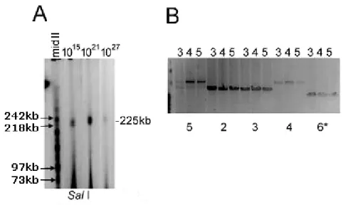
Após a identificação do clone positivo, o passo seguinte deveria ser a análise da sua estabilidade enquanto clone. Assim, durante o crescimento inicial da cultura mãe das bactérias portadoras do PAC CF225, foram plaqueados doze subclones individuais e analisados por PCR para avaliação da estabilidade da clonagem. Nove dos doze subclones continham os oito STSs testados por PCR e que cobriam o locus em diferentes posições ao longo do gene CFTR, incluindo sequências localizadas a montante do início da transcrição, a região da fusão entre os dois PACs e a região poli (A). Para analisar mais detalhadamente a estabilidade de clonagem, três culturas derivadas de clones individuais do PAC CF225 foram crescidas durante vários períodos de tempo, simulando um rendimento final potencial de 1015, 1021 e 1027 células de E. coli, contendo cada uma até ~10 cópias totalmente replicadas do constructo. A digestão do material contido em plugs de agarose, proveniente das colónias derivadas dos três subclones individuais, mostrou a presença, em todos os casos, de um inserto com o tamanho de 225 kb (SalI) e fragmentos idênticos e com os tamanhos previstos (BssHII), indicando uma estabilidade de clonagem global elevada para o locus. Os dados indicavam também a propagação fiel e estável do locus CFTR no PAC CF225 (inserto de 225,3 kb), demonstrando que o gene CFTR pode ser clonado de forma estável em E. coli.
Ainda continuando a análise estrutural do constructo, seguiu-se a sequenciação das suas extremidades, em que o inserto CFTR se liga ao vector. Assim, para determinar os extremos de CF225, foram efectuadas reacções de LR-PCR com primers que se ligam nas extremidades 5’ e 3’ da junção inserto/vector. Os fragmentos amplificados foram sequenciados e comparados por meio do programa BLAST com o hg built 37.1 no site NCBI. O locus CFTR reconstruído vai do nucleótido -60651 relativamente ao início da tradução ao nucleótido +9767 relativamente ao final da tradução. Ambas as extremidades coincidem com um local de restrição para Sau3AI, o que está de acordo com o facto de, para a preparação da biblioteca RPCIP704, para a qual foi utilizado o PAC pCYPAC2, que serviu de
fonte para a construção de CF225, o DNA genómico ter sido parcialmente digerido e clonado no local BamHI do vector.
Como resultado do procedimento da clonagem, CF225 tem duas deleções nos intrões 9 e 10 as quais representam regiões que não estavam incluídas nos PACs originais CF1 (intrão 9) e CF6 (intrão 10) e que foram omitidas pela reconstrução do exão 10 e das suas sequências intrónicas flanqueantes. Para localizar com precisão e determinar a extensão daquelas deleções, foram realizadas reacções de PCR com primers que hibridam em regiões que flanqueiam as duas deleções. Os produtos de PCR foram sequenciados e comparados por meio do programa BLAST com BACs portadores de sequências genómicas humanas publicadas. Ambas as deleções foram localizadas com precisão, tendo a deleção no intrão 9 5058 pb, ao passo que a deleção no intrão 10 tem 27128 pb.
Para conseguir a incorporação do locus CFTR num cromossoma humano artificial (HAC, na terminologia inglesa) formado de novo, foram efectuadas experiências de co-transfecção do locus CFTR clonado com um constructo linearizado portador de sequência de DNA alfa-satélite do centrómero e a expressão dos clones celulares obtidos analisada. Quatro ensaios independentes de co-lipofecção do inserto de 225 kb do PAC CF225 com o constructo TTE1 (fragmento de 133 kb) contendo um gene marcador de resistência à blasticidina S (BS) duplicado e o marcador EGFP, bem como sequências centroméricas, resultaram em 185 clones celulares, 122 dos quais foram expandidos e analisados por PCR com primers específicos para CF225. Cinco clones celulares individuais, BW24, BG32, CG13, DG27 e DG5 eram positivos para a reacção de PCR específica, indicando que apenas 1 em ~25 clones foram co-transfectados em simultâneo com CF225 e TTE1.
Foram realizadas reacções de RT-PCR com primers que geram um produto entre os exões 8 e 10 que, após splicing, tem 391 pb e representa uma mistura de produtos dos genes CFTR endógenos da linha celular HT1080 e do locus transgénico. Como resultado destas experiências, verificou-se que quatro (BG32, CG13, DG27 e DG5) das cinco linhas co-transfectadas expressavam o locus transgénico. Todas as linhas que expressavam evidenciavam níveis variáveis de expressão CFTR após 30 dias de crescimento sem selecção. Para distinguir entre a expressão endógena e a do transgene, os produtos de RT-PCR foram clivados com
XmaI que digere o exão 10 modificado de CF225 em dois fragmentos de 310 pb e
81 pb. Nas quatro linhas celulares, proporções variáveis do transcripto CFTR resultavam do transgene, o que demonstrava, em muitos casos, níveis de expressão do transgene acima dos dos genes endógenos (cujos produtos de RT-PCR não
eram clivados por XmaI) e mostravam a ocorrência de splicing correcto. As linhas celulares que expressavam e as células HT1080 parentais foram analisadas por sequenciação dos produtos de RT-PCR obtidos com os mesmos primers e também com o primer CFc3F (exão 3), demonstrando que todas as linhas continham tanto o polimorfismo 470M como a variante sintética XmaI no exão 10, confirmando que tinham origem no transgene.
Para verificar a integridade do constructo CF225 nas linhas celulares clonais, foram realizadas reacções de PCR com primers para a junção vector/CFTR a 5’ e para a junção vector/CFTR a 3’. Das cinco linhas celulares derivadas de HT1080 apenas DG27 manteve a extremidade 5’ de CF225, confirmada pela sequenciação do produto de PCR. Todas as 5 linhas foram negativas para a reacção de PCR relativa à extremidade 3’, bem como para duas outras reacções de LR-PCR que abrangiam aproximadamente 2 kb e 3 kb desde a extremidade 3’ do vector até ao locus CFTR, sugerindo que o DNA de CF225 a 3’ foi perdido em todas as quatro linhas que expressavam.
A fim de averiguar se se tinha formado um cromossoma de novo ou se havia ocorrido integração no genoma da célula, foram efectuados ensaios de FISH de tripla cor nestas linhas celulares após 30 dias de crescimento com e sem selecção por BS. Estas análises revelaram ou integração do locus CF225 num cromossoma do hospedeiro ou integração e truncação em todas as cinco linhas celulares clonais. Não se observaram HACs portadores do locus CFTR. A linha clonal DG27 mostrou co-integração estável próximo do gene CFTR endógeno no cromossoma 7. A linha BG32 revelou uma integração distal/telomérica num cromossoma que não o 7. A linha celular DG5 mostrou a integração de sinais de CF1, CF6 e E1 (centrómero) numa posição distal do cr19q e a linha CG13 mostrou integração de porções de CF1 e CF6 no braço p de um cromossoma metacêntrico que não o 7, acompanhada por truncação. Na linha celular BW24 apenas foram detectadas sequências de CF6 num pequeno cromossoma truncado. No geral, podemos concluir que CF225 e o centrómero E1 não formaram eficientemente em conjunto uma estrutura de replicação estável. Em vez disso, foram seleccionados raros clones estáveis que continham pelo menos o marcador BS e várias porções do locus CF225 sem a extremidade 3’, que todavia mostraram expressão e splicing correcto da sequência do exão 10 em 4 das 5 linhas obtidas.
No presente trabalho foram também feitas tentativas de construção de um HAC
de novo (pré-fabricado) por uma abordagem de recombinação in vitro por meio de
digestão enzimática e ligação, seguida de electroporação em E. coli, de um constructo portador do locus CF225 e de TTE1, contendo sequências de DNA
centromérico. Escolheu-se uma abordagem de recombinação in vitro para a construção porque esta técnica é mais reprodutível e menos propensa a rearranjos do que as abordagens de recombinação in vivo. Não foi possível obter o HAC pré-fabricado e é sugerida uma estratégia para uma futura obtenção do mesmo. A produção de um HAC de novo, com todas as vantagens que este tem sobre os vectores virais, é muito importante para o desenvolvimento de uma terapia génica de sucesso para o tratamento da Fibrose Quística. Este trabalho representa mais um passo em frente no sentido de tornar a terapia génica para a Fibrose Quística uma realidade e os resultados obtidos aqui deixam antever a possibilidade de criação de um cromossoma artificial humano portador do gene CFTR e respectivas sequências reguladoras o qual representaria um vector ideal e uma promessa de cura para a Fibrose Quística, independentemente da mutação causadora de doença.
Palavras-chave: Fibrose Quística, CFTR, Terapia Génica, Cromossoma Artificial
A
BBREVIATIONSA Adenine (base) residue; alanine (amino acid) residue
aa Amino acid
AAV Adeno-associated virus
ABC ATP-binding cassette
ACH Active chromatin hub
Ad Adenovirus
ASL Airway surface liquid
ATP Adenosine-5’- triphosphate
BAC Bacterial artificial chromosome
bp Base pairs
BS Blasticidin S
C Cytosine residue
C-terminus Carboxyl terminus
cAMP Cyclic adenosine 3’,5’-monophosphate
cDNA mRNA-complementary DNA
cen Centromere
CENP-A Centromere protein A
CENP-B Centromere protein B
CF Cystic fibrosis
CF HAE Human CF ciliated surface airway epithelium
CFTR Cystic Fibrosis Transmembrane Conductance Regulator
CIP Calf intestinal phosphatase
CTCF CCCTC-binding factor
D Aspartic acid residue
DEAC Diethylaminocoumarine
del Deletion
DHS DNase I-hypersensitive site
DMEM Dulbecco’s modified Eagle Medium
DNA Deoxyribonucleic acid
dNTP Deoxynucleotide Triphosphate
dUTP Deoxyuridine triphosphate
EBNA1 Epstein-Barr virus nuclear antigen 1
EBV Epstein-Barr virus
EDTA Ethylenediamine tetraacetic acid
ENaC Epithelial sodium channel
ER Endoplasmic reticulum
F Phenylalanine residue
FCS Fetal calf serum
FISH Fluorescence in situ hybridization
FITC Fluorescein isothiocyanate
G Guanine (base) residue; Glycine (amino acid) residue
GCH1 Guanosine triphosphate cyclohydrolase 1
GDS Gene delivery systems
HAC Human artificial chromosome
HAE Human airway epithelium
HPRT Hypoxanthine guanine phosphoribosyltransferase
HSP Heparan sulfate proteoglycan
HSV Herpes simplex virus
I Isoleucine residue
IBMX 3-isobutyl-1-methylxanthine
IPTG Isopropyl β-D-1-thiogalactopyranoside
K Lysine residue
kb Kilobase (1000 base pairs)
LB Luria Bertani medium
LINE Long interspersed element
LMP Low melting point
LPS Lipopolysaccharide
LR-PCR Long range-PCR
M Methionine residue
MC Minichromosome
MMCT Microcell-mediated chromosome transfer
MSD1 and MSD2 Membrane spanning domain 1 and 2
N Amino terminus; asparagine residue
NBD1 and NBD2 Nucleotide binding domain 1 and 2
NHERF-1/-2 Na+/H+ exchanger regulatory factor isoform-1/-2
NPD Nasal potential difference
ORCC Outwardly rectifying Cl- channel oriP Epstein-Barr virus origin of replication
P Proline residue
PBS Phosphate buffered saline
PCR Polymerase chain reaction
PEG Polyethylene glycol
PEI Polyethylenimine
PFGE Pulsed field gel electrophoresis
PGK Phosphoglycerine kinase
PIV Human parainfluenza virus
PKA Protein kinase A
PKC Protein kinase C
PLGA Poly lactic-co-glycolic acid
RD Regulatory domain
RNA Ribonucleic acid
ROMK Renal outer medullary K+ channel
RT-PCT Reverse transcription polymerase chain reaction
SDS Sodium dodecyl sulfate
SeV Sendai virus
SINE Short interspersed element
Sp1 Specificity protein 1
STS Sequence-Tagged Sites
T Thymidine (base) residue; threonine residue (amino acid)
TAE Tris/acetate/EDTA buffer
Taq Thermus aquaticus
Tris Tris(hydroxymethyl)aminomethane
TM Transmembrane segments
UTR Untranslated region
UV Ultraviolet
V Valine residue
W Tryptophan residue
wt Wild-type
X Any amino acid residue
C
HAPTER
I
C
HAPTERI.
G
ENERALI
NTRODUCTIONI.OVERVIEW OF CYSTIC FIBROSIS (CF) AND THE CFTR GENE AND PROTEIN
I.1.CYSTIC FIBROSIS
I.1.1HISTORICAL BACKGROUND
Cystic fibrosis (CF) was first recognized as a separate disease from celiac syndrome in 1938 by Dr. Dorothy Hansine Andersen (1901-1963) based on autopsy studies of malnourished infants, which allowed to distinguish a disease of mucus plugging of the glandular ducts, termed “cystic fibrosis of the pancreas” (Andersen, 1938). This thick, sticky mucus clogging the ducts of mucus glands throughout the body gave rise to the alternative designation “mucoviscidosis” (state of thick mucus) (Farber, 1944).
An important discovery was made during the 1948 heat wave in New York by Dr. Paul di Sant’Agnese (1914-2005) who was the first to recognize that babies with CF were at increased risk for heat prostration. This observation led to his discovery that the sweat is abnormal in patients with CF, presenting a fivefold excess of sodium and chloride, which persisted after the heat wave subsided (di Sant'Agnese et al., 1953). Standardization of the sweat test, which became the primary diagnostic test, in 1959 (and still in use today) allowed identification of milder cases, and CF was found to be not only a disorder of mucus (Davis, 2006).
Investigations proceeded in the CF field and in the 1980’s major breakthroughs were accomplished. Thus, in 1983, Paul Quinton, studying the ducts of sweat glands, identified chloride (Cl-) transport as the basic defect in CF (Quinton, 1983). In another line of investigation, Michael Knowles and Richard Boucher found diminished chloride movement from epithelia into the airway lumen, accompanied by increased sodium reabsorption in the epithelium (Knowles et al., 1983; Boucher et al., 1986).
The discovery of the CF gene by positional cloning, in 1989, resulted from the joint efforts of three research groups, those of Lap-Chee Tsui (Kerem et al., 1989) and Jack Riordan (Riordan et al., 1989) at the Hospital for Sick Children in Toronto, and Francis Collins (Rommens et al., 1989) at the University of Michigan. The discovery of the CF gene led to the demonstration that the impaired chloride transport is due to the failure of a cAMP-regulated Cl- channel. Since then,
substantial progress in basic and clinical research raised the median survival age of CF patients to ~37 years (Cohn et al., 2005; Wang et al., 2005; Davis, 2006). After 1989, CF diagnosis could also be made by direct identification of two mutant alleles, namely for borderline cases detected by the sweat test (Davis, 2006).
I.1.2 DESCRIPTION OF THE DISEASE
CF (MIM no. 219700), the most common life-threatening genetic disease of Caucasians (Welsh et al., 2001) is inherited in a Mendelian autosomal recessive pattern (Andersen and Hodges, 1946). The disease is caused by mutations in the Cystic Fibrosis Transmembrane Conductance Regulator (CFTR; ABCC7). This anion selective ion channel is required for the normal function of epithelia lining the airways, intestinal tract as well as ducts in the pancreas, salivary and sweat glands (Ameen et al., 2007) and the absence of its activity results in the failure of ionic and water homeostasis at exocrine epithelial surfaces (Riordan, 2008).
While practically all exocrine glands are affected in CF, the three main organs of greatest clinical importance are the sweat gland (diagnosis), the pancreas (malnutrition), and the lung (morbidity/mortality) (Quinton, 2007).
Clinically, CF disease is characterized by exocrine pancreatic insufficiency, due to failure of bicarbonate-rich fluid and enzyme secretion which impair intestinal digestion and absorption. CF also causes an increase in sweat NaCl concentration [3-5 times higher than in unaffected subjects (Shwachman and Antonowicz, 1962; di Sant'Agnese and Powell, 1962)] and male infertility. However, the major cause of morbidity and mortality is pulmonary disease due to recurrent bacterial infections. In fact, the pulmonary manifestations of CF are responsible for more than 90% of CF-related mortality (Pilewski and Frizzell, 1999).
Significant reduction (>90%) of functional CFTR in the plasma membrane of airway epithelial cells results in a defect in Cl- secretion, hyperabsorption of sodium and other changes that reduce the capacity of cilia to clear bacteria from the airways (Gibson et al., 2003; Boucher, 2004). In the lung, there is generation of thick and dehydrated mucus and subsequent chronic bacterial infections (mainly by
Pseudomonas aeruginosa) which lead to bronchiectasis.
The loss of CFTR from airway epithelial cells also leads to altered regulation of other ion channels (e.g., ENaC), significant changes in the composition of airway surface liquid (ASL) (Boucher, 2003) and production of pro-inflammatory cytokines
(Terheggen-Lagro et al., 2005; Machen, 2006). The ultimate destruction of the CF lung is mainly due to inflammation (Chmiel and Davis, 2003).
The frequency of the disease varies among ethnic groups and is highest in individuals of Northern European origin, of which about 1 in 2500 newborns is affected. Similarly, in this population the heterozygote frequency reaches the rather remarkable value of about 1 in 25 individuals (Collins, 1992).
In Portugal, the estimated CF incidence is 1 in 6000 newborns (Farrell, 2008). Recently, in one study by Lemos and colleagues (Lemos et al., 2010), the CF incidence in the central region of the country was calculated as 1 in 14000 newborns, i.e., lower than in the country as a whole.
I.2THE CFTR GENE
The CF transmembrane conductance regulator (CFTR) gene is located on the long arm of chromosome 7, at the region 7q31.2 (Kerem et al., 1989;Riordan et al., 1989;Rommens et al., 1989). The gene, and the corresponding mRNA, are both relatively large, spanning ~190 kb and 6129 bp, respectively (Collins, 1992), including 5’ and 3’ untranslated regions (UTR), of which 4443 bases are amino acid coding sequence. The gene consists of a TATA-less promoter and 27 exons (Figure I.1), whose sizes vary greatly from 38 bp (exon 14b) to 724 bp (exon 13) (Zielenski et al., 1991). It comprises 26 introns ranging in size from 600 bp (intron 22) to 28085 bp (intron 10).
I.2.1 THE CFTR GENE PROMOTER AND TISSUE-SPECIFIC CFTR EXPRESSION
As the characteristics of the CFTR gene, namely the elements that drive its expression, are important for gene therapy, a detailed description of the locus will be given here.
Promoter deletion experiments have defined the minimal promoter as -226 to +98 with respect to the transcription start site (+1) at -132 bp 5’ to the first methionine codon (Chou et al., 1991). Position +1, defined by Riordan and co-workers (1989) in their original description of the CFTR cDNA, is located 121 bp upstream of the ATG translation initiation codon and corresponds to position +11 in the numbering system used by Chou and co-workers (1991).
The CFTR gene has different transcription start sites which vary among cell lines expressing it. In one study (Koh et al., 1993) using the numbering system of Riordan and co-workers (1989), it was found that low abundance transcripts initiate at position -32, while the start site + 50 appears to be the major initiation point for
CFTR transcription in highly expressing cell lines.
Figure I.1 Scheme illustrating the CFTR gene, mRNA, and protein. MSD –
membrane-spanning domain (MSDs 1 and 2); NBD – nucleotide-binding domain (NBDs 1 and 2); R – regulatory domain; N – amino terminal; C – carboxyl terminal; aa – amino acid residue. [Adapted from (Zielenski and Tsui, 1995) by MD Amaral].
CFTR exhibits tightly regulated expression, both temporally during development
and spatially in different tissue types (Crawford et al., 1991; Trezise et al., 1993; Broackes-Carter et al., 2002). However, the CFTR promoter resembles that of a housekeeping gene: it is CpG-rich, contains no TATA box, has multiple transcription start sites and has several putative binding sites for the transcription factor Sp1 [Specificity protein 1; (Yoshimura et al., 1991)]. Consistent with promoters of this type, the CFTR promoter is weak and demonstrates no apparent tissue-specificity, suggesting the involvement of distal regulatory elements in control of CFTR expression. These elements are associated with DHS (DNase I hypersensitive sites) within the genomic region encompassing the CFTR locus, both upstream (at -20.9 kb) and downstream of the coding region (at +5.4, +6.8, +7.0,+7.4 and +15.6 kb) and in various introns (Smith et al., 2000; Phylactides et al., 2002).
A cell-type-specific DHS was identified within the first intron of CFTR, at 185 + 10 kb (where 185 is the last base in CFTR exon 1) (Smith et al., 1996), which corresponds to a regulatory element that functions as a classical, tissue-specific enhancer and can also independently recruit general factors necessary for transcription initiation (Smith et al., 1996; Mogayzel, Jr. and Ashlock, 2000; Rowntree et al., 2001; Ott et al., 2009b; Ott et al., 2009c). This element was shown to positively regulate CFTR promoter activity specifically in intestinal cells both in vitro and in vivo (Rowntree et al., 2001).
Blackledge and colleagues (Blackledge et al., 2007) identified two enhancer-blocking insulators, also DHS, located upstream and downstream to the CFTR gene that have distinct properties. The insulator located at -20.9 kb from the CFTR translation start site was associated with a classical CTCF (CCCTC-binding factor)-dependent insulator element. A second element, located 3’ to CFTR, within a DHS at +15.6 kb (with respect to the translational end point) also demonstrated enhancer-blocking activity but this was independent of CTCF binding.
Recently, two works (Blackledge et al., 2009; Ott et al., 2009a) reported structural and functional evidence for a CFTR transcriptional hub in which intronic enhancer elements are brought into close proximity to the CFTR promoter to activate cell-type-specific transcription (Figure I.2). This complex looped structure of the
CFTR locus occurs in cells that express the gene and is absent from cells in which
the gene is inactive. Close interaction of the CFTR promoter with sequences in the middle of the gene about 100 kb from the promoter and with regions 3’ to the locus that are about 200 kb downstream was demonstrated. These interacting regions correspond to prominent DHS within the locus, which recruit proteins that modify chromatin structure (Ott et al., 2009a).
Other features of the CFTR promoter, which may contribute to both the temporal and spatial regulation of gene expression, are the use of multiple transcription start sites for the gene (Yoshimura et al., 1991; Koh et al., 1993) and the recruitment of alternative upstream exons (Broackes-Carter et al., 2002; Mouchel et al., 2003; Lewandowska et al., 2009). Lewandowska and colleagues (Lewandowska et al., 2009) identified a novel cis-acting element that contributes to the activity of the basal CFTR promoter in airway epithelial cells and showed that a combination of epigenetic modifications contribute to the multiple mechanisms regulating the promoter of the CFTR gene.
CFTR exhibits a complex pattern of tissue-specific expression being expressed
at low levels [in normal individuals, CFTR mRNA transcripts are expressed at 1-2 copies per cell (Trapnell et al., 1991)] in specialized epithelial cells of gut, airways,
pancreas, sweat gland ducts and the male reproductive tract (Crawford et al., 1991; Trezise and Buchwald, 1991; Engelhardt et al., 1992; Trezise et al., 1992).
Figure I.2 A looping model for the active CFTR gene. In CFTR-expressing cell types,
elements in the CFTR 3’ flanking region are in close proximity to the CFTR promoter. This 3’ flanking region includes the tissue-specific +6.8 kb DHS, shown here to bind CTCF, as well as other previously described DHS (Nuthall et al., 1999; Blackledge et al., 2007). Protein factors bound at each of these sites interact with the promoter-bound transcription machinery, thus forming an active chromatin hub (ACH) and helping regulate expression of the CFTR gene. Besides the DHS from the 3’ flanking region, intronic DHS such as the intestine-specific intron 1 element and others may also contribute to the CFTR ACH in a tissue-specific manner (Ott et al., 2009b).
In the airways, CFTR expression depends on the cell type: high levels have been found in serous cells of submucosal glands (Engelhardt et al., 1994). A more recent study (Kreda et al., 2005) showed that significant levels of CFTR are found in the apical plasma membrane of all ciliated epithelial cells in the superficial epithelium, and at the apical surface of ciliated cells in submucosal gland ducts.
I.3 THE CFTR PROTEIN
The product of the CFTR gene is a transmembrane glycoprotein of 1480 amino acids (Riordan et al., 1989) that functions as a plasma membrane chloride (Cl-) channel activated by cyclic AMP (cAMP).
CFTR plays an important role in both secretion and reabsorption of ions and fluid at epithelial surfaces, depending on the electrochemical gradient present. To perform this task, i.e., to respond to cAMP-stimulation following phosphorylation by protein kinase A (PKA) and protein kinase C (PKC), it should be correctly localized at the lumen-facing or apical membrane of epithelial cells (Riordan, 1993).
I.3.1 CFTR: A MULTIDOMAIN MEMBRANE PROTEIN
CFTR consists of 2 repeated motifs, each composed of a hydrophobic membrane spanning domain (MSD1 and MSD2) containing six helices [transmembrane segments (TM)] each which compose the core structure of the pore (Tabcharani et al., 1991), and a cytosolic hydrophilic region for binding with ATP, that is, nucleotide binding domain [NBD1 and NBD2 (Riordan, 1993)]. These 2 motifs are linked by a cytoplasmic regulatory domain (RD), which contains a number of charged residues and multiple consensus phosphorylation sites (substrates for PKA and PKC) (Figure I.3).
Figure I.3 Simplified topological model of the CFTR chloride channel. The channel is
anchored through the membrane with 12 linked membrane spanning domains (MSD) interrupted between the sixth and seventh domains by an intracellular nucleotide binding domain (NBD1) and a putative “regulatory” domain (R). A second intracellular nucleotide binding domain (NBD2) occurs near the C terminus. During processing of the protein, two glycosylated side chains are added to the mature protein to the extracellular loop between transmembrane domains (TM) seven and eight. [Adapted from (Chen and Hwang, 2008)].
I.3.1.1 THE ABC SUPERFAMILY
CFTR, or ABCC7, is a member of the superfamily of ATP-binding cassette (ABC) transporters, the largest class of proteins encoded by the human genome (Amaral, 2006). The family name ABC was applied to reflect the presence in all members of two homologous NBDs (Holland et al., 2003). In many ABC transporters, both of the ATP-binding sites are hydrolytic, whereas in others, including the human ABCC subfamily, to which CFTR belongs, hydrolysis occurs at only one of the sites, in the case of CFTR, at NBD2 (Aleksandrov et al., 2002).
In humans, 48 ABC proteins, grouped into 7 different classes, have been identified (Klein et al., 1999; Dean and Annilo, 2005). CFTR is unique among ABC transporters because it has a RD that is phosphorylated by PKA and PKC. CFTR is functionally distinct from the other ABC transporters because it permits bidirectional permeation of anions rather than vectorial transport of solutes. This adaptation of the ABC transporter structure can be rationalized by considering CFTR as a hydrolysable-ligand-gated channel with cytoplasmic ATP as ligand (Riordan, 2005; Riordan, 2008).
I.3.2 MOLECULAR FUNCTION
Based on the high NaCl concentration in the sweat of CF patients, the first cellular defect demonstrated in CF was found in the sweat duct, which proved to be impermeable to Cl- (Quinton, 2007). Consistently, the most documented function of the normal CFTR protein is that of an anion conducting channel. Patch-clamp studies have established that the single channel has an anion selectivity pattern of Cl->I->Br
->NO3->HCO3->gluconate (Gray et al., 1989; Berger et al., 1991; Linsdell et al., 1997).
CFTR is also reported to be involved in the function or regulation of a number of other channels, transporters and mechanisms (see section I.5.1 for more details).
I.4 CFTR FUNCTION AS A CHLORIDE/BICARBONATE CHANNEL
On the basis of current knowledge only the CFTR protein is required to form an ATP- and PKA-dependent low-conductance Cl- channel of the type present in the apical membrane of many epithelial cells (Bear et al., 1992).
CFTR mediates transepithelial salt and water secretion into the lumen of kidney tubules, pancreatic ducts, and the intestine (Guggino and Stanton, 2006).
In the sweat duct the opposite mechanism occurs. The CFTR anion channel normally is expressed abundantly in the luminal membrane of the absorptive duct, where it absorbs salt (Cohn et al., 1991; Kartner et al., 1992). Thus CFTR provides for passive conductance of Cl- ions during reabsorption from the lumen back into the extracellular fluid across the cell. During absorption Na+ sets up an electrical driving force for the movement of Cl-. That is, Na+ passively enters the duct cell from the lumen down its electrochemical gradient through the epithelial Na+ channel (ENaC) in
the apical membrane (Quinton, 1981). Simultaneously, the transport of positive charge through the cells creates sufficient electrochemical gradients for transcellular electroconductive transport of Cl- from the lumen through CFTR in the apical membrane and then again through CFTR in the basal membrane to the serosa. Since the sweat duct is one of the few epithelia of the body that is relatively water-impermeable, as Na+ and Cl- leave the duct, water cannot follow, and a steep osmotic gradient develops across the duct that parallels the absorption of salt (Quinton, 2007). Thus CFTR plays a major role in preventing the body from losing too much salt during perspiration.
In CF, the reabsorptive mechanism fails due to the lack of functioning CFTR (Quinton, 1983; Quinton and Bijman, 1983). When the CFTR anion channel is absent or inactive, Cl- cannot follow Na+ out of the lumen, and both Na+ and Cl- absorption are impeded, an effect understood in terms of electroneutrality. If Cl- cannot be removed from the lumen, an equivalent of Na+ must remain with it. Thus, in CF patients, neither Cl- nor Na+ can be effectively reabsorbed from the duct, and salty sweat appears on the skin surface (Quinton, 2007).
In CF, the exocrine pancreas produces too little HCO3-, whose transport also
fails in this disease. This causes macromolecules and enzymes (which, under normal conditions, should be diluted and kept inactive) to aggregate and block the small ducts so premature proteolysis and inflammation destroys individual units until the exocrine pancreas becomes inadequate for normal digestion (Hadom et al., 1968; Johansen et al., 1968).
I.5 OTHER CFTR FUNCTIONS –ONE CHANNEL TO RULE THEM ALL
I.5.1 CFTR AS A REGULATOR OF OTHER CHANNELS AND TRANSPORTERS
In addition to its role as a secretory Cl- channel in epithelial cells, CFTR also regulates several transport proteins, including the epithelial sodium channel, ENaC (Stutts et al., 1997; Ji et al., 2000; Jiang et al., 2000), the outwardly rectifying Cl -channels, ORCCs (Gabriel et al., 1993; Jovov et al., 1995; Schwiebert et al., 1995; Schwiebert et al., 1999), renal outer medullary K+ channels, such as ROMK1 and ROMK2 (Yoo et al., 2004) or inwardly rectifying K+ channels (Schwiebert et al., 1999), ATP-release mechanisms (Schwiebert et al., 1995), anion exchangers (Lee et al., 1999; Ko et al., 2004), sodium-bicarbonate transporters (Shumaker et al., 1999),
and aquaporin water channels (Schreiber et al., 1999; Cheung et al., 2003). Figure I.4 illustrates some of CFTR multifunctions.
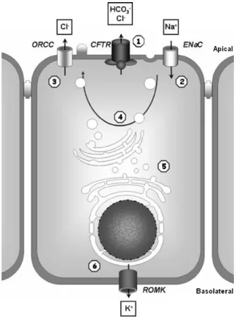
Figure I.4 Overview of CFTR functions and interactions. CFTR functions as a regulator of
other ion channels and affects numerous cellular processes: 1) Cl- channel function, which facilitates the release of Cl-, HCO3
-, and ATP-, 2) negative regulation of epithelial Na+ channels (ENaC), 3) positive regulation of outwardly rectifying Cl- channels (ORCC), 4) regulation of vesicle trafficking, 5) regulation of intracellular compartment acidification and protein processing, 6) modulation of the renal outer medullary potassium channel’s (ROMK) sensitivity to sulfonylureas [adapted from (Mueller and Flotte, 2008)].
Importantly for the pathophysiology of CF lung disease, CFTR co-regulates Na+ transport through an epithelial Na+ channel, ENaC. Wt-CFTR inhibits ENaC Na+ transport (except in sweat ducts where CFTR activates ENaC), whereas ENaC activates CFTR, and mutant CFTR allows enhanced Na+ transport, with a subsequent increase in Na+ absorption (Stutts et al., 1995; Mall et al., 1996; Reddy et al., 1999; Guggino and Stanton, 2006). This interaction between CFTR and ENaC is biologically relevant because the balance between CFTR-mediated Cl- secretion and ENaC-mediated Na+ reabsorption regulates the net amount of salt and water in airway periciliary fluid, and thereby the capacity to clear bacteria and other noxious agents from the lungs (Boucher, 2004).
Wt-CFTR activates the outwardly rectifying Cl- channels [ORCCs; (Gabriel et al., 1993; Schwiebert et al., 1995; Schwiebert et al., 1999)] through the release of ATP as an agonist into the extracellular milieu. This CFTR-dependent release of ATP out of the cell, allowing interaction with ORCCs, could be conducted by CFTR itself or via a closely associated ATP channel. The incidence of ORCCs has been reported to be enhanced in the presence of a functional CFTR Cl- channel (Jovov et al., 1995). Inwardly rectifying K+ channels (ROMKs) have been identified on the basolateral cell membrane in airway epithelia, where they are believed to play a role in K+ recycling (Schwiebert et al., 1999). NHERF1 and NHERF2 (Na+/H+ exchanger regulatory factor isoform-1/-2) increase the physical interaction between one member of this family, ROMK2 (renal outer medullary K+ channel) and CFTR (Yoo et al., 2004). The NHERF-facilitated interaction between ROMK2 and CFTR enhances glibenclamide-induced activation of ROMK2.
With CFTR regulating so many channels and processes, the thought “one channel to rule them all” comes to mind.
I.5.2 CFTR AS A PUTATIVE PSEUDOMONAS AERUGINOSA RECEPTOR
CF is characterized by the emergence and persistence of (and, ultimately, the inability to clear) chronic infection with a variant of Pseudomonas aeruginosa (mucoid
P. aeruginosa) that over-produces a surface polysaccharide known as alginate,
which protects the bacteria from antibiotics and other antimicrobial agents, making the infection very difficult if not even impossible to eradicate (Emerson et al., 2002; Li et al., 2005).
Attachment of P. aeruginosa in CF-airways was explained by a mechanism that proposes CFTR as a receptor for P. aeruginosa in the airways (Pier et al., 1997), indicating an additional function for the CFTR protein. According to some authors, CFTR is a cellular receptor for binding, endocytosing, and clearing P. aeruginosa from the normal lung. Once P. aeruginosa is bound to epithelial cells, CFTR accumulates in the cell membrane at a specific point of contact with the bacterial surface. Overall, according to a proposed model (Pier et al., 1997), a specific interaction between P. aeruginosa and the first extracellular domain of CFTR triggers CFTR-mediated resistance to infection in individuals who have wt-CFTR. Lack of this interaction and lack of a functional CFTR protein in most CF patients could contribute significantly to the respiratory manifestations of CF.
In CF, the diminished or non-existent binding of P. aeruginosa to the CF epithelium leads to a reduced initial clearance, allowing the organisms sufficient time to take advantage of the dehydrated ASL and remain within the airway lumen by binding to mucins via the bacterial FliD protein (Arora et al., 1998). Subsequently increased production of alginate occurs (Worlitzsch et al., 2002; Bragonzi et al., 2005), further serving to protect the microbe from host defences.
Recently, it was shown that P. aeruginosa chemically modifies lipid A (contained in bacterial LPS) and muropeptides (contained in peptidoglycan) as a strategy to evade immune system and detection, favouring survival in patients with CF (Cigana et al., 2009).
I.6 CFTR MUTATIONS
To date, more than 1700 variants have been identified in the CFTR gene (http://www.genet.sickkids.on.ca/cftr/StatisticsPage.html), most of them causing CF disease.
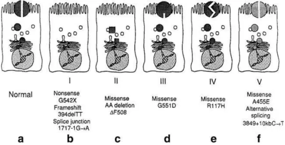
Figure I.5 Molecular consequences of mutations in the CFTR gene. a) CFTR protein correctly
positioned at the apical membrane of an epithelial cell, functioning as a chloride channel. b) Class I. No CFTR mRNA or no CFTR protein formed (e.g., nonsense, frame shift or splice site mutation). c) Class II. Trafficking defect. CFTR mRNA formed, but protein fails to traffic to cell membrane. d) Class III. Regulation defect. CFTR reaches the plasma membrane but fails to respond to cAMP stimulation. e) Class IV. Channel defect. CFTR functions as an altered chloride channel. f) Class V. Synthesis defect. Reduced synthesis of defective processing of normal CFTR. Chloride channel properties are normal (Proesmans et al., 2008).
Mutations in the CFTR gene can be grouped into five different classes according to their effect on CFTR function (Gibson et al., 2003) (Figure I.5). Class I mutations lead to a premature termination codon (PTC) that results in an unstable truncated CFTR transcript and/or no CFTR expression. Missense mutations (Class II), including F508del, cause protein misfolding that leads to the retention of the misfolded protein in the endoplasmic reticulum and premature degradation. Class III mutations result in the reduced capacity of CFTR to secrete Cl- due to abnormal channel activation by ATP. Class IV mutations cause a reduced capacity to conduct Cl- across membranes. Class V mutations cause abnormal or alternative splicing, which reduces the amount of functional protein.
I.6.1 F508DEL-CFTR
The most common CF mutation is loss of a phenylalanine (F) residue at position 508 (F508del). Up to 70% of individuals with CF are homozygous for the F508del mutation, and almost 90% of patients may have at least one F508del allele. The finding that F508del is responsible for such a high percentage of all CF mutations suggests that there may have been some heterozygote selection or a very strong founder effect for this particular mutation in the Northern European population (Tsui and Buchwald, 1991; Morral et al., 1994; Alfonso-Sanchez et al., 2010).
The F508del mutation: (a) retains CFTR in the endoplasmic reticulum (ER) where it is subsequently degraded by the proteasome; (b) reduces the capacity of CFTR to transport Cl- ions (Gibson et al., 2003; Boucher, 2004; Davis, 2006), (c) decreases the plasma membrane half-life of CFTR in polarized human airway epithelial cells (Swiatecka-Urban et al., 2005), and (d) reduces the levels of transcripts (Ramalho et al., 2002).
Most F508del-CFTR protein is rapidly removed from the cell through the cellular disposal machinery (Amaral, 2005). This mechanism substantially prevents F508del from reaching its correct cellular location, the apical membrane of epithelial cells, and explains why this mutation is included in class II (Cheng et al., 1990). Additionally, F508del causes major defects in channel regulation that interfere with channel opening (Wang et al., 2000), and therefore it can also be considered a class III mutation.
In native tissues from F508del-homozygous patients, however, CFTR has been described as having an apical localization (Kalin et al., 1999; Penque et al., 2000),