A Time to Remember: The Role of Circadian Clocks in
Learning and Memory
Benjamin L. Smarr, Kimberly J. Jennings, Joseph R. Driscoll, and Lance J. Kriegsfeld
University of California, BerkeleyThe circadian system has pronounced influence on learning and memory, manifesting as marked changes in memory acquisition and recall across the day. From a mechanistic perspective, the majority of studies have investigated mammalian hippocampal-dependent learning and memory, as this system is highly tractable. The hippocampus plays a major role in learning and memory, and has the potential to integrate circadian information in many ways, including information from local, independent oscillators, and through circadian modulation of neurogenesis, synaptic remodeling, intracellular cascades, and epige-netic regulation of gene expression. These local processes are combined with input from other oscillatory systems to synergistically augment hippocampal rhythmic function. This overview presents an account of the current state of knowledge on circadian interactions with learning and memory circuitry and provides a framework for those interested in further exploring these interactions.
Keywords: daily, recall, hormones, neurogenesis, hippocampus
To appropriately coordinate behavior and physiology with daily changes in their ecosystem, animals utilize an endogenous circa-dian timing system (Sharma, 2003). By employing an internal timing system, animals can anticipate environmental change and prepare accordingly, rather than respond only after the fact to a given event (Antle & Silver, 2009). Such an internal timing system should allow the formation of associations between a given stim-ulus with circadian phase, so that predictable daily events, or shifts thereof (i.e., changes following migration or alteration of temporal niche), can be accommodated (Daan, 2000; Smarr, Schwartz,
Wotus, & de la Iglesia, 2013). Given the extent of physiological
modulation by the circadian system, it not surprising that the ability to make these associations varies across the day.
Although the present overview focuses on mammals, the ability to phase behavior with appropriate times of day is common across taxa. For example, honey bees (Apis mellifera) can learn to use different floral landing strategies when collecting pollen based on time of day and associated changes in blossom position (J. L.
Gould, 1987). Fish can be trained to swim to different sides of an
aquarium for evening versus morning meals (Reebs, 1996). Gar-den warblers (Sylvia borin) learn to forage in different locations at different times of day based on daily rhythms in food availability
(Biebach, Falk, & Krebs, 1991). Finally, rodents rapidly learn food
(Bolles & Stokes, 1965;Carr & Wilkie, 1997;Holmes &
Mistl-berger, 2000; Marchant & Mistlberger, 1997; Mistlberger, de
Groot, Bossert, & Marchant, 1996;Mistlberger, 1993) and water
(Mistlberger, 1992,1993) spatial associations specific to time of
day (Cain, Chou, & Ralph, 2004;Cain, McDonald, & Ralph, 2008;
Carr & Wilkie, 1997;Holloway & Wansley, 1973a,1973b;
Hun-sicker & Mellgren, 1977;Ko, McDonald, & Ralph, 2003;
Mistl-berger et al., 1996;Ralph et al., 2002;Stephan & Kovacevic, 1978;
Wansley & Holloway, 1975). Pairings of specific spaces with
specific times can be learned through associations with both pos-itive (Carr & Wilkie, 1997;Hunsicker & Mellgren, 1977;Ko et al.,
2003; Mistlberger et al., 1996; Ralph et al., 2002; Wansley &
Holloway, 1975) and negative (Cain, Chou, et al., 2004;Cain et
al., 2008;Holloway & Wansley, 1973a,1973b;Stephan &
Kova-cevic, 1978) outcomes.
In addition to learning associations between time of day and resources, there are pronounced daily changes in the ability to acquire new memories. For example, aplysia (Aplysia californica) show enhanced sensitization of gill withdrawal during subjective day (Fernandez, Lyons, Levenson, Khabour, & Eskin, 2003). Cockroaches (Leucophaea maderae) learn to discriminate olfac-tory cues more effectively in subjective night (the phase of activity for nocturnal animals) than subjective day when held in constant conditions (Decker, McConnaughey, & Page, 2007). Likewise, mice acquire maze navigation memory (Hoffmann & Balschun, 1992) and contextual fear conditioning (CFC;Valentinuzzi et al., 2001) faster in the dark phase of the light– dark (LD) cycle. Interestingly, mice acquire tone-cued fear conditioning more rap-idly when trained in the subjective day (Chaudhury & Colwell, 2002). It is likely that these disparities in the timing of learning efficacy are stimulus specific, as similar discrepancies are ob-served across species. In rats, for example, the acquisition of maze navigation is better in the dark phase (Hauber & Bareiss, 2001) or subjective night (Valentinuzzi, Menna-Barreto, & Xavier, 2004), This article was published Online First April 7, 2014.
Benjamin L. Smarr and Kimberly J. Jennings, Department of Psychol-ogy, University of California, Berkeley; Joseph R. Driscoll, The Helen Wills Neuroscience Institute, University of California, Berkeley; Lance J. Kriegsfeld, Department of Psychology and The Helen Wills Neuroscience Institute, University of California, Berkeley.
Support during the writing of this review was received from National Institutes of Health Grant R01 HD050470.
Correspondence concerning this article should be addressed to Lance J. Kriegsfeld, Neurobiology Laboratory, Department of Psychology and Helen Wills Neuroscience Institute, 3210 Tolman Hall, #1650, University of Cali-fornia, Berkeley, CA 94720-1650. E-mail:[email protected]
This document is copyrighted by the American Psychological Association or one of its allied publishers. This article is intended solely for the personal use of the individual user and is not to be disseminated broadly. 283
whereas active avoidance is learned better at the end of a 12-h light phase than at the beginning (Pagano & Lovely, 1972). Hamsters demonstrate enhanced T-maze alternation and novel object dis-crimination performance during and just before the dark phase
(Ruby et al., 2008,2013). These time-of-day differences are not
observed in the acquisition of more complex operant conditioning tasks (Ghiselli & Patton, 1976;Stroebel, 1967; cf.Mistlberger et
al., 1996). These latter findings suggest that the cognitive load of
a task might overshadow changes from circadian timing, such that time-of-day modulation in acquisition may be more apparent on simpler tasks, but this hypothesis requires further investigation. Interestingly, tasks with a high cognitive load can themselves serve as zeitgebers (from German for “time giver,” meaning they can shift circadian phase), apparently through cholinergic signaling
(Gritton, Kantorowski, Sarter, & Lee, 2012; Gritton, Stasiak,
Sarter, & Lee, 2013; Gritton, Sutton, Martinez, Sarter, & Lee,
2009). These findings provide the basis for further dissecting the interaction of cognitive load and circadian modulation of learning and memory-dependent behavior.
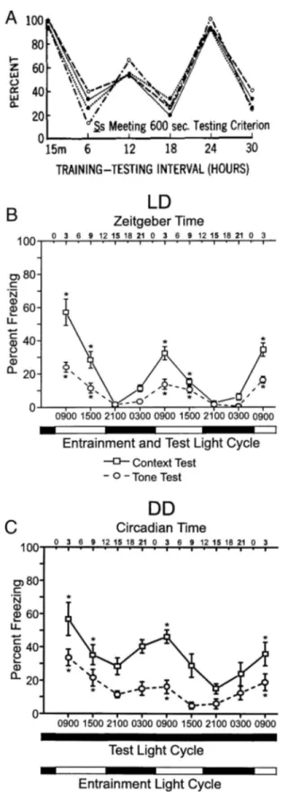
Once acquired, the ability to recall and apply learned informa-tion peaks periodically following training. Early studies suggested that there was a 12-h period to recall efficacy: Rats were found to perform best on active avoidance, passive avoidance, and appeti-tive tests every 12 h after training (Holloway & Wansley, 1973a,
1973b;Hunsicker & Mellgren, 1977;Wansley & Holloway, 1975,
1976;Figure 1A). Although the rate of acquisition was not found
to change across the day, rats trained at the end of the light phase lacked 12-h peaks in recall performance, showing peaks only at 24-h intervals (Holloway & Wansley, 1973b). Given that memo-ries are most vulnerable to extinction during recall (Bridge &
Paller, 2012), one might expect to observe increased susceptibility
to extinction with the same intervals posttraining, which is exactly what was observed (Holloway & Sturgis, 1976). In contrast to this earlier work, more recent studies report a 24-h periodicity in recall efficacy following acquisition, without a 12-h intermediate peak
(cf.Chaudhury & Colwell, 2002;McDonald, Hong, Ray, & Ralph,
2002; Figure 1B). For example, cockroaches exhibit peaks in
operant conditioning every 24 h following training (Garren,
Sex-auer, & Page, 2013). Hamsters show peak performance in
appet-itive (Ko et al., 2003;Ralph et al., 2002) and aversive (Cain, Chou,
et al., 2004; Cain et al., 2008; Stephan & Kovacevic, 1978)
conditioning 24 h after training. Likewise, mice show peaks of recall in CFC 24 h following training (Loh et al., 2010). Whether rhythms in recall peak every 12 or 24 h following learning requires further investigation, as discrepancies between studies have not been explored systematically. It is possible that the disagreement with earlier works showing 12-h periodic enhancement is due to rat-specific ultradian rhythms, but this possibility has not been examined.
As with daily rhythms in memory acquisition ability, recall and extinction also show a time-of-day dependence, independent of the time of training. Mice show more freezing when tested for con-textual or tone-cued fear conditioning in the early subjective day, regardless of time of training (Chaudhury & Colwell, 2002;
Eckel-Mahan et al., 2008). Similarly, rats display a peak in recall for
passive avoidance in the light phase when housed in an LD cycle
(Davies, Navaratnam, & Redfern, 1974). In contrast, rates of
extinction are greatest during the dark phase, consistent with the notion that acquisition (i.e., learning to disassociate a previously
learned association) is acquired faster during the night, at least in nocturnal rodents. Under natural light, rats show faster extinction to conditioned taste aversion when extinction-trained at night (12:00 a.m. and 6:00 a.m.) than during the day (12:00 p.m. and 6:00 p.m.; note that the exact sunrise and sunset times for these data are not indicated in the manuscript; the designation of night and day comes from the author observing the natural LD cycle;
Ternes, 1976). Similarly, mice exhibit a faster rate of extinction in
CFC when conditioning and testing took place in subjective night
(Valentinuzzi et al., 2001).
The ubiquity of circadian and daily changes in learning and memory does not permit an exhaustive review of the literature in the present overview (seeGerstner et al., 2009;Lyons, 2011; and
Mulder, Gerkema, & Van der Zee, 2013, for related reviews).
However, from the examples given, it should be clear that there is pronounced circadian impact on the efficacy of acquisition, recall, and extinction, at least for some tasks. Given that the vast majority of studies on the circadian control of learning, memory, and recall have been performed in rodents, investigations into the similarities and differences across taxa represent an important area for further inquiry.
The Master Clock and Circadian Changes in Learning
and Memory
In mammals, the circadian timing system is composed of a hierarchy of oscillators controlled by a central, master pacemaker in the suprachiasmatic nucleus (SCN) of the anterior hypothala-mus. Lesions of the SCN abolish circadian rhythmicity (Moore &
Eichler, 1972; Stephan & Zucker, 1972) and lead to a loss of
synchrony among independent, subordinate oscillators throughout the CNS and periphery (Welsh, Yoo, Liu, Takahashi, & Kay, 2004). Together, these findings suggest that the SCN communi-cates timing information to central and peripheral systems to maintain cohesion among independent cellular clocks required for system-specific rhythmicity (Figure 2).
At the cellular level, circadian rhythms are generated by⬃24-hr autoregulatory transcriptional–translational feedback loops con-sisting of “clock” genes and their protein products (see Ko &
Takahashi, 2006, andMohawk & Takahashi, 2011, for review). In
mammals, the feedback loop begins in the cell nucleus where CLOCK and BMAL1 proteins heterodimerize and drive the tran-scription of the Period (Per1, Per2, and Per3) and Cryptochrome (Cry1 and Cry2) genes by binding to the E-box (CACGTG) domain on their gene promoters. Once translated, PER and CRY proteins build in the cytoplasm of the cell over the course of the day, and eventually form hetero- and homodimers that feed back to the cell nucleus to inhibit CLOCK:BMAL1-mediated transcrip-tion. The timing of nuclear entry is balanced by regulatory kinases that phosphorylate the PER and CRY proteins, leading to their degradation (Lowrey et al., 2000; G.-Q. Wang, Du, & Tong, 2007). Two other promoter elements, DBP/E4BP4 binding ele-ments (D boxes) and REV-ERB␣/ROR binding elements (RREs;
Ueda et al., 2005), also participate in cellular clock function.
REV-ERB␣, an orphan nuclear receptor, negatively regulates the activity of the CLOCK:BMAL1.
Findings on the necessity of the SCN in mediating rhythms in the ability to learn or recall are equivocal. In rats, SCN lesions eliminate the enhancement in recall every 24 h following training
This document is copyrighted by the American Psychological Association or one of its allied publishers. This article is intended solely for the personal use of the individual user and is not to be disseminated broadly.
in a passive avoidance task (Stephan & Kovacevic, 1978). In hamsters made arrhythmic without ablating the SCN, daily vari-ance is lost in novel object recognition and T-maze alternation
(Ruby et al., 2008, 2013). However, rats trained to perform a
specific operant task depending on time of day are able to perform the correct lever operations at the correct time, even after SCN lesion (Mistlberger et al., 1996; Figure 3). Similarly, in some cases, SCN-ablated hamsters show conditioned place preference (CPP) only at the time during which training previously occurred
(Ko et al., 2003), suggesting that the SCN was not needed for
learning the “time stamp” of reward or, alternatively, for the 24-h periodicity of recall enhancement following training. Finally, whereas normal daily patterns of food and water foraging lose circadian rhythmicity following SCN ablation, animals under food or water restriction maintain the ability to learn time and place associations necessary to secure food or water when available
(Marchant & Mistlberger, 1997;Mistlberger, 1993).
The previous findings suggest that animals might be able to rely on periodic enhancement of recall when the SCN is absent, or that other subordinate circadian oscillators maintain the ability to track time for some tasks. For example, in hamsters trained on the CPP task discussed previously (Ko et al., 2003), olfactory cues and spatial cues were used together to establish the place memory. Olfactory information strongly modulates memory formation and recall, and the olfactory bulb (OB) is itself a circadian oscillator
(Granados-Fuentes, Tseng, & Herzog, 2006). Therefore, the OB
might serve as a source of circadian information for timed, scent-specific memories. Establishing time-coupled CPP in hamsters without scent cues would help to clarify the necessity of the SCN in circadian spatial memory formation and recall.
One other caveat that should be considered when evaluating these disparate findings is that periodic availability of food or water can entrain (synchronize) daily rhythms in physiology and behavior. Most species acquire food anticipatory behavior when food is restricted to a daily temporal window (reviewed in
Ara-gona, Curtis, Davidson, Wang, & Stephan, 2002; Carneiro &
Araujo, 2012;Mistlberger, 2011;Schibler, Ripperger, & Brown,
Figure 1. Twelve- and 24-h periodic recall enhancement. Passive avoid-ance testing session, percent of sessions meeting the 600-s session crite-rion, showing 12-h periodic enhancement. From “Multiple Retention Def-icits at Periodic Intervals After Active and Passive Avoidance Learning,” by F. Holloway and R. Wansley, 1973, Behavioral biology, 9, p. 5. Copyright 1973 by Academic Press. Reprinted with permission. (A) Rhythms in recall in C-3H mice trained in the day (ZT/CT 3), showing 24-h periodic enhancement. In all experiments, animals were first tested for context 24-h posttraining then repeatedly tested every 6 h, for 3 days. On Day 4, animals were tested for tone every 6 h for another 3 days. Testing was done at ZT/CT 3, 9, 15 and 21. (B) Mice were maintained on a light– dark (LD) cycle. (C) Mice were maintained in constant darkness (DD). Times of prior LD cycle are indicated. Each group contained eight animals. Within-population one-way repeated measures ANOVA at the first and second 24-h periods for both the context and tone showed significant differences in recall at different times of test, where the asterisk (ⴱ) denotes p⬍ 0.05. (B) and (C) from “Circadian Modulation of Learning and Memory in Fear-Conditioned Mice,” by D. Chaudhury and C. Colwell, 1973, Behavioral Brain Research, 133, p. 100. Copyright 1973 by Elsevier Science B.V. Reprinted with permission.
This document is copyrighted by the American Psychological Association or one of its allied publishers. This article is intended solely for the personal use of the individual user and is not to be disseminated broadly.
2003). Importantly, these food-seeking oscillations persist even in SCN-ablated animals (Stephan, Swann, & Sisk, 1979), as does water seeking (Mistlberger, 1993). When investigating the circa-dian basis of appetitive learning in the absence of the SCN, food delivered as a reward might entrain the food entrainable oscillator (FEO) and confound interpretation if the task is performed at the same time each day. In agreement with this possibility, the three articles mentioned previously that established a strong link be-tween the SCN and phasic recall enhancement (Ruby et al., 2008,
2013;Stephan & Kovacevic, 1978) used purely spatial tasks, not
likely vulnerable to the engagement of the olfactory oscillator or food–water entrainment. Careful conditioning paradigm selection will be needed to tease apart the potential roles of different circadian oscillators as cue-specific sources of temporal informa-tion for learning and memory.
Circadian Disruption Impact Learning and Memory
Circadian rhythms can be perturbed, without being eliminated, by a range of LD manipulations. These findings yield unique insights into the role of circadian information in memory forma-tion in animals with an otherwise intact circadian system. In addition to altering the phase of the SCN, adjustments to the LD cycle also shift non-SCN oscillators (e.g., Smarr, Gile, & de la
Iglesia, 2013;Yamazaki et al., 2000). The SCN (at least the central
core subregion) adjusts to pronounced phase shifts relatively rap-idly (Best, Maywood, Smith, & Hastings, 1999), compared with extra-SCN oscillators, which require considerably longer (
Ya-mazaki et al., 2000). Phase shifts that precede training by 2 to 3
days result in recall deficits (Fekete, van Ree, Niesink, & de Wied, 1985), a finding inconsistent with a model in which the SCN both provides a time stamp for new associations, and modulates recall efficacy. It is possible that extra-SCN oscillators (e.g., OB, FEO,
etc.) contribute to memory acquisition and recall, as these oscil-lators may remain out of phase with the environment longer than the SCN. If extra-SCN oscillators provide time-stamped informa-tion for acquisiinforma-tion or recall of a memory, then a phase shift preceding a task should cause a phase mismatch between that memory’s subjective time and the environment’s or SCN’s time. Because there is a periodic enhancement of recall following ac-quisition, a phase mismatch would result, following reentrainment between the time-stamped memory and environmental time, pro-portional to the magnitude of the phase shift. This being the case, an examination of recall efficacy at regular intervals following recovery from a phase shift should result in predictable mis-matches between peak recall and environmental time. Alterna-tively, if phasic enhancements of recall were found to be lost in such experiments, this would imply that circadian disruption fun-damentally impairs memory consolidation or recall. Such a sys-tematic investigation has not yet been conducted, but ground work has been laid.
In mice trained on a CFC task within a day (⫹ or –) of a 12-h phase shift, acquisition is normal, but major recall deficits are apparent at intervals of 24 h following the training. The mag-nitude of the deficit is proportional to both the size of the shift and to the distance in time from the shift. Either phase advances or phase delays cause these deficits (Loh et al., 2010). Rats also show deficits in recall when phase shifted, either advances (Davies
et al., 1974;Fekete, Van Ree, & De Wied, 1986;Tapp &
Hollo-way, 1981) or delays (Tapp & Holloway, 1981), within a day of
training on a passive avoidance task, and these deficits are pro-portional to the magnitude of shift (Fekete et al., 1986) and time since the shift (Fekete et al., 1985). Phase advances 5 days after training also impose recall deficits (Fekete et al., 1986). Rats trained in active avoidance tasks show enhanced extinction when Figure 2. The suprachiasmatic nucleus (SCN) synchronizes extra-SCN oscillators throughout the body. A
stereotyped mammal body showing that light input to the eyes is carried by the optic nerve to the master circadian pacemaker in the SCN. The SCN then sends phase information to synchronize the circadian oscillations of other neural nuclei and somatic systems. It accomplishes this task through many pathways, including spinal relay to the adrenals to regulate glucocorticoid rhythms, modulation of pituitary hormones to organ systems, and heretofore undefined signals to regulate muscle metabolism and activity output. The SCN also receives modulatory input from other oscillators, such as scent cues from olfaction, satiety cues from the gastrointestinal (GI) tract, and neural input from cognitive centers. A great deal remains to be examined in the mechanisms and effects of these interactions.
This document is copyrighted by the American Psychological Association or one of its allied publishers. This article is intended solely for the personal use of the individual user and is not to be disseminated broadly.
phase advances follow training (Fekete et al., 1986). In rats, at least following advances, time of day interacts with time since training, with normal acquisition occurring when trained in the early dark phase, and disrupted acquisition occurring when trained in the early light phase (Davies et al., 1974). Rats trained in a Morris water maze (MWM) following several days of phase ad-vances show no deficit in acquisition (Craig & McDonald, 2008;
Devan et al., 2001), but day-to-day retention is negatively
im-pacted (Devan et al., 2001). In Syrian hamsters, twice weekly phase advances (for 25 days) causes gross deficits on a CPP task; these deficits in the ability to learn the association persist even 4 weeks after the cessation of the phase advances (Gibson, Wang,
Tjho, Khattar, & Kriegsfeld, 2010). Finally, rats first subjected to
several weeks of repeated phase advances show both acquisition and retention deficits when trained and tested in an MWM (Craig
& McDonald, 2008) and show deficits in CPP (McDonald et al.,
2013), though, in the latter case, it is not clear if acquisition, recall, or both are impaired. Interestingly, phase shifts do not impair
tone-cued conditioning (Craig & McDonald, 2008) or social ex-ploration (Fekete et al., 1985), and in aged hamsters, the magni-tude of circadian rhythm degradation is not correlated with deficits in sexual behavior (Antoniadis, Ko, Ralph, & McDonald, 2000), suggesting that only specific learning and memory tasks, but not all behaviors, are impacted by disruption of circadian rhythms. Taken together with the preceding behavioral examples, these phase-shifting experiments suggest that external time manipula-tions impose proportional deficits on recall, but only chronic phase shifting affects acquisition, particularly for spatial tasks, but not necessarily nonspatial tasks.
The Hippocampus as an Integrator of Circadian
Information With Learning and Memory
The hippocampus (HC) is critical for forming an array of memories, including spatial associations (Anagnostaras, Gale, &
Fanselow, 2001;Corcoran & Maren, 2001;Frohardt, Guarraci, &
Figure 3. Conditioned place preference remains following suprachiasmatic nucleus (SCN) lesion. Group mean (⫾ SEM) discrimination ratios (number of correct lever presses divided by total lever presses, prior to the first reward of the session) for intact rats (top, n⫽ 5) and SCN-ablated rats (bottom, n ⫽ 5) during Stage 4 of the experiment (two-arm free choice,⬎1 lever press required for first reward). Sessions that were omitted are indicated by the squares along the abscissa. The first test following an omission test is indicated by a solid square. From “Discrimination of Circadian Phase in Intact and Suprachiasmatic Nuclei-Ablated Rats,” by R. Mistl-berger, M. de Groot, J. Bossert, and E. Marchant, 1996, Brain Research, 739, p. 14. Copyright 1996 by Elsevier Science B.V. Reprinted with permission.
This document is copyrighted by the American Psychological Association or one of its allied publishers. This article is intended solely for the personal use of the individual user and is not to be disseminated broadly.
Bouton, 2000), CFC (Anagnostaras et al., 2001), and, in some instances, tone-cued fear conditioning (Bast, Zhang, & Feldon,
2003;Maren, Aharonov, & Fanselow, 1997). As such, the HC is
likely necessary for most of these tasks (excluding odor or gusta-tory cues). The HC may integrate circadian information through direct and indirect input from the SCN or other oscillator outputs, rhythms in hippocampal neurogenesis, hippocampal clock gene oscillations, and genetic modifications downstream of the molec-ular clockwork.
Direct and indirect circadian input to the HC. Few studies have investigated the specific means by which circadian disruption affects hippocampal functioning. One pair of studies suggest that the SCN may provide information to the HC through rhythms in GABAergic activity, as the administration of a GABA receptor A antagonist restores circadian rhythms in novel object recognition and T-maze alternation performance in arrhythmic, SCN-intact hamsters (Ruby et al., 2008, 2013). The specific mechanism(s), and neural pathway(s), by which arrhythmicity results in enhanced GABAergic communication to the HC requires further investiga-tion.
An indirect pathway by which the HC might gain circadian information is adrenal glucocorticoids (CORT). Though CORT is not required for memory formation (Bialik, Pappas, & Roberts, 1984), CORT modulates hippocampal neurogenesis (discussed in the section “Neurogenesis rhythms in the HC”), memory forma-tion, and activation (reviewed inMaggio & Segal, 2010;
Schoen-feld & Gould, 2012). Plasma CORT concentrations exhibit
circa-dian rhythms, and SCN lesions eliminate (Abe, Kroning, Greer, &
Critchlow, 1979;Buijs, Kalsbeek, van der Woude, van
Heerikhu-ize, & Shinn, 1993;Moore & Eichler, 1972), and circadian
dis-ruptions disturb (Lilley, Wotus, Taylor, Lee, & de la Iglesia, 2012;
Loh et al., 2010; Weinert, Eimert, Erkert, & Schneyer, 1994;
Wotus et al., 2013), normal CORT rhythms. Aging also disrupts
and flattens circadian CORT rhythms (Cain, Karatsoreos, et al.,
2004;Gartside, Leitch, McQuade, & Swarbrick, 2003). The degree
of flattening strongly correlates with deficits in CPP training (Cain,
Karatsoreos, et al., 2004) and may cause structural changes in the
HC (Gartside et al., 2003). These findings further suggest a role for
CORT rhythms in memory acquisition and recall. Rhythms in CORT appear to be indirectly driven by arginine-vasopressin (AVP) from the SCN, acting to inhibit ACTH (Gomez et al., 1997;
Kalsbeek, Buijs, van Heerikhuize, Arts, & van der Woude, 1992;
Kalsbeek, van Heerikhuize, Wortel, & Buijs, 1996), a
neuropep-tide that drives CORT release from the adrenals. Administration of ACTH (Pagano & Lovely, 1972) or its analog ORG-2766 (Fekete
et al., 1986), or the AVP analog
desglycinamide9-(Arg8)-vasopressin (Fekete et al., 1986), mitigate the time-of-day effect on learning (as do lesions of the adrenal gland and its innervation;
Bialik et al., 1984), and the negative impact of phase shifts on
memory. Adrenalectomy impairs CFC recall, and CORT replace-ment rescues this effect (Pugh, Tremblay, Fleshner, & Rudy, 1997). Augmenting conditioning with anxiolytic treatments elim-inates time-of-day variance in long-term recall (Coll-Andreu,
Martí-Nicolovius, & Morgado-Bernal, 1991), whereas CORT
con-centrations in response to CFC training predicts the strength of CFC retention (Cordero, Merino, & Sandi, 1998). Finally, circa-dian changes in CORT concentrations may interact with stress-induced increases in CORT to mediate changes in learning
strength in response to stress at different times of day (Kelliher et
al., 2000;Rudy & Pugh, 1998).
Clock gene rhythms in HC. Further evidence for the role of the HC as a center for circadian influence on learning and memory comes from the observation that hippocampal cells express clock genes and keep circadian time. As mentioned previously, clock genes are expressed ubiquitously in the CNS and periphery, al-lowing for tissue-specific rhythms in activity and the maintenance of optimal health and functioning. Clock genes are expressed rhythmically in the HC, including the dentate gyrus (Jilg et al., 2010). Although a few studies have failed to observe hippocampal rhythms in Per2 expression in mice (Albrecht, Sun, Eichele, & Lee,
1997;Borgs, Beukelaers, Vandenbosch, Nguyen, et al., 2009),
per-haps due to its relatively low amplitude (Jilg et al., 2010), rhythms in Per1, Cry1, Cry2, Clock, and Bmal1 mRNA, and protein in the HC, have been consistently observed in rats, mice, and hamsters (Amir,
Harbour, & Robinson, 2006;Chaudhury, Loh, Dragich, Hagopian, &
Colwell, 2008;Duncan, Prochot, Cook, Tyler Smith, & Franklin,
2013;Gilhooley, Pinnock, & Herbert, 2011;Ikeno, Weil, &
Nel-son, 2013;Jilg et al., 2010;Lamont, Robinson, Stewart, & Amir,
2005; Segall, Milet, Tronche, & Amir, 2009; Segall, Perrin,
Walker, Stewart, & Amir, 2006;Valnegri et al., 2011;Wakamatsu
et al., 2001;L. M. Wang et al., 2009;Wyse & Coogan, 2010).
Furthermore, hippocampal slices continue to oscillate in culture
(L. M. Wang et al., 2009), suggesting the existence of independent
oscillations presumably synchronized by the SCN in vivo. The dentate gyrus rhythm of Per2 in rats persists even after electrolytic lesion of the SCN (Lamont et al., 2005) or after disruption of circadian CORT fluctuations (Segall et al., 2006, 2009), further suggesting the HC functions as an extra-SCN oscillator, potentially mediating maintenance of rhythms in learning and memory after SCN ablation.
Rhythms in cellular cascades in the HC. In addition to the rhythmic expression of clock genes, intracellular cascades impor-tant for transducing cellular responses oscillate with a circadian cycle (Dolci et al., 2003;Eckel-Mahan et al., 2008;Phan et al., 2011). For example, cyclic adenosine monophosphate (cAMP)/ cAMP response element binding protein (CREB) and mitogen activated protein kinase (MAPK) are rhythmic in the HC, and these rhythms are disrupted by exposure to constant light or SCN ablation (Eckel-Mahan et al., 2008;Phan et al., 2011). cAMP and MAPK pathways are necessary intracellular signaling cascades in the molecular clockwork (Antoun, Bouchard-Cannon, Cannon, &
Cheng, 2012;Butcher, Lee, Cheng, & Obrietan, 2005;Dziema et
al., 2003;Obrietan, Impey, & Storm, 1998;Tischkau, Gallman,
Buchanan, & Gillette, 2000). MAPK phosphorylates BMAL1, a
central component of the circadian clock, inhibiting BMAL1: CLOCK heterodimer-dependent transcription of the Per and Cry genes (Sanada, Okano, & Fukada, 2002). The Per1 and Per2 promoters contain cAMP response element (CRE) sites that bind CREB to enhance their protein production (Travnickova-Bendova,
Cermakian, Reppert, & Sassone-Corsi, 2002).
Given that cAMP/CREB and MAPK have been implicated in the formation and persistence of long-term memory (Athos,
Im-pey, Pineda, Chen, & Storm, 2002;Atkins et al., 2010;Kelleher,
Govindarajan, Jung, Kang, & Tonegawa, 2004;Kelly, Laroche, &
Davis, 2003;Lonze & Ginty, 2002;Scott, Bourtchuladze,
Goss-weiler, Dubnau, & Tully, 2002;Silva, Kogan, Frankland, & Kida,
1998; H.Wang, Ferguson, Pineda, Cundiff, & Storm, 2004), it is
This document is copyrighted by the American Psychological Association or one of its allied publishers. This article is intended solely for the personal use of the individual user and is not to be disseminated broadly.
likely that long-term memory may be modulated by the regulation of the circadian molecular clock by shared intracellular signaling pathways. In agreement with this possibility, the disruption of MAPK oscillations following training interferes with persistence of long-term memory (Eckel-Mahan et al., 2008). Likewise, Per1 and Per2 knockouts exhibit deficits in hippocampal-dependent learning tasks associated with abnormal long term potentiation (LTP; discussed below in the section “Circadian Influence on Long-Term Potentiation in the HC”) and reduced levels of phos-phorylated CREB in Per2 knockouts (L. M. Wang et al., 2009). These results have led to the suggestion that long-term memory may depend on the circadian-driven reactivation of the cAMP/ CREB and MAPK pathways required to consolidate memories
(Eckel-Mahan, 2012; Eckel-Mahan et al., 2008; Gerstner et al.,
2009).
Neurogenesis rhythms in the HC. A local mechanism by
which circadian information may influence HC functioning is through changes in adult hippocampal neurogenesis. New neurons are born from neural progenitor cells in the subgranular zone of the dentate gyrus. These adult-born neurons are eventually integrated into the granule layer, where they appear to contribute to spatial pattern recognition and hippocampal-dependent memory (re-viewed in Deng, Aimone, & Gage, 2010). Numerous lines of evidence support an association between adult neurogenesis and learning and memory. For example, neurogenesis is increased after dependent learning, but not after hippocampal-independent learning (Ambrogini et al., 2000;Epp, Spritzer, &
Galea, 2007; E.Gould, Beylin, Tanapat, Reeves, & Shors, 1999;
Leuner et al., 2004;van der Borght, Meerlo, Luiten, Eggen, & Van
der Zee, 2005). Furthermore, disrupting neurogenesis via a number
of techniques similarly disrupts performance on hippocampal-dependent tasks (Clelland et al., 2009;Dupret et al., 2007;
Farioli-Vecchioli et al., 2008;Shors et al., 2001). However, a growing
body of evidence suggests that adult hippocampal neurogenesis may only be necessary for relatively complex spatial learning
(Dupret et al., 2008;Saxe et al., 2006;Shors, Townsend, Zhao,
Kozorovitskiy, & Gould, 2002).
The dentate gyrus exhibits circadian rhythms in neurogenic proliferation under certain conditions (seeMueller, Mear, &
Mis-tlberger, 2011, for review). Briefly, rhythms in proliferation across
the day have been found in rats (Gilhooley et al., 2011;
Guzman-Marin, Suntsova, Bashir, Szymusiak, & McGinty, 2007), mice
(Tamai, Sanada, & Fukada, 2008), and hamsters (Smith,
Hecht-man, & Swann, 2010). Some studies in rats (Ambrogini et al.,
2002;Mueller et al., 2011) and mice (Holmes, Galea, Mistlberger,
& Kempermann, 2004; van der Borght et al., 2006) have not
observed rhythms in proliferation, whereas one study in mice observed rhythmic proliferation in the hilus but not in the sub-granular zone, indicating there may be a time-of-day effect in gliogenesis but not neurogenesis (Kochman, Weber, Fornal, &
Jacobs, 2006). These differences are likely due to differences in
experimental methodology, such as proliferative markers used or injection or survival protocols (seeMueller et al., 2011). Also of note, one study that reported no proliferative rhythms in nonma-nipulated animals did find an interaction between circadian time and exercise-induced increases in cell proliferation. Wheel running increased mouse hippocampal cell proliferation only when given during the active phase (Holmes et al., 2004). Thus, although the existence of adult neurogenesis was formerly controversial, it
seems clear that, at least under certain conditions, there is circadian regulation in the proliferative component of hippocampal neuro-genesis, with several thousand new cells generated daily in the rodent HC (Cameron & McKay, 2001;Rao & Shetty, 2004).
Perhaps not surprisingly, hippocampal neurogenesis is likely regulated by hippocampal clock genes. Many cell cycle-related genes show circadian patterns of expression, and at least some are under transcriptional regulation by the CLOCK-BMAL1 complex
(Matsuo et al., 2003; reviewed in Borgs, Beukelaers,
Vanden-bosch, Belachew, et al., 2009). Manipulations of Per1, both Cry
genes, or Clock in mice induce alterations in cell growth and cell cycle progression (Gery et al., 2006;Matsuo et al., 2003;Miller et
al., 2007). The cell cycle in neural progenitor cells is estimated to
be 24.7 h (Cameron & McKay, 2001; though this likely changes with the individuals’ and species’ free funning period), further suggesting an important role for circadian timing in progenitor cell maturation and differentiation. One study in mice found that neural progenitor cell expression of Per2 is maintained throughout dif-ferentiation into mature neurons (Borgs, Beukelaers,
Vanden-bosch, Nguyen, et al., 2009). However, Per2 might have
clock-unrelated functions in these cells, as this study found Per2 to be expressed constitutively in the dentate gyrus.
Outside of the HC, Clock and Bmal1 are also expressed in neural progenitor cells of the subventricular zone (SVZ) of mice, an area which gives rise to new neurons destined for the olfactory bulbs
(Kimiwada et al., 2009). These clock genes change expression
patterns as these adult-born cells differentiate into mature neurons, suggesting a role in neuronal differentiation. Furthermore, when Clock or Bmal1 are silenced by RNA interference, SVZ neurogen-esis is decreased (Kimiwada et al., 2009), further supporting their role as neurogenic regulators. It is currently unknown whether these genes serve the same function in hippocampal neurogenesis.
Rhythmic signals affecting neurogenesis in the HC. Because circadian rhythms are ubiquitous, pinpointing the specific neuro-chemical systems and cellular pathways by which the circadian system influences hippocampal functioning has been challenging. Nonetheless, a number of promising leads present themselves in the literature. CORT was described earlier and will not be revis-ited, except to point out its potential in this category. The brain-derived neurotrophic factor (BDNF) is a positive regulator of neurogenesis (Rossi et al., 2006;Sairanen, Lucas, Ernfors,
Cas-trén, & CasCas-trén, 2005;Taliaz, Stall, Dar, & Zangen, 2010) that acts
through its receptor TrkB, leading to downstream activation of MAPK. BDNF and TrkB are expressed rhythmically in the HC
(Bova, Micheli, Qualadrucci, & Zucconi, 1998;Dolci et al., 2003;
Ikeno et al., 2013), suggesting circadian regulation of BDNF
activity. Indeed, sleep and circadian disruption impacts BDNF levels in the HC, with the direction of change varying with disruption type and length (Fujihara, Sei, Morita, Ueta, & Morita,
2003;Guzman-Marin et al., 2006;Sei, Saitoh, Yamamoto, Morita,
& Morita, 2000; Sei et al., 2003). Melatonin and its precursor,
N-acetylserotonin (NAS), also show circadian rhythmicity, rising during nighttime and falling during daytime (Ganguly, Coon, &
Klein, 2002). Melatonin promotes immature neuron survival and
neurogenesis (Ramírez-Rodríguez, Klempin, Babu, Benítez-King, &
Kempermann, 2009;Ramirez-Rodriguez, Ortíz-López,
Domínguez-Alonso, Benítez-King, & Kempermann, 2011;Rennie, De Butte, &
Pappas, 2009), possibly through antioxidant activity (Manda & Reiter,
2010). On the other hand, NAS promotes proliferation by acting
This document is copyrighted by the American Psychological Association or one of its allied publishers. This article is intended solely for the personal use of the individual user and is not to be disseminated broadly.
through the BDNF receptor TrkB in the HC (reviewed inSompol et
al., 2011; Tosini, Ye, & Iuvone, 2012), which it activates in a
circadian manner (Jang et al., 2010).
Circadian Disruption Impairs Hippocampal
Neurogenesis
Consistent with a view in which clock gene signaling and neurogenesis in the HC are critical to some forms of learning and memory, disruptions in clock gene function or circadian rhythmicity are associated with impaired hippocampal neuro-genesis and learning and memory. Per2 mutant mice display increased proliferation and neuronal death, though this results in similar net numbers of adult-born mature neurons as wild-type mice (Borgs, Beukelaers, Vandenbosch, Nguyen, et al., 2009). The timing of survival, differentiation, and integration of new neurons to the granule layer is critical for functional hippocampal-dependent memory (Dupret et al., 2007;
Farioli-Vecchioli et al., 2008). Therefore, although total neurogenesis
in Per2 mutants appears unaffected (Borgs, Beukelaers,
Van-denbosch, Nguyen, et al., 2009), the neurogenic contribution to
learning and memory could still be disrupted by the clock gene mutation in these animals, and this possibility warrants inves-tigation. It is also important to consider that neural progenitor cells and immature neurons are exposed to daily rhythms in hormones, including CORT. Clamping glucocorticoids to a stable high level eliminates Per1-luciferase rhythms in the rat HC and reduces proliferation (Gilhooley et al., 2011), suggest-ing a potential mechanism by which chronic stress negatively impacts learning and memory.
Repeated phase shifts (i.e., jet lag) disrupt the circadian system and impair neurogenesis. In hamsters, for example, repeated 6-h phase advances grossly reduce hippocampal neu-rogenesis, as well as impair performance in a hippocampal-dependent memory task (Gibson et al., 2010;Figure 4). Jet-lag-induced reduction in cell proliferation is glucocorticoid dependent, and is abolished by adrenalectomy with CORT replacement. This fits with findings that stress and increased glucocorticoids impair neurogenesis (Mirescu & Gould, 2006). In contrast, jet-lag-induced reductions in total neurogenesis (as measured by labeling dividing cells repeatedly throughout 25 days of jet lag) are glucocorticoid independent and not affected by adrenalectomy, indicating that phase shifts can impair neu-rogenesis independent of the canonical stress response (Gibson
et al., 2010). Furthermore, the manipulation used in this
exper-iment resulted in animals that maintained circadian activity rhythms out of alignment with the external LD cycle. Thus, it is not necessarily circadian disruption that reduces neurogenesis, but rather a mismatch between internal physiology and external time (or the SCN, which more closely tracks external time). Similar results are observed in rats exposed weekly to 6-h phase advances or delays for 4 or 8 weeks (Kott, Leach, & Yan, 2012). In this study, the number of immature neurons decreased with increasing duration of jet lag, and was reduced by phase ad-vances more than phase delays. These results are consistent with previous findings indicating that phase advances require more time for reentrainment of the molecular clock than phase delays (Reddy, Field, Maywood, & Hastings, 2002).
Housing animals in constant bright light (LL) alters circadian rhythms in hormones and behavior in rats and mice, and is a useful tool to examine the impact of disruptions in circadian timing. Reports of the effects of housing in LL on neurogenesis have yielded equivocal results. Mice housed in LL exhibit a reduction in neurogenesis, along with impaired performance in a hippocampal-dependent task (Fujioka et al., 2011). However, rhythmicity was not characterized in these animals, precluding a determination of the extent of circadian disruption (Fujioka et
al., 2011;Mueller et al., 2011). In rats housed under LL for 4
days, 3 weeks, or 10 weeks, neither 4 days nor 10 weeks of continuous light affected cell proliferation, whereas 3 weeks of light did not affect cell survival (Mueller et al., 2011). Although these results are in apparent disagreement with studies of jet lag in hamsters and rats, it is noteworthy that jet lag results in mismatch between internal and external cues (Gibson et al., 2010), whereas in constant light there are no periodic environ-mental cues to impose acute mismatch (Mueller et al., 2011). It is possible that this periodic mismatched input is more detri-mental to neurogenesis than the stable disturbance to circadian rhythmicity resulting from LL exposure. Indeed, resonance between the circadian clock and environmental light cycle has been associated with increased survival and fitness in many organisms (Ouyang, Andersson, Kondo, Golden, & Johnson,
1998;Pittendrigh & Minis, 1972;Wyse, Coogan, Selman,
Ha-zlerigg, & Speakman, 2010). Resonance may also lend benefits
to hippocampal neurogenesis.
Sleep disruption impairs neurogenesis, but it is difficult to distinguish effects due to sleep disruption versus circadian disruption, as the two systems are intertwined. Studies of sleep disruption on neurogenesis have not traditionally controlled for differential effects on the circadian versus sleep systems. Nu-merous studies on sleep deprivation or fragmentation have shown that long-term sleep disruption impairs neurogenesis
(Guzman-Marin, Bashir, Suntsova, Szymusiak, & McGinty,
2007;Guzmán-Marín et al., 2003;Guzman-Marin et al., 2005,
2008;Mirescu, Peters, Noiman, & Gould, 2006;Mueller et al.,
2008, 2011; Sportiche et al., 2010), whereas short-term sleep
disruption may temporarily increase proliferation (see Junek,
Rusak, & Semba, 2010, and references therein). Sleep
depriva-tion has also been shown to block learning-induced increases in neurogenesis (Hairston et al., 2005). As this review is focused on the circadian system, these studies will not be addressed in depth here.
There is a growing body of evidence suggesting that circa-dian regulation of adult hippocampal neurogenesis may con-tribute to rhythms in learning and memory. Clock genes are expressed rhythmically in the dentate gyrus (Jilg et al., 2010), including hippocampal neural progenitor cells (Borgs,
Beuke-laers, Vandenbosch, Nguyen, et al., 2009), in which they may
act to generate proliferative rhythms (Gilhooley et al., 2011;
Goergen, Bagay, Rehm, Benton, & Beltz, 2002;
Guzman-Marin, Suntsova, et al., 2007;Smith et al., 2010;Tamai et al.,
2008) and regulate differentiation into mature neurons (
Kimi-wada et al., 2009). This hypothesis is supported by the findings
that disruption of the circadian system negatively impacts neu-rogenesis (Fujioka et al., 2011;Gibson et al., 2010;Kott et al., 2012), although mismatch between internal and external time,
This document is copyrighted by the American Psychological Association or one of its allied publishers. This article is intended solely for the personal use of the individual user and is not to be disseminated broadly.
Figure 4. Jet lag impacts hippocampal neurogenesis. (A) The number of PCNA-labeled cells in the granule cell layer was affected by the hormonal condition of the animal, F(2,20)⫽ 4.014, p ⫽ .03, with ovariectomy and estradiol replacement significantly increasing the number of labeled cells compared with intact hamsters (p⫽ .04). Jet lag resulted in a significant decrease in the number of proliferating cell nuclear antigen (PCNA)-labeled cells in both intact and OVX⫹ E2 hamsters (p ⫽ .007 and p ⫽ .05, respectively, by planned comparisons), whereas the number of PCNA-labeled cells in adrenalectomized animals was not affected by chronic temporal disruption (p⫽ .80). (B) Neurogenesis was decreased by jet lag, F(1,21)⫽ 20.147, p ⬍ .001, but there was no significant effect of hormone condition, F(1,21)⫽ 0.228, p ⫽ .80, and no interaction, F(2,21) ⫽ 0.231, p ⫽ .80. Chronic jet lag resulted in a decrease in neurogenesis by⬎50% in intact, ADX, and OVX ⫹ E2 hamsters (p ⫽ .01, p ⫽ .007, and p ⫽ .05, respectively;ⴱp⬍ .05; n ⫽ 4 to 5 animals per group). (C–F) Sections were processed for double-label BrdU (green) and NeuN (red), a marker for mature neurons, and quantified at 400⫻. (C) Photomicrograph of the dorsal and ventral blades of the dentate gyrus. Cells were considered double-labeled when BrdU (D) and NeuN (E) colocalized in the same focal plane (F; yellow). From “Experimental ‘Jet Lag’ Inhibits Adult Neurogenesis and Produces Long-Term Cognitive Deficits in Female Hamsters,” by E. Gibson, C. Wang, S. Tjho, N. Khattar, and L. Kriegsfeld, 2010, PLoS One, 5, p. 5. Open-source copyright 2010 by PLoS One. Reprinted with permission.
This document is copyrighted by the American Psychological Association or one of its allied publishers. This article is intended solely for the personal use of the individual user and is not to be disseminated broadly.
rather than arrhythmicity, may be of greatest concern (Mueller
et al., 2011).
Circadian Influence on Long-Term Potentiation
in the HC
LTP, the transient increase in synaptic strength between neuro-nal connections, is widely considered to be the canonical model of information storage among neural circuits, and thus is postulated as the underlying physiological mechanism of learning and mem-ory (for review, seeMalenka, 2003;Martin, Grimwood, & Morris,
2000;Martinez & Derrick, 1996;Sweatt, 2001).
Electrophysiolog-ical recordings in the HC show circadian variation in the induction of LTP in vivo (Barnes, McNaughton, Goddard, Douglas, &
Adamec, 1977;Cauller, Boulos, & Goddard, 1985;Dana &
Mar-tinez, 1984;Harris & Teyler, 1983;West & Deadwyler, 1980) and
in vitro (Chaudhury, Wang, & Colwell, 2005; Kole, Koolhaas,
Luiten, & Fuchs, 2001;Nishikawa, Shibata, & Watanabe, 1995;
Raghavan, Horowitz, & Fuller, 1999). In rats, for example,
excit-atory postsynaptic potentials (EPSPs) are 30% larger when LTP is induced during the night compared with the day (Barnes et al., 1977). This rhythm persists in enucleated rats and exhibits a “free running” rhythm, consistent with the notion that these rhythms are endogenously generated. Similarly, diurnal squirrel monkeys ex-hibit larger EPSP amplitude during the day, antiphase to that of nocturnal rats (Barnes et al., 1977). These results suggest the circadian system acts on the HC to maximize learning potential at times relevant to the life history of the animal.
In mouse hippocampal slice recordings of CA1, LTP induced during the dark phase results in an increase in amplitude of the population spikes with a slower decay of field EPSPs, relative to daytime induction (Chaudhury et al., 2005). When housed in constant darkness, this enhancement of LTP persists in subjective night, indicating endogenous control rather than a response to external temporal cues. Interestingly, when hippocampal slices are harvested during the light or dark phase, and then recorded in the opposite phase, the induction of LTP follows that of the light cycle rather than that of the circadian phase at the time the tissue was harvested (Chaudhury et al., 2005;Raghavan et al., 1999). This result is consistent with the idea that the HC oscillates in a circadian manner, independent of the SCN, providing a local mechanism to drive daily changes in LTP potential. Under natural circumstances, this independent hippocampal oscillation is likely synchronized to local time by the SCN and requires master clock input to maintain coherence among populations of hippocampal cells. Although these findings point to a role for the circadian timing system in hippocampal LTP, significant variability exists among studies, likely due to differences in experimental and re-cording strategies (Cauller et al., 1985; Harris & Teyler, 1983;
Raghavan et al., 1999;West & Deadwyler, 1980).
Although the evidence already presented suggests the potential for local, circadian control at the level of the HC, the fact that LTP expression can be altered by an array neuromodulators and hor-mones (Lisman, 2003;Silva, 2003) under potential circadian con-trol implies that neural and hormonal inputs impinging on hip-pocampal oscillators likely contribute to daily regulation of LTP. CORT can alter the expression of hippocampal LTP (Diamond,
Bennett, Fleshner, & Rose, 1992;Filipini, Gijsbers, Birmingham,
& Dubrovsky, 1991;Joëls & Krugers, 2007). Likewise,
adrena-lectomized rats exhibit a shift from a nocturnal to a diurnal enhancement of LTP, with the same amplitude of circadian vari-ation, suggesting an important role for CORT likely in hippocam-pal LTP rhythmicity (Dana & Martinez, 1984;Figure 5). Melato-nin has also been shown to be a modulator of LTP induction
(Collins & Davies, 1997; El-Sherif, Tesoriero, Hogan, &
Wi-eraszko, 2003). However, hippocampal slice recordings from C57
mice, which lack melatonin, maintain dark-phase enhancement of LTP (Chaudhury et al., 2005). The amplitude of the daily rhythm is smaller in C57 melatonin-lacking mice relative to CH3 mice, which do rhythmically secrete melatonin (Chaudhury et al., 2005), suggesting that melatonin may be working in parallel with the local hippocampal oscillations to adjust the gain of the LTP response under normal circumstances. These studies suggest that although circadian control of hormone release is a way of adjusting the rhythmic expression of LTP, the relationship between the circadian system and hippocampal synaptic connectivity is com-plex and needs further study.
Circadian Rhythms and Synaptic Morphology
Learning, in the HC and elsewhere, is associated not just with making new neurons or affecting the efficiency of existing syn-Figure 5. Long-term potentiation (LTP) daily oscillations are shaped by adrenals. The circadian rhythm of LTP in the dentate gyms. (A) Intact control animals. (B) ADX animals (adrenalectomy was performed at least 72 h prior to testing and animals were maintained on normal saline in their water bottles). Each point represents the mV increase in population spike amplitude (PS) for one animal measured 20 rain following tetanization. The time indicates when the conditioning train was delivered to the perforant path. A thin horizontal bar represents the mean mV increase for groups of animals in which LTP was studied during either the light or dark period. To the right of each thin bar is the mean plus or minus S.D. The dark horizontal bars represent the colony dark periods (19.00 – 07.00). From “Effect of Adrenalectomy on the Circadian Rhythm of LTP,” by R. Dana and J. Martinez, Jr., 1984, Brain research, 308, p. 393. Copyright 1984 by Elsevier Science Publishers B.V. Reprinted with permission.
This document is copyrighted by the American Psychological Association or one of its allied publishers. This article is intended solely for the personal use of the individual user and is not to be disseminated broadly.
apses, but more classically with modifying existing neural net-works through changes in synaptic morphology (reviewed in
Ca-roni, Donato, & Muller, 2012;Tronel et al., 2010). These changes
impact spine density and dendritic complexity, both of which provide additional targets mediating circadian influences on learn-ing and memory. These structural changes are associated with the formation of new synapses or the fine tuning of synaptic strength
(Caroni et al., 2012). Rats exhibit increases in dendritic length,
complexity, and spine density in the Layer III infralimbic cortex during the dark phase of the LD cycle, with greater changes seen in basilar dendrites relative to apical dendrites (Perez-Cruz, Simon,
Flügge, Fuchs, & Czéh, 2009). Diurnal rhythms in synaptic
mor-phology have also been observed in the HC in Siberian hamsters
(Ikeno et al., 2013). In this species, rhythms in basilar dendritic
complexity are observed in the CA1 subregion of the HC, but not the dentate gyrus; however, spine density fluctuates in both re-gions. Interestingly, these rhythms are photoperiod dependent; for example, only animals housed in long-day, summer-like photope-riods exhibited daily rhythms in the number of primary dendrites, whereas only animals housed in short-day, winter-like day lengths displayed rhythmic changes in dendritic length (Ikeno et al., 2013), indicating a potential interaction with circannual rhythms of sex hormones.
Circadian influences on synaptic morphology may be mediated through rhythmic changes in upstream neurochemicals, including glucocorticoids, melatonin, or BDNF. Glucocorticoids grossly im-pact synaptic morphology in the HC (Watanabe, Gould, &
McE-wen, 1992;Woolley, Gould, & McEwen, 1990) and were recently
shown to regulate spine formation in the mouse motor cortex after motor learning (Liston et al., 2013). Peaks in daily glucocorticoid levels promote postsynaptic spine formation via a rapid, nontran-scriptional mechanism, whereas troughs are important for stabiliz-ing newly formed spines. However, this effect has not been clearly demonstrated in the HC, and so whether this effect is present in the HC and hippocampal-dependent learning represents an important opportunity for further exploration. Separately, melatonin, which is released during the dark phase, positively regulates dendritic complexity in the HC (González-Burgos, Letechipía-Vallejo,
López-Loeza, Moralí, & Cervantes, 2007;Ramirez-Rodriguez et
al., 2011). Finally, BDNF is a well-established regulator of
syn-aptic plasticity and remodeling (reviewed inKoleske, 2013) and exhibits daily fluctuations in the HC (Bova et al., 1998;Dolci et
al., 2003), suggesting an important role in mediating daily changes
in hippocampal cell morphology and connectivity.
As with suppression of neurogenesis, circadian disruption neg-atively impacts synaptic morphology. Chronically phase-shifting mice, through housing in a 20-h 10:10 LD cycle, for example, reduces dendritic length and complexity in the prelimbic prefrontal cortex, in addition to disrupting metabolic and temperature rhythms. Additionally, this manipulation impairs the ability to relearn a task, while sparing initial learning, suggesting a reduction in behavioral flexibility (Karatsoreos, Bhagat, Bloss, Morrison, &
McEwen, 2011). Likewise, dim light exposure at night has recently
been shown to disrupt the circadian system (Fonken, Aubrecht,
Meléndez-Fernández, Weil, & Nelson, 2013;Fonken, Kitsmiller,
Smale, & Nelson, 2012;Shuboni & Yan, 2010) and reduce
den-dritic complexity (Bedrosian, Fonken, Walton, Haim, & Nelson,
2011;Fonken et al., 2012). Siberian hamsters exposed to dim light
at night exhibit reduced spine density in CA1, independent of
circulating cortisol levels (Bedrosian et al., 2011). Similarly, dim light at night reduces dendritic length in the dentate gyrus and CA1 in the diurnal Nile grass rat (Fonken et al., 2012). However, this manipulation does not impair hippocampal-dependent memory, despite diminishing melatonin and BDNF levels in the HC (
Bed-rosian et al., 2011;Fonken et al., 2013).
Although research on circadian regulation of synaptic mor-phology is relatively sparse, evidence to date does suggest a strong circadian component of this system. Rhythmic changes in dendritic complexity and spine density have been observed in rats and Siberian hamsters (Ikeno et al., 2013;Perez-Cruz et al., 2009), and many known regulators of synaptic morphology vary in a circadian fashion (Bova et al., 1998;Dolci et al., 2003;
González-Burgos et al., 2007; Liston et al., 2013;
Ramirez-Rodriguez et al., 2011). Furthermore, circadian disruption via
phase shifts or dim light exposure during the dark phase neg-atively impacts measures of synaptic complexity across species
(Bedrosian et al., 2011;Fonken et al., 2012;Karatsoreos et al.,
2011). Circadian influence on synaptic morphology likely un-derlies at least some of the circadian regulation of learning and memory.
Epigenetic Circadian Modulation in Learning and
Memory
Although it seems clear that the circadian system can influence the number and morphology of neurons to affect learning and memory, long-lasting modification of genomic expression may underlie many of these changes. DNA methylation and histone modification can augment or limit access to regulatory elements by changing the underlying architecture of chromatin, resulting in enhancement or repression of gene expression (Clapier & Cairns,
2009;Saha, Wittmeyer, & Cairns, 2006). Both circadian
physiol-ogy and synaptic plasticity are modulated by the dynamic expres-sion of a variety of genes, suggesting the potential for epigenetic control (Belden & Dunlap, 2008; Borrelli, Nestler, Allis, &
Sassone-Corsi, 2008; Masri & Sassone-Corsi, 2010; Sahar &
Sassone-Corsi, 2012;Sweatt, 2009;Figure 6). Consistent with this
line of reasoning, clock genes, including Per1, Per2, and Cry1 exhibit circadian rhythms in histone acetylation at their promoter regions that parallel the rhythmic expression of their respective mRNAs (Crosio, Cermakian, Allis, & Sassone-Corsi, 2000;Curtis
et al., 2004;Etchegaray, Lee, Wade, & Reppert, 2003;Katada &
Sassone-Corsi, 2010;Nader, Chrousos, & Kino, 2009;Naruse et
al., 2004;Ripperger & Schibler, 2006). Likewise, a key component
of the positive transcriptional loop of the molecular clockwork, CLOCK, has been found to act as a fundamental histone acetyl-transferase (HATs) enzyme, enhancing the transcription of clock genes through chromatin remodeling. Furthermore, CLOCK’s en-zymatic HATs function is necessary for the proper function of the molecular clock (Doi, Hirayama, & Sassone-Corsi, 2006;
Hi-rayama et al., 2007). The NAD⫹-dependent protein deacetylase,
SIRT1, acts to oppose CLOCK HAT activity, repressing transcrip-tion promoted by the CLOCK:BMAL1 complex by binding the heterodimer and promoting deacetylation (Asher et al., 2008;
Nakahata et al., 2008). With respect to learning and memory,
SIRT1 has been implicated in synaptic plasticity; deficiencies in SIRT1 result in the down regulation of phosphorylated CREB and BDNF, important regulators of synaptic plasticity
previ-This document is copyrighted by the American Psychological Association or one of its allied publishers. This article is intended solely for the personal use of the individual user and is not to be disseminated broadly.
ously described (Gao et al., 2010). SIRT1 knockout animals also exhibit deficits in hippocampal-dependent memory tasks
(Michán et al., 2010). Although correlative, these findings
underscore that there is enormous opportunity to investigate putative circadian-driven epigenetic modifications participating in learning and memory.
Conclusions
It is clear that some forms of learning and memory show circadian modulation, both in terms of when memories can be activated, and in terms of when they can be most effectively made. Though many forms of learning share these properties, the bulk of extant evidence points to hippocampal learning as particularly subject to circadian influence. This modulation may come from the SCN, but most likely involves input from multiple oscillators, including the OB, FEO, and the HC itself. Misalignment between the environment or SCN and the other oscillatory systems disrupts acquisition and recall more profoundly than loss of rhythmicity, suggesting nonredundant roles for encoding and storing time-of-day information (e.g., the SCN might encode time of time-of-day, whereas the HC recalls this information, with SCN-HC misalignment lead-ing to recall errors).
The HC has multiple levels at which it could potentially both encode time of day into new memories, and exhibit time-of-day modulation in encoding and recall efficiency. The HC shows
circadian rhythms in neurogenesis, and the magnitude of neuro-genesis correlates with learning behavior. The HC also shows rhythms in clock genes, which both influence and, in turn, are influenced by, central cell-signaling cascades (e.g., cAMP, MAPK). These cascades can affect cellular behavior in the form of receptor expression or synaptic remodeling, and can affect epige-netic regulation of gene expression, potentially enabling (or at least influencing) these cellular changes. These different levels of cir-cadian leverage on hippocampal function almost certainly overlap, complicating the dissection of any one particular piece or even level. And many of these leverage points are also influenced by hormonal signals (e.g., CORT, melatonin), adding still more com-plexity to the web of processes influencing daily modulation of learning and memory (Figure 7). Finally, differences in temporal niche (across species or even life stages within species) no doubt drive how and when learning and memory change across the day. Because of the breadth of systems involved in this circadian modulation of learning and memory, many opportunities exist for interested researchers. For example, most studies on the circadian control of learning, memory, and recall have been performed in nocturnal rodents. Given that light exposure can cause anxiety in nocturnal rodents, and that fear and anxiety can affect memory, specific lighting conditions could be confounding in a number of the studies reviewed here. In addition, to know if an effect is truly circadian, as opposed to light– dark driven, tests must be run in Figure 6. Epigenetic regulation by clock genes. During the activation phase, CLOCK-BMAL1 is associated
with the E-box promoter element and CLOCK mediates acetylation of histone tails. BMAL1 then becomes acetylated and recruits PER2-CRY1. PER2 is acetylated and curtails the activation phase. Systematic NAD⫹ -dependent deacetylation by SIRT of histones, BMAL1, and finally PER2 facilitates the establishment of a repressive chromatin state. For simplicity, SIRT1 is shown only when active. From “SIRT1 is a Circadian Deacetylase for Core Clock Components,” by W. Belden and J. Dunlap, Cell, 134, p. 213. Copyright 2008 by Elsevier Inc. Adapted with permission.
This document is copyrighted by the American Psychological Association or one of its allied publishers. This article is intended solely for the personal use of the individual user and is not to be disseminated broadly.
constant conditions appropriate to the species (e.g., ⬍1 lux red light for mice or rats)— only a subset of the work presented here was conducted under such conditions, precluding the ability to dissociate “circadian” versus “time of day” effects. Thus, species-specific lighting controls should be taken into account to disam-biguate such potential confounds in future work. Species and strain differences should also be explored, with greater consideration of species other than nocturnal rodents. Comparisons with other taxa and organisms occupying other temporal niches will yield impor-tant insights. Is there a consistent phase relative to activity that shows the greatest learning potential, and if so, what is the
func-tion? Are there life histories or temporal niches that tend to promote greater amplitude differences in circadian rhythmicity of learning and memory?
In addition to questions of behavioral differentiation of species, task, and niche, there are many open questions about the underly-ing neural architecture responsible for circadian control of such patterns. There is expansive work on “state dependent” memory systems, and perhaps “time of day” should be explored as a “state” variable within that infrastructure. In addition, the roles and re-dundancies of multiple oscillatory systems influencing memory must be studied systematically, at multiple levels of analysis, to be Figure 7. Circadian modulation of learning and memory in the hippocampus (HC), at anatomical,
physiolog-ical, cellular/molecular, and epigenetic levels. Hippocampal dependent (HCD) learning shows circadian oscil-lations in acquisition (top left). HCD learning shows circadian rhythms of recall following acquisition, with peaks 24 h periodically after acquisition (top right). Numerous oscillators likely contribute time stamps to mark time-of-day as part of the context of these memories (left; food entrainable oscillator: FEO; suprachiasmatic nucleus: SCN; olfactory bulb: OB), which manifest in the HC (center). The HC itself shows circadian rhythms of clock genes and cell-signal cascades (e.g., MAPK, center), synaptic remodeling (bottom left), epigenetic remodeling (bottom right), neurogenesis (NG) and neuroprotection (center), all underpinned by myriad inter-acting signaling pathways (right), including, but not limited to, brain-derived neurotrophic factor (BDNF), melatonin, and glucocorticoids (CORT). How all these pathways interact, and which are necessary or sufficient for specific circadian-based learning and memory effects, remains to be unraveled.
This document is copyrighted by the American Psychological Association or one of its allied publishers. This article is intended solely for the personal use of the individual user and is not to be disseminated broadly.