Microparticles for delivering therapeutic peptides and proteins to the
lumen of the small intestine
Maria JoaÄo Lopes GoncËalves de Brito Amorim, JoaÄo Paulo Medeiros Ferreira*
Escola Superior de Biotecnologia, Universidade CatoÂlica Portuguesa, Porto, PortugalAbstract
Several different peptides and proteins, such as the pancreatic trypsin inhibitor, growth factors and trefoil peptides, are known to play important roles in maintaining the structure and function of the gastrointestinal wall. With the advent of recombinant biotechnology, it has become feasible to test some of these proteins as therapeutics in different in¯ammatory conditions of the intestines. However, the harsh pH and enzymatic conditions of the stomach can lead to their inactivation. This research was aimed at the development of particulate, gastric-resistant pharmaceutical forms, incorporating those bioactive molecules. Mixtures of proteins in powder form were coated with cellulose acetate phthalate, EudragitwS100 or EudragitwRS PO, using simple preparation techniques based on single emulsion/solvent evaporation.
Using aprotinin as a model drug, it was found that these procedures were effective in microencapsulating protein in the solid form without affecting its biological activity. Furthermore, and in particular with the ®rst two polymers above, particles showed adequate in vitro release patterns for the applications envisioned. q 2001 Elsevier Science B.V. All rights reserved.
Keywords: Microparticles; Emulsion/solvent evaporation technique; Enteric forms; Protein stability; Proteolytic degradation; In¯ammatory conditions of the intestines
1. Introduction
Different in¯ammatory conditions of the gastrointestinal (GI) tract are among the most common clinical situations. Diseases of this type include gastric and duodenal ulcers, ulcerative colitis, or Crohn's disease [1]. In all of these conditions, the mechanisms of mucosal homeostasis have become impaired.
The normal structure and functioning of the GI epithelia are guaranteed, among others, by the action of over 20 different peptides and small molecular weight (MW) proteins, such as transforming growth factors, trefoil peptides, epidermal growth factor, and pancreatic secretory trypsin inhibitor [1]. Advances in biotechnology have lead to the availability of some synthetic and recombinant forms of these proteins and peptides. One may consider, then, the administration of these molecules in therapies of the above pathologies [2,3]. Due to their luminal action, oral admin-istration can be adequate and convenient. However, the stress conditions of the GI, such as the low pH in the
stomach and the proteolytic activities in both the stomach and the small intestine, may lead to their early and irrever-sible inactivation [4±6]. So, adequate pharmaceutical forms, that protect those proteins and release them at the damaged sites, are to be developed and tested.
Particulate pharmaceutical forms have shown several advantages over other forms, such as more uniform transit times through the GI tract, less variability among indivi-duals, and a smaller risk of dose dumping and high local concentrations.
In this study, trypsin inhibitor from bovine lung, aprotinin (Apro), was selected as the model drug. This molecule is similar to the trypsin inhibitor produced in the pancreas, which protects the organ from damage by prematurely acti-vated proteases, or produced in various other tissues, such as the foveolar cells of the GI lining, that protect the mucus layer from excessive digestion [1].
As the concentrations of the Apro needed at the mucosal surface are quite small, the formulations developed also include a bulking agent, in this case, casein from bovine milk. This selection was based on previous studies which showed that casein can protect other proteins from the action of pancreatic enzymes [5,6].
Among the coating materials that looked adequate for this application, the polymers cellulose acetate phthalate (CAP),
* Corresponding author. Escola Superior de Biotecnologia, U.C.P., Rua Dr AntoÂnio Bernardino de Almeida, 4200-072 Porto, Portugal. Tel.: 1351-22-5580027; fax: 1351-22-5090351.
and the polyacrylates EudragitwS100 (E S100) and Eudra-gitwRS PO (E RSPO) were selected. The ®rst two materials are gastro-resistant, and become soluble at neutral and alka-line pHs; the third one is insoluble in water and digestive juices, but it is permeable and swells in solution. Among the various alternative methods to produce small coated parti-cles, techniques of emulsion/solvent evaporation were employed.
2. Materials and methods 2.1. Materials
These included: CAP (Schultz & Co., Germany); Eudragitw RS PO (E RSPO), Eudragitw S100 (E S100; Rohm Pharma, Germany); technical grade casein from bovine milk, trypsin, benzoyl-dl-arginine-p-nitroanilide (BAPNA), Apro from bovine lung, Span 80, magnesium stearate (Sigma Chemicals, St. Louis, MO); silicon anti-foaming, cyclohexane, ethyl ether (Merck KGaA, Germany); acetone, liquid paraf®n and ethyl alcohol (Jose Vaz Pereira, Portugal).
2.2. Preparation of core materials
Core material was constituted by a bulking agent, casein from bovine milk, with or without the model drug Apro incorporated. Bulking agent and Apro powders, at 100:1± 300:1 weight ratios, were simply mixed thoroughly. The particle sizes of the powders were within the range of 250±500 mm.
2.3. Preparation of microcapsules
CAP microparticles were prepared by a simple emulsion, solvent evaporation method, based on the ones reported by Silva and Ferreira [7]. The polymer was dissolved in 10:1 mixtures (20 ml) of acetone/98% ethanol, at a 5±10% (w/v) concentration. Variable amounts of core material, in the powder form, were dispersed in this solution by magnetic stirring. Liquid paraf®n (120 ml) containing 1% (w/v) Span 80 was then placed in a 400 ml beaker, and agitated at 450 revs./min with a three-bladed propeller stirrer (diameter, 5 cm) linked to a stirring motor (Tecmatic SD2). The above dispersion was then poured into the paraf®n. The solvent was evaporated at room temperature for 5±7 h. The micro-capsules were collected in a Buchener ®lter, washed 2±3 times with ethyl ether, and dried at room temperature for 24 h.
E RSPO (2.5 g), followed by 0.15 g magnesium stearate, was dissolved in 30 ml acetone, and then 0.5±1.5 g of protein powder was dispersed in the solution. The suspen-sion was added to 150 ml paraf®n containing 0.5 ml Span 80, as above. Solvent evaporation took, on average, 4 h. Microcapsules were recovered by ®ltration, washed with
cyclohexane, and dried as before. This corresponds to a modi®cation of the techniques reported by Goto et al. [8].
The procedure for preparing the E S100 particles was based on the one employed by Lorenzo-Lamosa et al. [9]. The polymer (2 g) was dissolved in 30 ml of acetone/ethanol (2:1, v/v). Casein or core powder mixture (1±4 g) was dispersed in the polymer solution, and the suspension was then dispersed in 100 ml liquid paraf®n containing 1% (v/v) Span 80 and 0.1% (v/v) silicon antifoaming. The remaining steps were as in the previous protocol, but total evaporation of the solvent took, in this case, 15±17 h.
2.4. Particle sizing and morphological characterization of the particles
Particle size distributions were carried out by sieving analysis, using a series of laboratorial sieves with apertures between 125±1800 mm.
Examination of the surface morphology and cross-sections of the microparticles were performed by scanning electron microscopy (SEM; Jeol, JSM ± 5600 LV Scanning Electron Microscope). The microparticles had no special treatment for SEM analysis and were cross-sectioned by pressure with a sharp scalpel.
2.5. Release studies
Release studies of the proteins from the microcapsules were performed under sink conditions, in sealed plastic tubes immersed in a water bath (JULABO, SW-20C, Portu-gal), with the temperature maintained at 378C, and agitated at 100 revs./min. For the ®rst 2 h, microcapsules (,100 mg) were suspended in 20 ml of 0.1 M HCl, then this solution was decanted and replaced with 0.1 M HCl/2 M Na3PO4(pH 6.8). Samples (1 ml) were removed at appropriate time intervals, and passed through 45 mm ®lters.
2.6. Determination of total protein and Apro contents Total protein determinations were carried out by the Lowry method, using a calibration curve obtained with a standard solution of casein. Apro in solution was quanti®ed by a trypsin inhibition assay, that uses BAPNA as a substrate [10], and using calibration curves obtained with the same chemicals and reaction conditions. The validity of the method for the different conditions employed was always veri®ed.
3. Results and discussion 3.1. Particle morphology
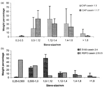
Fig. 1A illustrates size distributions for particles coated with CAP, at two different polymer/core ratios (1:3 and 1:1.7, g/g). In both cases, more than 90% (weight %) were in the range of 0.5±2 mm, with mean particle diameters of 1±1.2 mm. Regarding the acrylate particles, Fig. 1B shows
that the E S100 microcapsules were in the same size range as the CAP ones, but the E RSPO particles had a smaller mean particle diameter. Although only one batch is illu-strated in Fig. 1B for E RSPO, three other batches were sized with a different set of sieves, with identical conclu-sions. Nevertheless, it should be mentioned that, despite careful control of experimental variables, such as the stir-ring rate and temperature of solutions, particles of repetitive batches sometimes did not exhibit very reproducible size distributions.
The particles were regularly shaped (Fig. 2), with a more or less pronounced porous structure. SEM suggested that the E RSPO capsules were the least porous. Regarding the dispersion of the core material, their morphologies corre-spond to either the reservoir type, with single or multiple powder cores, or to the monolithic structure. A more de®nite conclusion is dif®cult to reach with this procedure, as powder leakage may occur when preparing the particles for the cross-sectional views.
3.2. Microencapsulation yields
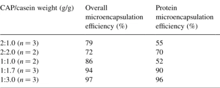
The overall yields of the procedures were evaluated by comparing the initial weight of polymer plus core material with that of the recovered, dried microparticles. The protein yields take into account only the protein content of the particles, as determined by complete dissolution studies. Table 1 lists the values of these parameters for the CAP-coated particles, at different initial weights of CAP and casein. Protein yields were above 50%, and overall process yields were above 80%, in all but one case. At low CAP/ casein ratios, these parameters were both above 90%.
Table 2 lists the same parameters for the E RSPO and E
S100 particles. For the former, and at the high polymer/core ratio of 2.5:0.5, an average of 76% of the protein was encap-sulated, but the overall yield was only 17%. This value is explained by the presence of polymer in excess, that was observed to deposit on the propeller and, to a lower extent, on the vessel wall during the preparation. At a 2:1 ratio, the yields were signi®cantly higher. With E S100, on the other hand, the protein was totally recovered in the particles, and excess of polymer occurred at ratios above 2:1 (g/g; data not shown).
3.3. Release studies
In order to simulate conditions through the GI tract, the microparticles were incubated for 2 h in acidic medium, followed by incubation in a phosphate buffer at neutral pH. Due to the high dilution of Apro in the powder mixtures with casein, the release of total protein (essentially casein) and that of Apro were measured separately, by distinct procedures. For Apro, an enzymatic method was used, which is more relevant than just peptide content, as any biological deactivation during particle preparation would be evaluated as well.
Fig. 3 shows the release pro®les for casein (total protein) from particles coated with CAP, at different weight ratios of the two materials. In all experiments, no release of protein was observed in HCl medium. After transferring to pH 6.8 buffer, the casein content in solution increased progres-sively, for approximately 3 h. There were no signi®cant differences in the release rates or in the time for total release among particles prepared with different amounts of CAP and casein (Fig. 3).
The release of Apro was followed for the particles with a CAP/casein ratio of 1:3, which were selected due to their higher yields. Fig. 4 shows that, in acidic medium, the Apro was retained in the particles to a great extent. After placing them in phosphate solution, over 90% of the Apro content was released in just half an hour. For comparison, Fig. 4 includes the release of casein.
The total protein release from the particles prepared with the two Eudragit types can be seen in Fig. 5. The release pro®les of casein from different E RSPO particles were similar, and slightly faster than the release from the E S100 ones. Analogous observation holds for the release of Apro (Fig. 6). However, while casein was completely retained in the particles in acidic medium, for Apro, some leaching was observed. During the 2 h incubation period, the E RSPO particles lost approximately half of the Apro, but the E S100 ones released only 20%, a value similar to the CAP particles. The results with E RSPO are not surpris-ing, since this polymer is not designed for enteric forms. Once in solution, it becomes slightly permeable and swells, therefore with increased pore size. Also, the smaller size of these particles (Fig. 1) may result in shorter release times.
The faster release of Apro, compared with that of casein, in phosphate buffer, is due to one or both of the following
Fig. 1. (A) Size distributions for microcapsules of casein coated with CAP, as determined by sieving analysis. Values are averages ^ SD of three batches, in each case. (B) Size distributions for microcapsules of casein coated with E RSPO or E S100, as determined by sieving analysis. The values for E S100 particles are averages ^ SD of three batches, and those for E RSPO particles are of a single representative batch.
factors. When the capsules are changed to this medium, the Apro is already hydrated, while the casein powder has to initiate its dissolution. The smaller size of Apro (MW, 6
kDa) enables it to diffuse out from the polymer structure (Fig. 2) faster or earlier than casein. The in¯uence of the MW in the release pattern was also observed in
microcap-Table 1
Yield determinations of casein microcapsules coated with CAPa
CAP/casein weight (g/g) Overall
microencapsulation ef®ciency (%) Protein microencapsulation ef®ciency (%) 2:1.0 (n 3) 79 55 2:2.0 (n 2) 72 70 1:1.0 (n 2) 86 52 1:1.7 (n 3) 94 90 1:3.0 (n 3) 97 96
a Values are averages of n batches, differing by no more than 10%.
Fig. 2. Scanning electron micrographs for microparticles of CAP, E RSPO and E S100. The voltage applied was 20 kV. Bars correspond to 100 mm: (A), 75 £ resolution; and (B), 100 £ resolution of microcapsules CAP/casein, 1:3. (C) 100 £ ; and (D), 100 £ resolution of microcapsules E RSPO/casein, 2.5:0.5. (E) 90 £ ; and (F), 85 £ resolution of microcapsules E S100/casein, 1:2. (A), (C) and (D) are entire particles, while (B), (D) and (F) are cross-sectional views.
Table 2
Yield determinations of casein microcapsules coated with E RSPO and E S100a
Eudragit type Eudragit/casein
weight (g/g) Overallmicroencapsulation ef®ciency (%) Protein microencapsulation ef®ciency (%) RSPO (n 4) 2.5:0.5 17 76 RSPO (n 2) 2:1 53 94 S100 (n 3) 2:4 98.5 98.5
sules containing casein hydrolysate as the major core mate-rial, instead of casein. That hydrolysate contains mainly single amino acids, with a few percent of short oligopep-tides. In this case, the hydrolysate was totally and quickly released in HCl medium, while the Apro was retained to a great extent (data not shown). These results, as regards CAP microcapsules in acidic solution, are also in agreement with previous ones for non-protein drugs [7], that showed that low MW molecules leach from the particles, while larger ones are retained by the polymeric structure.
We note that samples of hydrochloric acid solutions were evaluated for Apro bioactivity soon after being collected,
but that Apro is particularly resistant to inactivation by low pH environments. No loss of activity was observed after a 48 h incubation in 0.1 M HCl solution. However, with other proteins, different protocols for evaluating release and bioactivity in simulated gastric media might need to be adopted.
4. Conclusions
In this work, it was found that the single emulsion/solvent evaporation methods employed were adequate for
microen-Fig. 4. Comparison between release of total protein (as measured by the Lowry method), and release of Apro (as measured by the enzymatic assay) for microcapsules CAP/protein at 1:3 ratio. Results are averages ^ SD of three independent batches.
Fig. 5. Release of protein for microcapsules of E RSPO and E S100. The values for E RSPO/casein 2.5:0.5 and for E S100/casein 2:4 are averages ^ SD of three batches, and those for E RSPO/casein 2:1 are averages of two batches.
Fig. 3. Release of protein for CAP/casein microcapsules, as measured by the Lowry method. Results are averages ^ SD of three independent batches, in each case.
Fig. 6. Release of Apro for microcapsules coated with E RSPO and E S100. The values for E RSPO/protein 2:1 and E S100/protein 2:4 are averages of two batches, while those for E RSPO/protein 2.5:0.5 are averages of three batches.
capsulating casein and Apro in the powder forms. Also, the biological activity of Apro was not affected.
Both CAP and E S100 microcapsules represent interest-ing forms for deliverinterest-ing small MW proteins, such as Apro, to the intestine, avoiding exposure to gastric conditions. However, with CAP particles, release of the active agents can take place in a proximal part of the intestine, while the release can be delayed to lower parts of the intestine with E S100 ones. In vivo studies will elucidate the potential of these forms in dealing with the extremely common, diverse, and therapeutically challenging conditions of in¯ammation of the intestines.
Acknowledgements
This work was supported by the grant PRAXIS XXI, PCNA/C/BIO/66/96, from FundacËaÄo da CieÃncia e Tecnolo-gia, Portugal.
References
[1] R.J. Playford, C. Shaw-Smith, Growth factors and ulcerative gastro-intestinal disease, Balliere's Clin. Gastroenterol. 10 (1996) 135±149.
[2] A. Guglietta, P.B. Sullivan, Clinical applications of epidermal growth factor, Eur. J. Gastroenterol. Hepatol. 7 (1995) 945±950.
[3] W. Haedo, T. GonzaÂlez, J.A. MaÂs, S. Franco, B. Gra, G. Soto, A. Alonso, P. LoÂpez-Saura, Oral human recombinant epidermal growth factor in the treatment of patients with duodenal ulcer, Rev. Esp. Enferm. Dig. 88 (1996) 409±413.
[4] F. Araki, H. Nakamura, N. Nojima, K. Tsukumo, S. Sakamoto, Stabi-lity of recombinant human epidermal growth-factor in various solu-tions, Chem. Pharm. Bull. 37 (1989) 404±406.
[5] R.J. Playford, A.C. Woodman, P. Clark, P. Watanapa, D. Vesey, P.H. Deprez, R.C.N. Williamson, J. Calam, Efffect of luminal growth factor preservation on intestinal growth, Lancet 341 (1993) 843±848. [6] R.J. Playford, T. Marchbank, D.P. Calnan, J. Calam, P. Royston, J.J. Batten, H.F. Hansen, Epidermal growth factor is digested to smaller, less active forms in acid gastric juice, Gastroenterology 108 (1995) 92±101.
[7] J.P.S. Silva, J.P.M. Ferreira, Effect of drug properties on the release from CAP microspheres prepared by a solvent evaporation method, J. Microencapsul. 16 (1999) 95±103.
[8] S. Goto, M. Kawata, M. Nakamura, Y. Nagatsuma, K. Fujinaga, T. Aoyama, Evaluation of sustained release properties of Eudragit RS, RL and S (acrylic resins) microcapsules containing ketoprofen in beagle dogs, J. Microencapsul. 5 (1988) 343±360.
[9] M.L. Lorenzo-Lamosa, C. RemunÄaÂn-Lopez, J.L. Vila-Jato, M.J. Alonso, Design of microencapsulated chitosan microspheres for colo-nic drug delivery, J. Controlled Release 52 (1988) 109±118. [10] D.C. Egberg, R.H. Potter, G.R. Honold, The semiautomatic
determi-nation of trypsin inhibitors in textured soy protein, J. Agric. Food Chem. 23 (1975) 603±605.