Development, Characterisation and Application of
Calcium Phosphates Nanocrystals Aggregates in a
Collagen Matrix to be used as Biomaterial in Bone
Regeneration
Alis Yovana Pataquiva Mateus, MSc
.
Supervised by Professor Fernando Jorge Monteiro Mendes INEB – Instituto de Engenharia Biomédica and FEUP – Faculdade de Engenharia-Universidade do Porto Co-supervised by Dr. Maria Pia Ferraz INEB – Instituto de Engenharia Biomedica and Universidade Fernando Pessoa
Advisor: Dr. Myron Spector Senior Lecturer, Department of Mechanical Engineering Professor of Orthopaedic Surgery (Biomaterials), Harvard Medical School Submitted to the Faculdade de Engenharia –Universidade do Porto In Partial Fulfillment of the Requirements for the Degree of Doutoramento em Engenharia Biomédica April 2010
UNIÃO EUROPEIA Fundo Social Europeu
Thesis Supervisor: ________________________________________________________________
Fernando Jorge Monteiro Mendes INEB- Instituto de Engenharia Biomedica FEUP – Faculdade de Engenharia da Universidade do Porto
Thesis Co-Spervisor
________________________________________________________________ Maria Pia Ferraz INEB- Instituto de Engenharia Biomedica Universidade Fernando Pessoa
Development, Characterisation and Application of Calcium
Phosphate Nanocrystals aggregates in a Collagen Matrix to
be used as a biomaterial in Bone Regeneration
ABSTRACT
Three-dimensional scaffolds and bone proteins have shown to be important for bone tissue engineering. A scaffold for bone tissue engineering might have several applications and advantages such as: supporting cells for bone regeneration; being part of macromolecule delivery system and act as haemostatic sponge. The goal of this thesis was to develop a biomaterial to be applied for bone regeneration, as a novel means to guide bone cells towards tissue regeneration and/or to allow for local release of therapeutic macromolecules depending on what is the most adequate treatment strategy for each case.
In order to achieve this main goal, several stages were considered in this work. Production of nanometer-sized particles of hydroxyapatite at laboratory scale by chemical precipitation method. This method was selected after a thorough study of other nantechnology techniques for nanocrystals preparation and characterization. It was shown that CaP nanoparticles are similar to those present in human bone in morphology, dimensions and chemical composition. In collaboration with the Laboratory of Separations and Reaction Engineering - LSRE at Faculdade de Engenharia, Universidade do Porto, a patent on the production of nanohydroxyapatite in a novel mixed reactor was obtained.
Once the nanohydroxyapatite production method in the laboratory was made reproducible, microspheres based on nanohydroxyapatite particles were produced as a potential vehicle for transport and release of enzymes or antibiotics. Thus, in vitro studies were carried out to evaluate: nanosized hydroxyapatite performance in microspheres construction, nanohydroxyapatite microspheres as delivery system for antibiotics in periodontitis treatment, and collagen/nanoHA/SPARC (Secreted Protein Acidic and Rich in Cysteine) scaffold biocompatibility as a potential alternative biomaterial for bone cells. Results indicated that SPARC played an important role in the preparation of this new type of scaffold for tissue engineering due to its calcium and collagen binding sites. Its in vitro performance suggests a remarkable influence of SPARC on cellular adhesion and proliferation during the process of osteogenesis.
A collagen type I / III matrix supporting hydroxyapatite nanocrystals was synthesised, being a viable porous scaffold for cell proliferation. Additions of SPARC, (Secreted Protein Acidic and Rich in Cysteine) to this biomaterial is a novel approach in Bone Tissue Engineering. SPARC was used because it provides chemical binding between calcium phosphates and collagen fibers in the matrix, which make this biomaterial unique because the protein is not merely placed in the network but chemically attached, due to the SPARC ‘s calcium affinity domains.
Finally, in vivo experiments in rats were conducted to evaluate osteoinductive and osteoconductive properties of the nanoHA/Col/SPARC scaffolds. It was concluded that this biomaterial presented a similar nanostructure to bone in which hydroxyaptite nanocrystals 80x20 nm in size aggregating into microsized clusters, were well dispersed and bound to a collagen matrix through the presence of SPARC. The composite was incorporated into the remodeling
process of bone, resorbed by osteoclastic cells, and new bone was formed by osteoblasts after resorption, as if the composite were grafted into autologous bone. The highly-performing bioactivity of this composite might derive from its similarity in composition and nanostructure to bone. The nanoHA/Col/SPARC biomaterial showed to have the potential to be used as a highly demanding bioactive bone graft material.
Desenvolvimento, Caracterização e Aplicação de
agregados de nanocristais de fosfatos de cálcio numa
matriz de colagénio para ser usada como um biomaterial
para Regeneração Óssea
RESUMO
As matrizes tridimensionais (scaffolds) e as proteínas do osso demostram ter um papel relevante em engenharia do tecido ósseo. Uma matriz (scaffold) para engenharia de tecido ósseo pode apresentar várias aplicações e vantagens, tais como: Ser capaz de suportar células para a regeneração óssea; fazer parte de sistemas de libertação controlada de macromoléculas ou actuar como esponja hemostática. O objectivo principal desta tese foi o de desenvolver um biomaterial capaz de ser utilizado para a regeneração do tecido ósseo, como um novo meio de guiar células de tecido ósseo para a regeneração e/ou permitir a libertação local e controlada de macromoléculas terapêuticas, dependendo de qual é a mais adequada estratégia de tratamento em cada caso. Para se atingir este objectivo, várias fases de trabalho foram considerada, incluindo a obtenção à escala laboratorial, por precipitação química, de partículas de dimensão nanométrica de hidroxiapatite. Este método foi seleccionado após um aturado estudo sobre os diversos métodos alternativos para obter este tipo de nanocristais, e da sua caracterização. Verificou-se ser possível obter nanopartículas de fosfatos de cálcio, em particular de hidroxiapatite, semelhantes às presentes no corpo humano, que quanto à sua morfologia, quer em termos de dimensões e composição química. Em colaboração com o Laboratory of Separations and Reaction Engineering - LSRE , da Faculdade de Engenharia da Universidade do Porto, foi obtida uma patente internacional para a produção de nanohidroxiapatite num novo reactor de mistura optimizada. Quando o processo de obtenção de nanopartículas de hidroxiapatite foi tornado reprodutível, foram desenvolvidas microesferas baseadas em nanohidroxiapatite para funcionarem como potencial veículo para a libertação e transporte de enzimas e antibióticos. Foram realizados ensaios in vitro para avaliar a performance dos agregados de nanopartículas de hidroxiapatite nas microesferas, bem como a sua aplicabilidade a sistemas de libertação controlada de antibióticos para o tratamento da periodontite, e ainda a biocompatibilidade de scaffolds baseados em colagénio/ nanoHA/ SPARC, como potencial biomaterial alternativo para células de tecido ósseo. Os resultados mostraram que a SPARC desempenhou um papel importante na preparação deste novo tipo de scaffold para engenharia de tecidos dada a existência de pontos de ligação de cálcio ao colagénio e a sua performance in vitro sugeriu haver uma influência notável da SPARC na adesão celular e na proliferação durante o processo de osteogénese.
Foi sintetizada uma matriz de colagénio I/III para suportar nanocristais de hidroxiapatite, a qual se mostrou como alternativa viável de scaffold poroso para a proliferação celular. A adição de SPARC(Secreted Protein Acidic and Rich in Cysteine) a esta matriz é uma inovação em termos de aproximação à engenharia de tecido do osso. A SPARC foi utilizada por fornecer ligação química entre os fosfatos de cálcio e as fibras de colagénio, o que torna este biomaterial como
singular, dado que a proteína não está meramente presente na rede, mas antes está quimicamente ligada devido aos seus domínios de afinidade para o cálcio.
Por fim foram realizadas experiências in vivo, em ratos, para avaliar o potencial de osteocondução e osteoindução dos scaffolds de nanoHA/Col/SPARC. Concluiu-se que este biomaterial apresentava uma nanoestrutura semelhante à do osso, na qual os nanocristais de hidroxiapatite com 80x20 nm se reunem em agregados de dimensões micrométricas, bem dispersos e ligados a uma matriz de fibras de colagénio, através da presença da SPARC. O compósito foi incorporado no processo de remodelação óssea, re-absorvido pelos osteoclastos e formou-se osso novo através da acção dos osteoblastos após a reabsorção, como se o compósito estivesse integrado em osso autólogo. A elevada performance em termos de bioactividade deste compósito poderá também ter origem na sua semelhança com o osso quer em termos de composição quer de nanoestrutura. O biomaterial compósito nanoHA/Col/SPARC demonstrou ter potencial para ser utilizado como um material adequado ao preenchimento de defeitos ósseos.
Développement, caractérisation et application d’agrégats
de nanocristaux de phosphate de calcium inclus dans une
matrice de collagène pour être utilisés comme biomatériau
pour la régénération osseuse
RÉSUMÉLes matrices tridimensionnelles (scaffolds) et les protéines de l’os ont un rôle important dans l'ingénierie du tissu osseux. La matrice (scaffold) peut présenter plusieurs applications potentielles et avantages tels que la capacité de structure-support aux celules afin de promouvoir une régénération osseuse, faire partie d'un système permettant une libération contrôlée des macromolécules ou présenter un comportement similaire à celle d'une éponge hémostatique. Le principal objectif de ce travail de thèsea été de développer un biomatériau capable de guider les cellules osseuses vers une stimulation de la régénération de l’os et/ou permettre la libération contrôlée et localisée de macromolécules thérapeutiques.
Pour atteindre ce but, plusieurs étapes ont été considerées. Tout d’abord, des nanoparticules d’hydroxyapatite ont été obtenues et caractérisées au sein du laboratoire par un procédé de précipitation chimique après une étude de plusieurs méthodes alternatives pour obtenir ce type de nanocristaux. Il a été vérifié que ces nanoparticules de phosphates de calcium, en particulier d’hydroxyapatite, ressemblaient à celles présentes dans le corps humain, en termes de morphologie, de dimensions et de composition chimique. Grâce à une collaboration avec le Laboratory of Separations and Reaction Engineering - LSRE , de la Faculté d’Ingénierie de l’Université de Porto, un brevet international a été obtenu décrivant la production de nanoparticules d’hydroxyapatite au sein d’un nouveau réacteur de mélange optimisé.
Puis, des microsphères ont été développées à partir de ces nanoparticules d’hydroxyapatite, obtenues de manière reproductible, afin d’être utilisées comme véhicule pour la libération contrôlée d’enzymes ou d’antibiotiques. Des essais in vitro ont été réalisés afin d’évaluer la performance des agrégats de nanoparticles d’hydroxyapatite dans la synthèse des microphères, et d’étudier leur application comme système de libération contrôlée d’antibiotiques pour le traitement de la périodontite. La biocompatibilité de scaffolds composés de collagène/nanoHA/SPARC (Secreted Protein Acidic and Rich in Cysteine ) a été déterminée après contact avec des cellules de l’os. Les résultats ont démontré que la SPARC a un rôle très important dans la préparation de ce type de scaffold dû de l’existence de points de liaison entre calcium et collagène, et leur performance in vitro a paru indiquer qu’il y avait une influence remarquable de la SPARC sur l’adhérence et la prolifération cellulaires lors du procédé d’ostéogènese.
Permettant la prolifération cellulaire, une matrice poreuse composée de collagène de type I/III ainsi que des nanocristaux d’hydroxyapatite a été synthétisée. Le fait d’associer SPARC à ce biomatériau est une nouvelle approche en ingénierie du tissu osseux. La protéine SPARC n’est pas seulement séquestrée dans le réseau du biomatériau mais est chimiquement attachée grâce à son affinité particulière avec le calcium permettant de créer des liaisons chimiques entre le
phosphate de calcium et les fibres de collagène de la matrice.
Enfin, des expériences in vivo sur rats ont été réalisées afin d’évaluer les propriétés d’ostéoinductivité et d’ostéoconductivité des scaffolds composés de collagène/nanoHA/SPARC. Il a été montré que ces biomatériaux présentaient une nanostructure similaire à l’os dans lequel les nanocristaux d’hydroxyapatite, de taille de 80x20 nm, sont agrégés en microclusters, bien dispersés et liés à la matrice de collagène grâce à la présence de SPARC.
Après implantation, ce composite a été intégré comme s’il s’agissait d’un os autologue et soumis au processus de remodelage osseux, résorption par les cellules ostéoclastiques puis formation d’os par les ostéoblastes. Cette performance bioactive est probablement due à la forte similarité en termes de composition et nanostructure de ce composite avec l’os. Ainsi, il a été démontré que le biomatériau composé de collagène/nanoHA/SPARC peut-être utilisé comme matériau bioactif permettant la reconstruction osseuse.
A mi mamá y mi papá, Gloria y Juan Manuel, que han estado en cuerpo y alma junto a todos y cada unos mis sueños, A mi esposo, Herney, que ha sido el compañero ideal en la aventura de la vida, que es más maravillosa gracias a él A mi hija, Valeria, que tanto me ha incentivado y fortificado, incluso desde antes de nacer.
ACKNOWLEDGMENTS
After the preparation of this scientific study, many people are been present throughout this path, helping me in one way or another. To all of them my most sincere gratefulness and some day I hope to have the opportunity to be as helpful to them as they were to me.
During my research, I’ve had the wonderful opportunity to share time in Porto in Portugal at INEB and at the Tissue Engineering Lab in Boston, USA. In both places I found extraordinary people and friends.
In Porto, I would like particularly to thank my supervisor Fernando Jorge Mendes Monteiro, who was always a great guide, not only academically but also in life. Thanks for trying to rescue me in every single problem (and they were not few!) that I had to face with the scholarship and my training and staying abroad! I’m sure that I am never going to meet another young-hearted man as you in my life! Thanks for everything!
At INEB, I warmly thank Maria Pia Ferraz for giving her understanding support throughout my studies. Also, to Professor Mario Barbosa, Cristina Ribeiro, Cristina Barrias, Ana Paula Pego, Pedro Granja, Ana Queiroz, Carlos Fonseca, Merriem Lamghari and Isabel Amaral, always I’ll be very grateful with all of you. My dearest collegues: Silvia Bidarra, Hugo Oliveira, Maritie Grellier, Liliana Pires, Serafim Oliveira, Rui Azevedo, Alejandro Pelaez, Alexandra Bargue, Carla Moreira, Inês Gonçalves, Christiane Salgado, Sidonio Freitas, Judith Barbosa, Andreia Cabral, Marta Evangelista, in particular my biocomposite-buddy Sandrinha Teixeira, and all my lab mates that passed by our labs: Thank you so much! Sardinhas no molho! And, it would be impossible to pass over the huge help that I received from Ana Paula Filipe, Dulce Carqueijó, Virginia Fonseca, Ana Lopes, Patricia Cardoso, Eliana Vale and Manuela Brás.
In Boston, my scientific production was perfectly improved and shaped by Dr. Spector, who was a great mentor, a person who trusted me and my capabilities, who did cheer me up with his academic and scientific counselling. You pushed me up till getting the best of me. Thanks! On the other hand, my time at the VA (Veteran Affairs) Medical Center was very enriched thanks in particular to Dr. Hu-Ping Hsu and Alix Weaver, because they always found a way to help me; and to my colleagues from Harvard University and MIT (Massachusetts Institute of Technology): Rahmat Cholas, Kristy Shine, Paul Elias, Erica Ueda, Lily Jeng, Cathy Bolliet, Cathal Kearney, Leonide Saad, Xiaodan Sun, Tadanao Fukanoshi, Hsi-Chin Wu, Tzu-Wei Wang, Malcom Xing, and Saurab Lal.
I am deeply thankful to Dr. Eduardo Jorge Herrero from Hospital Puerta de Hierro in Madrid - Spain, for supporting me throughout experimental studies as professional and as a friend! Also I would like to thank to Dr. Antonio Lopez Bravo from Hospital Provincial de Avila - Spain, for his paramount contribution in the histological analysis.
I would like to thank also the other people who are close enough to me to support me when I’m tensed up or in a bad mood, such as all my friends, my closer family, and I think particularly of Maricel Klichuk Rocha, Maria Gonzalez y Sandra Barbosa.
Lastly, I would like to acknowledge the Portuguese Government Scholarship - FCT (Fundação para a Ciencia e a Tecnologia) for providing financial support for this amazing time of my life!
TABLE OF CONTENT
INTRODUCTION ... 23
CHAPTER I – BONE AND BIOMATERIALS BASED ON COLLAGEN AND HYDROXYAPATITE FOR BONE ENGINEERING ... 24
1. BONE ... 25
1.1CONCEPTOFBONE ... 25
1.2BONEFUNCTIONS ... 26
1.2.1 Mechanical support ... 26
1.2.2 Protection of vital structures ... 26
1.2.3 Hematopoiesis ... 26
1.2.4 Mineral homeostasis ... 28
1.2.5 Locomotion and others ... 28
1.3BONECOMPOSITION ... 28 1.3.1 Compact bone ... 28 1.3.2 Trabecular bone ... 29 1.3.3 Periosteum ... 29 1.3.4 Endosteum ... 29 1.4BONECELLS ... 29 1.4.1 Osteogenesis ... 29 1.4.2 Osteoblasts ... 30 1.4.3 Osteoclasts ... 30 1.4.4 Bone-lining cells ... 30 1.4.5 Osteocytes ... 31
1.4.6 What are Stem Cells? ... 31
1.4.7 Mesenchymal Stem Cells ... 32
1.5ANGIOGENESIS ... 33
1.5.1 Growth Factors ... 34
1.5.2 Hormones ... 37
1.5.2.1 Parathyroid hormone or PTH ... 37
1.5.2.2 Calcitrol and Vitamin D ... 37
1.5.2.3 Calcitonin ... 38
1.5.2.4 Sex hormones ... 38
1.5.2.5 Other important hormones ... 39
1.5.3 Chemotaxis ... 40
1.6MOLECULARSTRUCTUREOFBONE ... 40
1.6.1.1 Glycoproteins ... 41
1.6.1.2 Proteoglycans ... 42
1.6.2 Mineral phase and water ... 42
1.7BONEFORMATION ... 43 1.7.1 Intramembranous ossification ... 43 1.7.2 Endochondral Ossification ... 43 1.8FRACTUREHEALING ... 44 1.8.1 Reactive Healing ... 45 1.8.2 Reparative Healing ... 45 1.8.3 Remodeling ... 46 1.9BONEDISEASES ... 46 1.9.1 Osteoporosis ... 46 1.9.1.1 The problem ... 47
1.9.1.2 Symptoms and diagnosis ... 47
1.9.1.3 Clinical Management ... 48
1.9.2 Other bone diseases ... 49
1.9.2.1 Rickets and osteomalacia ... 49
1.9.2.2 Paget´s disease of bone (Osteitis deformans) ... 50
1.9.2.3 Osteogenesis imperfecta ... 50
1.9.2.4 Bone Cancer... 50
1.9.2.5 Oral health and bone disease ... 51
1.9.2.6 Osteonecrosis ... 51
2. SCAFFOLDING FOR BONE ENGINEERING ... 53
2.1 BIOCERAMICS ... 54
2.1.1 Calcium phosphates (CaP) ... 54
2.1.1.1 Dicalcium phosphate dehydrate(DCPD) ... 55
2.1.1.2 Dicalcium phosphate anhydrous (DCPA) ... 56
2.1.1.3 Tricalcium phosphate (TCP) ... 56
2.1.1.4 Hydroxyapatite ... 56
2.1.1.5 Hydroxyapatite in nanometric scale ... 57
2.1.1.6 Nanohydroxyapatite Synthesis Review ... 58
2.2 COLLAGEN ... 61
2.2.1 Structure and function ... 63
2.2.2 Collagen Porous scaffolds preparation ... 65
2.2.3 Collagen scaffolds Cross-linking ... 65
2.3 NANOCOMPOSITES ... 66
2.3.2 Nanohydroxyapatite association to Collagen ... 68
2.3.3 Bone Tissue Engineering using Col/HA composite scaffolds... 69
REFERENCES ... 70
CHAPTER II – NANOPARTICLES OF HYDROXYAPATITE: PREPARATION, CHARACTERISATION, AND CELLULAR APPROACH – OVERVIEW ... 85
1. INTRODUCTION ... 85
2. HYDROXYAPATITE PARTICLES ... 86
3. NANOHA APPLICATIONS ... 87
4. NANOHA PREPARATION METHODS ... 87
4.1 MECHANOCHEMICAL PROCESSING ... 87 4.2 SOL-GEL ROUTE ... 88 4.3 BIOMIMETIC PROCESS ... 88 4.4 HYDROTHERMAL METHOD ... 89 4.5 EMULSION TECHNIQUE ... 89 4.6 CHEMICAL PRECIPITATION ... 90
5. MATERIALS AND METHODS ... 91
5.1 NANOHA SYNTHESIS ... 91 5.2 NANOHA CHARACTERISATION ... 91 5.2.1 Physical Characterisation ... 91 5.2.2 Chemical Characterisation ... 92 5.2.3 In vitro performance ... 92 6. RESULTS ... 93 7. DISCUSSION ... 95 8. CONCLUSIONS ... 96 REFERENCES ... 97 ANEXI ...102
CHAPTER III - MICROSPHERES BASED ON HYDROXYAPATITE NANOPARTICLES AGGREGATES FOR BONE REGENERATION ...114
1. INTRODUCTION ...114
2. MATERIALS AND METHODS ...115
3. RESULTS ...116
4. DISCUSSION ...118
5. CONCLUSIONS ...118
REFERENCES ...118
CHAPTER IV– NANOHYDROXYAPATITE MICROSPHERES AS DELIVERY SYSTEM FOR ANTIBIOTICS: RELEASE KINETICS, ANTIMICROBIAL ACTIVITY, AND INTERACTION WITH OSTEOBLASTS ...120
2. MATERIALS AND METHODS ...122
2.1 MATERIALS PREPARATION AND CHARACTERIZATION ...122
2.1.1 Preparation of HA granules ...122
2.1.2 Synthesis of nanosized HA ...122
2.1.3 Alginate/ hydroxyapatite microspheres preparation ...122
2.1.4 Materials characterization ...123
2.2 DRUG LOADING ...123
2.3 RELEASE KINETICS AND BIOACTIVITY ...123
2.4 CYTOTOXICITY ...124
2.4.1 Growth of human osteoblasts - Qualitative cytotoxicity ...124
2.4.2 Direct growth of MG63 cells on the surface of materials loaded with antibiotics. ...125
3. RESULTS ...125
3.1 MATERIALS CHARACTERIZATION ...125
3.2 RELEASING PROFILE AND BIOLOGICAL ACTIVITY ...125
3.3 CYTOTOXICITY ...132
3.3.1 Growth of human osteoblasts - Qualitative cytotoxicity ...132
3.3.2 Direct growth and adhesion of MG63 cells on the surface of materials loaded with antibiotics. ...132
4. DISCUSSION ...133
5. CONCLUSION...135
REFERENCES ...136
CHAPTER V - COMPARATIVE STUDY OF NANOHYDROXYAPATITE MICROSPHERES FOR MEDICAL APPLICATIONS ...138
1. INTRODUCTION ...139
2. MATERIALS AND METHODS ...140
2.1 SYNTHESIS OF NANOSIZED HYDROXYAPATITE (HA) ...140
2.2 PREPARATION OF HA MICROSPHERES ...140
2.3 CHARACTERISATION OF NANOSIZED HA ...140
2.3.1 Infrared spectroscopy ...140
2.3.2 Transmission electron microscopy – TEM ...140
2.4 CHARACTERISATION OF NANOHA MICROSPHERES ...141
2.4.1 Inverted stand optical microscope analysis ...141
2.4.2 Infrared spectroscopy ...141
2.4.3 Scanning electron microscopy analysis – SEM ...141
2.4.4 Mercury porosimetry method ...141
2.4.5 X-ray diffractometry – XRD ...141
2.5.1 Cytotoxicity tests (Neutral Red) ...142
2.5.2 Cell morphology (SEM and CLSM) ...142
3. RESULTS ...143
4. DISCUSSION ...147
5. CONCLUSIONS ...151
REFERENCES ...151
CHAPTER VI - SPARC (SECRETED PROTEIN ACIDIC AND RICH IN CYSTEINE) IN COLLAGEN/CALCIUM PHOSPHATES SCAFFOLDS FOR BONE ENGINEERING ...155
1. INTRODUCTION ...155
2. MATERIALS AND METHODS ...156
2.1 SYNTHESIS OF HA ...156
2.2 PREPARATION OF COLLAGEN SCAFFOLDS ...157
2.3 PHYSICOCHEMICAL CHARACTERIZATION ...158
2.3.1 Analysis of pore structure ...158
2.3.2 Trace Element Analysis ...158
2.3.3 Mechanical compression testing ...159
2.4 IN VITRO CELL CULTURE STUDIES ...159
2.4.1 Seeding of GBMSCs on scaffolds ...159
2.4.2 Diameter contraction ...160
2.4.3 Cell viability ...160
2.4.4 Cell proliferation evaluation ...160
2.4.5 Histology studies ...160
2.4.6 Statistical Analysis ...161
3. RESULTS AND DISCUSSION ...161
3.1 ANALYSIS OF PORE STRUCTURE ...161
3.2 TRACE ELEMENT ANALYSIS ...161
3.3 MECHANICAL COMPRESSION TESTING ...163
3.4 DIAMETER CONTRACTION ...164 3.5 CYTOTOXICITY ASSESSMENT ...164 3.6 CELLULAR PROLIFERATION ...167 3.7 HISTOLOGY STUDIES ...167 4. CONCLUSIONS ...168 REFERENCES ...170 ANEXII ...172
HISTOLOGICAL STUDY OF GOAT BONE MARROW STEM CELLS CULTURE (GBMSCS) AND SCAFFOLDS BASED ON COLLAGEN I/III ...172
2. MATERIALS AND METHODS ...172
2.1 ISOLATION AND CELL CULTURING OF GBMSCS ...172
2.2 COLLAGEN SCAFFOLDS PREPARATION ...173
2.3 SEEDING GBMSCS ON SCAFFOLDS AND CELL CULTURE ...173
2.4 HISTOLOGY AND HISTOCHEMICAL STUDIES ...173
3. RESULTS ...174
4. DISCUSSION ...175
5. CONCLUSIONS ...177
REFERENCES ...177
CHAPTER VII - BONE REGENERATION USING A SCAFFOLD BASED ON COLLAGEN I/III, SPARC AND HIDROXYAPATITE NANOPARTICLES: AN IN VIVO STUDY ...179
1. INTRODUCTION ...179
2. MATERIALS AND METHODS ...180
2.1NANOPHASED HYDROXYAPATITE AGGREGATES/TYPE (I/III) COLLAGEN COMPOSITE SCAFFOLDS PROCESSING ...180
2.2ANIMAL EXPERIMENTATION AND OPERATIVE PROCEDURE...181
2.3HISTOLOGY ...181
2.3.1 Microscopic studies ...182
2.3.2 Statistical Analysis ...182
3. RESULTS ...182
3.1HISTOLOGICAL FINDINGS ...182
3.2CONTROL SAMPLES HISTOLOGY ...182
3.3SUBCUTANEOUS IMPLANTED SAMPLES - HISTOLOGY ...183
3.4INTRAOSSEOUS IMPLANTED SAMPLES - HISTOLOGY ...187
4. DISCUSSION ...190
5. CONCLUSIONS ...192
REFERENCES ...192
GENERAL CONCLUSIONS ...195
TABLE OF FIGURES
CHAPTER I
Figure 1. Bone composition: compact and spongy tissues. ... 25
Figure 2. Hematopoiesis pathway ... 27
Figure 3. Distribution by weight of the constituents of whole cortical bone [12]. ... 41
Figure 4. Collagen structure. ... 63
Figure 5. Collagen type I/III scaffolds following the above procedure. (Own work) ... 67
CHAPTER II Figure 1. SEM (left) and TEM (right) micrographs. NanoHA particles have been studied using these methods to characterize morphologically these nanocrystals that are presented in agglomerates of few microns. Nanometric particles of HA exhibit in both micrographs bar and needle-like shapes with 80x20nm dimensions in mean. (SEM, scale bar=500nm; TEM, 12000x) ... 94
Figure 2. XRD spectra of nanoHA en powder. It is indicated that results (blue line) fit the most of peaks (vertical lines) that exhibits natural hydroxyaptite. ... 95
Figure 3. SEM (left) and Confocal (right) micrographs of seeded microspheres with MG-63 osteoblast cells after 7 days of culture. In the left, osteoblasts proliferation was observed on nanoHA microspheres means scanning electron spectroscopy using backscattered electron imaging. In the right, a confocal laser scanning microscopy image shows aligned osteoblasts proliferating on microsphere surface where nuclei is stained in red and cytoplasm stained in green. ... 96
CHAPTER III Figure 1. SEM images from HA-1 and HA-2 powders ... 116
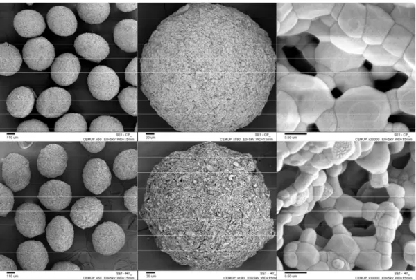
Figure 2. SEM images of HA-1 (up) and HA-2 (down) microspheres ... 117
(a, b: x50; c,d: x190 and e,f: x30000). ... 117
Figure 3. XRD spectra from HA powders and microspheres ... 117
(a) HA-1 powder, (b) HA-1 microspheres, (c) HA-2 powder, (d) HA-2 microspheres, : monoclinic HA, *:ß-TCP. ... 117
CHAPTER IV Figure 1. SEM micrograph of HA-1 microspheres (a, x50 and b, x30000) ... 126
Figure 2. Bacteria inhibition zones around the wells that were initially filled with 70 µL of amoxicillin released from HA-1 microspheres after different elution periods. ... 126
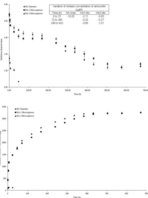
Figure 3. Bacteria inhibition zones diameter (A) and the mean cumulative concentration of release amoxicillin (B) as a function of release time. The variation of the released amoxicillin (µg/h) is inserted in (A) ... 128
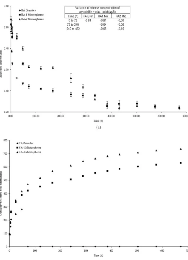
Figure 4. Bacteria inhibition zones diameter (A) and the mean cumulative concentration of release amoxicillin + clavulanic acid (B) as a function of release time. The variation of the released amoxicillin + clavulanic acid (µg/h) is inserted in (A) ... 129
Figure 5. Bacteria inhibition zones diameter (A) and the mean concentration of released erythromycin (B) as a function of release time. The variation of the released
erythromycin (µg/h) is inserted in (A) ... 130
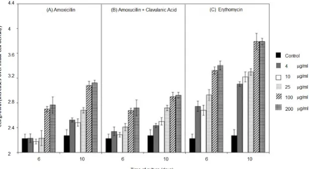
Figure 6. Effects of the presence of different concentrations (4, 10, 25 100 and 200 g/ml) of amoxicillin (A), amoxicillin + clavulanic acid (B) and erythromycin (C) on the growth of MG63 cells. MG3 cells were grown on TCPS with and without (control) antibiotics supplement, as described in materials and methods. Results are expressed as the average increase in the number of cells relative to the numbe present initially on day 0... 131
Figure 7. Effect of amoxicillin (A), amoxicillin + clavulanic acid (B) and erythromycin (C) adsorbed on HA granules, HA-1 and HA-2 microspheres, relative to the level expressed by control cells, grown on the same material without adsorbed antibiotic (defined as 100%) ... 131 CHAPTER V
Figure 1. FTIR spectra of HA-1 and HA-2 samples (powders and microspheres) ... 144
Figure 2. TEM images of HA-1 (a: x20000) and HA-2 (b:x20000) ... 145
Figure 3. SEM micrographs of of HA-1(a,c,e) and HA-2 (b,d,f) microspheres: a,b (x50) c,d (x190) and e,f (x30000)... 145
Figure 4. X-ray diffraction of powders and microspheres of HA-1 and HA-2. (a) HA-1 powder, (b) HA-1 microspheres, (c) HA-2 powder, (d) HA-2 microspheres. :
monoclinic HA, *: -TCP. ... 146
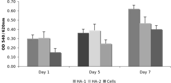
Figure 5. MG-63 cells culture on microspheres based on hydroxyapatite nanosized .... 147
Figure 6. SEM micrographs of 1 to 7 days of MG-63 cells culture on microspheres HA-1 and HA-2 (x180 and x1000). ... 148
Figure 7. SEM micrographs (left x110 and right x1000). Notice that osteoblast cells built bridges between neighbors microspheres. This image corresponds to HA-1 construct after 3 days. ... 148
Figure 8. CLSM images of HA-1 (left, x100) and HA-2 microspheres (right, x100) respectively) after 5 days of MG63 culture. ... 149 CHAPTER VI
Figure 1. Set-up nanoparticulate hydroxyapatite synthesis. ... 157
Figure 2. Multi-step process for Col/SPARC/CaP scaffolds preparation G4* and G7* groups were prepared for ICP analysis. ... 158
Figure 3. Microtomed section stained with Aniline Blue of 6 m of thickness, fom collagen scaffolds (G1). 4x, Pore diameter 208 m and apparent density of 2E-5 g/mm3, that corresponds to 89% of porosity ... 161
Figure 4. Calcium and phosphorous content into scaffolds (n=3). Controls: G1, G4* and G7*. ... 163
Figure 5. Different zones in the characteristic strain-stress curve (a). Linear elastic
modulus values (±SD) for all groups of samples (n=5) (b). ... 165
Figure 6. Diameter contraction in percentage of G1-G7 scaffolds (n=3) ... 166
Figure 7. Cell viability after 1 day and 2 weeks of GBMSCs in osteogenic medium (n=6, mean ±SD) ... 167
Figure 8. DNA content of GBMSCs cultured on the scaffolds 2 weeks of culture in osteogenic medium (n=6, mean ±SD) ... 168
Figure 9. Light micrograph of central region of goat bone marrow stem cells seeded on (a) G1 and (b) G7 scaffolds at 14 dys. H&E stain; scale bars, 100 m. ... 169
Figure A1. Scheme about goat bone marrow stem cells´ culturing. After isolation of 6 goats, cells were culture to primary culture (P0) and sub-cultured until osteoblasts differentiation (P2) and finally they were seeded on sterile scaffolds and TCPS, used as controls in a cellular density of 4x10⁶ cells/scaffold... 174
Figure A2. Light micrographs of osteblast-seeded scaffolds following 3 weeks (a, b) and 5 weeks (c, d) culture period. (a) osteoblasts only growing on scaffold surface, (b) cell concentration on scaffold surface, (c) and (d) higher population of cells conforming a expanded mass on scaffold surface where a oriented growth in layers parallel to surface is observed. In any case, cells could not colonize the inner portions of the scaffold due to the high crosslinking density (5:2:5) chosen for this test. ... 175
Figure A3. Light micrographs of histology and immunhistochemistry of osteblast-seeded scaffolds following 3 5 weeks. (a) Alizarin Red technique indicated positive result and it is observed calcium phosphate deposits inside the scaffold, (b) ALP by
immunohistochemistry where extracellular matrix formation is observed, (c) H&E staining indicating inside and superficial cell proliferation, (d) Negative Safranin-O staining to verify that stem cells did not differentiate into chondrocyte cells and did not produce cartilage tissue. Images were taken from central regions of the constructs, which appeared uniform throughout. ... 176
Figure A4. Light micrographs where different types of bone cells over 5 weeks of cell cultre in ostegenic medium are observed. OB: osteoblasts, OC: osteoclasts, OCy:
osteocytes, LC. Bone lining cells.H&E, Scale bar: 100 m. ... 177 CHAPTER VII
Figure 1. Collagen I/III control scaffolds were studied using Masson’s Trichromic acid stain differentiating collagen I to III fibers in the network, where fine and thick fibers that stain green and red, representing col I and col III respectively, were observed. ... 183
Figure 2. Small and well dispersed aggregates of small particles (black arrows) containing calcium (probably nanoHA aggregates) are observed along the collagen matrix. Mineralized and crosslinked control scaffolds (Collagen I/III + nanoHA) stained with Von Kossa. ... 184
Figure 3. Foreign body giant cells formation (black arrow) announcing the initial stages of foreign body reaction (FBR) of mineralized scaffold (Col I/III+nanoHA) after 7 days of subcutaneous implantation (H&E). ... 184
Figure 4. Foreign body giant cells formation (black arrow) announcing the initial stages of foreign body reaction (FBR), after 7 days of implantation subcutaneously. Scale bar: 40mm (H&E). ... 185
Figure 5. Firm collagen layer surrounding the mineralized scaffold created by neighbouring cells as a protective measure against the foreign body after 7 days of
subcutaneous implantation (H&E). ... 185
Figure 6. Fibroblasts proliferation conforming granulation tissue near new blood vessels, after 7 days of subcutaneous implantation (H&E). ... 186
Figure 7. Processes of fibrosis and scar formation are fully visible after 15 days of subcutaneous implantation (H&E). ... 186
Figure 8. At later healing stages after implantation, the collagen persistence is evident as well as the foreign body reaction (FBR) after 15 days of subcutaneous implantation (TC). ... 187
Figure 9. Mineralized sample (col I/III+nanoHA) implanted intraosseously after 45 days. There are three regions of the wounds: a deeper area where osteoclasts and osteoblasts coexist in large quantities, with osteoblasts depositing calcium phosphates on collagen fibers (A1), an intermediate area (A2) where the same process is occurring but delayed, and a top area where the inflammation reaction is severe, suggesting that the regeneration process has not yet occurred (A3)(RET) ... 188
Figure 10. Typical new bone formation for uncrosslinked mineralized samples implanted intraosseusly for 45 days. the presence of osteoclasts and osteoblasts in well-organized structures, divided by fibrosis trabecula (TC) is observed. ... 189
Figure 11. Structured bone between fibers with new blood vessels for uncrosslinked mineralized samples implanted intraosseusly for 45 days (H&E). ... 189
Figure 12. Old bone in the presence of increasing osseous medulla and newly organized bone and newly formed collagen fibers for uncrosslinked mineralized samples implanted intraosseusly for 45 days. (H&E). ... 190
TABLE OF TABLES
CHAPTER I
Table 1. Comparison between structures that compose skeleton [9] ... 25
Table 2. Growth factors - Overview ... 35
Table 2. Growth factors - Overview ... 36
Table 4. Apatitic and Non-apatitic calcium phosphates. [200,201]. ... 55
Table 5. Comparison of mechanical features of human bone and bioceramics [228]. ... 57
Table 6. Nanohydroxyapatite processing methods - Review. ... 59
Table 7. Various collagen types and their distribution in the body. Adapted from [283]. 62
Table 8. Tensile strength performance of different tissues - Comparison ... 65
Table 9. Tensile elastic modulus of various porous scaffolds. ... 68 CHAPTER II
Table 1. Ion concentrations of SBF solution and “human plasma”. ... 89
Table 2. Binding energies (eV) of core electrons and surface atomic ratios of apatite .... 94 CHAPTER V
Table 1. Mercury porosimetry ... 146 CHAPTER VI
Table 1. Groups of scaffold used in the presen study. G4* and G7* groups were
fabricated as controls for ICP analysis... 159 CHAPTER VII
INTRODUCTION
Transplantation provides hope for many patients with tissue loss or organ failure, but inherent limitations such as donor shortage and the required immunosuppression therapy seriously hinder the potential benefits that such an approach can provide for many patients. The problems of current transplantation therapy have stimulated research for alternative solutions. An emerging interdisciplinary and multidisciplinary field aims to recreate biologically functional tissues and organs. This field is either called tissue engineering or regenerative medicine, depending on what aspects are more deeply focused. Man-made materials, not necessarily originally designed for body part replacements, are being used as implants and prostheses. These foreign materials` lack of biological function imposes serious health risk in patients.}
Scaffolds play a critical role in tissue engineering by acting as temporary artificial extracellular matrices for cell accommodation, proliferation, and differentiated function as well as serving as three dimensional templates for some tissue/organ formation. As the knowledge in scaffolding design increases with the development of more tissue engineering research programs, there is increased need for a comprehensive studies focusing on scaffolding for tissue engineering. The present work, divided in six chapters, is intended to explore the design, synthesis and applications of a specific bone tissue engineering scaffold based on collagen and hydroxyapatite. First chapter is dedicated to bone and bone scaffolding mainly related to collagen and calcium phosphates where hydroxyapatite is outstanding; chapter two shows the design and synthesis of hydroxyapatite nanoparticles and microspheres, while chapters three and four are good examples of how nanoHA microspheres as a biomaterial could be explored as scaffolds for osteoblasts and potential drug delivery systems for macromolecules such as antibiotics for the treatment of periodontitis; finally, the potential of a composite scaffold based on type I/III collagen and nanoHA was evaluated in a rat model, as reported in chapter VII.
CHAPTER I – BONE AND BIOMATERIALS BASED ON
COLLAGEN AND HYDROXYAPATITE FOR BONE
ENGINEERING
In the last decades, the interest on new therapeutic strategies emerging from tissue engineering and gene therapy has increased in a vertiginous way, and likewise bone engineering has been performed daily with the hundreds of thousands of dental and orthopedic implants around the world, being bone the most implanted tissue after blood [1,2]. Millions of bone tissue replacements are required worldwide and for that reason many companies have had to develop new orthopedic materials. Therefore, there has been an evident research and development effort in the design of biomaterials used in substitution and bone repair. However, the current developments in the understanding of bone wound healing and the interface with artificial materials are still limited compared to what is required to engineer new materials that might provide scaffolds to grow new bone tissue in regenerative therapies [3-8].
Understanding the mechanisms of bone formation as well as the manner by which this extraordinary tissue repairs or regenerates is a good motivation for developing new and improved biomaterials aiming at mimiquing nature.
Even though the mechanisms that regulate the formation and maintenance of the skeleton are not yet well understood, it must be clarified throughout this chapter that cell-signaling molecules and receptors play critical roles in these processes; due to their number, diversity and complexity; such as therapeutic regulation of bone growth and homeostasis. Bone is not just calcium and phosphate, its proteins and macromolecules are artificers with specific call in time and space! A better understanding into the biomaterial development thought for bone regeneration consists in obtaining a conscious study of bone biology to identify “novel” solutions and possibly apply some to solve problems using synthetic materials. This subject will be discussed next.
1. BONE
1.1 CONCEPT OF BONE
Bone is a very hard and rigid organ of vertebrates due to a complex mineral substance, largely composed of calcium, phosphates and carbonates distributed throughout a soft organic matrix (Figure 1). The most remarkable fact about living bone is that it is living, that means blood circulation transports materials to and from bone (by arteries and veins, respectively), becoming it able to grow, change, remove, or suffer resorption, according to the stresses to which bone is being submitted. Besides, as a living tissue, bone is a highly specialized form of connective tissue characterized by large amounts of extra cellular matrix material (ECM), intracellular proteins and proteoglycans that hold tissue together, in the spaces between connective tissue cells. Although the skeleton is mostly constituted by bone tissue, tendons, ligaments and cartilage are also present, and all of them keep their own characteristics giving shape to the body (Table 1).
Figure 1. Bone composition: compact and spongy tissues. Source (www.wikipedia.org)
Table 1. Comparison between structures that compose skeleton [9]
Structure Water Composition Proteoglycans Composition Collagen Composition Mineral Composition Tendon/Ligame
nt Moderate Low High Low
Cartilage High High Low -
Moderate Low
In a comparison between the structures that conform the skeleton, it is interesting to note that high amounts of collagen make the tendon a tissue with good tensile strength; however, cartilage, is composed by proteoglycans that hold water and due to their presence, cartilage is stiff under compression. On the other hand, bone is more mineralized than tendon and cartilage and that induces bone to have higher compressive strength, while collagen helps bone to be tough. Sometimes, bone is called “calcified cartilage”. However, in arthroidal joints cartilage has a unique superior quality of lubrication and shock absorption, and it has a very low dry friction coefficient (≈0.0026), probably the lowest value of any know solid material [10].
1.2 BONE FUNCTIONS
Provision of support, protection, locomotion, red blood cells production, and mineral homeostasis are some of the tasks performed by bone tissue.
1.2.1 Mechanical support
Mechanical functions of bone are by far the most widely studied issue related to bone tissue. The mechanical nature of bone extends beyond basic description of strength and stiffness; it includes the developed mechanisms to avoid fatigue fracture, meaning fractures due to cyclic loading at physiologic load levels. It is interesting to highlight the fact that bone microstructure reflects the variety and diversity of mechanical requirements of each particular organism, for instance, a bovine femur can withstand considerable load and present rapid growth during lifetime, but in contrast, it is not prepared to decrease the potential for fatigue failure. On the other hand, canine femur are developed to prevent skeletal fatigue in bony structure, while not being adapted to massive weight or rapid growth [11].
1.2.2 Protection of vital structures
At the gross anatomical level, two cortical plates compose bone with a trabecular zone in between, so-called spongy or trabecular and cancellous structures. This complex array allows for maximum absorption of energy and minimum trauma to the bone itself [11,12]. Cranial bone and ribs are examples of this kind of bone that protect vital organs such as the brain in the case of skull, or the heart and lungs for ribs.
1.2.3 Hematopoiesis
Hematopoiesis is the process by which mammalian stem cells differentiate into several kinds of blood cell within the bone marrow [13]. In adults, hematopoiesis occurs in the spongy tissue at bone with large surface areas for the production of red blood cells, such as the iliac crest, vertebrae and proximal femur. These bones also have the responsibility for rapid bone turnover and play an important role in long-term control of calcium balance [14].
All lymphocytes are originated during this process from a common lymphoid progenitor before differentiating into their distinct lymphocyte types, which are Natural Killer cells (NK cells); and B and T cells [15].
Figure 2. Hematopoiesis pathway
Hemocytoblast or multipotential hematopoietic stem cells convert into the common lymphoid progenitors, and these turn into lymphoblast from which prolymphocytes and lymphoid are derived. Prolymphocytes give rise to small lymphocytes, such as B and T lymphocytes, and natural killer (NK) lymphocytes [16]. B cells mature into B lymphocytes in the bone marrow, whereas T cells migrate and mature in the thymus. After maturation these lymphocytes enter the circulation and peripheral lymphoid organs (spleen, lymph nodes) where they survey for invading pathogens and/or tumor cells [13].
NK cells are part of the innate immune system, whereas T and B cells are part of the adaptive immune response. While NK cells defend the host from both tumors and cells infected by viruses; T and B cells recognize “non-self antigens” and respond to pathogens [16]. Even, their pathway of attack is different: NK cells release small cytoplasmic granules of proteins called perforin and granzyme that cause the target cell to die by apoptosis or necrosis; B cells respond to pathogens by producing large quantities of antibodies which then neutralize foreign objects like bacteria and viruses; and T cells, such as helper T cells, produce cytokines that control the
immune response, others such as cytotoxic T cells, produce toxic granules that induce the death of pathogen infected cells [13,15,16].
1.2.4 Mineral homeostasis
Bone, with 99% of the body’s calcium, is not just the main storage for calcium but also for phosphorous ions, and many other ions that are needed for the adequate function of a variety of systems within the body, proper enzyme reactions, satisfactory blood clotting and transmission of nerve impulses [17]. Although some amount of calcium is found in trabecular bone, the largest volume comes from dense cortical bone.
1.2.5 Locomotion and others
Skeleton, mainly constituted by bone tissue, forms a system of levers to which the voluntary muscles are attached, allowing controlled and guided movement.
Bone also acts in the acid-balance of the body, buffering the blood against excessive pH changes, through the absorption or release of alkaline salts [11]. Also, bone tissue can store foreign elements like heavy metals, eliminating them from the blood for excretion and reducing their effect on other tissues. Besides, underwater hearing is by bone conduction [18,19] and localization of sound appears to depend on differences in amplitude detected by the conduction of sound to the inner ear through the bones of the skull.
1.3 BONE COMPOSITION
Bones are composed by two types of tissues: compact and trabecular, and are covered by two membranes: periosteum and endosteum. Compact bone forms a rigid outer shell that resists deformation, while trabecular network provides strength with low mass due to the complex system of internal support by lamelae.
1.3.1 Compact bone
Compact bone, also called dense or cortical bone is a hard outer layer, which gives a smooth, white and solid appearance, and minimal gaps and spaces are found in it. 80% of total bone mass of an adult skeleton corresponds to dense bone. At microscopic level, cortical structure has a characteristic pattern of arrangement: lamellae and ground substance are arranged in concentric layers; Haversian channels found parallel to the long axis of the bone; Volkmann’s channels that are interconnected with the Haversian channels and contain blood and lymph vessels and are innerved; lacunae are the cavities that occur at regular intervals in the concentrated layers; canaliculi interconnect several lacunae and Haversian channels; and the Haversian systems are the architecture formed by Haversian channels, lamellae, lacunae and canaliculi [11,12].
1.3.2 Trabecular bone
Trabecular bone, also named as cancellous or spongy bone, is the filling of the inner part of bone. It is arranged as open cell porous networks that generate room for blood vessels and marrow. Presenting a ten fold increased surface area with respect to cortical bone, spongy bone holds the remaining 20% of total bone mass [11]. In bones with a substantial weight-bearing function it is the trabecular pattern that offers the maximum resistance to physical stress.
1.3.3 Periosteum
Periosteum is a dense but thin fibrous membrane that covers the entire outer surface of a mature bone, not including the articulating joints, and serves as an attachment for tendons and muscles. The inner layer of periosteum, so-called osteogenic layer, contains the highly active cells that produce circumferential enlargement and remodeling of the growing of long bone. On the other hand, the outer layer is fibrous and comprises the entire periosteum [10]. Periosteum, attached to bone by strong collagenous fibers called Sharpey’s fibers, is irrigated by blood vessels that nourish the surrounding bone and nerves, making it very sensitive to manipulation [17]. In a injury, cells originating from the inner periosteal layer become osteogenic [10].
1.3.4 Endosteum
Endosteum, on the other hand, is the thin layer of vascular connective tissue lining the marrow cavity, and holds morphology and function similar to those of periosteum [14,17].
1.4 BONE CELLS
Bone, a highly vascularised, innervated and mineralized conjunctive tissue, is composed of many different types of cells such as osteoclasts, osteoblasts, lining cells and osteocytes; that build structures based on lamellae of calcified matrix. The arrangement of these lamellae determines whether the bone is cortical or cancellous. Both cortical and trabecular bone contain specialized cells, organic matrix and mineral phase.
1.4.1 Osteogenesis
Osteogenesis is dynamic process of laying down new bone material by osteoblasts. It occurs in two different ways: either by Intramembranous osteogenesis that is the direct laying down of bone into the primitive connective tissue (mesenchyme), or by endochondral osteogenesis that involves a cartilage precursor [20].
There are three main types of bone cells [21] (See Fig. 3, [22]): osteoblasts, responsible for bone formation; osteoclasts, responsible for bone resorption; and osteocytes, fully differentiated osteoblasts that have become embedded in matrix whose function is not clear [23]. Each type of cells will be discussed next.
1.4.2 Osteoblasts
Osteoblasts are small (20-30 m), polyedric, and mono-nucleated cells originated from bone marrow, and they are the responsible of producing new bone named osteoid, the calcified matrix is mainly based on collagen type I and osteonectin, controlling calcium and mineral deposition [12], carrying out these osteoblast communications by protrusions or cell-cell contacts [24]. Active osteoblasts cover the bone surface as a sheath as a layer of plump cells, closely together along the surface of bone [24].
The osteoblast structure is grossly based on nucleus, with a single nucleoli; Golgi area, responsible for collagen synthesis; and rough endoplasmatic reticulum, parallel to cellular membrane that contains ribosomes; these two last acquire a prominent size when osteoblasts evolve towards osteocytes [24], due to their new role as osteoid builders. The whole structure of osteoblasts plays an active role in the synthesis of the proteins and polysaccharides of the bone matrix.
The half life time of human osteoblasts goes from 1 to 10 weeks, when they become less active, turning into the so-called bone–lining cells, beginning to elongate, becoming flat and staying parallel to the bone surface, decreasing their volume and increasing their base, and from that moment on, they are not involved in bone formation anymore, producing gaps between cells [24]. At the end of their life cycle, osteoblast may disappear by apoptosis mechanisms, or transform into both bone-lining cells or osteocytes [25].
Functions of osteoblasts into the osteodynamics are synthesis of collagenous and non-collagenous proteins of the organic matrix of bone; to direct the fibrils of the ECM (extracellular matrix), mediate the resorption process carried out by osteoclasts by means of specific cytokines [26]; and synthesis of growth factors [27].
1.4.3 Osteoclasts
Large and multi-nucleated cells that dissolve bone are named osteoclasts (100 m) rich in mitochondrias and vacuoles, which are originated from bone marrow hematopoietic stem cells known as “Granulocyte-Macrophage Colony-Forming Units” (GM-CFU), precursors of macrophages and monocytes [28,29]. Osteoclasts have two special features in the membrane: a ruffled border, where resorption takes place, and a clear area rich in microfilaments, with integrins that serve as an anchor to the matrix. To this end, the osteoclasts move towards the area to be resorbed and then immediately adhere to the mineralized bone surface with the ruffled border and sealing the edges of the area with the integrins.
1.4.4 Bone-lining cells
Mature osteoblasts become bone-lining cells with flat appearance, and are thought to regulate the exchange of calcium and phosphorus ions into and out of the bone and respond to hormones by producing special proteins that activate osteoclasts.
1.4.5 Osteocytes
Osteocytes derive from mature osteoblasts and can sense pressure or cracks in bone and may help to direct osteoclasts to the locations where they will dissolve bone [30]. Cortical or compact bone, containing osteocytes, is arranged concentrically around Haversian Channels. Cancellous or trabecular bone is formed by a network of bone lamellae, delimiting areolar cavities where the bone marrow is found [31].
Originated from osteoblasts, osteocytes, decrease about 30% when compared to osteoblast volume, at the expense of the cytoplasm so that the nucleus becomes the outstanding feature of the cell surrounded by giant rough endoplasmatic reticulum and Golgi area structures [24]. Osteocytes get smaller while filling the lacuna in which they are lying with bone matrix and elongate and orientate along axis parallel to collagen fibrils [12]. In histology, the study of osteocytes depends on the way of cutting, whether the cut is done longitudinally or transversally. In the first case, osteocytes can be observed flat and elongated and in the second they appear rounded or in triangular shapes [24].
1.4.6 What are Stem Cells?
Stem cells, name proposed by the Russian histologist Alexander Maksimov in 1908, are non-specialized (undifferentiated) cells that have the extraordinary capacity to renew themselves for long periods, and develop into many different cell types in the body under certain physiologic or experimental conditions [32]. Each new cell resulting from the division of a stem cell has the potential to either remain a stem cell or become another type of cell but with a special function, i.e. brain cells, beating cells of the heart muscle, muscle cells, insulin-producing cells, bone cells, among others [33].
Research studies are conducted with two kinds of stem cells from animals and humans depending their source; these are embryonic stem cells, which are undifferentiated cells derived from embryos developed from eggs that have been fertilized in vitro and then donated for research purposes with informed consent of the donors; and adult or somatic stem cells, which are undifferentiated cells found in a differentiated tissue, or in the blood stream, that can renew themselves and differentiate (with certain limitations) to develop in all the specialized cell types of the tissue from which they are originated. However, scientists do not agree about whether or not adult stem cells may develop into all cell types.
A stem cell requires two properties, the first one is self-renewal capability that is the ability to go through numerous cellular cycles of division while remaining in the undifferentiated state; and the second is about potency that is the capacity to differentiate into specialized cell types.
In a rigorous sense, this requires stem cells to be either totipotent; being produced from the fusion of an egg and sperm cell that are able to construct a complete, viable, organism [33]; or pluripontent; descendants of totipotent cells; have the capacity to self-renew and be able to give
rise to any of the three somatic germ layers that comprise an organism (endoderm, mesoderm or ectoderm) [32].
Although multipotent; stem cells that can differentiate into those cells of a closely related family; or unipotent progenitor cells; stem cells that can produce only one cell type; are sometimes referred to as “stem cells” even when they are capable of differentiating into a limited number of tissue types.
Embryonic stem cells, the most studied pluripotent stem cells [34], and adult stem cells represent valuable sources of cells for applications in cell therapy, drug screening and tissue engineering. Fetal stem cells are primitive cell types found in the organs of fetuses [35]. The classification of fetal stem cells is often grouped within adult stem cells. However, a more clear distinction between the two cell types appears to be necessary.
Many efforts have been done to maintain their self-renewal and differentiation capabilities that are critical when expanding stem cells in culture.
The process of stem cells differentiation is conducted by internal signals that mean that a cell’s genes are interspersed across long strand of DNA and carry coded instructions for all the structures and functions of a cell. On the other hand, external signals for cell differentiation include chemicals secreted by other cells, physical contact with neighboring cells, and certain molecules in the microenvironment (extracellular matrix).
Certainly, biomedical research concerning cell-based therapies, treatments where stem cells are induced to differentiate into a specific cell type required to repair damaged or destroyed cells or tissues, is focused in establishing precisely how stem cells remain unspecialized and self renewing for many years; and in recognizing the signals that cause stem cells to become specialized cells. However, several questions remain requiring answers such as: why can embryonic stem cells proliferate for a year or more under experimental conditions without differentiating in contrast to adult stem cells; and what are the factors in living organisms that normally regulate stem cell proliferation and self-renewal. These questions focus nowadays the attention of many scientists.
1.4.7 Mesenchymal Stem Cells
Mesenchymal stem cells or MSCs are multipotent stem cells that can differentiate into a variety of cell types. Cell types that MSCs have been shown to differentiate into in vitro or in vivo include osteoblasts, chondrocytes, myocytes, and adipocytes [36].
MSCs are characterized morphologically by a small cell body with a few cell processes that are long and thin. Their cell body contains a large, round nucleus with a prominent nucleolus which is surrounded by finely dispersed chromatin particles, giving the nucleus a clear appearance [37]. The remaining cell body contains a small amount of Golgi apparatus, rough endoplasmic reticulum, mitochondria, and polyribosomes. The cells, which are long and thin, are widely
dispersed and the adjacent extracellular matrix – ECM is populated by a few reticular fibrils but is devoid of the other types of collagen [38].
1.5 ANGIOGENESIS
Angiogenesis is an important physiological process involving the growth and development of new blood vessels both in health and in disease, from pre-existing vessels as well as in wound healing, where various angiogenic proteins including several growth factors stimulate it [39]. Endothelial progenitor cells are bone marrow-derived cells that circulate in the blood and have the ability to differentiate into endothelial cells, the cell that make-up the lining of blood vessels. Endothelial cells are selective filters that regulate the passage of gases, fluid and various molecules across their cell membranes. Different organs have different types of endothelium: some leaky and some very tightly bound [39].
Vascular endothelial cells, which divide only about once every three years in average, form the walls of blood vessels; nevertheless angiogenesis can stimulate them to divide and regulate by both activator and inhibitor molecules when it is required.
Normal angiogenesis occurs in the human body at specific times in development and growth. A process called vasculogenesis creates the primary network of vascular endothelial cells that will become major blood vessels. Afterwards, angiogenesis remodels this network into the small new blood vessels or capillaries that complete the body’s circulation system [39].
In addition, angiogenesis is active in females a few days each month during the menstrual cycle, as new blood vessels form in the lining of the uterus to mature the egg during ovulation, and during pregnancy to establish the circulation between mother and fetus called placenta. As expected, angiogenesis is necessary for the repair or regeneration of tissue during wound healing [40].
The body controls angiogenesis by producing a precise balance of growth and inhibitory factors in healthy tissues. When this balance is disturbed, the result is either too much or too little angiogenesis. For instance, in response to hypoxia, or low O2 pressure, mammalian cells increase the expression of a multitude of genes to stimulate cellular processes blood vessels growth [41]. Abnormal blood vessel growth, either excessive or insufficient, is now recognized as a “common denominator” underlying many deadly and debiliting conditions, including cancer, skin diseases, age-related blindness, diabetic ulcers, cardiovascular disease, stroke, and many others [42]. The list of diseases that have angiogenesis as an underlying mechanism grows longer every year. How do new blood vessels grow? The cascade of events that leads to blood vessels growth starts with the release of angiogenic growth factors (proteins) that diffuse into the nearby tissues, and bind to specific receptors placed on the endothelial cells (EC) or nearby pre-existing blood
vessels. After this, EC become activated and signals are sent from the cell’s surface to the nucleus, where new molecules, such as enzymes, are produced to dissolve tiny holes in the basement membrane surrounding all existing blood vessels. Afterwards, endothelial cells proliferate and migrate towards the diseased tissue; and integrins, specialized molecules of adhesion, serve as grappling hooks to help pull the sprouting new blood vessel sprout forward. Extra enzymes, such as matrix metalloproteinases – MMP, are produced to dissolve the tissue in front of the sprouting vessel tip to accommodate it. As the vessel extends, the tissue is remolded around the vessel rolling up to form a blood vessel tube. Interconnection between blood vessels permits blood circulation, and specialized muscle cells provide structural support to the network and finally, blood flows then begins [40,42].
Some years ago, the controversy over whether blood stem cells were created, or born, in the endothelium or originated from another cell type in a nearby location, came to the end when scientists proved definitively that blood stem cells are made during mid-gestational embryonic development by endothelial cells.
Although normal tissues have an automatic mechanism to attract an increased blood supply when they require it, many tumors achieve rapid growth by switching on enhanced production of angiogenic signals. The new blood vessels supply the tumor with nutrients and oxygen, and they may also provide an easier escape route for metastatic cells [43]. Therefore, angiogenesis became one of the most studied biological processes by oncology scientists using in vitro techniques that require the addition of endothelial cells to the engineered skin. Endothelial cells may organize into vascular structures in culture with the aid of biomaterial scaffolds and coculture (i.e. fibroblasts and human umbilical vein endothelial cells HUVEC) with accessory cells, and vascular smooth muscle cells in a collagen matrix [44,45].
1.5.1 Growth Factors
Growth factors are proteins (see Table 2), which act as signaling molecules between cells, capable of stimulating cellular growth [46], proliferation, differentiation and maturation; and regulating a variety of cellular processes [47].
ABBR. NAME FUNCTION
BMP
BONE MORPHOGENETIC
PROTEINS
Group of twenty growth factors and cytokines known for their ability to induce the formation of bone and cartilage, interacting with specific receptors on the cell surface: bone morphogenetic protein receptors – BMPRs. BMPs have an important role during embryonic development on the embryonic patterning and early skeletal formation [47].
BMP CARTILAGE BONE Involved in development of
BMP-1
BMP-2 Osteoblast differentiation
BMP-3
BMP-4
BMP-5
BMP-6 Joint integrity in adults
BMP-7 Osteoblast differentiation
BMP-8a
FGF FIBROBLAST
GROWTH FACTOR
Heparin-binding proteins involved in angiogenesis, wound healing, and embryonic development that interact with cell-surface associated heparin sulfate proteoglycans. FGFs play an important role in the processes of proliferation and differentiation of a considerable variety of cells and tissues [48,49].
FGF Families Function
FGF-1 to FGF-10 Bind fibroblast growth factor receptors FGFRs
FGF-11 to FGF14 Named iFGF, are involved in intracellular processes unrelated to FGFs
FGF-16 to FGF-23 Have systemic effects and they are not well characterized
VEGF
VASCULAR ENDOTHELIAL GROWTH FACTOR
They are more specific of platelet-derived growth factor family of cystine growth factors and are important signaling proteins involved in both vasculogenesis and angiogenesis.
They stimulate cellular responses by binding to tyrosine kinase receptors – VEGFRs, on the cell surface, causing them to dimerize and become activated though trans-phosphorylation, although at different sites, times and extents.
VEGF type Function
VEGF-A It has been mostly studied on cells of the vascular endothelium, and it has shown to stimulate endothelial cell mitogenesis and cell migration. It is also vasodilator and increases microvascular permeability and increases the interior of a vessel named lumen.
VEGF-B Embryonic angiogenesis
VEGF-C It is involved in angiogenesis, lymphagiogenesis and endothelial cell growth and survival, and can also affect the permeability of blood vessels. VEGF-D This protein is structurally and functionally similar to vascular endothelial growth factor VEGF-C. It is active for the development of lymphatic vasculature surrounding lung bronchioles. PIGF Placenta growth factor is important for vasculogenesis, angiogenesis during ischemia, inflammation, wound healing and cancer.
ABBR. NAME FUNCTION
PDGF PLATELET-DERIVED
GROWTH FACTOR
PDGF, composed of two polypeptide chains that may exist as a homodimer (-AA, -BB) or heterodimer (-AB), enhance wound repair [50], support angiogenesis [51,52], and stimulate cell proliferation in the fetal rat calvarial system and in cultures of osteoblast-like cells derived from adult human bone explants [53].
The role of PDGF in osteoblast differentiation may be to increase the number of cells that can progress into osteoblastic lineage and express the osteoblast phenotype [54].
PDGF expression at fracture sites in addition to its mitogenic effects indicates a role for PDGF in wound healing and fracture repair [52]. PDGF and EGF are broad-specificity factors that can stimulate many types of cells to divide, such as fibroblasts, smooth muscle cells and neuroglial cells [55].
TGF TRANSFORMING
GROWTH FACTOR
TGF is used to describe two classes of polypeptide growth factors: TGF- and TGF-. Mainly found in bone, platelets, and cartilage TGF- triggers growth, differentiation, and extracellular matrix synthesis [56].
In addition to mitogens that stimulate cell division, there are factors, such as some members of the TGF- family, that act on some cell to stimulate cell proliferation and others to inhibit it, or that stimulate at one concentration and inhibit at another [57].
The effect of TGF over osteoclast differentiation, appears to depend on the mixture of cell types and cytokines that are present in the microenvironment in which osteoclast differentiation occurs [58]. Also, TGF- causes cell retraction, thus leading to greater bone surface exposure, increasing osteoclast precursor attraction to the bone surface [59].
![Figure 3. Distribution by weight of the constituents of whole cortical bone [12].](https://thumb-eu.123doks.com/thumbv2/123dok_br/15727191.1071102/41.892.170.726.162.566/figure-distribution-weight-constituents-cortical-bone.webp)
![Table 3. Calcium, phosphate and carbonate species present in bone. Modified from [12]](https://thumb-eu.123doks.com/thumbv2/123dok_br/15727191.1071102/43.892.203.702.180.332/table-calcium-phosphate-carbonate-species-present-bone-modified.webp)
![Table 5. Comparison of mechanical features of human bone and bioceramics [228].](https://thumb-eu.123doks.com/thumbv2/123dok_br/15727191.1071102/57.892.114.757.779.915/table-comparison-mechanical-features-human-bone-bioceramics.webp)
![Table 7. Various collagen types and their distribution in the body. Adapted from [283]](https://thumb-eu.123doks.com/thumbv2/123dok_br/15727191.1071102/62.892.124.777.369.959/table-various-collagen-types-distribution-body-adapted.webp)