*e-mail: [email protected]
1. Introduction
Typically, a bone cement is used to mechanically ix implants to bone. However, in order to have a cement able to bond chemically to bone it is necessary to develop a bioactive material that stimulate the regeneration of osseous tissue. The bond mechanism of implant-tissue is based on the formation of a bioactive calcium phosphate layer on the implant surface by reaction and ion exchange. The ability to form a bioactive layer at the interface between material and living tissue was observed in 1969 and this phenomenon was deined as bioactivity1.
Since the discovery of Bioglass, which promotes a rapid osseointegration2,3, a great variety of bioactive materials
for medical applications, such as glasses, glass-ceramics, ceramics and composites, have been developed4,5. The
main requisites of a bioactive material are biocompatibility, appropriate mechanical properties and the ability to form an HA layer on its surface6.
The application of biomaterials depends upon their interfacial properties and resultant interactions with cells and biological luids in vivo7. The interaction of red blood
cells with biomaterials or their extracts may be useful to evaluate the hemolytic activity of materials. For decades, evidence of the in vitro hemolysis has been used to identify the biocompatibility properties of biomaterials8.
A great variety of bone cements have been developed and they are used for ixing, reparation or replacement of bone9. In recent years, research has been focused on the
development of calcium phosphate cements10,11, however
the low mechanical strength of these materials limits their use to replace bone in high load bearing applications. Additionally, calcium aluminates cements have been used as iller materials in dental and orthopedic applications12.
These cements show long setting time (135 min) and low compressive strength (46.33 MPa for 24h), which limit their use in some medical applications13.
The calcium sulphoaluminate cement develops early high strengths14, higher than those reported for calcium aluminate
and calcium phosphate cements. This high strength is due to the formation of ettringite (Ca6Al2(SO4)3(OH)12.26H
2O)
during hydration of CSAC. In addition, these cements have a short setting time in the range of 45-60 min without any accelerating additive. Due to this and taking into account that no bioactive sulphoaluminate cements have been reported, in this work the in vitro bioactivity and hemocompatibility of a calcium sulphoaluminate cement were assessed.
2. Experimental
2.1. Cement preparation
The clinker of calcium sulphoaluminate (CSA) was obtained from a mixture of reagent grade chemicals (Sigma Aldrich) of calcium carbonate (CaCO3), alumina (Al2O3) and hemi-hydrated gypsum (CaSO4·1/2H2O). The mixture was homogenized in a plastic jar with alumina balls in acetone for 4 h using milling rods, then the mixture was dried at 80 °C, deagglomerated and heat treated at 1350 °C for 4 h. The obtained clinker was milled in a porcelain jar with alumina
Bioactive and Hemocompatible Calcium Sulphoaluminate Cement
Iván Omar Acuña-Gutiérreza*, José Concepción Escobedo-Bocardoa, José Manuel Almanza-Roblesa,
Dora Alicia Cortés-Hernándeza, Mirna María Guadalupe Saldívar-Ramíreza,
Perla Janet Reséndiz-Hernándeza, Alejandro Zugasti-Cruzb
aCINVESTAV-IPN Unidad Saltillo, Ave. Industria Metalúrgica No. 1062, Parque Industrial
Saltillo-Ramos Arizpe, CP 25900, Ramos Arizpe, Coah., México
bFacultad de Ciencias Químicas, Universidad Autónoma de Coahuila, Blvd. V. Carranza y
José Cárdenas, Apdo. Postal 935, CP 25280, Saltillo, Coah., México
Received: June 16, 2014; Revised: January 11, 2015
Calcium sulphoaluminate cement (CSAC) is an attractive candidate for biomedical applications due to its appropriate mechanical properties and high calcium content. In vitro bioactivity and hemocompatibility of calcium sulphoaluminate cement were assessed. The cement was prepared from a mixture of calcium sulphoaluminate (CSA) clinker, gypsum and water. Cement samples were immersed in a simulated body luid (SBF) at 37 °C for different periods of time (7, 14 and 21 days). The analyses of these samples after their immersion in SBF revealed the formation of a bonelike apatite layer on their surface. The hemolytic activity was assessed by measuring hemoglobin released from erythrocytes when they were exposed to the cement. The results showed that the cement is hemocompatible.
balls until reaching a surface area of 531 m2/kg with an
average particle size of 12 μm. The obtained clinker was analyzed by X-ray diffraction (XRD; Philips, Xpert). The cement (CSAC) was prepared by mixing the clinker (80 wt-%) with CaSO4·1/2H2O (20 wt-%), then water was added using a water/cement ratio of 0.5 and casted into nylamid molds of 1 cm in diameter and 0.5 cm in height. Samples were set for 1 h at room temperature and assessed for in vitro
bioactivity. After hydration the samples were immersed in ethanol for 1 h and dried for 24 h at room temperature and then were analyzed by XRD.
2.2. In vitro bioactivity assessment
The SBF was prepared following the procedure described by Kokubo & Takadama15. Appropriate amounts
of the following reagent grade chemicals were dissolved into deionized water: NaCl, NaHCO3, KCl, K2HPO4·3H2O, MgCl2·6H2O, HCl, CaCl2 and Na2SO4; tris(hidroximetil)-aminometane was used to adjust pH to 7.4 at 37 °C.
After setting, samples were immersed in 200 ml of SBF in a polyethylene lask and placed into an incubator at 37 °C for 7, 14 and 21 days under sessile conditions. The area to volume ratio was 1.57 mm2/ml. After immersion, samples
were soaked in ethanol for 1 h to stop any other reaction and then dried for 24 h and stored in a desiccator. Cement samples were analyzed before and after immersion in SBF by scanning electron microscopy (SEM; Philips, XL30 ESEM), energy dispersive spectroscopy (EDS; EDAX, Pegasus), Fourier transformed-infrared spectroscopy (FT-IR; Nicolet, Avatar 360) and XRD. For comparison purposes, a CSAC sample was hydrated in water for 21 days and characterized by FT-IR.
The calcium, phosphorus, sulphur and aluminum concentrations of the remaining SBF’s were evaluated after 1, 7, 14 and 21 days of immersion of CSAC samples by inductively-coupled plasma atomic emission spectrometry (ICP-AES; Thermo Elemental, IRIS Intrepid II XSP). The pH values of the remaining SBF’s were also monitored at different periods of time (Thermo Orion 420 pH meter).
2.3. Hemolysis tests
Hemolysis is deined as the release of hemoglobin due to the damage to the erythrocytes membrane. This type of testing allows the in vitro evaluation of the hemolytic activity of biomaterials or their extracts. In the hemolysis tests both extract and cement were assessed. The aim of hemolysis testing of the extract was to determine if the cement leachate products were hemolytic, while the objective of the hemolysis testing of cement was to determine if the direct contact between the cement surface and red blood cells may cause hemolysis.
Fresh human blood from a volunteer donor was obtained. The blood was collected in tubes with anticoagulant (EDTA) and centrifuged at 3200 rpm for 4 min. The obtained pellets were washed three times with phosphate-buffered saline (PBS). The supernatant was then removed and 1 ml of the puriied erythrocytes was diluted with PBS (1:9). The cement specimens were prepared by mixing clinker (80 wt-%) with CaSO4·1/2H2O (20 wt-%), then water was added using a water/cement ratio of 0.5 and the paste was casted into nylamid molds of 0.6 cm in diameter and 1.4 cm
in height. Specimens were hydrated in SBF for 24 h at 37 °C before testing. The hemolysis tests were performed placing the specimens into tubes with a volume of 1800 µl of PBS for 30 min at 37 °C in a water bath. In the case of the cement extract tests, the specimens were taken out of the tubes. Then, 200 µl of the blood solution were added to the tubes containing the extract or the specimens and were incubated for 1 h at 37 °C in a water bath. A positive and a negative controls are required in this test. The negative control (0% hemolysis) was prepared by adding an isotonic solution; in this case PBS was used. The positive control (100% hemolysis) was prepared by adding deionized water. According to the ASTM F756 standard, PBS is used as a blank in the hemolysis testing due to the fact that this solution keeps pH and the anticoagulant properties of the chelating agents used in collecting the blood. PBS is not hemolytic. Blood cells require an isotonic medium; it means a solution with the same amount of solute than cells. PBS is a common solution used to preserve cells. However, when cells are exposed to a hypotonic medium, a lower solute concentration outside the cells is present. This fact leads to a water absorption by cells in order to compensate the solute imbalance, leading to the expansion and bursting of cells, releasing hemoglobin. Deionized water is a hypotonic medium due to its very low amount of solutes.
Six experimental replicates were used for each group. After incubation, samples were centrifuged at 3200 rpm for 4 min at room temperature to collect the supernatant. The absorbance (A) value of the hemoglobin released from the erythrocyte cells was measured spectrophotometrically at 545 nm. Hemolysis percentage was calculated as follows:
(
)
(
samplepositive controlnegative controlnegative control)
A A /
Hemolysis (%) 100
A A − =× − (1)
3. Results and Discussion
Figure 1 shows the XRD patterns corresponding to the clinker of CSA (1a) and to the cement after hydration (1b). The only detected phase in the clinker was calcium
sulphoaluminate (4CaO·3Al2O3·SO3) (Figure 1a) and the main hydration product corresponded to ettringite (Ca6Al2(SO4)3(OH)12.26H
2O) (Figure 1b). There was an
indication that the hydration was not completed since some CSA remained without reaction.
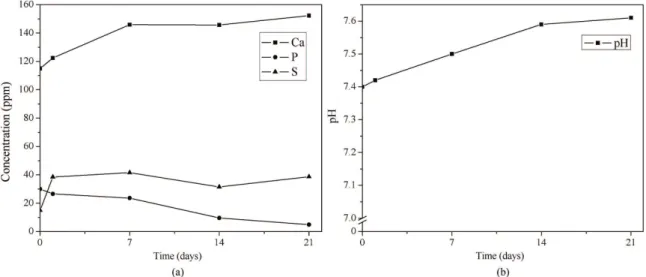
Figure 2 shows the calcium, phosphorus and sulphur concentrations (Figure 2a) and the pH behavior (Figure 2b) of the remaining SBF’s as a function of immersion time. Initially, the calcium and sulphur concentrations increase in the SBF and then keep almost constant up to 21 days of immersion. This increase is due to the partial dissolution of ettringite in SBF. In contrast, the concentration of phosphorus decreases. The depletion of phosphorus in SBF may be due to the formation of a Ca, P-rich compound on the CSAC. On the other hand, aluminum in the remaining SBF’s was detected after only 21 days of immersion showing a concentration of 0.015 ppm. Reported values of Al in human plasma of healthy subjects indicated that the content of this element varied from 0.003 to 0.039 ppm16. The Al
concentration in the remaining SBF of this work, 0.015 ppm, is within this range. The ettringite dissolution leads to the formation of Al(OH)3 (solid)17 and Ca2+ and SO
4 2-. The
pH values of SBF increased from 7.4 (before immersion) to 7.6 after 21 days of immersion. This slight change in pH indicates that an ionic exchange has occurred.
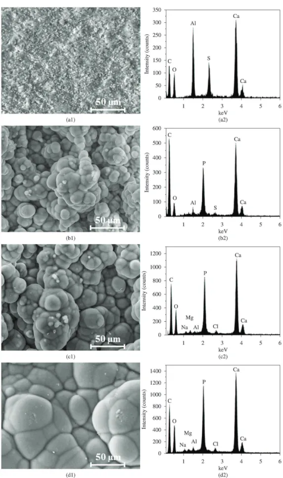
SEM images and corresponding EDS spectra of the cement surface before and after immersion in SBF (for 7, 14, and 21 days) are shown in Figure 3. Figure 3a1 shows the CSAC surface before immersion in SBF. A compact and rough morphology is observed. The corresponding EDS spectrum (Figure 3a2) shows the main elements of this kind of cement, Ca, S, Al and O. Figures 3b1, 3c1 and 3d1 show that a Ca, P-rich layer has been formed on the cement surface after 7, 14 and 21 days of immersion in SBF. In all the cases, the morphology of this compound consists of spherical agglomerates similar to those formed on the existing bioactive systems. As the immersion time increases, the spherical agglomerates increase in size. The EDS spectrum of each sample (Figures 3b2, 3c2 and 3d2)
showed that the peak corresponding to Al decreases as the immersion time is increased and, at the same time, that of P increases. The materials that are able to form a Ca, P-rich compound while in contact with SBF are considered as bioactive.
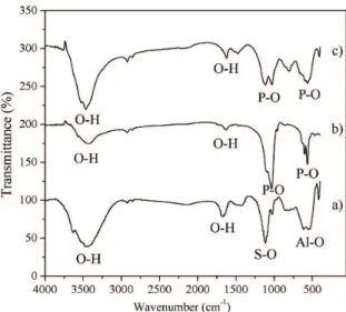
Figure 4 shows the FT-IR spectra of the CSAC hydrated in water for 21 days (Figure 4a), the stoichiometric hydroxyapatite (Figure 4b) and the CSAC after immersion in SBF for 21 days (Figure 4c). In Figure 4a the bands within the ranges of 1100-1200 cm–1 and 600-700 cm–1 correspond
to S-O and Al-O bonds, respectively. These bonds are present from octahedrally co-ordinated AlO6 units in ettringite and to the CSAC18,19. In this spectrum (Figure 4a),
the bands observed in the ranges of 1600-1700 cm–1 and
3400-3600 cm–1, corresponding to O-H vibrations and bound
water, are attributed to ettringite. In Figure 4b (HA spectrum) the presence of wavebands in the range of 1000-1100 cm–1 correspond to the P-O stretching vibrations20, as well
as the bands located within 565-601 cm–1 (P-O bending
modes). In this spectrum, the bands corresponding to the O-H vibrations are found in the range of 3400-3600 cm–1.
In Figure 4c (spectrum of the cement after immersion in SBF) the wavebands in the range of 1025-1040 cm–1 and
within 565 and 601 cm–1 correspond to the P-O bending
modes. Bands corresponding to the O-H vibrations in the range of 3400-3600 cm–1 were also observed. According
to these two spectra (4b, c), it is evident that the formation of a bonelike apatite on the CSAC after immersion in SBF has been occurred.
Figure 5 shows the XRD patterns corresponding to the CSAC after 14 and 21 days of immersion in SBF (Figures 5a, b, respectively). After 14 and 21 days of immersion in SBF, the phase corresponding to hydroxyapatite [024-0033; Ca5(PO4)3(OH)] was identiied, Figures 5a, b. The ettringite phase was not detected after these immersion time periods. A further research is needed to completely understand the mechanism of apatite formation on this calcium sulphoaluminate cement. However, the release of Ca ions from the cement to the SBF due to the partial dissolution of
ettringite increases pH leading to the nucleation of apatite on the cement. Kirsimäe et al.17 studied the phosphorous
elimination from solutions using hydrated calcareous ash sediments that contains ettringite. They stated that agglomerates of calcium phosphate may be formed on the ettringite crystals. Also, they suggested that, as the ettringite structure consists of columns and channels, there are suitable spaces for a number of ions and ion complexes
Figure 4. FT-IR spectra of (a) CSAC hydrated in water for 21 days, (b) stoichiometric hydroxyapatite and (c) CSAC after immersion in SBF for 21 days.
Figure 5. XRD patterns of CSAC after immersion in SBF for (a) 14 and (b) 21 days.
Figure 6. Hemolysis percentages of the extract and the specimen of the calcium sulphoaluminate cement.
(PO4–3, AsO 4
–3, etc.) to either interact with the ettringite
surface or to be ionically exchanged for sulphate ions into the structural tunnels.
Figure 6 shows the results of hemolysis percentage for the extract and the specimens of the calcium sulphoaluminate cement. It is observed that there is no hemolytic activity for the CSAC extract (0.6% of hemolysis) and only a slight hemolytic activity for the specimens (4.4% of hemolysis). These results demonstrated that the CSAC is hemocompatible (less than 5% hemolysis) according to the ASTM F756 standard21.
4. Conclusions
A Ca, P-rich compound was formed on the cement as early as 7 days of immersion in SBF. The morphology of this compound is similar to that formed on the existing bioactive systems. Furthermore, the Ca, P-rich layer was identiied as apatite. The bioactivity of this cementive system makes this material a potential novel bone cement. The Ca ions release from the cement to the SBF due to the partial dissolution of ettringite and the increase in pH enhances the bonelike apatite formation. Ettringite crystals may be acting as preferred sites for apatite nucleation. Calcium sulphoaluminate cement demonstrated to be hemocompatible (less than 5% hemolysis21) after the in
vitro hemolysis testing.
Acknowledgements
The authors gratefully acknowledge CONACYT, México for the provision of the scholarship for I.O. Acuña Gutiérrez.
References
1. Coleman NJ, Nicholson JW and Awosanya K. A preliminary investigation of the vitro bioactivity of white Portland cement.
Cement and Concrete Research. 2007; 37(11):1518-1523.
http://dx.doi.org/10.1016/j.cemconres.2007.08.008. 2. Ohtsuki C, Kokubo T and Yakamuro T. Mechanism of apatite
formation on CaO-SiO2-P2O5 glasses in a simulated body
fluid. Journal of Non-Crystalline Solids. 1992; 143:84-92. http://dx.doi.org/10.1016/S0022-3093(05)80556-3.
3. Kokubo T. Bioactive glass ceramics: properties and applications. Biomaterials. 1991; 12(2):155-163. http://dx.doi. org/10.1016/0142-9612(91)90194-F. PMid:1878450 4. Oyane A, Onuma K, Ito A, Kim H-M, Kokubo T and Nakamura
new kinds of simulated body fluids. Journal of Biomedical
Materials Research. Part A. 2003; 64(2):339-348. http://dx.doi.
org/10.1002/jbm.a.10426. PMid:12522821
5. Siriphannon P, Kameshima Y, Yasumori A, Okada K and Hayashi S. Formation of hydroxyapatite on CaSiO3 powders in simulated body fluid. Journal of the European Ceramic
Society. 2002; 22(4):511-520.
http://dx.doi.org/10.1016/S0955-2219(01)00301-6.
6. Sandra E. Rodil. Modificación superficial de biomateriales
metálicos. Revista Latinoamericana de Metalurgia y
Materiales. 2009; 29(2):67-83.
7. Cai X, Yuan J, Chen S, Li P, Li L and Shen J. Hemocompatibility improvement of poly(ethylene terephthalate) via self-polymerization of dopamine and covalent graft of zwitterions.
Materials Science and Engineering: C. 2014; 36:42-48. http://
dx.doi.org/10.1016/j.msec.2013.11.038. PMid:24433885 8. Henkelman S, Rakhorst G, Blanton J and van Oeveren W.
Standardization of incubation conditions for hemolysis testing of biomaterials. Materials Science and Engineering: C. 2009; 29(5):1650-1654. http://dx.doi.org/10.1016/j. msec.2009.01.002.
9. Santos LA, Carrodéguas RG, Rogero SO, Higa OZ, Boschi AO
and Arruda ACF. α-tricalcium phosphate cement: “in vitro”
cytotoxicity. Biomaterials. 2002; 23(9):2035-2042. http:// dx.doi.org/10.1016/S0142-9612(01)00333-7. PMid:11996045 10. Guo H, Wei J and Liu CS. Development of a degradable cement
of calcium phosphate and calcium sulfate composite for bone reconstruction. Biomedical Materials (Bristol, England). 2006; 1(4):193-197. http://dx.doi.org/10.1088/1748-6041/1/4/003. PMid:18458405
11. Hernández Q and Piña Barba C. Caracterización física y química de pastas de cementos óseos con ZrO2. Revista
Mexicana de Física. 2003; 49:123-131.
12. Medri V, Mazzocchi M and Bellosi A. Doped calcium-aluminium-phosphate cements for biomedical applications.
Journal of Materials Science. Materials in Medicine. 2011;
22(2):229-236. http://dx.doi.org/10.1007/s10856-010-4205-3. PMid:21165760
13. Oh SH, Choi S, Lee YK and Kim KN. Preparation of calcium aluminate cement for hard tissue repair: effects of lithium fluoride and maleic acid on setting behavior, compressive
strength, and biocompatibility. Journal of Biomedical Materials
Research. 2002; 62(4):593-599. http://dx.doi.org/10.1002/
jbm.10347. PMid:12221708
14. Liao Y, Wei X and Li G. Early hydration of calcium sulfoaluminate cement through electrical resistivity measurement and microstructure investigations. Construction
& Building Materials. 2011; 25(4):1572-1579. http://dx.doi.
org/10.1016/j.conbuildmat.2010.09.042.
15. Kokubo T and Takadama H. How useful is SBF in predicting in vivo bone bioactivity? Biomaterials. 2006; 27(15):2907-2915. http://dx.doi.org/10.1016/j.biomaterials.2006.01.017. PMid:16448693
16. Wawschinek O, Petek W, Lang J, Pogglitsch H and Holzer H. The determination of aluminium in human plasma.
Mikrochimica Acta. 1982; 77(5-6):335-339. http://dx.doi.
org/10.1007/BF01197113.
17. Kirsimäe K, Kaasik A, Vohla C, Mõtlep R and Mander U. Hydrated calcareous oil-shale ash as potential filter media for phosphorus removal in constructed wetlands. Water Research. 2008; 42(4-5):1315-1323. http://dx.doi.org/10.1016/j. watres.2007.10.002. PMid:17959214
18. Gastaldi D, Canonico F and Boccaleri E. Ettringite and calcium sulfoaluminate cement: investigation of water content by near-infrared spectroscopy. Journal of Materials Science. 2009; 44(21):5788-5794. http://dx.doi.org/10.1007/s10853-009-3812-1.
19. Fernández-Carrasco L, Torrens-Martín D, Morales LM and Martínez-Ramírez S. Infrared spectroscopy in the analysis of building and construction materials. In: Theophile T, editor. Infrared spectroscopy: materials science, engineering
and technology. In Tech; 2012. p. 369-382. Chapter 19.
Available from: <http://www.intechopen.com/books/infrared-spectroscopy-materials-science-engineering-and-technology/ infrared-spectroscopy-of-cementitious-materials>.
20. Fathi MH, Hanifi A and Mortazavi V. Preparation and bioactivity evaluation of bone-like hydroxyapatite nanopowder. Journal
of Materials Processing Technology. 2008; 202(1-3):536-542.
http://dx.doi.org/10.1016/j.jmatprotec.2007.10.004.
21. American Society for Testing and Materials - ASTM. ASTM F-756: standard practice for assessment of hemolytic